HAL Id: tel-01480950
https://tel.archives-ouvertes.fr/tel-01480950
Submitted on 2 Mar 2017HAL is a multi-disciplinary open access
archive for the deposit and dissemination of sci-entific research documents, whether they are pub-lished or not. The documents may come from teaching and research institutions in France or abroad, or from public or private research centers.
L’archive ouverte pluridisciplinaire HAL, est destinée au dépôt et à la diffusion de documents scientifiques de niveau recherche, publiés ou non, émanant des établissements d’enseignement et de recherche français ou étrangers, des laboratoires publics ou privés.
delivery of vaccines
Beatrice Bernocchi
To cite this version:
Beatrice Bernocchi. Porous maltodextrin nanoparticles for the intranasal delivery of vaccines. Hu-man health and pathology. Université du Droit et de la Santé - Lille II, 2016. English. �NNT : 2016LIL2S010�. �tel-01480950�
1
Université Lille 2 Droit et Santé
Ecole doctorale Biologie-Santé de Lille
THESE
Pour l’obtention du grade de
DOCTEUR DE L’UNIVERSITE DE LILLE 2
Discipline : Physiologie, Physiopathologie et Biologie systémique médicale
Présentée et soutenue publiquement par
Beatrice BERNOCCHI
Le 18 Juillet 2016
POROUS MALTODEXTRIN NANOPARTICLES FOR THE INTRANASAL
DELIVERY OF VACCINES
Composition du jury:
Rapporteurs : Pr Gillian BARRATT Institut Galien-Paris Sud, CNRS UMR 8612
Pr Patrick MIDOUX Université de Orléans, CNRS UPR 4301
Examinateurs : Pr Pierre DESREUMAUX Université de Lille2, Inserm - UMR 995
Pr Philippe CHAVATTE Université de Lille 2, Inserm - UMR 995
Directeur de Thèse : Pr Didier BETBEDER Université de Lille 2, Inserm - UMR 995
2
TABLE OF CONTENTS ... 2
ACKNOWLEDGMENTS ... 4
LIST OF PUBLICATIONS ... 6
ABBREVIATIONS ... 7
LIST OF FIGURES AND TABLES... 9
AIM AND OUTLINE OF THE THESIS ... 10
ABSTRACT ... 12
RÉSUMÉ ... 13
RÉSUMÉ DE LA THÈSE ... 14
PART I: INTRODUCTION ... 22
1. NANOPARTICLES ... 23
1.1 NANOPARTICLES AS DRUG DELIVERY SYSTEM ... 23
Publication 1: Endocytosis of nanoparticles ... 25
1.1.1 Nanoparticles for vaccine and macromolecules delivery ... 78
Publication 2: Nasal nanovaccines ... 81
1.1.2 NPL positively charged porous nanoparticles with a lipid core ... 112
1.2 BIOLOGICAL FATE OF NASALLY ADMINISTERED NANOPARTICLES ... 113
1.2.1 Anatomy of the nose ... 113
1.2.2 Nasal immune system: NALT ... 115
1.2.3 Fate of nanoparticles after intranasal administration ... .117
2. INFLUENZA AND VACCINATION ... 118
2.1 HISTORY OF VACCINATION…..………..…....……118
2.2 INFLUENZA DISEASE………..……….……..……...118
2.2.1 Influenza virus: classification and structure ... 119
2.2.2 The infection mechanism ... 121
2.3 INFLUENZA VACCINES: INACTIVATED AND LIVE ATTENUATED INFLUENZA VACCINES ... 124
2.3.1 Administration routes of influenza vaccines ... 125
3
2.3.4 CTA1-3M2e-DD ... 132
2.4 INFLUENZA TREATMENT: ANTIVIRAL DRUGS ... 133
PART II: RESULTS ... 135
NANOPARTICLES FOR PROTEIN DELIVERY IN THE NASAL MUCOSA ... 137
Publication 3:Mechanisms allowing protein delivery in nasal mucosa using NPL nanoparticles ... 138
NPL endocytosis by THP-1 derived macrophages ... 148
Conclusion ... 149
Choice of the cell model: In vitro models of airway barriers ... 150
Ovalbumin as model antigen for vaccine delivery ... 151
NANOPARTICLES FOR UNIVERSAL INFLUENZA VACCINE DELIVERY ... 153
Porous nanoparticles for the mucosal delivery of an adjuvanted universal influenza vaccine 154 PART III: DISCUSSION ... 172
PART IV: CONCLUSION AND PERSPECTIVES ... 180
4 Firstly, I would like to express my gratitude to Professor Didier Betbeder for welcoming me in his research group as PhD student and particularly for his teachings, support and patience during these years. I will always be grateful for this opportunity.
Besides my advisor, I would like to thank all the members of the jury for accepting being part of my thesis committee and for evaluating this work.
I would like to thank the funding from the People Programme (Marie Curie Actions) of the European Union's Seventh Framework Programme FP7/2007-2013/ under REA grant agreement n°607690.
My sincere thanks also go to Dr Rodolphe Carpentier for his guidance, encouragements and support. Thank you for your constant help and fruitful discussions.
I wish to express my appreciation to Professor Dimier-Poisson for her presence during these years and for hosting me in her laboratory. I also wish to thank all the UMR1282 of the University Francois Rabelais of Tours, especially Céline Ducournau and Dr Isabelle Lantier for the productive collaboration.
I would like to express my thanks also to all the members of the UniVacFlu group, especially to the coordinator Professor Nils Lycke, but also to Dr Drubavka Grdic Eliasson and Valentina Bernasconi for the work done on the European project.
I am also very grateful to Fatima Dahmani for all her teachings, technical guidance and also constructive dialogues.
I would like to thank Michael Howsam for proofreading the publications and Laura Ravasi for checking the English of this manuscript.
I would like to thank the members of the LIRIC-UMR 995 for their support.
Thanks to Audrey Langlois, Madjid Djouina, Laura Choteau, Anthony Martin-Mena, Nicolas Esquerre, Mohamed Bousaleh, François Maggiotto and Didier Lefranc. Thanks for your assistance, encouragement and friendship.
5 technicians with whom I shared the daily life in the laboratory. To Helena, Audrey, Coralie, Minh Quan, Amelie and Clement thanks for the nice moments together.
Finally special thanks go to my family, Manuel and all my friends. Thanks for your presence, support and encouragements during these years.
6
Mechanisms allowing protein delivery in nasal mucosa using NPL nanoparticles,
B. Bernocchi, R. Carpentier, I. Lantier, C. Ducournau, I. Dimier-Poisson, D. Betbeder, Journal of Controlled Release 232 (2016) 42-50
Nasal Nanovaccines,
B.Bernocchi, D. Betbeder Submitted to Biomaterials
Endocytosis of Nanoparticles,
B.Bernocchi, R. Carpentier, D. Betbeder Submitted to Nanoscale
7 (except of publications)
16HBE14o-, human bronchial epithelial cell line
ADP, adenosine diphosphate APC, antigen presenting cell
API, active pharmaceutical ingredient B cell, lymphocyte B
BM2, ion channel influenza B virus BSA, bovine serum albumin
cAMP, cyclic adenosine monophosphate CD4, cluster of differentiation 4
CD8, cluster of differentiation 8 CM2, ion channel influenza C virus CME, clathrin mediated endocytosis CTA1, cholera toxin subunit A1 CTL, cytotoxic T lymphocytes
DAS181, sialidase inhibitor of influenza virus attachment
DC, dendritic cell
DC-SIGN, dendritic cell-specific
intercellular adhesion molecule-3-grabbing non-integrin, C-type lectin receptors
DD, dimer of the D-fragment of the protein A from Staphylococcus aureus
DPPG, dipalmitoylphosphatidylglycerol EMA, European Medicine Agency FDA, Food and Drug Administration FITC, fluorescein isothiocyanate
GCN4, transcriptional activator protein GM1, monosialotetrahexosylganglioside GRAS, generally recognized as safe Gsα, G-protein α-subunit
GTMA, glycidyltrimethylammonium chloride
GTP, guanosine triphosphate HA, hemagglutinin
hAEpC, primary human alveolar type I-like cells
HBc, hepatitis B virus core protein HBsAg, hepatitis B virus antigen
HLA-DR, human leukocyte antigen, antigen D related
HSP70, heat shock protein 70 IFN-γ, interferon gamma Ig, immunoglobulin
IIV, inactivated influenza virus IL-2, interleukin 2
ISCOM, ImmunoStimulating COMplex LAIV, live attenuated influenza virus LD50, median lethal dose
L-SIGN, lectin-specific intercellular
adhesion molecule-3-grabbing non-integrin, C-type lectin receptors
LTR192G, non-toxic form of the heat labile toxin
8 M2, matrix protein 2 of influenza virus
M2e, matrix protein 2 extracellular domain of influenza virus
M-cells, microfold or membranous cells MDCK, Madin-Darbey canine kidney cells MHC, major histocompatibility complex NA, neuraminidase
NALT, nose-associated lymphoid tissue NB, ion channel influenza B virus NP, nucleoprotein of influenza virus NP+, maltodextrin nanoparticles
NPL, maltodextrin nanoparticles containing a lipid core
OVA, ovalbumin
PA, Influenza RNA polymerase subunit A PB1, Influenza RNA polymerase subunit B1 PB2, Influenza RNA polymerase subunit B2 PLGA, polylactic-co-glycolic acid
QH-A, QH-B, QH-C, fraction of Quil A QS-21,purified fraction of Quillaja saponaria
RNA, ribonucleic acid RNPs, ribonucleoproteins RSV, respiratory syncytial virus sIgA, secretory immunoglobulin A SMBV, supramolecular biovectors T cell, lymphocyte T
TEER, transepithelial electrical resistance Th, T helper
THP-1: monocyte cell line TJ, tight junction
TLR, toll like receptor TMC, N-trimethyl chitosan
TNF-α, tumor necrosis factor alpha WHO, world health organization
9 (except of publications)
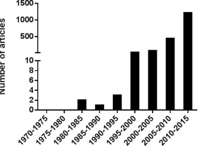
Figure 1 Publications about “vaccine nanoparticle”. ... 78
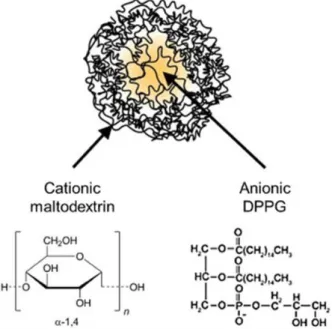
Figure 2 Representation of NPL. ... 112
Figure 3 Sagittal section of the nasal cavity. ... 114
Figure 4 Mucosal immune response. ... 116
Figure 5 Influenza A virus nomenclature. ... 120
Figure 6 Schema of Influenza A virus. ... 121
Figure 7 Influenza virus replication mechanism. ... 123
Figure 8 Adjuvanted antigen CTA1-3M2e-DD. ... 132
Figure 9 NPL endocytosis inTHP1-derived macrophages... 148
Figure 10 Mechanism of NPL interaction with the nasal mucosa. ... 149
Figure 11 Scanning electron microscopy of airway epithelial cells. ... 150
Figure 12 Characterization of CTA1-3M2e-DD NPL. ... 160
Figure 13 Characterization of CTA1(R9K)-3M2e-DD NPL. ... 160
Figure 14 Characterization of HA CTA1- 3M2e-DD NPL. ... 161
Figure 15 Characterization of the stability of CTA1- 3M2e-DD NPL: native PAGE... 162
Figure 16 Characterization of the stability of CTA1- 3M2e-DD NPL 12 months. ... 163
Figure 17 Characterization of the stability of CTA1- 3M2e-DD NPL: SDS PAGE. ... 164
Figure 18 Characterization of the stability of CTA1- 3M2e-DD NPL 12 months. ... 164
Figure 19 Characterization of the stability of CTA1- 3M2e-DD. ... 165
Figure 20 Characterization of the size and charge stability of CTA1- 3M2e-DD: NPL. ... 166
Figure 21 Characterization of the colloidal stability of CTA1- 3M2e-DD NPL12 months. ... 167
Figure 22 Characterization of the NPL size and charge stability. ... 168
Figure 23 Characterization of the stability of CTA1- 3M2e-DD. ... 169
Figure 24 Antigen delivery in airway epithelial cells (a) and macrophages (b). ... 170
10
Aim of the thesis
The aim of these studies is the evaluation of cationic maltodextrin nanoparticles with a lipid core (NPL) as drug vector. We intend to elucidate the mechanism of nanoparticles interaction with the nasal mucosa and evaluate their application as protein delivery system for a universal influenza vaccine.
The UniVacFlu Consortium
This manuscript describes the thesis work performed under the supervision of Professor Didier Betbeder, head of the laboratory of Nanomedicine. This laboratory is part of the group of Therapeutic Innovation Targeting Inflammation of the INSERM unit LIRIC-UMR 995 of the University of Lille 2.
We evaluated the mechanism implied in NPL protein delivery after intranasal administration. These studies are prerequisite for developing a new adjuvant system for vaccine application. Hence we used this NPL technology in the project UniVacFlu, financed by the European Union Seventh Framework Programme FP7/2007/2013, part of the People Programme, Marie Skłodowska-Curie Actions. This International Training Network focuses on the development of a mucosal universal Influenza vaccine.
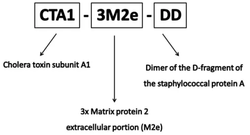
The UniVacFlu project is coordinated by Professor Lycke from the University of Gothenburg. Lycke and co-workers developed the non-toxic mucosal adjuvant CTA1-DD, to circumvent the safety problems of the cholera toxin. Lycke et al., together with Pr. Fiers and Pr. Saelens from the University of Ghent, developed the fusion protein CTA1-3M2e-DD, where M2e is a conserved epitope of influenza virus A. Therefore to optimize the mucosal vaccine efficacy we combined CTA1-3M2e-DD with nanoparticles technology. This vaccine is currently under evaluation for the induction of protective immunity after intranasal administration in the
Consortium.
We also investigated the oral administration of the nanoparticle vaccine in collaboration with Rescigno et al., form the European Institute of Oncology in Milan. Eventually we addressed the protection from virus transmission with Pr. Staeheli and co-workers, from the University of Freiburg.
The role of our laboratory in the UniVacFlu Consortium is (i) to prepare and characterize nanoparticles and vaccine formulations, (ii) to analyze the interaction of these nanoparticles with
11 stability and (iiii) to supply the vaccine formulations to the partners.
Outline of the thesis
This document is organized in four principal chapters:
The PART I is a general introduction about the state of the art of the nanotechnology applied to drug delivery. This chapter is arranged in two main sections. The first section focuses on the nanoparticles interaction with cells and their use in vaccinology. A review (submitted) on the literature concerning the study of nanoparticles endocytosis is included. This section contains also a review entitled “Nasal Nanovaccines” (submitted), which discusses the types of nanocarriers studied in literature for the nasal vaccination and controversies regarding these studies. The importance of the knowledge of vector and antigen biodistribution is also introduced. In the second section, the main features of Influenza virus and vaccines are described.
The PART II presents the results on the NPL evaluation for nasal drug delivery. In the first part we present the published article about the mechanism of nanoparticles interaction with the nasal mucosa. In the second part the results about the development of nanoparticle formulation for mucosal influenza vaccine are described. The characterized formulations were sent to our partners of the UniVacFlu Consortium for evaluation.
The PART III is a general discussion concerning the obtained results and the application of the nanoparticles in vaccinology.
12 Nanoparticles technology for mucosal delivery of vaccines received a growing interest in the last decades. Intranasal administration owns great advantages for immune system stimulation, such as local and systemic protection against infectious diseases. However delivery systems and adjuvants are often required to efficiently trigger mucosal and systemic immune responses. In this thesis, nanoparticles (NP) have been evaluated as delivery system for a nasal universal influenza vaccine in a People Program of the European Union Seventh Framework Program FP7 called UniVacFlu. The aim of the UniVacFlu network is to develop a universal influenza vaccine administered through the mucosal route. We used porous maltodextrin nanoparticles with a lipidic core (NPL). We loaded an adjuvanted antigen named CTA1-3M2e-DD in the NPL. CTA1-3M2e-DD is composed of the A1 subunit of the cholera toxin and a conserved epitope of influenza A virus (M2e), while DD, dimer of the synthetic analogue of the Staphyloccous aureus protein A, targets B cells. Interestingly the antigen loading in NPL was quantitative for the antigen: NPL 1:5 mass ratio and the formulation was stable for at least six months at 4°C. We assessed the successful delivery of the antigen by NPL in airway epithelial cells and macrophages. These formulations are currently evaluated by the UniVacFlu consortium in mice. One of the main issues of intranasal vaccines is the toxicity that can be elicited by the nose-brain passage of one of their components. We investigated the loading of antigens in NPL and their delivery in airway mucosa. We observed a high endocytosis of NPL and an increased protein delivery into the cells. On a transwell model of the airway mucosa we assessed the absence of transcytosis and paracellular passage of the NPL. In vivo results confirmed the lack of nose-brain passage of the NPL, as NPL were found not to cross the mucosa. Interestingly, we observed an increased nasal residence time of the protein targeted by NPL. The particles after having delivered their payload are totally eliminated through the gastrointestinal tract, making these nanoparticles good candidates for mucosal delivery system. These results highlight the interest of NPL as vectors for mucosal delivery of drugs.
13 Au cours des dernières décennies, la technologie des nanoparticules pour la délivrance des vaccins au niveau de muqueuses a reçu un intérêt croissant. L’administration intranasale possède de grands avantages pour la stimulation du système immunitaire, telles que la stimulation d’une immunité protectrice locale et systémique. Cependant des systèmes de délivrance et des adjuvants sont souvent nécessaires pour déclencher efficacement la réponse immunitaire. Nous avons appliqué la technologie des nanoparticules en tant que système de délivrance d'un vaccin universel nasal contre la grippe dans un projet européen FP7 appelé UniVacFlu. Nous avons formulé un antigène adjuvé CTA1-3M2e-DD avec les NPL. Cet antigène est composé de la sous-unité A1 de la toxine du choléra et d’un épitope conservé du virus de la grippe A (M2e), ainsi que du dimère de l’analogue synthétique de la protéine A de Staphylococcus aureus (DD). Les nanoparticules utilisées sont poreuses et constituées de maltodextrines réticulées ayant un cœur lipidique (NPL). L’association de cet antigène avec les NPL est quantitative et la formulation est stable pendant au moins six mois à 4°C. Les NPL permettent également de délivrer d’une manière accrue cet antigène dans les cellules épithéliales des voies respiratoires et les macrophages. Actuellement ces formulations sont évaluées chez la souris par le consortium UniVacFlu.
L'un des principaux problèmes des vaccins nasal est la toxicité qui peut être provoquée par le passage nez-cerveau de l'un de ses composants. Le but de ce travail est d'évaluer le potentiel des NPL, en tant que vecteurs pour la délivrance des vaccins nasal. Ainsi, nous avons étudié le chargement d’un antigène dans les NPL et sa délivrance dans les cellules épithéliales des voies respiratoires. Notre étude révèle que les NPL interagissent fortement avec les muqueuses et délivrent d’une manière accrue les antigènes dans les cellules. Nous avons également montré l'absence de transcytose et de passage paracellulaire des NPL ou des antigènes délivrés dans un modèle de barrière épithéliale in vitro. Les résultats in vivo confirment l'absence de passage nez-cerveau des NPL et montrent qu’elles prolongent fortement le temps de résidence nasale des antigènes qui sont ensuite éliminés par le tractus gastro-intestinal.
Ces résultats mettent en évidence l'intérêt des NPL comme vecteurs pour la prochaine génération de médicaments et de vaccins.
14
RÉSUMÉ DE LA THÈSE
Préambule
Ces travaux ont été effectués sous la direction du Professeur Betbeder au sein de l’équipe de Nanomédecine de l’unité Inserm LIRIC-U995 de l’Université de Lille 2. Nous avons évalué les nanoparticules comme système de délivrance des protéines dans la muqueuse nasale. Ces études sont un prérequis pour le développement d’un nouveau système adjuvant pour application dans le domaine du vaccin, et plus particulièrement pour le développement d’un vaccin universel contre la grippe administré par les voies de muqueuses. Ces travaux sont insérés dans un vaste projet européen nommé UniVacFlu. Le projet UniVacFlu a été financé par l’European Union Seventh Framework Programme FP7/2007/2013. Le consortium UniVacFlu est coordonné par Professeur Lycke de l’Université de Gothenburg et il est composé par plusieurs partenaires : l’Université de Ghent, l’Université de Freiburg, l’Institut Européen d’oncologie de Milan et le Kings College de Londres. Notre rôle dans le consortium UniVacFlu est de (i) préparer et caractériser les nanoparticules et les formulations vaccinales, (ii) analyser les interactions des nanoparticules avec la muqueuse nasale, (iii) étudier la stabilité des vaccins développés et (iiii) fournir les formulations à nos collaborateurs.
15
Introduction
Ces dernières années la technologie des nanoparticules a suscité de plus en plus d’intérêt. Ce sont des objets ayant au moins une dimension inférieur à 100nm (International Organization for Standardization, 2011). Les nanoparticules ont des applications dans plusieurs domaines. Cependant, dans le cadre de cette thèse, nous nous intéressons à l’application de cette technologie innovante pour la délivrance de protéines par voie nasale. Dans la première partie de ce travail nous nous intéresserons aux mécanismes d’interaction des nanoparticules avec la muqueuse nasale, puis dans la deuxième partie nous utiliserons cette stratégie pour le développement d’un vaccin universel contre la grippe.
Les nanoparticules ont plusieurs avantages, comme l’amélioration de la solubilité et de la stabilité des molécules, ainsi que la possibilité de cibler certains organes et tissus une fois administrées. Concernant les vaccins, les nanoparticules peuvent avoir différentes fonctions : adjuvant ou immunomodulateur (Zazo et al., 2016; Zhao et al., 2014). Quand les nanoparticules agissent comment adjuvant, elles améliorent l’immunogénicité de l’antigène par action locale simultanée avec l’antigène même. Néanmoins elles peuvent agir comme immunomodulateur par stimulation directe et systémique du système immunitaire.
Différents types de nanoparticules ont été utilisées pour délivrer des vaccins dans la muqueuse nasale. Nous avons groupé ces types des nanoparticules sur la base des matériels utilisés : polysaccharidiques, polymériques, lipidiques et protéiques (Publication «Nasal Nanovaccine »). Toutefois des systèmes plus complexes, qui mélangent différents matériaux, ont été aussi développés.
Des nanoparticules cationiques poreuses ont été utilisées comme vecteur nasal d’antigènes. Celles-ci sont constituées de maltodextrines, des polysaccharides dérivés de l’hydrolyse de l’amidon. Les maltodextrines sont hydratées et réduites, puis réticulées grâce à l’épichloridrine jusqu’à obtenir un gel. Le gel est ensuite rendu cationique par addition de Glycidyl Tri-Méthyl Ammonium chloride (GTMA) qui fixe les groupes d’ammonium quaternaire sur le polymère. Ce gel est broyé par homogénéisation à haute pression et les particules obtenues (NP+) sont filtrées pour éliminer les résidus de la synthèse (Castignolles et al.1994). Il est possible d’introduire des phospholipides dans la structure des NP+, afin d’obtenir des nanoparticules lipidées (NPL). Dans le cadre de ce travail le dipalmitoyl phosphatidyl glycérol (DPPG) a été introduit dans le cœur des NP+.
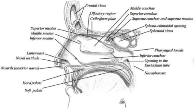
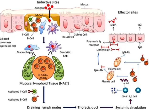
16 La voie nasale est très avantageuse pour l’administration des vaccins, car le système lymphatique associé au nez (NALT) est directement accessible. L’interaction des antigènes avec la muqueuse nasale, plus précisément le NALT, peut déclencher une réponse immunitaire locale et systémique, ainsi qu’une protection contre les agents infectieux. Cette voie est facile d’accès et plus confortable pour les patients (aucune aiguille n’est nécessaire). La cible principale des vaccins à délivrance nasale est l’anneau du Waldeyer, qui est composé par l’ensemble des amygdales, situées autour des cavités nasales et buccale (Gizurarson, 2012). Le tissu épithélial de la cavité nasale se compose principalement de cellules pseudo-stratifiées. Au niveau de la partie supérieure de la cavité nasale se trouve la région olfactive, constituée par l’épithélium olfactif, qui peut être une voie directe vers le cerveau grâce aux neurones olfactifs insérés dans l’os cribriforme.
Le NALT est un organe lymphatique secondaire, site inductif du système immunitaire muqueux. A ce niveau l'administration d'antigènes déclenche l'initiation de la réponse immunitaire. Le système d'échantillonnage de l'antigène (par exemple, les cellules M) capte l'antigène dans l'épithélium associé aux follicules et le transfère aux cellules présentatrices d'antigène (APC), telles que les cellules dendritiques (DC). Les DC induisent la réponse de cellules T naïves CD4 + et CD8 +.
Les lymphocytes T CD8 + maturent en cellules T cytotoxiques (CTL). Le rôle des CTL est de tuer les cellules infectées et de lutter contre l'infection virale.
Grâce à la présentation de l'antigène par les DC aux cellules T CD4 +, les réponses Th1, Th2 et Th17 sont activées ainsi que la commutation des immunoglobulines de classe IgA et l’hypermutation de cellules B dans les centres germinaux. Les cellules B IgA + migrent vers les sites effecteurs à travers les ganglions lymphatiques cervicaux et le sang périphérique. Dans le cas d'antigènes administrés par voie nasale, les sites effecteurs sont la lamina propria des voies respiratoires supérieures, de l’intestin et de l'appareil génital.
Une fois atteint le site effecteur, les cellules B sécrètent des IgA (sIgA). Les slgA sont transcytosées sur le côté luminal de l'épithélium par le récepteur polymèrique d'Ig ce qui permet de bloquer l'entrée d'agents pathogènes (Kiyono et al., 2004; Lamichhane et al., 2014; Lycke, 2012).
Les cellules épithéliales respiratoires sont impliquées dans la régulation de la réponse immunitaire (Pichavant et al., 2003). En effet ces cellules expriment le complexe majeur d’histocompatibilité de classe II (MHCII), spécifique des cellules présentatrices d’antigènes. La présence de MHC II a été observée au niveau du cornet nasal (Kalb et al., 1991). Par conséquent
17 les cellules épithéliales respiratoires peuvent avoir une fonction de cellules présentatrices d’antigènes et constituer une cible supplémentaire pour les vaccins.
Les vaccins activant des mécanismes en cascade, ceux de transport de la formulation nanoparticulaire doivent être étudiés afin d’éviter les effets secondaires toxiques.
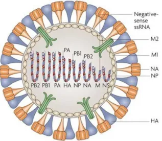
Des virus, comme par exemple le virus d’Influenza à l’origine de la grippe, infectent les animaux et les humains par voie aérienne. Le virus de la grippe peut avoir une forme sphérique avec une taille de 100nm de diamètre ou une forme filamenteuse avec une taille majeure, dans l’ordre des micromètres (Rossman et al., 2012). Avec des nanoparticules de propriétés similaires et chargées des antigènes viraux, il serait possible, d’un point de vue immunologique, de mimer la forme sphérique de ce virus.
Le type majoritaire des virus de la grippe est l’Influenza A, à l’origine de pandémies. Le virus de la grippe est constitué de plusieurs protéines dont les hémagglutinines (H) et les neuraminidases (N) qui déterminent la classification des virus de type A.
L’association des protéines H et N est très variable et chaque année, de nouvelles combinaisons apparaissent causant les grippes saisonnières. Plusieurs inconvénients aux vaccins actuels existent: (1) Le caractère saisonnier des vaccins empêche une protection contre toutes les souches de virus grippaux de type A. (2) L’injection intramusculaire est relativement efficace mais ne reproduit pas la voie d’entrée naturelle du virus. Les vaccins muqueux présentent de meilleures réponses immunitaires (cellulaires et humorales), plus proches d’une primo-infection. (3) L’utilisation de virus vivant atténué souffre de problèmes de stabilité et peut causer une réversion vers une forme pathogénique. (4) La méthode de production prévoit l’utilisation d’œufs et la formulation vaccinale finale peut être contaminée par l’ovalbumine, un allergène reconnu.
Plusieurs nouveaux vaccins sont en phase de développement. Ceux-ci sont produits par incubation du virus dans des cellules ou par technologie recombinante.
Pour les vaccins muqueux et notamment pour les vaccins recombinants, il est nécessaire d’utiliser des adjuvants afin d’obtenir une réponse immunitaire plus efficace, spécifiquement une réponse cellulaire. Les adjuvants acceptés ne sont pas nombreux mais restent nécessaires surtout pour les vaccins muqueux et recombinants. Parmi les adjuvants les plus puissants, on trouve des toxines bactériennes que nous avons modifiées afin de limiter leur toxicité. La sous-unité A1 de cette toxine a été conjuguée au canal ionique de Influenza (M2e), une protéine très conservée du virion et au fragment de la protéine A du Staphylococcus aureus (D), qui lie les cellules B
18 (Agren et al., 1997; Eriksson et al., 2004; Lycke, 2004a). Cette protéine recombinante CTA1-3M2e-DD a été synthétisée pour être utilisée comme vaccin adjuvanté contre la grippe.
But de la thèse
L'objectif de cette thèse est l'évaluation des NPL comme vecteur de médicament. Nous étudierons les mécanismes d'interaction des NPL avec la muqueuse nasale et évaluerons leur application en tant que système de délivrance des protéines pour un vaccin universelle contre la grippe.
19
Résultats et discussion
Dans la première partie de ce travail nous avons cherché à mieux comprendre les interactions des nanoparticules poreuses (NPL) avec les cellules épithéliales des voies aériennes, leur capacité à traverser cette barrière, et d'analyser l'impact des lipides au sein de ces nanoparticules sur la délivrance et la transcytose d'antigènes dans les cellules épithéliales. Les études in vivo ont été réalisées pour suivre la délivrance de l'antigène dans les muqueuses respiratoires et sa biodistribution après administration nasale.
Les nanoparticules NP+ possèdent un diamètre de 70nm et un potentiel zeta de +45.9 mV. Des phospholipides, notamment le DPPG, peuvent être inclus dans la structure poreuse de ces particules, donnant les NPL. Les NPL ont une taille de 76 nm et une charge de +44.2 mV. Il est possible de marquer, avec des marqueurs fluorescents, la partie polysaccharidique du NPL, ainsi que la partie lipidique. Suite au marquage des NPL, nous avons suivi l’endocytose des nanoparticules dans les cellules épithéliales des voies aériennes (16HBE14o-) par cytométrie en flux. Nous avons observé que les NPL sont endocytosées par les cellules épithéliales et ce mécanisme atteint un plateau après 3 h. Constatant la similitude du profil d’endocytose de la partie polysaccharidique et de la partie lipidique, nous suggérons que les lipides ne sont pas délivrés par les nanoparticules pendant l’endocytose mais restent associés à celle-ci.
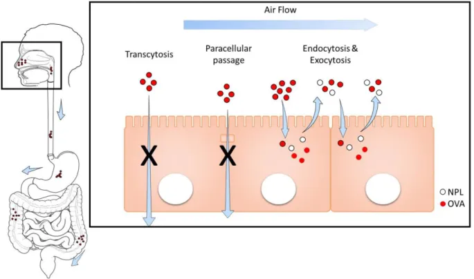
Ensuite nous avons évalué la transcytose de ce vecteur à travers un modèle in vitro d’épithélium (Transwell®). Nous avons montré que les NPL n’ouvrent pas les jonctions serrées entre les cellules épithéliales et ne traversent pas la barrière épithéliale ni par voie paracellulaire ni par transcytose.
Afin d’évaluer la capacité des NPL à délivrer des antigènes, nous avons préparé des formulations on utilisant l’ovalbumine (OVA) comme antigène modèle. La protéine est chargée dans les NPL par simple mélange à température ambiante. Différentes quantité de NPL ont été utilisées pour formuler la protéine. Nous avons observé que dans la formulation, ayant le rapport protéine : NPL 1 :3 (poids :poids), toutes les protéines sont associées et incorporées dans les NPL. Ensuite nous avons étudié la délivrance de la protéine modèle marquée dans les cellules respiratoires 16HBE par cytométrie en flux. L’OVA est efficacement délivrée dans les cellules épithéliales grâce aux NPL (14 fois plus que l’OVA seule après 24h). La transcytose de l’OVA libre ou formulée avec les NPL a été examinée sur le modèle de Transwell®. Nous avons conclu que les NPL ne favorisent pas le passage des protéines à travers la barrière épithéliale in vitro. Nous avons aussi étudié la biodistribution de l’OVA après administration nasale dans un modèle animal murin. L’imagerie in vivo montre que les NPL prolongent le temps de résidence de
20 l’antigène dans la muqueuse nasale. Cependant l’OVA libre est dégradée et éliminée totalement après 1.5h, tandis que l’OVA encapsulée dans les NPL reste dans le nez jusqu’à 6h et est ensuite éliminée par le tractus gastro-intestinal. Enfin nous avons vérifié la résidence dans la muqueuse de l’OVA et des NPL. L’endocytose des NPL dans les cellules de l’épithélium nasal a été confirmée in vivo, ainsi que l’absence de passage transcellulaire. Nos travaux précédents ont montré que ces NPL peuvent être chargées avec une grande quantité d’antigènes et induire efficacement des réponses immunitaires humorales, cellulaires et de la muqueuse après administration nasale (Dimier-Poisson et al., 2015). Par ailleurs, dans la cadre de ce travail nous avons montré que les NPL ne franchissent pas la barrière nez-cerveau. Ceci supporte les résultats obtenus précédemment (Merhi et al., 2012) et confirment que ces NPL ne sont pas toxiques. Les NPL sont des vecteurs idéaux pour les vaccins car ils délivrent l’antigène dans la muqueuse et sont totalement éliminées.
Dans la deuxième partie de cette thèse nous avons utilisé les NPL pour le développement d’un vaccin universel mucosal contre la grippe. Ces travaux ont été réalisés en collaboration avec les partenaires du projet Européen UniVacFlu. En conséquence nous avons étudié l’association de l’antigène CTA1-3M2e-DD aux NPL. Différents rapports en masse d’antigènes (CTA1-3M2e-DD et OVA) et NPL ont été évalués. L’antigène est complètement associé aux NPL à partir du rapport en masse testé antigène : NPL 1:3. A l’inverse, une fraction des protéines libres a été détectée en électrophorèse natif pour le rapport 1 :0.5 Antigène :NPL. En complément, nous avons étudié la stabilité des formulations 1 :0.5 et 1 :5 à 40°C pendant 3 mois et à 4°C pendant 12 mois, notamment en termes de taille, de potentiel zêta, d’association antigène : NPL et de dégradation de l’antigène. Nous avons observé que la formulation 1 :0.5 n’est pas stable contrairement à la formulation 1 :5 (4°C pendant 12 mois). Par contre, à 40°C, la dégradation partielle de l’antigène seul et associé aux NPL a été observée après 3 mois. Ensuite, nous avons vérifié la délivrance de l’antigène par les NPL dans les cellules épithéliales des voies aériennes (16HBE) et dans les macrophages (THP1). Des résultats similaires à ceux trouvés pour l’OVA ont été obtenus pour CTA1-3M2e-DD. Les NPL augmentent la délivrance du CTA1-3M2e-DD de douze et neuf fois par rapport à l’antigène libre dans les cellules épithéliales et les macrophages respectivement. Enfin nous avons évalué la transcytose des formulations et de l’antigène libre à travers le modèle Transwell® de l’épithélium nasal. Nous n’avons pas observé de passage transcellulaire des formulations ou de l’antigène CTA1-3M2e-DD qui n’ouvrent pas les jonctions serrées. Ces formulations sont en cours d’évaluation pour la stimulation de
21 l’immunité humorale et cellulaire ainsi que la protection contre un challenge viral dans le modèle murin de transmission.
Conclusion
Les nanoparticules sont des outils prometteurs pour la délivrance de vaccins dans les muqueuses. Elles sont utiles pour la stabilisation des protéines, augmenter la délivrance des médicaments dans les cellules et pour fournir un effet dépôt, qui permet d’éviter les administrations multiples. Nous avons étudié les mécanismes d’interaction des nanoparticules de maltodextrine poreuses et cationiques avec la muqueuse nasale. Les NPL sont des vecteurs idéaux pour l’administration des vaccins, capables d’associer une grande quantité d’antigènes, de les délivrer efficacement dans les cellules et d’être totalement bio-éliminés.
22
23
1. Nanoparticles
A nanoparticle is defined as an object that has at least one dimension measuring between 1-100nm (International Organization for Standardization, 2011). The discovery of nanoparticles came along with the development of suitable detection techniques and technological advances. We may attribute the introduction of the nanotechnology concept to Richard Feynmann in 1959, with his remarkable speech “There’s plenty of room at the bottom”. However the term nanotechnology was abandoned for about a decade and introduced back in the industry of electronics by Taniguchi in Tokyo (Bassi et al., 2013).
In the last 35 years we have seen a dramatic growth of nanotechnology in multiple fields. Several disciplines such as chemistry, physics, material sciences, electronics, biology and medicine have been affected by the introduction of nanotechnology. In particular in medicine, nanotechnology finds different applications in medical devices, diagnosis and imaging, radiation therapy, theranostic, tissue regeneration and drug delivery. In this thesis we will discuss about nanoparticles used as delivery systems in nanomedicine.
1.1
Nanoparticles as drug delivery system
Nanoparticles are suitable drug delivery as they improve drug stability, counterbalance drug solubility issues and reduce drug toxicity (W. H. De Jong et al., 2008). In fact, the use of nanoparticles does not only enable the administration of poorly soluble drugs, but also the one of nucleic acids and proteins. Bioavailability of nucleic acids and proteins is improved and their degradation is slowed down. At the nanoscale a high surface area to volume ratio is observed, thus favouring the chances of drug absorption. The application of nanoparticles may also modify drug pharmacokinetics and tissue distribution leading to drug targeting. As theorized by Paul Ehrlich, the ideal “magic bullet” is able to transport active molecules to action site with no side effect. Despite the non-“magic bullet” presence, many advances have already been accomplished, especially in cancer therapy (Couvreur et al., 2006). Doxorubicin (Doxil®) is the first drug licensed as liposomal formulation and it is used for AIDS associated with Kaposi’s sarcoma, ovarian cancer and multiple myeloma. Its encapsulation into stealth liposomes prolongs its half-life, enhancing tumor targeting and reducing the drug toxicity. The risk of cardiotoxicity, one of the principal adverse effects of doxorubicin, is reduced by the administration of doxorubicin-loaded liposomes (Kubecek et al., 2015; Xing et al., 2015).
24 Other nanoparticles formulations currently used are AmBisome®, amphotericin B liposomes used for fungal infections, and Abraxane®, albumin-paclitaxel conjugates for breast cancer (L. Zhang et al., 2008).
However safety is the main concern of nanoparticles technology. Several materials, in particular polymers and lipids, have been used to prepare nanoparticles for drug delivery. The use of biodegradable compounds is always highly suited for pharmaceutical formulations. Hence the possible side effect of the degraded material has to be considered. Moreover the knowledge of the in vivo fate of the carrier, e.g. its elimination, degradation or potential accumulation in the body should be investigated.
There are many reasons to deliver drugs into cells. Firstly the drug bioavailability can be improved and the adverse effects reduced. For instance paclitaxel loaded into PLGA nanoparticles increases drug efficiency of two fold (Betbeder et al., 2015; Le Broc-Ryckewaert
et al., 2013).
Drugs should reach a specific target localized in a defined cell compartment (e.g. endosome, cytoplasm) to be functional. Hence nanoparticles can direct drug delivery towards the targeted cell area. When cytoplasm delivery is required, pH responsive polymers can be applied. For instance polyethylenimine can induce the osmotic lysis of the endosomes through the “proton sponge effect” allowing cytoplasm drug release. Peptide modified particles have been investigated to achieve nuclear or organelle drug targeting (e.g. neurogenerative diseases) (Parodi et al., 2015).
Nevertheless when the drug has to enter the cell to reach the aimed intracellular target, nanoparticles can assist its entrance into the cell using endocytosis mechanisms. In the review entitled “Endocytosis of nanoparticles” we describe the main mechanisms of endocytosis and we review the literature about the endocytosis of different vectors.
25
PUBLICATION 1:
26
Endocytosis of nanoparticles
B.Bernocchi
a,b,c, R. Carpentier
a,b,c, D. Betbeder
a,b,c,da
Inserm, LIRIC—UMR 995, F-59000 Lille, France
b
Université de Lille, LIRIC—UMR 995, F-59000 Lille, France
c
CHRU de Lille, LIRIC—UMR 995, F-59000 Lille, France
d
University of Artois, 62000 Arras, France
Abstract
Endocytosis of nanoparticles is influenced by several factors related to the particle itself, like size, surface charge and shape or to the cell like type, phase and differentiation. The nanoparticles composition also affects the cell uptake. We briefly describe the main mechanisms of endocytosis used by different cells types, hence we review the recent findings on nanoparticles endocytosis. Besides the nanomaterial composition is just one of the parameters affecting the endocytosis, we propose here a qualitative and quantitative analysis of the current literature based on this aspect.
Key words: endocytosis, nanoparticles
1. INTRODUCTION ... 27 2. ENDOCYTOSIS ... 28 2.1 PHAGOCYTOSIS ... 29 2.2 PINOCYTOSIS ... 29 2.2.1 Clathrin-dependent endocytosis... 29 2.2.2 Caveolae-dependent endocytosis ... 30 2.2.3 Clathrin- and caveolae-independent endocytosis ... 31 2.2.4 Macropinocytosis ... 32
3. EXOCYTOSIS... 32 4. TRANSCYTOSIS AND PARACELLULAR PASSAGE ... 33 5. ENDOCYTOSIS OF NANOPARTICLES ... 33 5.1 POLYSACCHARIDE NANOPARTICLES ... 34
27 5.1.2 Maltodextrin ... 34 5.1.3 Hyaluronic acid ... 35 5.1.4 Cellulose ... 35 5.1.5 Other polysaccharides ... 36 5.2 POLYMER NANOPARTICLES ... 37 5.2.1 Polystyrene ... 37 5.2.2 PLGA ... 37 5.2.3 Polyacrylates ... 38 5.2.4 Polyethylenimine ... 39 5.2.5 Poly -ε-caprolactone ... 39 5.2.6 Other polymers ... 40 5.3 LIPID NANOPARTICLES ... 40 5.3.1 Liposomes ... 40 5.3.2 SLN and NLC ... 40 5.3.3 Lipid nanocapsules ... 41 5.3.4 Other lipids ... 42 5.4 INORGANIC NANOPARTICLES ... 42 5.4.1 Silica ... 42 5.4.2 Calcium phosphate ... 43 5.4.3 Gold ... 43 5.4.4 Magnesium and Aluminium ... 43 5.4.5 Other metals... 44
6. DISCUSSION ... 45 7. CONCLUSION ... 48
1. Introduction
Endocytosis is a class of highly regulated heterogeneous mechanisms that allows outer material to enter cells. This process is essential for cell’s communication with the surrounding environment. Cells have various possibilities to interact with the external milieu, like receptors and ion channels. Unfortunately not all these complexes processes have been completely elucidated yet.
The knowledge of these mechanisms is a considerable advantage when talking about identifying new therapeutic targets and mechanisms dysfunctions (e.g. Alzheimer, Huntington) [1].
28 Endocytosis is also used to direct drug delivery in a specific compartment of the cell, thus improving drug efficacy. To attain this goal, drugs can be chemically modified or delivered by a suitable targeted carrier. Therefore nanoparticles are used, not only to direct drug delivery into the cell, but also to improve the amount and the kinetics of the drug released, leading to a reduction of the dose administered and limiting toxicity issues. Nanoparticles, as drug delivery systems interact with the endocytic machinery of the cell, mimicking viral pathogens, and may reach subcellular compartment (e.g. the nucleus) to deliver drugs. Additionally the use of nanocarriers can modify the intracellular fate of drugs, leading to its lysosomal degradation rather than its cytosolic delivery.
Interest should be payed also to the nanoparticle fate, once they are endocytosed by the cell. Different scenarios are possible: the carrier may be transcytosed or metabolized. Exocytosis may occur towards the luminal side of the cell, the same side of nanoparticle entry, leading to the carrier elimination [2].Some nanoparticles may cross the cell barrier, reaching other targets present in the underlying tissue [3]. In this case we talk about transcytosis.
With this review we aim to describe general endocytosis mechanisms and possibilities of nanoparticle interactions with the cellular barrier: nanoparticles alternatives to enter the cell by endocytosis, to exit the cell by exocytosis and to eventually overcome the cell’s barrier by transcytosis are reported. Moreover we describe different nanoparticles materials in order to establish general rules for nanoparticles uptake.
2. Endocytosis
Endocytosis is the transport of solid or liquid matter intoa cell by means of a vesicle. It is divided into two major categories: phagocytosis (also called cell eating) that involves larger particles and pinocytosis (or cell drinking) that includes solutes and particles of smaller sizes. The phagocytosis is typically operated by specific cells (e.g. macrophages and DCs) while the pinocytosis by almost all eukaryotic cell types [4].
Macropinocytosis is considered as a pinocytosis mechanism [5] since it is operated by all the cells, however Underhill et al. defined it as a “triggered phagocytosis“ [6].
Nanoparticles are internalized by pinocytosis or following interaction with the cell membrane. This interaction may be non-specific, caused by the charge or hydrophobic interactions, or specific, such as a receptor-mediated binding [7].
29 Rab GTPases have a pivotal role in the intracellular vesicle trafficking and their multiple roles in the regulation of these mechanisms are reviewed elsewhere [8].
Mechanisms of cellular uptake are typically elucidated by the use of pharmacologic inhibitors. These allows to exclude an endocytosis pathway in favor of another. However particles use often more than one internalization mechanism to enter the cell [9, 10]. This depends on several characteristics of particles and cells: the composition, shape, charge, size, elasticity, porosity of the nanoparticles, the cell’s type, the medium composition, the protein corona are just some elements influencing this interaction [11].
2.1 Phagocytosis
Phagocytosis occurs primary in professional phagocytes such as macrophages, neutrophils and DCs for particles of bigger sizes and cell debris. Particles are opsonized predominantly by immunoglobulins (IgG and IgM) and complement proteins to induce phagocytic recognition [12]. Phagocytosis is also enhanced by surface receptors. This interaction induces actin rearrangement and the consequent particle engulfment into phagosome. The phagosome undergoes maturation and it is finally merged to a lysosome to form a phagolysosome and next a late endosome [5, 13]. This pathway is used for the uptake of large particles and bacteria [14]. However, opsonin-independent phagocytosis was observed in alveolar macrophages due to the relatively low presence of opsonins in the airways [11, 15].
2.2 Pinocytosis
This mechanism is subclassified on the basis of the protein or lipid involved. The mechanisms of pinocytosis differ in the composition of the coat of the endocytic vesicle (if present), the size and the fate of the internalized particles [16]. On the other hand a common feature of these mechanisms is the involvement of the actin cytoskeleton, necessary for the vesicles to move in the cytoplasm and reach the targeted cell compartment [4, 16-18].
2.2.1 Clathrin-dependent endocytosis
Clathrin-coated vesicles are the first vesicles identified able to mediate the sorting and the transport of membrane-bound protein [19].
The recycling of activated G-coupled receptors is the most known mechanism of clathrin-mediated endocytosis (e.g. β2 adrenergic receptor) [20, 21].
30 Clathrin-mediated endocytosis (CME) may be either receptor-dependent or independent [22]. Clathrin is a protein, firstly identified in 1975 by Pearse, composed by a triskelion. Each leg of the triskelion is formed by a heavy chain and a light chain. Three legs are linked at the c-terminal domain of the light chains by a central hub [23, 24].
Briefly, in case of agonist-receptor binding, when the endocytosis is receptor-dependent, the beta arrestin is bound to the G-coupled receptor. Then the adaptor protein 2 (AP2) bounds the beta arrestin [25](or directly the plasma membrane, for a receptor-independent stimuli [26]). The AP2-beta arrestin complex recruits and assembles a clathrin lattice at the level of the plasma membrane to form a clathrin-coated pit. The clathrin-coated pit is finally detached from the membrane thanks to the action of the dynamin that assembles at the neck of invaginated coated pits causing constriction. Subsequently GTP hydrolysis causes a conformational change necessary to generate the force required for membrane fission and release of the vesicle in the cytoplasm [27]. Afterwards the vesicle evolves into an early endosome thanks to the clathrin depolymerization.
Early endosomes develop into different intracellular paths, depending on the nature of the cargo. The payload of early endosome leads either to degradation or to plasma membrane recycling. Early endosomes may fuse with each other forming mature endosomes that, following further fusion, result into lysosomes. During this process the pH drops from neutral to 6 in early endosomes, then to 5 in late endosomes and lysosomes [16, 28]. In the endosomes, the cargo is sorted to different cellular compartment like lysosomes for degradation, the Golgi network, the nucleus or the plasma membrane for recycling. Retrograde trafficking from early or late endosomes to the trans-Golgi network (TGN) has been also observed as an alternative to the degradation [1, 29].
Cargo recycling to the plasma membrane is mediated by Rab4. This process may be direct and fast from the early endosome to the membrane [1]. Alternatively, the payload may be deviated from the early endosome to the endocytic recycling compartment before ending up to the plasma membrane [30].
2.2.2 Caveolae-dependent endocytosis
Caveolae are flusk-shaped invagination of the plasma membrane enriched in cholesterol and glycosphingolipids [31]. Caveolin are a family of membrane protein present in almost all mammalian cells but more abundantly in adipocytes, endothelial cells and fibroblast as well as in
31 pneumocytes. These proteins, named Caveolin 1, 2 and 3, are differently distributed in tissues where their abundance is variable [32].
Caveolae-dependent endocytosis is generally recognized for its ability to avoid the lysosomal degradation [33-35] .
Caveolae, once they engulfed the extracellular material, are detached by the membrane by the action of the dynamin. They then fuse with caveoseomes or multivesicular bodies, having a neutral pH, or with the early endosome, in a Rab5-dependent manner [36]. Caveosomes are then transferred to the endoplasmic reticulum (ER) while endosomes are transported to the Golgi. The caveolar unit are then recycled back to the plasma membrane [36, 37].
2.2.3 Clathrin- and caveolae-independent endocytosis
These pathways are included in a heterogeneous class and have been classified in four main categories, considering the effector proteins: RhoA-dependent, Arf6-dependent, flotillin-dependent and CDC42-flotillin-dependent [4, 18]. Mayor and Pagano gave a noteworthy classification of the clathrin- and caveolae-independent mechanisms. They distinguished these diversified categories in pathways that use a dynamin-mediated scission mechanism (dynamin-dependent) and those that require other processes to separate the vesicle from the plasma membrane (dynamin-independent). RhoA-mediated endocytosis is dynamin-dependent while Arf6-, flotillin- and CDC42-dependent mechanisms instead, are dynamin-independent. All these pathways seem to require specific lipid compositions and are dependent on cholesterol [38]. Studying the pathway responsible for the interleukin-2 receptor internalization, it was shown that the GTPase RhoA35 regulates a clathrin- and caveolae-independent endocytosis [16, 38]. While the cell division cycle 42 (CDC42) has a role in the internalization of glycosphingolipids anchored proteins. This pathway is associated with the formation of tube-like invagination of the plasma membrane that has been observed for the cholera toxin B [4, 39]. The role of the ADP-rybosilation factor 6 (Arf6) and flotillin in endocytosis needs to be further clarified [4]. Arf6 is found in clathrin vesicles and interacts with the dynamin [40]. Flotillin role in endocytosis is controversial. Meister and Tikkanen suggested a flotillin-assisted mechanism, instead of a flotillin-driven mechanism, since the presence of an actual pathway has not been proved yet [41].
When internalization by these pathways occurs, the cargo merges into early endosomes and here it is sorted to the different intracellular compartments [38].
32
2.2.4 Macropinocytosis
Macropinocytosis is a route for the non-selective endocytosis of solutes macromolecules [16, 17]. This mechanism may be triggered by the transient activation of receptor tyrosine kinases by growth factors but also by virus and bacteria [42, 43]. The activation of receptors induce membrane ruffles that are described as actin-driven membrane protusions, similarly to phagocytosis [35]. Depending on cells type and activation pathway, the ruffles can have different shapes: ruffles can be planar folds (lamellipodia-like), circular cup-shaped extensions (circular ruffles) or large plasma extrusions (blebs) [42, 44]. These protusions fuse with the plasma membrane and form large vesicles called macropinosomes (0.5–10 μm) [17, 22, 42]. Macropinosomes have no coat and do not concentrate receptors [16]. The intracellular fate of macropinosomes depends again on the cell type but often they acidify [38]. They may also fuse with lysosomes or recycle the cargo to the plasma membrane [22, 45].
Macropinocytosis mediates antigen sampling by antigen-presenting cells of the innate immune system: macrophages and activated dendritic cells operate extensive and prolonged macropinocytic activity [17, 43, 46].
3. Exocytosis
Exocytosis is the opposite mechanism of endocytosis, used to actively export molecules outside the cell. These two systems are perfectly balanced in the cell and are employed as intercellular communication tool. During this process the membrane of intracellular organelles fuses with the cellular membrane. Exocytosis can be secretory, when neurotransmitters and proteins use this pathway to be released in the extracellular medium, or non-secretory, when membrane-anchored receptors are transferred on the cell surface [47, 48]. Freshly synthesized proteins in the cell are translocated in the endoplasmic reticulum. Consequently they are transported to the Golgi apparatus in COPII-coated vesicles, to be finally sorted at the TGN. The sorting is necessary to address the vesicle cargo to the right cellular compartment or for secretion. However this mechanism of TGN sorting remains unclear. Proteins can follow different paths as being transported to the lysosomes through a clathrin-mediated pathway, stored into secretory granules or transported outside the cell [47].
Once arrived at the plasma membrane, SNARE (Soluble N-ethylmaleimide sensitive factor protein receptor) complexes form between the vesicle and the plasma membrane. This produces
33 the membrane fusion and cargo delivery [49]. Moreover the actin activity influences many steps in exocytosis [50].
Neurotransmitters, for example, are released in the synapsis by a calcium-dependent mechanism of exocytosis [51].
Materials that reach the early endosomes can be recycled back to the plasma membrane or delivered to the Golgi. Early endosomes fuse with late endosomes followed by lysosomes but some material may escape to the cytosol. In a typical exocytosis process, the substances are entrapped in lysosomes before being transported to the cell membrane for excretion [52].
4. Transcytosis and paracellular passage
There are two possible pathways to overcome biological (mainly endothelial and epithelial) barriers. The transcellular route involves both endocytosis and exocytosis processes. It enables substances to pass through the cell membrane from one side of a cell to the other. The paracellular passage instead allows molecules to pass through the tight junctions between epithelial or endothelial cells [52]. These processes may allow materials to reach the basolateral side of the barrier, but in other cases (e.g. sIgA) the inverse direction of translocation is followed, i.e. from the basolateral to the luminal axis of epithelial cells [53].
Not only the physicochemical characteristics of the materials may influence their transcytosis but also the characteristics of the barrier. The possibility of molecules transcytosis depends also on the complexity of the barrier encountered by nanoparticles. Importantly it has been observed that transcytosis is also species-specific [54].
5. Endocytosis of nanoparticles
Nanoparticles endocytosis and trafficking is dependent on several characteristics of particles and cells involved in the interaction. Concerning the particle physicochemical characteristics, the size and the charge play a pivotal role but the elasticity and the composition of the nanosized material are also important [55-57]. Moreover the type of cell is relevant, since certain cells (e.g. macrophages) are professional phagocytic cells as they are specialized in engulfment of cell debris.
34
5.1 Polysaccharide nanoparticles
Polysaccharides are biocompatible materials broadly applied for nanoparticles preparation. In this section we reported the main polysaccharide used in nanoparticles development and their endocytosis mechanisms in different cells types.
5.1.1 Chitosan
Chitosan is a class of co-polymers derived from chitin and it is one of the most used polysaccharide for drug delivery. It is composed by N-acetylglucosamine monomers linked by β-1,4 bonds [58]. Several forms of modified chitosans have been proposed for nanoparticles preparation and targeting.
Many nanocarriers made of chemically modified chitosan and trimethyl chitosan (TMC) are endocytosed mainly by the clathrin-mediated pathway by epithelial cells (e.g. HeLa, Caco-2) [59, 60], glioma cells [61], embryonic and transformed (COS-7) kidney cells [62-64].
Interestingly chitosan oligomer polyplexes (SBTCO) forms positive particles of 76 nm that are endocytosed to a higher extent than linear chitosan (LCO) by HeLa cells; in addition LCO polyplexes are unable to escape lysosomes while SBTCO successfully from endocytic vesicles. Garaiova et al. showed that different pathways, clathrin-dependent and independent, are involved in the uptake of these polyplexes [59].
Similarly to other polysaccharides and polymers, chitosans have been further functionalized to obtain receptor-mediated endocytosis. Han L. et al. showed that galactosylation of positively charged TMC nanocomplexes increases the receptor-mediated endocytosis up to 2.4 fold in hepatocarcinoma cells after 6 h [65, 66].
Other endocytic receptors that have been exploited for nanoparticles development are megalin and cubilin. These receptors are expressed on the apical membrane of polarized epithelial cells and binds molecules such as transferrin and vitamin B12 [67, 68]. Hence Gao S. et al. showed that 200nm chitosan/siRNA nanoparticles accumulates in the kidney and their uptake is megalin-mediated [69].
5.1.2 Maltodextrin
Maltodextrin is a polysaccharide derived from the partial hydrolysis of starch and consists of α-1,4 linked glucose units [70]. Maltodextrin have been used to prepare biocompatible non-toxic
35 nanoparticles [71]. Interestingly these particles can be modified by adding a lipid inside or outside the polysaccharide network [72, 73].
Porous positively charged maltodextrin nanoparticles are found colocalized with clathrin vesicles but not with ER and Golgi apparatus suggesting a clathrin-dependent endocytosis path in airway epithelial cells [72]. Porous positive maltodextrin nanoparticles are exocytosed by airway epithelial cells (i.e. 16HBE14o- [2]) and do not to cross the airway epithelial barrier [74]. However transport studies of polysaccharide nanoparticles across BBB in an in vitro model evidenced that neutral and cationic particles can cross this barrier [75].
Polycaprolactone, a biodegradable, hydrophobic polyester has been used to modify maltodextrin nanoparticles. These negatively charged vectors are internalized by different mechanisms in prostate cancer cell lines. However Korang-Yeboah and co-workers identified multiple mechanism of uptake of these carriers. Although the clathrin-independent pathway is mainly responsible for polycaprolactone maltodextrin particles endocytosis in prostatic LNCaP cells, CME is the main uptake mechanism used by other prostatic cells (PC3 and DU145). Moreover macropinocytosis is involved in the internalization of these carriers, as it is a critical mechanism in cancer cells [76].
5.1.3 Hyaluronic acid
Hyaluronic acid (HA), a naturally occurring polysaccharide composed of N-acetyl-d-glucosamine and d-glucuronic acid, is receiving a lot of attention because it has a strong affinity for cell-specific surface marker CD44, which is overexpressed on the surface of malignant cells [77]. Singh et al., similarly to Zhao and co-workers, prepared hyaluronic acid modified silica nanoparticles and confirmed their CD44 receptor-mediated endocytosis in human colon carcinoma cells. These particles measure 70-80 nm of average diameter and have a surface charge (Z-potential) about -26 mV [78, 79]. In a like manner Mezghrani et al. showed the same mechanism of uptake for hyaluronic acid-glycyrrethinic acid conjugates in hepatocellular and breast cancer cells [80]. Receptor-mediated endocytosis was evidenced also by Yang et al.. They prepared positively charged hyaluronic acid/chitosan carriers and found that these nanoparticles can enter C6 glioma cells by multiple endocytosis mechanisms [81].
5.1.4 Cellulose
Cellulose is one of the most abundant polysaccharide in nature. It is composed by repeated units of cellobiose. Cellobiose is a disaccharide formed by glucose linked by a β-1,4 bond [82]. Many
36 chemically modified cellulose have been used as pharmaceutical excipient and are currently applied for nanoparticles preparation. Interestingly Pan-In and co-workers encapsulated Garcinia mangostana Linn extract in ethyl and methyl cellulose nanoparticles obtaining a 250nm negatively charged formulation for anticancer purpose. These particles are endocytosed by a clathrin-mediated mechanism by HeLa cells and take an endo-lysosomal pathway [83]. Hoang et al. showed that Cellax, a carboxymethylcellulose-docetaxel conjugate, is also uptaken by murine mammary carcinoma and human leukemic monocyte lymphoma cells through CME [84].
5.1.5 Other polysaccharides
Chondroitin sulfate is a glycosaminoglycan consisting of a protein core modified by tetrasaccharide linkers [85]. Chondroitin sulfate has been combined to other polymers such as chitosan or polyamidoamidine. Hagiwara and co-workers used chondroitin sulfate as coating agent, to ameliorate the transfection efficiency of pDNA/chitosan complex [86]. The negatively charged particles (-38 mV) are internalized by fibroblast through macropinocytosis. Similarly Imamura et al. prepared positively charged dendrimers-chondroitin sulfate complexes that improved the transfection efficiency of plasmid DNA in mouse melanoma cells [87].
Chen and co-workers covalently modified the heparosan, the exopolysaccharide of E. coli, by linking doxorubicin. These negatively charged conjugated have an average diameter of 140nm and showed to be endocytosed by multiple pathways by HeLa cells; the major pathway was CME followed by micropinocytosis [88].
Folic acid, a vitamin of the B group, has been linked to the nanoparticles surface to stimulate receptor-mediated endocytosis. Lee et al. fabricated chitosan-folic acid conjugates to ameliorate doxorubicin uptake by human carcinoma cells [89]. Similarly to CD44, folate receptor is highly expressed on cancer cells. Therefore Su and co-workers conjugated folic acid with carboxymethylcellulose to target folate receptor-positive tumors. These negatively charged carriers are successfully endocytosed by HeLa cells [90].
For cancer theranostic purpose Nagahama et al. conjugated curcumin with dextran, an α-1,6 glucose polymer, and showed high endocytosis of these conjugates in HeLa cells but not in normal fibroblast and epithelial cells [91].
Alginate is a linear polysaccharide containing β-D-mannuronic acid and α-L-guluronic acid. The endocytosis of alginate nanoparticles of different sizes in epithelial cells (Caco-2) was shown to