HAL Id: hal-01959403
https://hal.archives-ouvertes.fr/hal-01959403
Submitted on 14 Nov 2019HAL is a multi-disciplinary open access
archive for the deposit and dissemination of sci-entific research documents, whether they are pub-lished or not. The documents may come from teaching and research institutions in France or abroad, or from public or private research centers.
L’archive ouverte pluridisciplinaire HAL, est destinée au dépôt et à la diffusion de documents scientifiques de niveau recherche, publiés ou non, émanant des établissements d’enseignement et de recherche français ou étrangers, des laboratoires publics ou privés.
A new species of the gobiid fish g enus Pseudogobiopsis
( Teleostei, Gobiidae, Gobionellinae) from Indonesia
Helen Larson, Renny Hadiaty, Nicolas Hubert
To cite this version:
Helen Larson, Renny Hadiaty, Nicolas Hubert. A new species of the gobiid fish g enus Pseudogob-iopsis ( Teleostei, Gobiidae, Gobionellinae) from Indonesia. The Raffles Bulletin of Zoology, National University of Singapore, 2017, 65, pp.175-180. �hal-01959403�
A new species of the gobiid fish g enus Pseudogobiopsis ( Teleostei,
Gobiidae, Gobionellinae) from Indonesia
Helen K. Larson1*, Renny K. Hadiaty2 and Nicolas Hubert3
Abstract. A new species of Pseudogobiopsis is described, solving the identity of a goby from Java in a Kuhl and van
Hasselt painting from Java. The species reached the European aquarium trade since at least 2001 but its identity had remained unknown due to lack of preserved, scientific specimens. Recent collections in Sumatra and Java included specimens of this new goby, which resembles P. oligactis. A revised key to the species of the genus is provided.
Key words. Gobiidae, Gobionellinae, Pseudogobiopsis, new species
RAFFLES BULLETIN OF ZOOLOGY 65: 175–180
Date of publication: 1 June 2017
http://zoobank.org/urn:lsid:zoobank.org:pub:37CF3BF7-BE06-4D4C-B0F5-F5E648108351
© National University of Singapore
ISSN 2345-7600 (electronic) | ISSN 0217-2445 (print)
1Museum and Art Gallery of the Northern Territory, P.O. Box 4646, Darwin, Northern Territory 0801, Australia; Museum of Tropical Queensland, 102 Flinders Street, Townsville, Queensland 4810, Australia.; Email: helen.larson@nt.gov.au (*corresponding author)
2Museum Zoologicum Bogoriense (MZB), Division of Zoology, Research Center for Biology, Indonesian Institute of Sciences (LIPI), Jalan Raya Bogor Km46, Cibinong 16911, Jawa Barat, Indonesia.
3Institut de Recherche pour le Développement (IRD), UMR226 ISE-M, Bât. 22 - CC065, Place Eugène Bataillon, 34095 Montpellier cedex 5, France.
INTRODUCTION
The gobionelline genus Pseudogobiopsis was reviewed recently by Larson (2009). The genus has attracted little attention other than that given by European aquarists, who have had access to wild-caught specimens (mostly P. oligactis and P. siamensis). Several aquarists had sent HKL photographs of an odd Pseudogobiopsis for identification from about 2001 onwards, but no specimens were available for study. It was not until HKL visited the United States National Museum in Washington, D.C. in 2012 and met Lynne Parenti’s PhD student Daniel Lumbantobing who had made a collection of Indonesian gobiids (including this odd Pseudogobiopsis) during his dissertation research, that specimens were first examined and data taken. In 2015, a joint French-Indonesian DNA barcoding project was able to collect additional specimens of the new species in the rivers of western Java and southern Sumatra. Larson (2009: 162) referred to an illustration of a goby by Kuhl and van Hasselt during 1820–1923, reproduced in Tyson Roberts’ paper on freshwater fishes (Roberts, 1993: fig. 51) as probably being an undescribed species. Our examination of the new material shows that appears to be the same as that painted by Kuhl and van Hasselt. We describe the new species below.
MATERIAL & METHODS
Measurements were taken using electronic callipers and dissecting microscope. Counts and methods generally follow Hubbs & Lagler (1958), except as indicated below. Papillae pattern terminology is based on that of Sanzo (1911). Pectoral fin ray counts from each side of body were taken; only the count of the right-hand side is used in the description. Transverse scale count (TRB) is the number of scale rows from directly in front of the anal-fin origin diagonally upward and back toward the second dorsal fin base. Circumpeduncular scale count is taken beginning at the first scale on top of the caudal peduncle immediately in front of the caudal fin, following the scale rows down and forward to the ventral edge of the peduncle, then around and back to the original scale. Interorbital width is least fleshy width (not least bony width). Head length (HL) is taken to the upper attachment of the opercular membrane. Numbers in parentheses after counts indicate the number of specimens with that count, or the range of counts. Abbreviations for institutions are as in Sabaj Pérez (2014); note that ZRC is the Zoological Reference Collection of the Lee Kong Chian Natural History Museum, National University of Singapore.
TAXONOMY
Pseudogobiopsis lumbantobing n. sp.
Gen. & spec. undet. – Roberts, 1993: 44, Fig. 51 (Tjisekat).
Material examined. HOLOTYPE – MZB 23017, 29.5 mm
SL male, Sungei Kluet, northwest Sumatra, Indonesia, BDS 17, D. Lumbantobing and party, 13 July 2006. PARATYPES – INDONESIA: MZB 23018, 14(16–33.5), Batu Garirgis River (near Barus), Kabupaten Tapanuli Tengah, West Sumatra, BDS 35, D. Lumbantobing and party, 23 July 2006; USNM 432514, 14(16–33.5), Batu Garirgis River (near Barus), Kabupaten Tapanuli Tengah, West Sumatra, BDS 35, D. Lumbantobing and party, 23 July 2006; QM I.39794 (ex MZB-BIF 1437–1438), 2(27–28.5), Cibeber,
Kabupaten Pandeglang, Banten Province, Java, N. Hubert, F. Busson, S. Sauri, 7 December 2013; MNHN 2016-0195–96 (ex MZB-BIF 1439–1440), 2(26–27), Cibeber, Kabupaten Pandeglang, Banten Province, Java, N. Hubert, F. Busson, S. Sauri, 7 December 2013; MZB-BIF 1559, 1(27.5), Cikareo, Kabupaten Pandeglang, Banten Province, Java, N. Hubert, F. Busson, S. Sauri, 7 December 2013; MZB-BIF 4138–4140, 3(18–21), Cicamara, Banten, Banten Province, Java, N. Hubert, F. Busson, S. Sauri , 20 May 2015; AMS I.47145-001, (ex MZB-BIF 4141), 1(20.5), Cicamara, Banten, Banten Province, Java, N. Hubert, F. Busson, S. Sauri , 20 May 2015; MNHN 2016-0197 (ex MZB-BIF 4138), 1(20.5), Cicamara, Banten, Banten Province, Java, N. Hubert, F. Busson, S. Sauri , 20 May 2015; AMS I.47144-001, (ex MZB-BIF 4229), 1(33), Wai Bambang, Lampung Barat, Sumatra, H. Darhuddin, N. Hubert, F. Busson, 23 May 2015; ZRC 54925 (ex MZB-BIF 4856–57), 2(27–33), Sungai Tumbuan, Bengkulu, Sumatera Selatan, Sumatra, 22 November 2015; MZB-BIF 4858, 60-61, 3(24–33), Sungai Tumbuan, Bengkulu, Sumatera Selatan, Sumatra, 22 November 2015; ZRC 55524, 3(28.0–31), hill stream along road Subulussalam-Singkil, Trumon, Kabupaten Aceh Singkil, Aceh, T. Sim and company, 18 April 2009; ZRC 55525, 7(13–25), stream along road Singkil-Subulussalam, outskirts of Singkil town, Desa Kampung Baru, Kecamatan Gosong Telaga, Kabupaten Aceh Singkil, Aceh, T. Sim and company, 19 April 2009.
Material examined (but no data taken). MZB-BIF 1441–
1442, 2, Cibeber, Kabupaten Pandeglang, Banten Province, Java, N. Hubert, F. Busson, 7 December 2013 (both with most of body missing); MZB-BIF 4859, 1, Sungai Tumbuan, Bengkulu, Sumatera Selatan, Sumatra, 22 November 2015.
Diagnosis. A moderately slender Pseudogobiopsis with
second dorsal-fin rays always I,6; anal-fin rays always I,6; pectoral-fin rays 17–18; longitudinal scales 21–24; TRB 6–8; predorsal scales 5–6, large, reaching up to close behind eyes; jaws enlarged in male; three preopercular pores present, posterior portion of oculoscapular canal present, but no canal or pores over opercle; most scales on body ctenoid; first spine of dorsal fin longest in both sexes, greatly elongate in males; five elongate dark blotches along midside of body, with five indistinct dusky short saddles crossing dorsum, black spot behind anus and four internal black blotches along midventral line, commencing at anal fin origin, black spot on upper part of pectoral fin base and chin with blackish mental frenum.
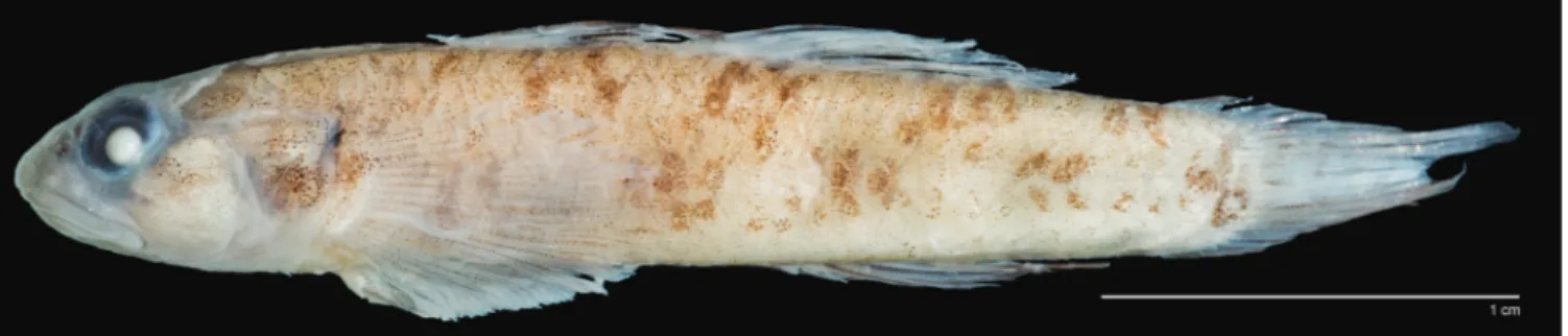
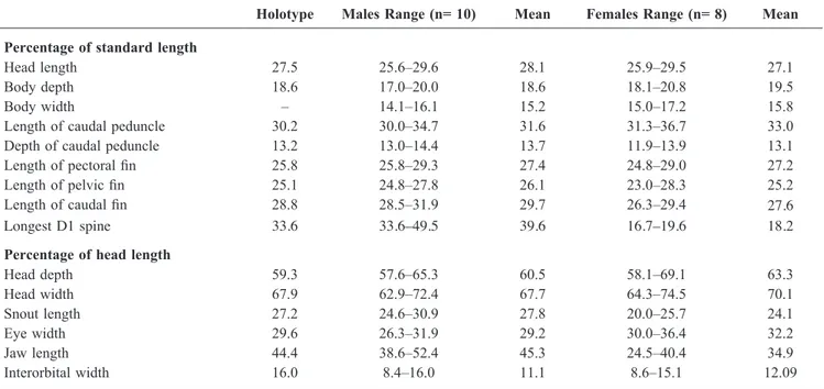
Description. Based on 18 specimens, 18–33.5 mm SL. An
asterisk indicates counts of holotype (Fig. 1). See Table 1 for morphometric data.
First dorsal-fin VI*; second dorsal-fin I,6*; anal-fin I,6*, pectoral-fin rays 16–18* (modally 18), segmented caudal-fin rays 16–17*; caudal-fin ray pattern 9/8; branched caudal-fin rays 11–14* (modally 7/7); longitudinal scale count 21–24* (modally 23*); TRB 6–8 (modally 7*); predorsal scale count 5*–6; circumpeduncular scales 12*.
Body compressed posteriorly, rounded anteriorly. Body depth at anal-fin origin 17.0–20.8% (mean 19.1%) of SL. Head length 25.6–29.6% (mean 27.6%) of SL; wider than deep, but not flattened or depressed even in large males. Head depth at posterior preopercular margin 57.9–69.1% (mean 62.1%) of HL; head width at posterior preopercular margin 62.9–74.5% (mean 69.1%) of HL; cheeks may be inflated in adult males. Mouth terminal, slightly oblique; jaws enlarged in males, reaching back to below mid-eye; females with jaws reaching to below front half of eye. Lips smooth, no fleshy fimbriae on inner edge of upper lip; lower lip free at sides, fused across anterior edge, with slightly raised fleshy symphysis. Upper jaw 24.5–52.5% (mean 34.9% in females, 45.3% in males) of HL. Eyes dorsolateral, forming part of dorsal profile, 26.3–36.4% of HL. Snout short, rounded and fleshy, curving anteriorly to slightly overhang upper lip in large specimens, 20.0–30.9% (mean 26.1%) of HL. Interorbital very narrow, 8.4–16.0% (mean 11.3%) of HL. Caudal peduncle long, compressed, length 30.0–36.7% (mean 32.3%) of SL. Caudal peduncle depth 11.9–14.4% (mean 13.4%) of SL.
First dorsal fin triangular, first spine always longest in males, spine elongate and filamentous, reaching well past insertion of second dorsal fin when depressed; females without elongate first dorsal fin spines, usually first spine longest; fin barely reaching second dorsal fin origin when depressed. First dorsal-fin spine 33.6–49.5% (mean 39.6%) in males, 16.7–19.6% (mean 18.2%) in females, of SL. Second dorsal-fin spine longest in one female, length 17.3% of SL. Second dorsal and anal fins short-based, posteriormost fin rays longest, pointed, rays falling well short of caudal fin base when depressed. Pectoral fin slender, pointed, central rays longest, 24.8–29.3% (mean 27.4%) of SL; fin rays branched but for two or three uppermost. Pelvic fins slender to oval, reaching to or just past anus, 23.0–28.3% (mean 25.7%) of SL. Caudal fin relatively short, rounded to slightly pointed posteriorly, 26.3–31.9% (mean 28.8%) of SL.
No distinct mental frenum, but chin smooth, with rounded to almost triangular fleshy pad behind symphysis (most developed in males). Anterior nostril placed just above preorbital edge, in very short tube. Posterior nostril rounded, placed just above front centre margin of eye. Gill opening moderate, usually extending forward to just under opercle. Inner edge of pectoral girdle smooth with no ridge or flange. Tongue short, tip usually blunt (rarely slightly concave to rounded). Teeth in both jaws small, curved and sharp, in 2–4 rows across front and only one row at side; outermost row teeth at front of jaws largest, curved and pointed (teeth larger in males and usually in more rows at front).
Predorsal scales moderately large, evenly sized, reaching to close behind eyes; anteriormost scale broader than those posterior to it. Operculum with 2–3 rows of large cycloid scales. Cheek naked. Pectoral-fin base covered with few large cycloid scales. Prepelvic area covered with cycloid scales. Belly covered with cycloid or ctenoid scales, may have some ctenoid scales near anus. Ctenoid scales on side of body extending forward to pectoral-fin base. Anteriormost scales on body larger than those posteriorly.
Genital papilla in male elongate, flattened, narrowing toward tip; in female, papilla rounded and bulbous.
Head pores present (Fig. 2). Pair of nasal pores, pair of anterior interorbital pores, a median posterior interorbital pore, postorbital and infraorbital pore behind each eye, oculoscapular canal and pore over preoperculum and three preopercular pores.
Sensory papillae pattern longitudinal, as in Fig. 2. Cheek rows a, c and cp composed of large, widely spaced papillae, and rows b and d of very small, closely spaced papillae. Two to three s rows on snout, of one papilla each (upper
row by nostril may have two papillae); middle row often absent. Mandibular f row of two papillae, one on each side of fleshy frenum/pad on chin.
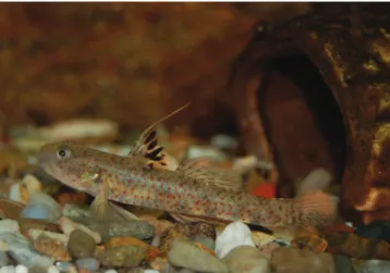
Colouration of live material. Based on photographs of
captive and fresh dead specimens (Figs. 3–6). Head and body translucent pearly grey, ventrally whitish, body and nape scattered with small rounded orange spots; in some individuals, spots smaller posteriorly and more brownish-orange on dorsum and nape; scales on dorsal third of body with very thin dark brown margins. Side of head with scattered small orange spots; spots on snout brownish-orange. Upper part of cheek, mid-opercle and most of pectoral-fin base with fine opalescent speckles. Underside of head white with distinct black patch on chin and tip of lower jaw. Some photographs show a diffuse blackish area along anterior part of pectoral-fin base. First dorsal fin transparent, with first spine whitish and a few brown to black spots along edge, nearly half of fin occupied by black to dark brown vertical blotches extending out to fin margin (blotches smaller and Table 1. Morphometrics of Pseudogobiopsis lumbantobing n. sp.
Holotype Males Range (n= 10) Mean Females Range (n= 8) Mean Percentage of standard length
Head length 27.5 25.6–29.6 28.1 25.9–29.5 27.1
Body depth 18.6 17.0–20.0 18.6 18.1–20.8 19.5
Body width – 14.1–16.1 15.2 15.0–17.2 15.8
Length of caudal peduncle 30.2 30.0–34.7 31.6 31.3–36.7 33.0
Depth of caudal peduncle 13.2 13.0–14.4 13.7 11.9–13.9 13.1
Length of pectoral fin 25.8 25.8–29.3 27.4 24.8–29.0 27.2
Length of pelvic fin 25.1 24.8–27.8 26.1 23.0–28.3 25.2
Length of caudal fin 28.8 28.5–31.9 29.7 26.3–29.4 27.6
Longest D1 spine 33.6 33.6–49.5 39.6 16.7–19.6 18.2
Percentage of head length
Head depth 59.3 57.6–65.3 60.5 58.1–69.1 63.3 Head width 67.9 62.9–72.4 67.7 64.3–74.5 70.1 Snout length 27.2 24.6–30.9 27.8 20.0–25.7 24.1 Eye width 29.6 26.3–31.9 29.2 30.0–36.4 32.2 Jaw length 44.4 38.6–52.4 45.3 24.5–40.4 34.9 Interorbital width 16.0 8.4–16.0 11.1 8.6–15.1 12.09
Fig. 2. Sensory pores and papilla pattern in Pseudogobiopsis lumbantobing. Composite drawing based on holotype and two other male specimens (MZB-BIF 4229 and MZB-BIF 4856). Drawing by Helen Larson.
more brownish in females); one photograph of a male shows the fin mostly translucent pinkish-orange to reddish, grey along base, with greyish blotches on outer part of fin membrane. Second dorsal fin transparent with about 3–6 rows of dusky yellow to pale brown spots on spines, fin margin transparent to pinkish. Caudal fin translucent, with 5 or 6 irregular vertical bands or rows of irregular spots of pale pinkish brown, bands breaking up into spots ventrally and fading posteriorly; one adult male has most of caudal fin pinkish-orange with whitish centre, and rows of brownish spots. Anal fin whitish to pink; one adult male displaying pinkish-orange fin rays with whitish membrane. Pectoral fin transparent, whitish near base. Pelvic fins translucent whitish to transparent.
Colouration of preserved material. In males, head and body
whitish to pale speckled yellowish, white on ventral third, with five elongate, rectangular, diffuse dark grey blotches along midside of body, posteriormost blotch just before hypural crease and may be darker than others; body with scattered small grey-margined pale spots, becoming darker and arranged more regularly on lower part of body (Figs. 1, 7). Five diffuse dusky grey saddles usually visible. Nape and dorsum often with scale margins greyish, giving net-like appearance. Pectoral fin base with dense black irregular blotch on upper portion, blotch may partly extend upward onto body and under opercle toward pectoral girdle. Ventral midline of caudal peduncle with four evenly spaced, partly internal, narrow black blotches present. Belly with partly internal black spot just behind the anus in both sexes. Male urogenital papilla pale to dark grey.
Head dusky grey dorsally with short dark grey triangular to linear mark extending from front of eye to almost straight back to end on cheek well above corner of mouth; second dark grey broad bar extending from rear part of eye obliquely across cheek. Small blackish blotch around and including posterior nostril by eye. Opercle with large rounded grey blotch or horizontal bar across centre. Upper lip plain dusky grey, lower lip paler in most specimens. Chin with black blotch on mental frenum; black pigment extending up to centre of lower lip. Breast and belly whitish to faintly greyish.
First dorsal fin in males grey to blackish with two to three rows of black spots on spines and fin membranes (spots on membrane joining to form irregular blackish rows), first dorsal-fin spine mostly blackish. Second dorsal fin dusky grey, with 5–7 rows of blackish spots forming rows, becoming darker posteriorly. Anal fin plain dark grey, blackish posteriorly. Caudal fin translucent to dusky with 7–12 rows of small dark grey spots or fine irregular dusky bars. Pectoral fin translucent to dusky grey, darker ventrally. Pelvic fins and frenum dusky grey.
Females similar to males but paler and markings less pronounced. Black blotch on pectoral fin base smaller, less distinct; first dorsal fin transparent with large black blotch on rear third of fin; mental frenum paler, never black; pelvic fins transparent; caudal fin with 3–5 rows of brownish grey spots coalescing to form irregular oblique bands; urogenital papilla white.
Comparisons. This species is most similar to P. oligactis,
differing in that P. lumbantobing has a pale body scattered with small orange spots when live; has a grey to blackish blotch on the chin; a black blotch on the upper part of pectoral fin base; and the snout is rounded over the anterior part of the upper lip (snout inflated in males) versus P. oligactis Fig. 3. Captive specimen of Pseudogobiopsis lumbantobing,
supposedly imported from Myanmar. Photograph by Christophe Maillet.
Fig. 4. Captive specimen of Pseudogobiopsis lumbantobing. Photograph by Thomas Ackerman.
Fig. 5. Captive specimen of Pseudogobiopsis lumbantobing, imported via Singapore. Photograph by Emma Turner.
which has a greyish body with internal grey blotches when live; the lips are plain dusky grey without a black blotch on the chin; the pectoral-fin base is grey but without a distinct black blotch; and the snout is pointed and flat. See Larson (2009) for comparison with congeners.
Distribution. Known only from freshwater drainages
of western Sumatra and western Java, Indonesia. Some specimens in the aquarium trade are said to be imported from Myanmar (Fig. 3), but this is yet to be confirmed.
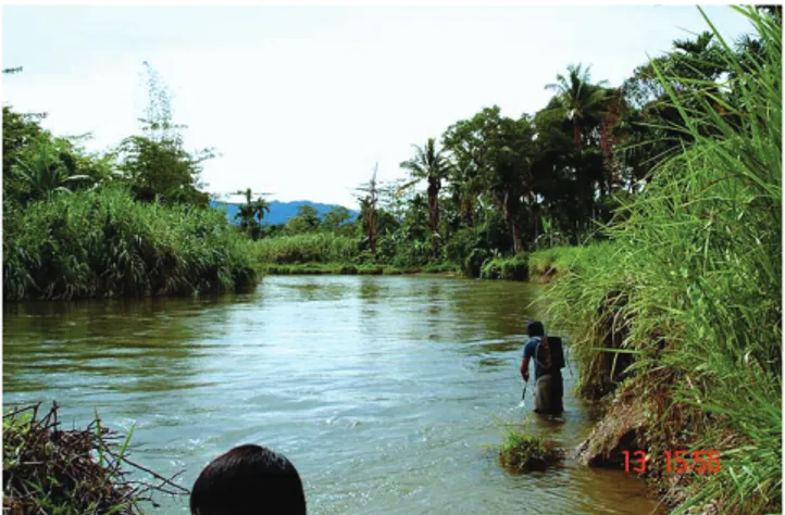
Ecology. Freshwater, found in rivers and streams at altitudes
of 5–22 m, with a substratum of sand, gravel, rock and boulders; algae and aquatic macrophytes may be present (Figs. 8, 9).
Etymology. This species is named for Daniel Lumbantobing
of Jakarta, who collected the first specimens and showed them to HKL in 2012, which solved the mystery of the orange-spotted goby of which aquarists had been sending her photos. Daniel is an ichthyologist who specialises in freshwater fishes. Name used as a name in apposition.
Remarks. The identity of a Kuhl and van Hasselt watercolour
painting, done during 1820–1823, of a goby from Java, illustrated in Roberts (1993), has been a mystery for some
time (Larson, 2009: 162). Now that specimens are available, we conclude from the body and fin shape and colour pattern, including the dark grey chin (Roberts, 1993: Fig. 51), that the figure is of P. lumbantobing. It was hoped to reproduce the painting here, but access to the Naturalis Library is not possible until 2018 (undergoing renovation).
It should also be noted that in Larson (2009: Plate B) that the photograph of P. oligactis in Plate 2B was inadvertently attributed to Gerhard Ott; it is by Rainer Hoyer.
REVISED KEY TO SPECIES OF PSEUDOGOBIOPSIS
1. Preopercular pores absent, pores on top of head present or absent ...4 – Preopercular pores present, pores on top of head always
present ...2 2. Dorsal-fin rays I,7, and anal-fin rays I,6–7; 15–16 pectoral-fin rays; body with 12-13 narrow vertical blackish lines; no elongate first dorsal fin spines ... ...P. tigrellus (Nichols, 1951) (Papua New Guinea) – Dorsal and anal fin rays modally I,6; 16–19 pectoral-fin rays;
first dorsal-fin spine usually elongate in males ...3 3. Body pale, scattered with small orange spots when alive; lips
pale with dusky grey to black blotch on chin; first dorsal fin with Fig. 6. Freshly dead male (upper) and female (lower) specimens
of Pseudogobiopsis lumbantobing (MZB-BIF 4856 and MZB-BIF 5859). Photograph by Nicolas Hubert.
Fig. 7. Preserved specimens of Pseudogobiopsis lumbantobing, ZRC 55524 (both males, upper 31.4 mm SL, lower 28.0 mm SL; not to scale). Photograph by Tan Heok Hui.
Fig. 8. Sungei Kluet, Sumatra. Photograph by Daniel Lumbantobing.
Fig. 9. River at Cibeber, Kabupaten Pandeglang, Banten Province. Photograph by Nicolas Hubert.
row of black bars (forming broken stripe) on membranes near margin; black blotch on upper part of pectoral-fin base; snout rounded over anterior part of upper lip (inflated in males) ... ... P. lumbantobing (Indonesia) – Body with internal dusky grey blotches when live; lips plain
gey without distinct black blotch on chin; first dorsal fin with two dusky sgrey tripes and may have blackish spot posteriorly; pectoral fin base dusky grey but without distinct blotch; snout pointed and flat in males ...P. oligactis (Bleeker, 1875) (Thailand, Malaysia, Singapore, Indonesia)
4. Headpores present; pectoral-fin rays 17–19; cheeks, preorbital and upper lip crossed with rows of irregular, oblique, distinct black lines ...P. festivus Larson, 2009 (Malaysia). – Head pores absent; pectoral-fin rays 13–16; side of head with
dark spots and blotches, forming two broad bars, one across preorbital and one to end of jaws ... ...P. paludosus (Herre, 1940) (Malaysia, Indonesia)
ACKNOWLEDGEMENTS
We wish to thank Philippe Keith and Frédéric Busson of Muséum National d’Histoire Naturelle, and Jean-Paul Toutain and Jean-François Agnèse for their support, and thank Bambang Dwisusilo, Sumanta, Ujang Nurhaman and Daisy Wowor for their help during field sampling. Thanks also to Sopian Sauri and Hadi Darhuddin of MZB for their work with the specimens and in the field. Part of the present study was funded by the MNHN (UMR 7208 BOREA), the ‘Institut de Recherche pour le Développement’ (UMR ISEM), the Indonesian Institute of Sciences (LIPI), the French Ichtyological Society (SFI) and the Fondation de France. This study has been approved by the Indonesian Ministry of Research and field sampling was conducted according to the research permits 097/SIP/FRP/SM/IV/2014 for Philippe Keith, and research permit 41/EXT/SIP/FRP/SM/VIII/2014 for Nicolas Hubert. We wish to thank MENRISTEK staff
as well as Mohammad Irham, Rosichon Ubaidillah, Hari Sutrisno and Witjaksono (LIPI) for research permits and supporting letters. And thanks to reviewers Lynna Parenti and Kelvin Lim, for their constructive comments.
Daniel Lumbantobing collected his specimens with the assistance of Deden Rudaya and Ni Made Ray during 2006 fieldwork financially supported by the Leonard P. Schultz Fund, Division of Fishes, Smithsonian National Museum of Natural History Museum, Washington, DC.
Our thanks to Sandra Raredon (USNM), who photographed the holotype. And thank you to the aquarists who sent HKL photographs of living P. lumbantobing, which kick-started the quest for its identity: Thomas Ackerman, Christophe Maillet, Angela Schneider and Emma Turner. Thanks to Tan Heok Hui and Kelvin Lim, for additional material and photographs. This publication has ISEM number 2017-040 SUD.
LITERATURE CITED
Hubbs CL & Lagler KF (1958) Fishes of the Great Lakes Region. University of Michigan Press, Ann Arbor, 213 pp.
Larson HK (2009) Review of the gobiid fish genera Eugnathogobius and Pseudogobiopsis (Gobioidei: Gobiidae: Gobionellinae), with descriptions of three new species. Raffles Bulletin of Zoology, 57(1): 127–181.
Roberts TR (1993) The freshwater fishes of Java, as observed by Kuhl and van Hasselt in 1820–23. Zoologische Verhandelingen, 285: 1–94.
Sabaj Pérez MH (2014) Standard symbolic codes for institutional resource collections in herpetology and ichthyology: an online reference. Version 5.0 (22 September 2014). American Society of Ichthyologists and Herpetologists, Washington, D.C. http:// www.asih.org/. (Accessed 17 February 2017).
Sanzo L (1911) Distribuzione delle papille cutanee (organi ciatiformi) e suo valore sistematico nei Gobi. Mitteilungen aus der Zoologischen Station zu Neapel, 20: 249–328.