HAL Id: tel-03246771
https://tel.archives-ouvertes.fr/tel-03246771
Submitted on 2 Jun 2021HAL is a multi-disciplinary open access archive for the deposit and dissemination of sci-entific research documents, whether they are pub-lished or not. The documents may come from teaching and research institutions in France or abroad, or from public or private research centers.
L’archive ouverte pluridisciplinaire HAL, est destinée au dépôt et à la diffusion de documents scientifiques de niveau recherche, publiés ou non, émanant des établissements d’enseignement et de recherche français ou étrangers, des laboratoires publics ou privés.
Interaction between the influenza replication
machineries and the cellular nuclear import system
Amelie Donchet
To cite this version:
Amelie Donchet. Interaction between the influenza replication machineries and the cellular nuclear import system. Biomolecules [q-bio.BM]. Université Grenoble Alpes [2020-..], 2021. English. �NNT : 2021GRALV003�. �tel-03246771�
Remerciements
Je tiens à remercier tous ceux qui m’ont accompagnée lors de cette aventure intense, palpitante et riche en enseignements.
Je souhaite tout d’abord remercier Thibaut Crépin, mon directeur de thèse, pour sa confiance, ses conseils, et le précieux temps qu’il m’a toujours accordée. Merci de m’avoir guidé sur ce chemin ces quatre années. Merci d’avoir relevé ce défi et de m’avoir permis de devenir une vraie biochimiste !
Je veux également remercier Rob Ruigrok, pour tous les riches échanges que l’on a pu avoir, pour ses conseils, sa présence, sa bienveillance et son soutien.
Merci à Laura, Christopher et Alice, pour m’avoir accueillie, formée et présentée leurs projets. Merci de m’avoir mis le pied à l’étrier !
Un grand merci à tous les membres du laboratoire, pour tous les bons moments. Plus particulièrement, merci à Jean-Marie, Francine, Pascal, Emilie, Philippe, Nicolas, Caroline pour leur temps, pour toutes nos discussions, scientifiques et un peu moins, et pour leur aide très précieuse.
Je souhaite aussi adresser un merci spécial à Carla "Carliña" et Nicolas "Jojo" sans qui ces années au laboratoire auraient été bien différentes et bien plus pâles. Merci pour ces bouffées d’oxygène, pour les rires et les blagues, pour le soutien essentiel lorsque la science faisait de la résistance !
Merci à tous les petits piou-pious qui sont passés en stage au laboratoire (et particulièrement à Mai, Justine et Bastien pour leur aide) et aux grands piou-pious qui ont commencé leur thèse. Les prochains, c’est vous ! ;)
Je souhaite remercier également Antoine (parmi tant d’autres, le Atlas de mon P-body, qui défie littéralement les lois de la probabilité et retire cruellement le pont sous ses pieds !), Audrey (ma force tranquille dans le chaos). Il n’y a pas besoin de mot, je pense, car cela va au-delà de ça, pour te remercier Alice. Merci pour tout.
Merci aux copains de promo, aux thésards d’ici et d’ailleurs, à la team Glob’Alps et BIOTechno pour tous les bons instants.
Merci à mes parents qui m’ont permis d’arriver jusqu’ici de la meilleure des façons, et à toute la famille qui a toujours essayé de comprendre ce que je pouvais bien faire au labo, mais qui a adoré les photos de cristaux et de protéines fluo !
Merci d’avoir fait un bout de chemin avec moi, et quel chemin ! Thank you all!
Amélie
A mes étoiles,
List of abbreviations... 9 List of figures ... 13 List of tables ... 17 Résumé détaillé ... 21 Chapter 1: Introduction ... 31 I. Influenza viruses ...33
A. Features of influenza viruses ...33
i. Taxonomy ... 33
ii. Viral particle structure ... 34
iii. The ribonucleoproteins, the replicative units of the virus ... 37
iv. Genome organization, protein content and host tropism ... 37
v. Evolution: antigenic drift and antigenic shift ... 42
vi. Disease and epidemiology ... 43
B. Influenza virus cycle ...45
C. Viral transcription and replication machinery ...48
i. Nucleoprotein ... 48
ii. Heterotrimeric RNA-dependent RNA-polymerase ... 51
II. Nucleocytoplasmic shuttling of proteins ...56
A. Nuclear transport pathways ...57
B. The nuclear transport pathway based on Ran and karyopherins ...58
i. The Ras-like small GTPase Ran ... 58
ii. The nuclear import and export: importins-α, importins-β, and exportins ... 58
iii. The nuclear localization and export signals ... 60
C. Similarities and specificities of importins-α ...61
D. In vitro and in cellulo interaction parameters of the complex between cargo and importins: affinity and rates ...66
E. Nuclear import regulation through cellular machinery ...67
i. Importin-α:importin-β1 sub-complex versus importins-β as transport receptors ... 67
ii. Post-translational modifications ... 68
iii. Other regulatory mechanisms ... 69
III. Nuclear import of the influenza replication machinery ...70
A. Nucleoprotein ...70
B. Heterotrimeric RNA-dependent RNA polymerase ...73
C. Ribonucleoproteins ...74
Objectives of the PhD thesis ... 77
Chapter 2: Structural analysis of the influenza D nucleoprotein and characterization of its nuclear localization signal ... 81
Chapter 3: Comparative analysis of the interaction involving each influenza nucleoprotein types and importins-α involved in influenza nuclear import ... 99
Chapter 4: Structural and biochemical characterization of the Asiatic toad influenza NP ... 119
I. Characterization of the interaction with importins-α ... 122
II. Characterization of the interaction with RNAs ... 125
III. Structural characterization of Toad/NP ... 128
IV. Discussion ... 131
Conclusions ... 137
Annexes ... 141
Annexe 1: Sequence identities between importins-α ... 142
Annexe 2: Conservation of NPTAIL within each influenza type ... 143
Annexe 3: Protein accession numbers ... 147
Annexe 4: Supplementary Data Chapter 2 ... 148
Annexe 5: Supplementary Data Chapter 3 ... 154
List of abbreviations
9
List of abbreviations
List of abbreviations
11 ARM repeats: Armadillo repeats
BLAST: Basic Local Alignment Search Tool BRD: Bovine Respiratory Disease
CAS: Cellular Apoptosis Susceptibility
CCP4: Collaborative Computational Project Number 4 CCP4i: CCP4 Interface
cNLS: conventional/classical Nuclear Localization Signal COOT: Crystallographic Object-Oriented Toolkit
Crm1: Chromosome region maintenance 1 protein cRNA: complementary RNA
CTD: C-terminal domain DNA: Deoxyribonucleic Acid EM: Electron Microscopy
EMBL: European Molecular Biology Laboratory
ESCRT: Endosomal Sorting Complex Required for Transport FA: Fluorescence Anisotropy
FAM: Fluorescein
FG-repeats: phenylalanine-glycine repeats GDP: Guanosine DiPhosphate
GTP: Guanosine TriPhosphate HA: Hemagglutinin
HEAT repeats: Huntington, Elongation factor 3, PR65/A, TOR HEF: Hemagglutinin-Esterase-Fusion
HPAIs: Highly Pathogenic Avian Influenza viruses HSQC: Heteronuclear Single Quantum Correlation HTX: High Throughput Crystallization laboratory IAV: Influenza A Viruses
IBB domain: Importin-β Binding domain IBV: Influenza B Viruses
ICTV: International Committee on Taxonomy of Viruses ICV: Influenza C Viruses
IDV: Influenza D Viruses
IPTG: isopropyl-β-D-thiogalactopyranoside IRD: Influenza Research Database
Isavirus: Infection salmon anemia virus ISBG: Integrated Structural Biology Grenoble Kd: Dissociation constant
kDa: kilo Daltons
LPAIs: Low-Pathogenic Avian Influenza viruses M1: Matrix protein 1
M2: Matrix protein 2
MAD: Multi-wavelength Anomalous Dispersion MDa: Mega Daltons
mRNA: messenger RNA MW: Molecular Weight
MxA: Human myxovirus resistance protein 1 NA: Neuraminidase
NCBI: National Center for Biotechnology Information
ncNLS: non-conventional/non-classical Nuclear Localization Signal NEP: Nuclear Export Protein
NES: Nuclear Export Signal Neu5Ac: N-Acetylneuraminic acid NLS: Nuclear Localization Signal
List of abbreviations
12 NMR: Nuclear Magnetic Resonance
NP: Nucleoprotein
NPC: Nuclear Pore Complex NS1: Non-Structural protein 1 PA: Polymerase Acidic protein PB1: Polymerase Basic protein 1 PB2: Polymerase Basic protein 2 PEG: PolyEthylene Glycol
pH: potential of Hydrogen pHi: isoelectric point
PolII: DNA-dependent RNA polymerase II PTM: Post-Translational Modification
PY NLS: Proline-Tyrosine rich Nuclear Localization Signal RanBP1: Ran-Binding Protein 1
RanBP5: Ran-Binding Protein 5
RanGAP: Ran GTPase-Activating Protein
RCC1/RanGEF: Regulator of Chromosome Condensation 1 / Ran Guanine nucleotide Exchange Factor
RdRp: RNA-dependent RNA polymerase RNA: Ribonucleic Acid
RNP: Ribonucleoprotein
SAXS: Small Angle X-rays Scattering SEC: Size Exclusion Chromatography
SEC-MALLS: Size Exclusion Chromatography coupled with Multi Angle Laser Light Scattering
SNP: Single Nucleotide Polymorphism SPR: Surface Plasmon Resonance
STEM: Scanning Transmission Electron Microscopy SUMO: Small Ubiquitin-like Modifier
TEV: Tobacco Etch Virus
ToadV: Wuhan Asiatic Toad influenza Virus TEM: Transmission electron microscopy vRNA: viral RNA
WHO: World Health Organization YFP: Yellow Fluorescent Protein
List of figures
13
List of figures
List of figures
15
Figure 1: Viral particle structure and genome organization. ...35
Figure 2: Main and accessory proteins encoded by influenza A viral mRNAs. ...38
Figure 3: Host tropism of influenza viruses. ...39
Figure 4: Structure of influenza HA, HEF, NA, M2 and M1 proteins. ...41
Figure 5: Major historical outbreaks in the shared human and influenza history. ...44
Figure 6: Influenza viral cycle. ...46
Figure 7: Biochemical features of influenza nucleoproteins. ...49
Figure 8: Influenza nucleoprotein structures and structural features. ...50
Figure 9: Structure of Isavirus NP. ...51
Figure 10: Bat IAV, IBV, ICV and IDV RdRp structures. ...52
Figure 11: Reconstituted model of influenza RdRp transcription. ...53
Figure 12: Replication of the influenza genome. ...54
Figure 13: Nuclear pore complex (NPC). ...56
Figure 14: The nuclear transport pathway based on Ran. ...59
Figure 15: Evolution, diversification and global organization of importins-α. ...62
Figure 16: Structure of importins-α. ...63
Figure 17: Diversity of the roles played by importins-α in cells. ...65
Figure 18: Summary of the nuclear import of the influenza replication machinery. ...72
Figure 19: Toad/NP constructs. ... 121
Figure 20: Profiles of the SEC purification step for each Toad/NP constructs. ... 122
Figure 21: Interaction assays between Toad/NPconstructs and human importin-α7 by size exclusion chromatography. ... 123
Figure 22: Characterization of the interaction between Toad/NPTAIL-YFP and the human importin-α7 by SPR. ... 124
Figure 23: Titration against FAM-labelled RNAs of Toad/NPFULL and Toad/NPCORE by fluorescence anisotropy at 150 mM NaCl. ... 126
Figure 24: 1H-15N HSQC spectra of 15N-labelled Toad/NP TAIL. ... 129
Figure 25: Crystals of Selenomethionine-labelled Toad/NPTAIL and Toad/NPTAIL solved structure. ... 130
Figure 26: Crystals of Selenomethionine-labelled Toad/NPCORE and its crystallization condition. ... 131
Figure 27: Structural features and comparison between Toad/NP and B/NP. ... 132
Toad/NP N-terminal structure is a result of this PhD work whereas B/NP modelling was published by 177. ... 132
17
19
Table 1: Full taxonomy of the Orthomyxoviridae family members. ...33
Table 2: Summary of the influenza main features depending on the virus type. ...36
Table 3: Sequence identities between influenza types. ...48
Table 4: Reference NLS sequences and consensus sequences for 6 classes of conventional and nonconventional basic NLSs interacting with importins-α. ...60
Table 5: NLS sequences of the influenza A replication machinery. ...70
Table 6: Sequence identities between Toad/NP and the other influenza NPs. ... 120
Table 7: Toad/NPTAIL sequences. ... 121
Table 8: Biacore steady state affinities between Toad/NPTAIL or B/NPTAIL and human ΔIBB importins-α. ... 125
Table 9: Toad/NP:RNA apparent dissociation constants determined by fluorescence anisotropy. ... 127
Table 10: Sequence identities between influenza NP types. ... 134
Table S1: Sequence identities between importins-α paralogs of distinct species of interest. ... 142
Table S2: Consensus residues and frequencies of A/NPTAIL. ... 143
Table S3: Consensus residues and frequencies of B/NPTAIL. ... 144
Table S4: Consensus residues and frequencies of C/NPTAIL. ... 145
Table S5: Consensus residues and frequencies of D/NPTAIL. ... 146
Résumé détaillé
21
Résumé détaillé
Résumé détaillé
23
Les virus grippaux font partie de la famille des Orthomyxoviridae. Cette famille comporte sept genres de virus, dont les virus de la grippe de type A, B, C et D (IAV, IBV, ICV et IDV). Cette classification est destinée à évoluer, car la recherche n’a fait qu'effleurer la surface de la diversité virale : une étude de 2018 a identifié 3 nouveaux membres de la famille des Orthomyxoviridae, notamment un virus de crapaud à Wuhan. Ces trois virus présentent des identités de séquence plus importantes avec IBV qu’avec les autres virus grippaux. Le virus de crapaud, étudié durant ce travail de thèse, sera appelé ToadV. Les virus grippaux possèdent une solide base commune en terme de structure et de fonctionnement, mais ils ont également de claires spécificités.
Les virus grippaux sont des virus enveloppés, à génome ARN segmenté, simple brin et de polarité négative. L'unité réplicative du virus, appelée ribonucléoprotéine (RNP), est composée du segment génomique, encapsidé par de multiples copies de la nucléoprotéine (NP) et associé par ses extrémités 3' et 5' à une ARN polymérase ARN dépendante (RdRp) virale hétérotrimérique. NP est la protéine la plus abondante des RNPs et elle est nécessaire aux activités de transcription et de réplication virales. NP est également impliquée dans la protection du génome viral, dans l'interaction avec les partenaires cellulaires et dans le trafic cellulaire des RNPs. NP est une protéine virale centrale, dont les fonctions clés sont conservées entre les NPs de IAV, IBV, ICV et IDV (A/NP, B/NP, C/NP et D/NP) mais avec des spécificités structurales et fonctionnelles dépendantes du type viral.
Les ions et les petites molécules (< 30-60 kDa) diffusent librement dans le noyau des cellules. Cependant, les protéines plus grosses nécessitent d’être importées par transport actif. La voie la plus étudiée est le système impliquant la petite GTPase Ran et les récepteurs de transport nucléaire appelés importines-α et -β. Les importines-α, agissant comme des adaptateurs, interagissent avec un signal spécifique de la protéine à importer (cargo), appelé signal de localisation nucléaire (NLS), et avec l’importine-β, via son domaine d’interaction avec l’importine-β (domaine IBB). Ce domaine IBB, en absence de l’importine-β, agit comme auto-inhibiteur en se repliant dans les deux sillons de l’importine-α où les NLSs peuvent se fixer. Certaines importines-β n’ont pas besoin d’adaptateurs et peuvent directement interagir avec le NLS du cargo. Il existe deux types de NLS, les NLS classiques/conventionnels et les NLS non classiques/non conventionnels. Un NLS est généralement riche en Arginine
Résumé détaillé
24
et en Lysine, et généralement composé d’un ou deux motifs basiques (NLS monopartite ou bipartite respectivement). Les importines sont très conservées au cours de l’évolution. Chez l’homme, il y a sept paralogues d’importines-α et au moins dix importines-β responsables de l’import nucléaire. Cette diversité s’accompagne d’une spécificité de cargos, de comportement et d’expression dans les cellules et tissus : les importines ne sont pas interchangeables.
Les virus grippaux font partie des rares virus à ARN à répliquer et transcrire leur génome dans le noyau de la cellule hôte. La machinerie réplicative ainsi que le génome ont donc besoin d’être importés dans le noyau, et pour cela détournent la machinerie des importines-α et -β. PB1, PB2 et NP possèdent des NLS pour interagir avec les importines : PB2 et NP avec les importines-α, PB1 avec une importine-β RanBP5, et PA est essentielle pour l’import efficace de PB1. L’import initial du génome dans le noyau sous forme de RNPs est supposé être médié par la NP, un autre des rôles essentiels de cette protéine virale dans le trafic viral.
Le NLS fonctionnel de A/NP et B/NP a été identifié sur l’extrémité intrinsèquement désordonnée N-terminale d’une longueur de 21 et 71 acides aminés respectivement (A/NPTAIL et B/NPTAIL). Le NLS de A/NP appartient aux NLS non conventionnels,
interagit avec les importines-α1, -α3, -α5 et -α7, notamment avec une faible affinité pour l’importine-α1 (constante de dissociation entre 1 et 5 µM). De nombreux motifs basiques sont présents sur B/NPTAIL et les résidus 30 à 71 ont été identifiés comme
participant à l’interaction avec l’importine-α7, qui est caractérisée par une constante de dissociation de 844 nM. C/NP et D/NP, de leur côté, présentent une longue extrémité intrinsèquement désordonnée de 62 et 52 acides aminés respectivement, mais en C-terminale. C/NPTAIL et D/NPTAIL possèdent des motifs basiques qui
ressemblent clairement à des NLS conventionnels bipartite. Des travaux publiés durant ce travail de thèse par une équipe concurrente ont montré que C/NPTAIL interagit
avec les importines-α1, -α3, -α4, -α5 and -α7, et son affinité pour l’importine-α1 est de 50 nM.
Au début de ce travail de thèse, la caractérisation de la machinerie réplicative des virus de type D, et plus particulièrement de D/NP, était déjà initiée par l’équipe, en parallèle de ses travaux sur A/NP et B/NP. Ces dernières présentaient des affinités pour les importines-α trop faibles pour être considérées complétement fonctionnelles.
Résumé détaillé
25
L’existence de ToadV et de sa NP n’était pas encore connue. Malgré le travail effectué sur les NPs grippales et leurs interactions avec les importines-α, de nombreuses questions restaient en suspens, d’autant plus que le mécanisme de fonctionnement d’une NP, ainsi que ses caractéristiques structurales et biochimiques, ne sont pas forcément transposables aux autres. Il a été décidé de caractériser dans un premier temps D/NP, structuralement ainsi que dans ses fonctions clés. Par la suite, dans l’optique de pouvoir comprendre comment les différents types grippaux accédaient au noyau et en quoi leurs stratégies convergeaient et/ou divergeaient, une stratégie expérimentale a été mise au point dans le but de pouvoir comparer aussi fiablement que possible les différentes NPTAILS et les différentes importines-α humaines. Enfin,
avec la découverte inédite du virus grippal de crapaud, proche phylogénétiquement du virus de type B strictement humain, il a semblé judicieux d’initier sa caractérisation, aussi bien structurale que fonctionnelle, afin d’intégrer une vision évolutive aux fonctions cœur de la NP grippale et de mieux comprendre le fonctionnement de chaque NP individuellement.
Découvert en 2011 et classifié comme un nouveau genre grippal en 2013, le virus de la grippe de type D circule largement au niveau mondial chez les bovins. Peu de choses sont connues sur les protéines virales et son cycle.
A l’aide de la diffraction des rayons-X, de la microscopie électronique, du dichroïsme circulaire, de la chromatographie d’exclusion de taille couplée ou non à la diffusion de la lumière à plusieurs angles, de tests de stabilisation thermique, de l’anisotropie de fluorescence et de la résonance des plasmons de surface, la caractérisation de D/NP, à savoir ses activités d’interactions avec l’ARN et avec l’importine-α7, son comportement oligomérique et sa structure, a été réalisée.
D/NP forme principalement des tétramères en solution, et la concentration en sel n’a pas d’impact sur cet état oligomérique. La structure a été résolue à une résolution de 2.4 Å, montrant un corps replié très conservé. D/NP se localise dans le noyau de cellules infectées et/ou transfectées et, contrairement à A/NP et B/NP, D/NP possède une extrémité flexible intrinsèquement désordonnée en C-terminale, avec un évident NLS bipartite. L’interaction entre l’importine-α7 et D/NPTAIL se caractérise par une
affinité environ dix fois plus forte que celles déterminées pour A/NPTAIL et B/NPTAIL, de
Résumé détaillé
26
C-terminal du NLS conduit à une interaction défectueuse avec l’importine-α7 et une localisation nucléaire affectée, tandis que la délétion des deux motifs conduit à une abolition complète des deux mécanismes. Ces résultats ont fait l’objet d’une publication rattachée à ce travail de thèse.
Les fonctions clés de la nucléoprotéine grippale sont retrouvées avec D/NP : sa structure, son état d’oligomérisation, son NLS sur une extrémité désordonnée, sa localisation cellulaire. Cependant, chacun de ces mécanismes montrent aussi de claires spécificités, propre à D/NP.
Un large nombre d’études se sont intéressées à la caractérisation de l’interaction NP:importine-α, apportant de précieux renseignements au point de vue fonctionnel, moléculaire et atomique. Cependant, ces connaissances restaient lacunaires, autant du côté des importines-α que des NPs. Les seuls complexes étudiés dans le détail et impliquant A/NP et B/NP, A/NPTAIL:importine-α1 et B/NPTAIL:importine-α7, montraient
une faible affinité, probablement au-delà de la limite de fonctionnalité d’un NLS. Au contraire, C/NPTAIL:importine-α1 et D/NPTAIL:importine-α7 présentent des affinités
consistantes avec une activité fonctionnelle in cellulo, respectivement 50 et 100 nM. Cependant, alors que toutes les NPs grippales ont commencé à être investiguées, il y avait toujours un manque criant d’intégration des différentes importines-α, alors même qu’il est bien connu désormais qu’il s’agit d’entités uniques, spécifiques, avec leurs propres régulations et partenaires.
Grâce à la chromatographie d’exclusion de taille couplée (ou non) à la diffusion de la lumière, à la résonance des plasmons de surfaces et à l’anisotropie de fluorescence, la première stratégie comparative complète, impliquant toutes les NPTAILS déjà décrites
et toutes les importines-α, a pu être mise au point et a révélé des spécificités surprenantes. Les NPTAILS ont été fusionnées à la YFP, de manière à conserver un
contexte globulaire sans contribution de l’oligomérisation propre à la NP. Toujours capables d’interagir avec les importines, la caractérisation cinétique a pu être réalisée. Les vitesses d’interaction impliquant d’une part, les importines-α3, -α5 et -α7, et d’autre part, A/NPTAIL, C/NPTAIL and D/NPTAIL se sont révélées extrêmement rapides, tant
l’association que la dissociation. D/NPTAIL est capable d’interagir avec une forte affinité
avec toutes ces importines-α, sans spécificité identifiable. A/NPTAIL et C/NPTAIL
Résumé détaillé
27
importine-α particulière, respectivement l’importine-α7 et l’importine-α3. B/NPTAIL s’est
montrée extrêmement surprenante: les vitesses d’association et de dissociation se sont révélées encore plus rapides, et les affinités pour toutes les importines-α testées se sont avérées très faibles.
L’importine-α1, présentant une propension à dimériser en solution, n’a pas pu être testée par la stratégie mise en place. A l’aide d’un test de compétition et d’anisotropie de fluorescence, des valeurs d’affinités ont pu être obtenues et ont révélées une forte affinité pour D/NPTAIL et C/NPTAIL, une affinité basse pour A/NPTAIL et une affinité
médiocre pour B/NPTAIL.
Ainsi, alors que des différences étaient attendues dans les caractéristiques de ces complexes, elles se sont avérées encore plus importantes, avec une contribution égale de chacun des partenaires à l’interaction. Ces résultats ont fait l’objet d’une seconde publication au cours de travail de thèse. Ils établissent une base solide pour continuer à disséquer et détailler cette interaction hôte:pathogène extrêmement complexe et essentielle.
En 2018, une étude métagénomique a identifié plus de 200 nouveaux virus à génome à ARN, et notamment 3 nouveaux virus appartenant à la famille des Orthomyxoviridae. Un de ces nouveaux virus infecte un crapaud et, de façon assez surprenante, est plus proche des virus de type B que des autres virus grippaux. Sa NP (Toad/NP), de la même façon, est phylogénétiquement plus proche de B/NP que des autres NPs grippales. Une prédiction structurale a mis en évidence une architecture potentiellement similaire à celle de B/NP, avec une longue extrémité N-terminale désordonnée (Toad/NPTAIL), un corps central replié de taille similaire (Toad/NPCORE) au
autres NPs et une courte extrémité C-terminale désordonnée. Cette prédiction a aussi identifié des traits très particuliers de Toad/NPTAIL : elle pouvait potentiellement
posséder un domaine replié. De plus, longue d’environ 130 acides aminés, cette extrémité N-terminale l’est près de deux fois plus que celle de B/NP et présente de nombreux motifs basiques. Il a donc été décidé de commencer à caractériser l’interaction de Toad/NP avec les importines-α via la stratégie SPR mise en place précédemment, ainsi que son interaction avec l’ARN et sa structure. La cristallographie, la microscopie électronique, la chromatographie d’exclusion de taille,
Résumé détaillé
28
l’anisotropie de fluorescence et la résonance des plasmons de surface ont été utilisés à ces fins.
Toad/NP a tendance à former des tétramères en solution, et son état d’oligomérisation n’est pas impacté non plus par la concentration en NaCl, de façon similaire à D/NP. Toad/NPTAIL interagit avec les importines-α d’une façon très similaire
à B/NPTAIL, avec des vitesses d’association et de dissociation très rapides, hors des
limites de détection de l’appareil. Son affinité pour les importines-α5 et -α7 est très basse, hors des limites supposées d’un NLS fonctionnel, mais son affinité pour l’importine-α3 indique par contre qu’il pourrait s’agir d’une interaction fonctionnelle pour l’import nucléaire. Toad/NPCORE interagit avec l’ARN de façon similaire à A/NP et B/NP,
avec coopérativité, sans spécificité de séquence et avec une potentielle saturation du site de fixation de l’ARN avec un ARN 12-mer. Toad/NP possède en revanche une spécificité intéressante : son extrémité N-terminale semble réguler négativement l’interaction avec l’ARN, peut-être via une région riche en glutamate. La structure de Toad/NPCORE n’a pas encore été résolue à ce jour mais le travail est encore en cours.
Il est hautement probable que ce corps, comme le corps de toutes les autres NPs, soit structuralement très conservé. En revanche, des cristaux du domaine N-terminal ont permis de résoudre la structure d’un domaine replié en 3 longues hélices-α antiparallèles (acides aminés 4 à 68). Il s’agit du premier domaine replié identifié dans l’une des extrémités désordonnées des NPs des virus grippaux. Le rôle et la fonction de ces deux domaines (le domaine replié et le domaine désordonné) ne sont pas encore clairement identifiés et nécessite davantage de travaux.
La découverte et la caractérisation de Toad/NP a mis en évidence une forte conservation du fonctionnement de la NP malgré son tropisme vers un animal à sang froid et a également révélé des traits uniques de cette NP, comme ce domaine N-terminal très long, et partiellement replié.
En conclusion, ce travail de thèse a permis de disséquer les interactions entre les différents types de NP grippales et les importines-α de manière comparative, en évaluant la contribution de deux partenaires. Il s’agit d’une base importante et essentielle pour finalement comprendre cette interaction complexe et très régulée. Il a aussi permis de caractériser les NPs de deux nouveaux membres de la famille des virus de la grippe et mis en évidence à la fois la forte base conservée structurelle et
Résumé détaillé
29
fonctionnelle des NPs grippales et des caractéristiques très spécifiques, propres à chaque type grippal et adaptés à son hôte.
Chapter 1: Introduction
31
Chapter 1: Introduction
Chapter 1: Introduction
Influenza viruses|
33
I.
Influenza viruses
The results detailed in this manuscript concern all influenza virus types. However, because influenza A is a major concern for the human health, the majority of the publications is based on influenza A viruses.
A. Features of influenza viruses
i. Taxonomy
Influenza viruses, as viruses with a segmented negative-sense single-stranded RNA genome, belong to the Riboviria realm, the Negarnaviricota phylum, the Articulavirales order and are part of the Orthomyxoviridae family (Table 1). According to the last release of the International Committee on Taxonomy of Viruses in July 2019 134, the
Orthomyxoviridae family contains seven genera, all composed by one or two species. Amongst these seven genera, 55 Orthomyxoviridae viruses are currently unclassified.
Table 1: Full taxonomy of the Orthomyxoviridae family members.
Taxonomy Features
Riboviria realm RNA viruses
Orthornavirae kingdom
Negarnaviricota phylum Negative single-stranded RNA genome
Polyploviricotina sub-phylum Cap-snatching RNA viruses
Insthoviricetes class Contraction of influenza, isavirus and thogotovirus
Articulavirales order Segmented genome
Orthomyxoviridae family Alphainfluenzavirus genus Betainfluenzavirus genus Gammainfluenzavirus genus Deltainfluenzavirus genus Isavirus genus Quaranjavirus genus Thogotovirus genus
This classification is bound to evolve, as researchers have only scratched the surface of the viral diversity. The science enters the area of metagenomics and new studies are currently focusing on virus sampling in organisms that have been
Chapter 1: Introduction
Influenza viruses|
34
overlooked before 151,188. As an example, in 2018 a study identified 214 new RNA
viruses found in reptiles, amphibians and fishes 329. In this study, three new
Orthomyxoviridae family members were identified: the Wuhan Asiatic toad influenza virus, the Wuhan spiny eel influenza virus and the Wenling hagfish influenza virus. None of these viruses has been included in the ICTV taxonomy yet. Unexpectedly, these viruses present sequence identities closer to the narrow-host range influenza B viruses rather than to the wide-host range influenza A viruses. It has been liberally chosen to use Toad influenza virus (ToadV) to refer to the Wuhan Asiatic toad influenza virus studied in this manuscript, due to a clear lack of insights on the virus cycle and specificities.
In the Orthomyxoviridae family, only influenza A viruses (IAV) are further classified into subtypes, based on the antigenicity of their two surface glycoproteins, the hemagglutinin (HA) and the neuraminidase (NA). Up to now, 18 different hemagglutinins (H1 - H18) and 11 neuraminidases (N1 - N11) subtypes have been identified. Influenza B viruses (IBV) present only one HA and NA subtype while influenza C and D viruses (ICV and IDV) possess only one surface glycoprotein, the hemagglutinin-esterase-fusion (HEF), combining both HA and NA activities 117,120,335.
The current nomenclature 393 for influenza virus includes the influenza type
(influenza A, B, C or D), the host species from which it has been isolated (except for human strains), the geographical area, the strain number, the year of isolation and, for influenza A viruses, the HA and NA subtypes (HxNx).
-A/WSN/1933(H1N1), -B/Memphis/13/03, -C/Ann-Arbor/1/50/ and
-D/bovine/France/2986/2012 influenza strains
were used for this PhD thesis work along with the newly discovered Wuhan Asiatic toad influenza virus (ToadV). Proteins will be labelled as X/Y, with X the influenza type (A, B, C, D or Toad) and Y the name of the protein.
ii. Viral particle structure
Influenza viruses (Fig. 1A and 1B) are enveloped viruses with a lipid envelop deriving from the plasma membrane of the host cell, and so enriched in host lipids such
Chapter 1: Introduction
Influenza viruses|
35
as cholesterol 317. During the virion release, several cytoplasmic and membrane host
proteins are integrated into the envelope, such as lipid raft-associated proteins and cytoskeleton proteins 327. By electron microscopy (EM), envelop-inserted HA and NA
can be distinguished by their specific features, respectively a smaller globular head and stalk and a high flat head (Fig. 1A and 1B) 31,229,308,402. Approximately 350 trimeric
HA and 50 tetrameric NA are inserted into a single viral particle envelop; this HA/NA ratio allows the functional receptor binding of HA 115. Balance in HA and NA activities
seems required for the fitness of the viral particles 376. C/HEF and D/HEF are
distributed hexagonally at the virion surface, with a distinct mushroom shape 121,234.
Influenza virions are pleomorphic 43,115. All influenza viral particles present similar
sizes and structures, with both spherical and filamentous phenotypes. The size of the viral particles is usually around 100-120 nm in diameter (Table 2) 26,117,155,254. In
addition, ICV filamentous particles can present an assembly in cord-like structures, up
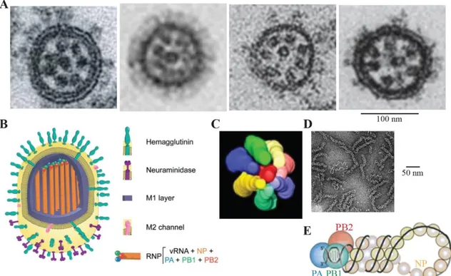
Figure 1: Viral particle structure and genome organization.
(A) Electron microscopy (EM) pictures of IAV, IBV, ICV and IDV particles, from left to right 234,235,250,
(B) schematic representation of influenza A virus particle 62, (C) modelization from STEM tomography
of the packaging of the genome inside virions 234, (D) EM picture of influenza ribonucleoproteins
(RNPs) purified from virions 310, (E) schematic representation of a RNP, with the viral genomic
segment in black, the nucleoprotein in light yellow and the three subunits of the polymerase in blue, green and red 231.
Chapter 1: Introduction
Influenza viruses|
36
to several micrometers in length 247. IAV, IBV and IDV particles occasionally form such
structures, seemingly without packaged RNPs 42,200,234. The morphology of the viral
particle seems to be cell- and/or strain-dependent and is modified by serial passages in culture. Viral particles isolated from clinical samples tend to form more filamentous structures while cell culture-adapted or eggs-adapted virus shed virions far more homogeneous and spherical, as a result of host adaptation 42,159,300.
Using electron tomography and EM, each segment of the viral genome, can be visualized as a dense rod-like structure (Fig. 1A and 1D). Eight of these structures, called ribonucleoproteins (RNPs), are localized at the distal end of influenza virions (Fig. 1A, 1B and 1C) 234,250.
Table 2: Summary of the influenza main features depending on the virus type.
Sph, Fil and Cord respectively stand for Spherical, Filamentous and Cord-like. * While reassortment can occur for IBV due to the two circulating lineages, this does not give rise to an antigenically distinct virus, only to a genetically distinct virus.
Virus type IAV IBV ICV IDV ToadV
Viral particle phenotypes Sph: 120 nm Fil: 120 nm Cord: several µm Sph: 120 nm Fil: 150 nm Cord: several µm Sph: 100 nm Fil: 100 nm Cord: several µm Sph: 100 nm Fil: 100 nm Cord: several µm ? Genomic segments and main proteins Eight segments S1: PB2 S2: PB1 S3: PA S4: HA S5: NP S6: NA S7: M1/M2 S8: NS1/NEP Eight segments S1: PB2 S2: PB1 S3: PA S4: HA S5: NP S6: NA S7: M1/M2 S8: NS1/NEP Seven segments S1: PB2 S2: PB1 S3: P3 S4: HEF S5: NP S6: M1/M2 S7: NS1/NEP Seven segments S1: PB2 S2: PB1 S3: P3 S4: HEF S5: NP S6: M1/M2 S7: NS1/NEP Eight segments? Viable reassortment Yes, except some strain combinations No* No No ?
Reservoir Aquatic birds Humans Humans Cattle Toad?
Tropism Wide spectrum Mammals and birds Narrow spectrum Mainly humans Narrow spectrum Humans and swines Medium spectrum Mammals ?
Chapter 1: Introduction
Influenza viruses|
37
iii. The ribonucleoproteins, the replicative units of the virus
The functional replicative unit is composed by the viral RNA genome (vRNA) with viral proteins necessary and sufficient for an efficient replication and transcription. In influenza viruses, such a macromolecular complex is called an RNP (Fig. 1). One RNP is composed by a single genomic segment, encapsidated by multiple copies of the nucleoprotein (NP) and with one heterotrimeric RdRp (PA:PB1:PB2) associated to both 3’ and 5’ extremities (Fig. 1E) 7,230. This structure is helicoidal, supercoiled and
highly flexible (Fig. 1D and 1E) 46,162,277. The dimensions of an RNP are respectively
approximatively for 30-120 nm x 10-15 nm, depending on the encapsidated segment
46,162,277,309. The oligomerization of RNA-bound NP gives RNPs their rod-like structure
280,353. Influenza viruses possess 7 or 8 genomic segments and consequently 7 or 8
unique, distinct RNPs. Interestingly, even if ICV and IDV possess only seven viral genomic segments, it has been shown that all influenza viruses present a 7 + 1 packaging pattern of RNPs in virions (Fig. 1C) 115,234. Whether ICV and IDV package
an extra vRNA segment or one of the host RNA is still unclear 249.
iv. Genome organization, protein content and host tropism
IAV and IBV possess eight viral RNA genomic segments whereas ICV and IDV genomes are composed by seven segments 117,265,298. Each segment encodes for one
or several proteins. Some segments are known to contain distinct open reading frames, encoding distinct proteins. Transcribed mRNAs can also be processed by the cellular splicing machinery, to generate other proteins (Fig. 2) 67,90,233,408. The segments are
numbered and named depending on their length and the main protein they encode (Table 2, Fig. 2). The difference in segment number is due to the difference in the viral glycoprotein composition: IAV and IBV possess HA and NA while ICV and IDV only possess HEF. While the full genome is not yet available for ToadV, two glycoproteins (HA and NA) are encoded by the genome, meaning that it seems to be made of eight segments, like both IAV and IBV. The details of the proteins is shown with the Figure
Chapter 1: Introduction
Influenza viruses|
38
Beside the main proteins, several accessory type-dependent proteins have been identified. Up to now, seven non-essential proteins have been described for IAV, which seem to play many roles in the regulation, the virulence and/or the host adaptation processes (Fig. 2) 370,409.
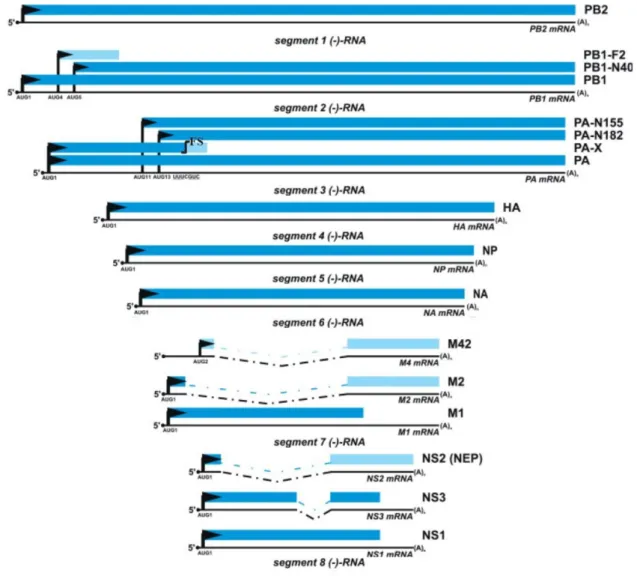
Figure 2: Main and accessory proteins encoded by influenza A viral mRNAs.
Adapted from 370. Black arrow and dotted lines represent respectively the mRNA translation start and
spliced parts. Each blue shade corresponds to one of the 3 open reading frames. For segment 3 PA-X mRNA generation, one frameshift occurs (FS).
Chapter 1: Introduction
Influenza viruses|
39
Each influenza virus possesses its own specific host tropism, which relies partly on the behaviour and specificities of its proteins (Fig. 3, Table 2). Aquatic birds are the reservoir for IAV but they are able to infect a wide spectrum of animals. Some serological reports show evidence of infection in reptiles and amphibians or at least some sort of cross-reactivity, in addition to mammals, bats and birds 36,369,387,415. IBV
and ICV present a restricted host spectrum: IBV mainly infect humans, but some cases have been reported in seals, and ICV can infect swine in addition to humans 255,260,417.
IDV have been discovered in 2011, initially identified as an ICV subtype, but officially considered as a new influenza genus in 2013. IDV show a large spectrum of host organisms in mammals, from camelids to pigs and goats, but their reservoir is suspected to be the cattle 68,116,117,285. They can replicate in ferrets, the animal model
for human infection 117. Humans, and more particularly people working closely to the
cattle, have been shown to seroconvert by producing anti-influenza D antibodies
117,363,391. Because of its recent discovery, ToadV has not been studied so far, without
any further information in terms of reservoir and tropism.
Several factors restrict the host and the cell tropisms. The growth temperature is one of them. IDV have been found to efficiently replicate at both 33°C and 37°C, likely
Figure 3: Host tropism of influenza viruses.
The known host range of each influenza virus is labelled as a coloured background, with blue for IAV, light green for IBV, red for ICV, purple for IDV and dark green for ToadV. Adapted from 171.
Chapter 1: Introduction
Influenza viruses|
40
helping in its broad host range 117,124,391. IAV and IBV present an optimal growth
temperature of 37-39°C and 35°C respectively, in agreement with the temperature of their respective reservoir hosts and cell environments 200. ICV tend to grow better at
lower temperatures (i.e. 33°C rather than 37°C) 117,254.
In term of cell tropism, the main and most studied factor is the HA/HEF glycoprotein, which binds receptors modified with sialic acids (Fig. 4A). The A/HA activation seems to be done through a restricted number of proteases and its optimal temperature ranges from 33 to 39°C depending on the HA type and the targeted host. In contrast, B/HA demonstrates a large panel of activating proteases with an optimal temperature of 33°C, well adapted to human airways 182. D/HEF and C/HEF are structurally well
conserved and are both able to bind to a large spectrum of host tissues, but with a better D/HEF attachment to the cell surface than C/HEF. D/HEF presents a more open binding cavity, likely allowing more diverse receptors and/or partners interactions 335.
C/HEF is rather sensitive to the temperature, with an optimal close to 33°C 348.
Interestingly, IDV is the most resistant of all influenza viruses to an acidic or hot environment and its stability was shown to be mainly due to D/HEF 416.
Several other viral proteins are involved at host:pathogen interfaces and are responsible for these differences in host and cell tropisms such as NS1, the polymerase subunits and NP.
The subunits of the RdRp, and in particular PB2, are considered as determinants of the host tropism 23,351. It is now well-known that the A/PB2 E627K substitution is an
important marker of the mammal adaptation, for the replication at lower temperatures
343,352. When residue 627 is in its avian form, the D701N substitution can compensate,
allowing a successful replication, probably in its interaction with the cellular nuclear import machinery 24,341. Other discreet substitutions can also compensate for a fully
avian phenotype at these two positions, as seen in the case of the pandemic 2009 H1N1 strain 214.
NP can be found at several host:pathogen interfaces. Several mutations (N319K, G102R, M105K or D375N) have been identified as positions involved in nuclear import
93,94,291,292. Other mutations (G16D, L283P, F313Y or Q357K) allow the viral escape
Chapter 1: Introduction
Influenza viruses|
41
Outside of a few specific mutations, the diversity of adaptive mutations suggests that adaptation to a new host is a largely synergic mechanism, with several distinct mutations likely required to overcome this barrier 203,319.
NS1 is strongly involved in the host defences regulation, and more particularly against interferon signalling 110. In H5N1 viruses, the residue 92 tends to be a
glutamate or an aspartate, and could lead to a stronger resistance to antiviral responses and to higher virulence 271,324. A/NS1, B/NS1, C/NS1 and D/NS1, while
sharing the same antiviral activities, are not completely interchangeable for the replication and the host response inhibition purposes 251.
While not directly involved in the host tropism, M1 and M2 seem to possess a very specific behaviour depending on their viral type.
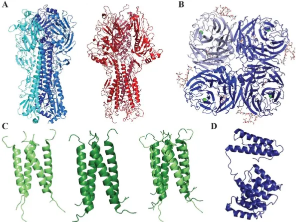
Figure 4: Structure of influenza HA, HEF, NA, M2 and M1 proteins.
(A) Crystal structures of trimeric A/HA (blue, PDB 3LZG) and C/HEF (red, PDB 1FLC). (B) Crystal structure of A/NA (PDB 5HUK). (C) Crystal structures of the open channel of B/M2 (left), the close conformation (middle) and the superimposition (right) (PDB 6PVR and 6PVT). (D) Cryo-EM structure of the A/M1 matrix protein (PDB 7JM3).
Chapter 1: Introduction
Influenza viruses|
42
The influenza M2 protein is an ion channel essential during the viral cycle and more particularly for the cytoplasmic release. A/M2 and B/M2 (Fig. 4C) share some features, such as a mostly proton channel role, histidines playing the part of proton sensor and activator and a similar transmembrane domain length. They also seem to present specificities, with a different pH of activation and a different direction of the proton flux
179,198,420. In contrast, C/M2 and D/M2 are primarily chloride ion channel with a slight
proton permeability, suggesting a different mechanism in the viral cycle 21,125,158,227.
The highly flexible influenza M1 protein (Fig. 4D) is heavily involved in the structure of the viral particle and the budding process 303. M1 is often found at the interface
between viral and host partners, and it is known to interact with viral RNPs, several viral and cellular factors, and lipids.
v. Evolution: antigenic drift and antigenic shift
Influenza viruses are known for their fast continuous evolution, depending on two mechanisms: the antigenic drift and/or the antigenic shift.
Like the majority of RNA virus RdRp, the influenza RdRp does not possess a proofreading mechanism. Therefore, the influenza RdRp integrates about one error every genome replication, at a rate of about 10-5-10-6 substitution/nucleotide site/cycle 23,66,269,340. It has been shown previously that IAV present a higher mutation rate than
IBV, with rates of about 2.10-3 and 0.6.10-3 substitutions/nucleotide site/year
respectively 248,390. Influenza viruses are under a strong selective pressure from both
the host immune system and the host machineries they highjack. NA and HA (Fig. 4A
and 4B) are in the front line, recognized swiftly by the immune system and the main
target for host antibodies 141,276. This requires a fine balance between escape
mutations and/or mutations increasing the virus fitness and non-deleterious mutations to preserve the protein functions. This process is called the antigenic drift and occurs also in host adaptation in cell culture 334.
The antigenic shift is a brutal event that is due to the direct jump from one host species to another and/or the reassortment. The latter is specific to the segmented nature of the viral genome. It occurs when two viruses co-infect the same cell and exchange one or several genomic segments during the packaging step. However, it is
Chapter 1: Introduction
Influenza viruses|
43
required for the two viruses to be of the same type and compatible 187. Genomic
segment exchange often leads to pandemic strains of IAV 319,384. As an example, the
pandemic 2009 H1N1 genome came from four distinct IAV parental strains 225,253.
vi. Disease and epidemiology
Influenza viruses are mostly known due to the seasonal epidemic and sporadic pandemic they cause in humans. Seasonal influenza is an acute and contagious disease affecting the human respiratory tract with several non-specific symptoms, such as high fever, cough, asthenia, headaches and muscular aches. In most cases, no complication arises. However, severe or deadly outcomes can befall to high-risk people, including pregnant women, immunocompromised or people affected by a chronic disease, the elderly and new-borns. Upon influenza virus infection, they could develop severe respiratory tract infection, associated or not with coinfection, and extreme inflammatory reaction. The severity is intrinsically linked to the virus and the host immune response 149. IAV and IBV are responsible for these yearly events,
infecting around 10% of the world population, resulting in 3 to 5 million severe cases and 290 000 to 650 000 respiratory-related deaths each year 138. The average annual
economic burden of a seasonal outbreak has been recently estimated at $11.2 billion in the USA or €78.3 million in Germany 284,318.
Seasonal epidemics occur during fall and winter in temperate climates 400. In the
tropical and equatorial parts of the globe, influenza viruses circulate all around the year, with unpredictable epidemics, assuring virus constant exchange between the Northern and the Southern hemispheres 312,374. The virus is mainly transmitted through
infectious droplets emitted by infected patients, but it can also be spread through the contact with an infected surface or person 213,252.
IAV and only IAV can cause pandemics, corresponding to worldwide sporadic occurrences: only IAV possess several antigenically distinct HA and NA proteins, leading to large-scale outbreaks for a lack of previous immunity.
Chapter 1: Introduction
Influenza viruses|
44
The human history is tightly intertwined with influenza viruses (Fig. 5): in 412 B.C. in Ancient Greece, Hippocrates described a disease strikingly similar to influenza and no less than 13 pandemics have been identified these last 400 years 174,279,315. The
worst pandemic in modern history was caused by a H1N1 influenza virus, submerging the world in three waves in 1918-1920. As World War I was raging across the world, involved belligerents did not report the pandemic. Spanish censorship-free newspapers did, and thus the pandemic was wrongly named “the Spanish flu”. The mortality rate was estimated at least 2.5-5%, far higher than any other influenza outbreak. The virus infected almost half of the world population, killing at least 50 million people, mainly young adults 145,330,400. Beyond the new antigenic variant and
the virus intrinsic virulence, other reasons can explain this catastrophe 135. In 1918, the
responsible agent was unknown and therapeutic, prophylactic and sanitary measures were lacking 144,208,336. Antibiotics to treat severe secondary bacterial infections were
not discovered yet, becoming widely available from 1939 onwards.
Since the 1920’s, four pandemics occurred, with a high death toll but far from the level of 1918-1919 (Fig. 5). Nowadays, experts are closely monitoring several influenza viruses, and more particularly zoonotic viruses of the H5Nx, H6Nx, H7Nx, H9Nx and H10Nx subtypes 222,400. Sporadic transmission of these viruses to humans
occurs regularly, without sustainable human-to-human transmission. However, the
Figure 5: Major historical outbreaks in the shared human and influenza history.
Putative and confirmed pandemics (Pdm) and epidemics (Edm) are illustrated as red bars. Sporadic outbreaks of deadly highly pathogenic avian influenza viruses are shown as spiky forms of distinct colours for distinct HA types. This schematic representation is not at scale.
Chapter 1: Introduction
Influenza viruses|
45
mortality rate in these infections can be up to 50%, suggesting that a pandemic could result in a disaster 147,323,394.
In addition to human infections, influenza viruses widely circulate in animals, more particularly in birds for IAV 169,262. While wild aquatic birds tend to be asymptomatic or
present little symptoms, this is not the case in poultry 226. Low-pathogenic avian
influenza viruses (LPAIs) and highly pathogenic avian influenza viruses (HPAIs) tend to spread quickly in bird populations. LPAIs tend to induce in worse cases mild symptoms, such as a decrease in egg production and slight respiratory issues; HPAIs, in contrast, usually result in a severe disease, killing domesticated birds in a few days, with a mortality rate that can be close to 100% 262,347,392,401. The severity of the IAV
infection depends on the strain and the host and ranges from little to no symptom to systemic infections and high mortality rates 338.
Even if its epidemic and circulation patterns remain largely unknown, ICV infect humans and more particularly children, leading to a mild respiratory disease 321. Swine
are also susceptible but with no evident signs of the disease.
IDV reservoir seems to be the cattle. The clinical symptoms for IDV alone are typically weak but IDV coinfection with other pathogens seems to be involved in a more severe disease, called the bovine respiratory disease (BRD) 80,216. Swine and various
farm animals such as goat but also camel present an anti-IDV serology but the pathology has not been extensively studied. Swine and animal models like ferret do not present influenza-like symptoms and lesions 79,117.
Regarding the newly identified influenza viruses infecting cold-blooded animals, it is not known if they can induce symptoms, infect other animals or cause any diseases.
B. Influenza virus cycle
The first step of the viral cycle is the attachment to the host cell through HA/HEF (Fig. 6 and 4A). Both recognize sialic acid molecules, highly expressed and displayed on cell surfaces.
A/HA and B/HA recognize the N-Acetylneuraminic acid (Neu5Ac) sialic acid, found on glycolipids or glycoproteins 344. Human A/HA bind preferentially to α2,6-linked
Chapter 1: Introduction
Influenza viruses|
46
Neu5Ac, abundant on human epithelial cells in the upper respiratory tract 47,375. In
contrast, avian A/HA interact with α2,3-linked Neu5Ac, predominantly found in the digestive tract of aquatic bird and the human lower respiratory tract 137. Both α2,3- and
α2,6-linked sialic acids are highly expressed in the pig trachea 136,375. ICV and IDV HEF
bind to the N-Acetyl-9-O-acetylneuraminic acid 301. The high HA/HEF density could
compensate the weak binding affinity by a high avidity 316,349.
The viral entry in cells mainly occurs through the clathrin-dependent mechanism but clathrin-independent endocytosis and macropinocytosis have also been reported
210,270,331,377. Influenza virions then go through the endosomal/lysosomal pathway.
From early endosomes to lysosomes, the internal pH of the endosomal particles drops from 6.5 to 4.5. The ion channel M2, inserted into the virion envelop, leads to the internal acidification of the viral particle, resulting in the M1 dissociation from the RNPs (Fig. 4C) 204. HA/HEF goes through a conformational change, triggering the viral
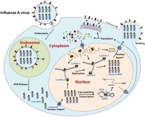
Figure 6: Influenza viral cycle.
The viral proteins are represented with different forms and colours: NP, PA, PB1 and PB2 are represented as round forms in blue, brown, green and red respectively. M1 and NEP are triangular forms, in orange and yellow respectively. HA, NA and M2 are cylindrical forms, red, yellow and purple respectively .Adapted from 423.
Chapter 1: Introduction
Influenza viruses|
47
particle envelop fusion with the endosomal membrane and the RNPs release in the cytosol 96,210,424.
The RNPs then require access to the nucleus to initiate the transcription/replication steps. Influenza viruses hijack the host nuclear import machinery, and in particular the importin-α/β pathway (cf. part. II) 28,54. RNPs are rapidly translocated through the
nuclear pore complex, and transcription is initiated by the viral polymerase 205. Viral
messenger RNAs (mRNAs) are directly transcribed from the viral genome. They are exported in the cytoplasm, where they will be translated by the host machinery 54.
Newly synthesized subunits of the RdRp, NP, M1, NS1 and NEP will be imported or will diffuse in the nucleus to contribute to the synthesis of viral RNAs 28,57,93,381.
Replication is the other important process in the nucleus. For this, negative-sense genomic RNAs are firstly used to generate positive-sense RNA (cRNA) which is used as a template for genomic negative-sense RNA synthesis. As the viral RNAs are never naked, the encapsidation by both NP and the RdRp occurs simultaneously to their synthesis. Once reconstituted, RNPs are exported in the cytosol by using the cellular nuclear export machinery, M1 and NEP. NEP interacts with the cellular Chromosome Region Maintenance 1 (Crm1) export protein and M1, which in turn interacts with RNPs
2,37,72,126,240,267,385. RNPs are then addressed to the lipid rafts at the plasma membrane,
where HA/HEF, NA and M2 are already localized to be packaged into virions 132.
A functional viral particle requires the seven/eight individual genomic segments packaged in a single particle. While the molecular basis behind this step is still unclear, selective packaging of RNPs is the favoured hypothesis 25,97,98,235. A viral RNA segment
seems to present at its extremities packaging signals, which interact directly with other distinct RNP packaging signals 88,89,197,228. In addition, parts of coding regions are
suspected to enhance a correct packaging 99,105. They bring together distinct
segments, resulting in a close parallel alignment of distinct RNPs. The budding of infectious virions is a mechanism poorly understood. However, the budding seems to be initiated at lipid rafts by the presence of the RNP complexes, HA/HEF, M1 and NA, in an ESCRT-independent manner 238,313,317,388. M2, localized at the border of lipid
rafts, is responsible for the membrane scission 204,302. NA then cleaves sialic acids from
Chapter 1: Introduction
Influenza viruses|
48
C. Viral transcription and replication machinery
i. Nucleoprotein
The nucleoprotein is the most abundant protein of the RNPs and also in the infected cells. NP is required for both viral transcription and replication activities and could act as a switch between the two processes 15,325. NP is also involved in the viral RNA
genome protection, in the interaction with cellular partners and in the cellular trafficking.
Table 3: Sequence identities between influenza types.
Sequences were recovered from the Influenza Research Database 419. The
strains used for alignment are those used in this work.
A/NP B/NP C/NP D/NP Toad/NP A/NP 100 37.2 21.8 21.1 22.5 B/NP 100 23.1 23.8 28.7 C/NP 100 37.5 22.1 D/NP 100 20.1 Toad/NP 100
Globally, sequence identities between all NP types range from 20 % to 38 %, with biochemical properties and structure predictions rather well conserved (Fig. 7, Table
3). A/NP, B/NP, C/NP, D/NP and Toad/NP molecular mass and protein chain length
vary between 56 and 67 kDa, ranging from 498 to 607 amino acids.
NP is a highly basic protein with a basic groove, a feature to be linked to its RNA binding activity (Fig. 8B) 242,243. NP does not seem to bind to RNA bases but rather to
the phosphate skeleton 13. It is still unclear how NP and RNA interact, as recent studies
showed that NP was not distributed homogeneously on viral genomic segments with a preferential binding mode 183,184,396. The monomeric form of NP is probably the active
form in the interaction with RNAs, with the NP oligomerization triggering the formation of the RNP architecture; the high affinity binding of the trimeric A/NP to RNA could impair the oligomerization process required for the RNP formation 178. A/NP and B/NP
bind RNAs with nanomolar affinity, with cooperativity, but behave differently depending on the salt concentration and the RNA length 13,70,71,178,407. The hypothetical length of
Chapter 1: Introduction
Influenza viruses|
49
24 nucleotides 46,207,259. Interestingly, NP-bound RNAs are still susceptible to RNases,
consistently with the suspected binding pattern of RNAs, wrapped around NP 143,207.
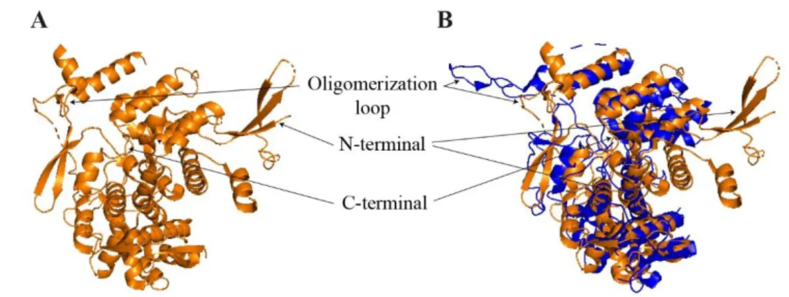
Influenza NPs are globular, tightly folded proteins. To date, several influenza NP structures have been solved, but none of them with RNA (Fig. 8). More particularly, two A/NP and one B/NP have been crystallized and solved as trimers and tetramers respectively, highlighting the NP tendency to form oligomers in solution and in absence of RNA. NP oligomerization is affected by the RNA, the salt concentration, its concentration, its phosphorylation state and the temperature 39,178,353,365.
While A/NP and B/NP share only 37% of sequence identity, they are structurally well conserved (Fig. 8C). They are folded mainly in α-helices. The two major differences come from i) the oligomerization loop orientation in the crystal structure, due to the oligomeric state (trimer v.s. tetramer) and ii) a longer N-terminal (N-ter) domain not visible in the structure and intrinsically disordered (i.e. 21 residues for A/NP against 71 for B/NP). The structure of the monomeric R416A mutant gives precious insights into the oligomerization mechanism (Fig. 8D). In the native structure, the Arg-416 is localized in a protruding loop (from residue 402 to 428) making the interaction interface with a second protomer. The Arg-416 makes an essential salt bridge with the side chain of the Glu-339. The R416A mutant loop, in contrast, was not extended and was folded into its own NP-binding groove (Fig. 8A, 8C and 8D). Putatively due to the oligomerization loop conformation, the monomeric mutant is drastically impaired in its interaction with RNA (micromolar affinity) 27,40,353. The B/NP oligomerization
Figure 7: Biochemical features of influenza nucleoproteins.
Intrinsically disordered tails and structured domains are represented as thin lines and boxes respectively.
Chapter 1: Introduction
Influenza viruses|
50
mechanism seems similar to the A/NP one, forming a conserved salt bridge between residues R472 and E395.
In the Orthomyxoviridae family, another virus, the infectious salmon anemia virus (Isavirus) and its NP (ISA/NP) have been studied. ISA/NP has been crystallized as a dimer. It is structurally well conserved with influenza NPs, but it possesses a supplementary partly structured N-ter domain (Fig. 9) 422. ISA/NP seems to be mainly
dimeric in solution. Each monomer binds to 12 nucleotides long RNA molecules and 24 nucleotides long RNAs seem wrapped around ISA/NP, as it has also been suggested for influenza NPs. The activity or the functionality of the unique N-terminal domain of ISA/NP has not been elucidated yet. It is interesting to note that ISA/NP N-ter extremity (NPTAIL) is longer than those of A/NP and B/NP (respectively 111, 21 and
71 amino acids). Furthermore, A/NPTAIL and B/NPTAIL have a well-defined function,
playing an essential role in the nuclear translocation (cf. part III.). Although, due its
Figure 8: Influenza nucleoprotein structures and structural features.
(A) Crystal structure of trimeric influenza A/NP with the essential salt bond responsible for the oligomerization process, (B) Electrostatic potential of a protomer of the trimeric A/NP, with positive charges in blue and negative charges in red (C) Surimposition of A/NP (PDB 2IQH), coloured in blue; with B/NP (PDB 3TJ0), in green (D) Monomeric R416A mutant of A/NP, adapted from 40.
Chapter 1: Introduction
Influenza viruses|
51
size, B/NPTAIL could probably have a yet unravelled function.
While no structure for C/NP or D/NP was solved at the beginning of this PhD thesis work (cf. Chapter 2), it was possible to make assumptions on their potential folding. The disorder prediction indicates that in contrast to A/NP and B/NP, C/NP and D/NP present only a few unstructured N-terminal residues and a long intrinsically disordered C-terminal (C-ter) region spanning about 50-60 residues (Fig. 7). This change from an N-ter NPTAIL to a C-ter one is puzzling. Identity between C/NP and D/NP sequences is
38%, similar to the one between A/NP and B/NP, suggesting that their folding could very well be alike.
ii. Heterotrimeric RNA-dependent RNA-polymerase
The influenza RdRp is an intricate heterotrimeric complex of PA, PB1 and PB2, each of them being composed of 700 to 800 residues with a total molecular mass close to 270 kDa.
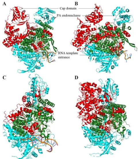
The influenza polymerase structure has eluded researchers for a long time, due to issues in producing it in a soluble amount. The issue was tackled by firstly solving individual domains of the complex, like the PB2-627 domain 311,352. Subunit interfaces
were initially roughly determined: PAC-ter interacting with PB1N-ter, which in turn interacts
with PB2N-ter through its C-ter part. EM analysis showed that the polymerase is a tightly
folded complex, suggesting supplementary inter-subunit interaction regions. These last
Figure 9: Structure of Isavirus NP.
(A) Crystal structure of ISA virus NP (PDB 47EWC) (B) Surimposition of A/NP (PDB 2IQH), coloured in blue, with ISA/NP (PDB 47EWC), in orange.