SCIENTIFIC ARTICLE
Magnetic resonance imaging frequently changes classification
of acute traumatic thoracolumbar spine injuries
Sebastian Winklhofer&Merly Thekkumthala-Sommer&
Diethard Schmidt&Kaspar Rufibach&
Clément ML Werner&Guido A Wanner&
Hatem Alkadhi&Jürg Hodler&Gustav Andreisek
Received: 8 October 2012 / Revised: 4 November 2012 / Accepted: 6 November 2012 / Published online: 27 December 2012 # ISS 2012
Abstract
Objective To evaluate the influence of additional (MRI) com-pared with computed tomography (CT) alone for the classifi-cation of traumatic spinal injuries using the Arbeitsgemeinshaft für Osteosynthesefragen (AO) system and the Thoraco-Lumbar Injury Classification and Severity (TLICS) scale. Materials and methods Images from 100 consecutive patients with at least one fracture on CT were evaluated retrospectively by three radiologists with regard to the AO and TLICS classification systems in 2 steps. First, all images from the initial CT examination were analyzed. Second, 6 weeks later, CT and MR images were analyzed together. Descriptive statistics and Wilcoxon tests were performed to identify changes in the number of fractures and ligamentous lesions detected and their corresponding classification. Results CT and MRI together revealed a total of 196 fractures (CT alone 162 fractures). The AO classification changed in 31 %, the TLICS classification changed in 33 % of the patients compared with CT alone. Based on CT and MRI together, the
TLICS value changed from values <5 (indication for conser-vative therapy) to values≥5 (indication for surgical therapy) in 24 %.
Conclusion MRI of patients with thoracolumbar spinal trau-ma considerably improved the detection of fractures and soft tissue injuries compared with CT alone and significantly changed the overall trauma classification.
Keywords Spine . Trauma . Fracture . Classification . Magnetic resonance imaging . Computed tomography
Introduction
Computed tomography (CT) plays a key role in the initial diagnostic work-up of traumatic spinal injuries in most mod-ern emergency departments. Osseous injuries, as well as mis-alignment, can be reliably evaluated with this modality [1–4]. However, accompanying lesions of ligamentous structures, as well as surrounding soft tissues, can be detected with a higher sensitivity by magnetic resonance imaging (MRI) [5].
As a consequence, MRI is considered the standard or reference imaging modality for visualizing the posterior mentous complex (PLC), consisting of the supraspinous liga-ment (SSL), the interspinous ligaliga-ment (ISL), the ligaliga-mentum flavum (LF), and the facet joint capsules (FJC) [6–8].
Spinal stability is determined by the intactness of the osseous components, as well as by the integrity of the ligamentous structures [9,10]. Thus, the analysis and eval-uation of the latter is essential for decision-making and further treatment of the trauma patient [11–13], as patients with PLC lesions often undergo surgery with posterior in-strumentation and possibly fusion.
S. Winklhofer
:
M. Thekkumthala-Sommer:
D. Schmidt:
H. Alkadhi
:
J. Hodler:
G. Andreisek (*)Institute for Diagnostic and Interventional Radiology, University Hospital Zürich, Rämistrasse 100, 8091 Zürich, Switzerland
e-mail: gustav@andreisek.de K. Rufibach
Division of Biostatistics, Institute for Social and Preventive Medicine, University of Zürich, Hirschengraben 84, 8001 Zürich, Switzerland
C. M. Werner
:
G. A. WannerDivision of Trauma Surgery, Department of Surgery, University Hospital Zürich, Rämistrasse 100, Zürich, Switzerland
In clinical practice, the imaging data, in combination with the clinical condition of the patient, are used to categorize traumatic spine injuries. Two classification systems are com-monly used: the Arbeitsgemeinshaft für Osteosynthesefragen (AO) system and the Thoraco-Lumbar Injury Classification and Severity (TLICS) scale (the AO and TLICS systems have been described elsewhere in detail, please refer to Magerl et al. [14] and Vaccaro et al. [15]). In addition to serving as inter-nationally accepted classification schemes for nomenclature, both are used to indicate the severity of a spinal lesion and thus help stratify the patient work-up into surgical and non-surgical treatment groups [16]. The AO classification describes the severity of a spinal lesion with respect to the complexity of the fracture. It is divided into three groups: type A (compression), B (flexion/distraction), and C (rotation), each with seven to eight subgroups. The TLICS classification sys-tem is based largely on three components: the morphology of the injury, the integrity of the PLC, and the neurological status of the patient. Compared with the AO system, the TLICS system includes additional clinical information and provides, depending on the score, recommendations for therapy (con-servative versus surgical management).
A recent study with a relatively low number of patients (n030) indicated a high rate of change in the classification of spinal injuries after performing MRI compared with CT alone [6]. However, the results of that study are limited owing to the small sample size and the fact that further analyses of the PLC components, which are essential to the stability of the spine, were not made [6].
The aim of our study was to evaluate the influence of additional MRI compared with CT alone for the classifica-tion of traumatic spinal injuries using the AO and the TLICS, systems and to consider possible reasons for mod-ifying the classifications.
Materials and methods
Patients
As this was a retrospective study, conducted in accordance with the Declaration of Helsinki, the local ethics committee waived the requirement for informed patient consent.
CT and MRI of the spine were performed routinely in 100 consecutive trauma patients: 24 women (median age 41 years, range 21–55 years) and 76 men (median age 47 years, range 21–70 years). Inclusion criteria were a spinal trauma with one or more thoraco-lumbar fractures seen on the CT performed initially and on subsequent MRI within 10 days of CT. Table1
shows the trauma mechanisms of the 100 patients.
Exclusion criteria for enrollment in the study were age younger than 18 years and over older than 55 years in women (men >70 years), with the latter to exclude latent tumor disease
or osteoporotic changes. Individuals with anamnestic known primary tumor disease or anamnestic known osteoporosis; osteoporotic or pathological fractures; a Glasgow Coma Scale (GCS) score of < 13; surgery between the CT and MRI; or any general MRI-related exclusion criteria such as metallic implants, foreign bodies, or claustrophobia, were also exclud-ed. The data sets were collected over 3 years (2009–2011).
The clinical information including the GCS and the neu-rological status were taken from patient records.
CT and MRI
The initial diagnosis of spinal injury was based on either the whole-body CT examination, as routinely performed in our emergency department in trauma patients (Somatom Defi-nition, Siemens Healthcare, Erlangen, Germany) using axi-al, coronaxi-al, and sagittal reformations of the spine, or on CT examinations taken at other facilities if the quality fulfilled the criteria of our department. CT imaging parameters in-cluded tube voltage of 120 kVp; use of automatic exposure control with tube current–time modulation (CareDose4D, Siemens Healthcare, Forchheim, Germany); 0.6-mm config-uration for slice acquisition; rotation time, 0.5 s; and pitch, 0.6. Data reconstruction included a sharp, as well as a soft, tissue convolution kernel (B50f and B30f respectively), slice thickness of 2 mm, and slice increment of 1.6 mm.
All MRI examinations were performed on a 1.5-T MRI (Excite HDx, GE Healthcare, Waukesha, WI, USA) using the standard MR protocol for spinal trauma as established in our department. It includes a sagittal T1-weighted (w) [repe-tition time/echo time (TR/TE), 500/13 ms; field of view (FOV), 240 mm], a sagittal T2-w (TR/TE, 3160/112 ms; FOV, 240 mm), an axial T2-w (TR/TE, 3,160/112 ms; FOV, 160 mm), and a sagittal short tau inversion-recovery (STIR) (TR/TE, 4,760/44 ms; inversion time, 200 ms; FOV, 240 mm) MRI sequence. Slice thickness for all acquisitions was 3 mm, spacing 1 mm, and number of excitations 2.
CT and MR image analysis
All images were analyzed on a workstation (AW 3.2; GE Medical Systems, Waukesha, WI, USA) by two senior
Table 1 Main causes of trauma in our study sample Trauma mechanism n (%) Fall (>3 m) 27 (27) Car accident 11 (11) Bicycle accident 10 (10) Ski accident 7 (7) Motorcycle accident 5 (5)
Traffic accident pedestrian 2 (2)
radiologists (DS and GA, with 7 and 9 years’ experience in trauma radiology respectively), and one resident in radiology (SW) in consensus.
First, the initial CT examination of each patient was analyzed blinded to the MR images and without knowledge of the MRI findings. Second, 6 weeks later, CT and MR images were analyzed in combination in random order, again blinded to the initial CT results, patient information, or knowledge of the findings from the first read-out.
The following variables were assessed in each patient according to the AO and the TLICS classifications: number of fractures of each patient, as well as the levels of those lesions; the morphology of the fracture pattern; and the integrity of the PLC. Fracture patterns were categorized according to TLICS as compression, translation/rotation, or distraction, and according to AO as compression, flex-ion/distraction or rotation. The PLC was classified as intact, suspected/indeterminate or injured. On CT, injured was defined when one of the following abnormalities was pres-ent: diastasis of the facet joints, avulsion fracture of the superior or inferior aspect of contiguous spinous processes, vertebral translation, or an interspinous spacing greater than that of the level above or below [17, 18]. The PLC was classified as intact on CT if no remarkable structures or aforementioned abnormalities were found.
On MRI the PLC was divided in its constituent parts— SSL, ISL, LF, and the FJC. These components were evalu-ated individually as intact, suspected/indeterminate, or in-jured. On MRI, intact was defined as no change in the signal of these structures. The PLC was classified as injured when complete or incomplete discontinuity of one or more liga-mentous structures or a clear change in the MR signal was seen [16,17,19]. If more than one spinal level was affected, the different lesions and levels were analyzed separately.
Finally, the TLICS and AO classifications for the two read-outs were compared, and changes in classification were analyzed. If there were two or more injured levels in one patient, the lesion with the highest severity defined the injury grade. If there was a burst and compression fracture seen in the same patient, burst was ranked higher according to the TLICS classification. The changes in the classification grades also refer to the most severe lesion classified by CT.
The TLICS classification combines the imaging findings and the neurological status with values that are added to a composite injury severity score (ISS; see Table2). A score higher than 5 indicates an unstable spinal injury requiring surgery [20]. Additional findings, such as intervertebral disk herniation, myelopathy, or bone bruise were noted. The latter was defined on the MR images as high signal intensity on the STIR sequence and low signal intensity on the T1-weighted images without an apparent fracture line. Bone bruises were not rated as fractures.
Examples of CT and MRI analysis are shown in Figs.1,2
and3.
Statistical analysis
Descriptive statistics were performed to describe distribu-tion of the number of observadistribu-tions (n), minimum, median, maximum, and interquartile range (IQR). The effect of additional MRI on the ordinal variable TLICS was com-pared using a Wilcoxon test. Confidence intervals (CIs) were based on inversions of the Wilcoxon tests [21]. For proportions, we used Wilson CIs. All CIs were computed at a level of 95%. All computations were performed by a statistician (KR) using R (R Development Version 2.14.0, Core Team, 2010) [22,23].
Results
In 41 out of 100 (41%) patients the thoracolumbar spinal injury was the only lesion, whereas 37 patients (37%) had one or more injuries in other locations. Twenty-one (21%) patients suffered polytrauma, including head injury. Ninety-two out of 100 patients (92%) had a Glasgow Coma Scale (GCS) score of 15, 6 out of 100 (6%) a GCS of 14, and 2 out of 100 patients (2%) had a GCS of 13.
Median time between initial CT and additional MRI was 26 h (range 2–240; IQR 16–46 h).
CT alone revealed a total of 162 fractures in the thoraco-lumbar spine (n0107; 66% thoracic and n055; 34% lum-bar). Most fractures were seen at level L1 (n031; 19%), Th12 (n021; 13%), and Th7 (n015; 9%). Sixty-four of the 100 patients (64%) sustained 1 fracture, whereas 36 patients
Table 2 Injury severity score (ISS) for the Thoraco-Lumbar Injury
Classification and Severity scale according to Vaccaro et al. [15]
Injury morphology Points
Compression 1
Burst 1
Translational/rotational 3
Distraction 4
Integrity of posterior ligamentous complex
Intact 0 Suspected/indeterminate 2 Injured 3 Neurological status Intact 0 Nerve root 2
Cord, conus medullaris, complete 2
Cord, conus medullaris, incomplete 3
(36%) showed 2 or more fractures. CT and MRI together revealed 34 (21%) new fractures, totalling 196 fractures (n0 136; 69% in thoracic- and n060; 31% in the lumbar spine).
Most fractures were seen at level L1 (n032; 16%), Th12 (n 022; 11%), and Th8 (n 016; 8%). Bone bruise was detected by MRI in 39 vertebral bodies in 24 patients (24%) at levels that were classified as unremarkable on CT alone. In 41 patients one fractured level was found, whereas 59 patients suffered from 2 or more injured segments.
Classification according to AO
On CT alone 136 out of 162 fractures (84%) were classified as AO type A fracture and 26 out of 162 (16%) as type B. There were no AO type C fractures (rotation) detected. Most common AO subgroups were wedge-fractures A1.2 (45 out of 162; 28%), as well as incomplete burst fractures A3.1 (43 out of 162; 27%). Using CT and MRI together, endplate impactions AO type A1.1 - (48/196; 24%) and wedge-fractures A1.2 (41/ 196; 21%), as well as incomplete burst fractures A3.1 (33/ 196; 17%), were detected most frequently. Table 3 shows the overall distribution of the AO classification for CT and CT/MRI for all detected fractures.
Changes in the AO classification after MRI
Based on CT and MRI together, the highest AO classifica-tion of each patient changed in 31 out of 100 patients [31%; unchanged 69 out of 100 (69%)], 95% Wilson CI (0.59, 0.77). Of those that changed, 28 out of 100 (28%; 0.20, 0.37) were upgraded and 3 out of 100 (3%; 0.01, 0.08) were downgraded compared with CT alone. Of the 28 upgrades a change from AO type A to B was found in 23 cases, a change within type A was seen in 2 cases and an upgrade within type B was detected in 3 cases. All three downgrades were made in type B lesions, which were re-classified as A lesions after additional MRI.
Fig. 1 A 37-year-old woman after a bicycle accident. a Compression fracture at Th11 without any obvious lesion of the posterior ligamen-tous complex (PLC) on sagittal computed tomography (CT). Lesion was classified as Arbeitsgemeinshaft für Osteosynthesefragen (AO) A1.2 and the Thoraco-Lumbar Injury Classification and Severity (TLICS) injury severity score (ISS) 1 based on CT alone. b After magnetic resonance imaging (sagittal short tau inversion-recovery im-age) a lesion of the interspinous ligament is visible (arrowhead). Subsequently, the lesion was upgraded to AO B1.2 and TLICS ISS 7 respectively. Change in TLICS ISS includes recommendation of sur-gery instead of conservative treatment. Note the additional newly detected bone bruise at Th10 (arrow)
Fig. 2 A 42-year-old man after fall from a window. a Compression fracture at Th8 (arrow) without any obvious lesion of the posterior ligamentous complex in sagittal computed tomography. Lesion was classified as Arbeitsgemeinshaft für Osteosynthesefragen (AO) A1.2 and Thoraco-Lumbar Injury Classification and Severity (TLICS) injury
severity score (ISS) 1. b After MRI, sagittal T2-weighted image) a lesion of the ligamentum flavum (arrow) was seen with subsequent change to AO B1.2 and TLICS ISS 7. Change in TLICS ISS includes recommendation of surgery instead of conservative treatment
Classification according to the TLICS
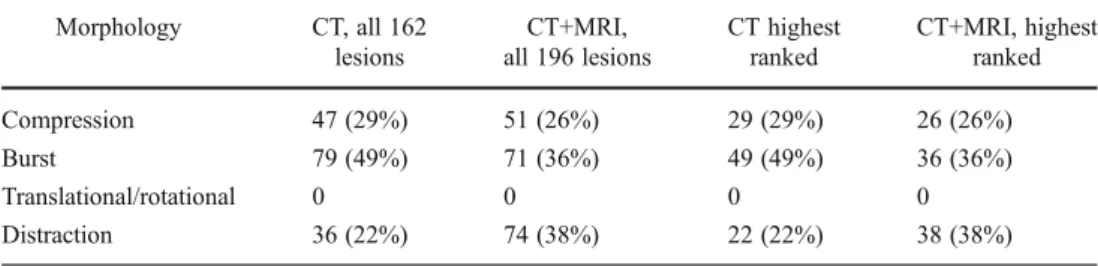
Distribution and classification of fracture morphology re-garding the TLICS classification was similar to the AO classification, as shown in Table4.
With CT alone the integrity of the PLC was defined as intact in 80 (80%), suspect in 2 (2%), and injured in 18 (18%) of the 100 patients. Reasons for an injured assess-ment on CT was avulsion fracture of the spinous processes 10 out of 18 (56%), interspinous spacing 2 out of 18 (11%), fracture of spinous processes in combination with interspi-nous spacing 3 out of 18 (17%), diastasis of the facet joints 1 out of 18 (6%), fracture of spinous processes in combination with diastasis of the facet joints 1 out of 18 (6%), and interspinous spacing in combination with diastasis of the facet joints 1 out of 18 (6%). The reason for a suspect classification on CT was a suspected fracture of the spinous process in two cases.
With CT and MRI together the PLC was assessed as intact in 55 out of 100 (55%), suspect in 3 out of 100 (3%), and injured in 42 out of 100 (42%) patients. Most of the PLC lesions (29 out of 42, 69%) were combinations of injuries of the different components: [8 out of 42 (19%); 7 out of 42 (17%) ISL+SSL; 7 out of 42 (17%) ISS+LF+FJC; 3 out of 42 (7%) ISL+FJC; 3 out of 42 (7%) ISL+SSL +FJC]. In 13 out of 42 patients (31%) only one structure of the PLC was injured [7 out of 42 (17%) ISL; 4 out of 42 (10%) SSL; 2 out of 42 (5%) FJC].
Calculated from these data, the most frequently affected PLC structure was the ISL, which was injured in 36 out of 42 cases (86%). This was followed by the SSL and the FJC, each with 23 out of 42 (55%), and, finally, by the LF with 16 out of 42 injuries (38%). The suspected/indeterminate PLC lesions were assessed for the ISS in two cases and once for the FJC.
Fig. 3 A 53-year-old man after a car accident. a Sagittal computed tomography shows an incomplete burst fracture L1 (arrow) without any obvious lesion of the posterior ligamentous complex. Lesion was classified as Arbeitsgemeinshaft für Osteosynthesefragen (OA) A3.1 and Thoraco-Lumbar Injury Classification and Severity (TLICS) injury severity score (ISS) 1. b, c After magnetic resonance imaging (b,
sagittal T2-weighted image; c, sagittal STIR image) a lesion of the supraspinous ligament was seen (arrows), which subsequently changed the classification systems to AO B1.2 and TLICS ISS 7. Change in TLICS ISS includes recommendation of surgery instead of conserva-tive treatment
Table 3 Number and percentage of fractures according to the Arbeitsge-meinshaft für Osteosynthesefragen (AO) classification system on comput-ed tomography (CT) and on CT and magnetic resonance imaging (MRI)
CT, n 0 162 (%) CT+MRI, n 0 196 (%) Type A 136 (84) 143 (74) Group A1 67 (41) 91 (46) Subgroup A1.1 17 (10) 48 (24) Subgroup A1.2 45 (28) 41 (21) Subgroup A1.3 5 (3) 2 (1) Group A2 10 (6) 8 (4) Subgroup A2.1 5 (3) 5 (3) Subgroup A2.2 4 (2) 3 (2) Subgroup A2.3 1 (0.6) 0 Group A3 59 (36) 44 (22) Subgroup A3.1 43 (27) 33 (17) Subgroup A3.2 2 (1) 0 Subgroup A3.3 14 (9) 11 (7) Type B 26 (16) 53 (27) Group B1 11 (7) 30 (15) Subgroup B1.1 0 0 Subgroup B1.2 11 (7) 30 (15) Subgroup B1.3 0 0 Group B2 14 (9) 22 (11) Subgroup B2.1 4 (2) 4 (2) Subgroup B2.2 0 0 Subgroup B2.3 10 (6) 18 (9) Group B3 1 (0.6) 1 (0.5) Subgroup B3.1 1 (0.6) 1 (0.5) Subgroup B3.2 0 0 Subgroup B3.3 0 0 Type C 0 0
Eighty-six of 100 patients (86%) were neurologically asymptomatic, whereas 10 out of 100 patients received 2 points and 4 patients (4%) received 3 points according to the TLICS classification (see Table2).
Changes in grading the integrity of the PLC assessing CT and MRI together
The proportion of patients whose PLC assessment did not change using CT and MRI data together amounted to 68 out of 10000.68 with 95% Wilson CI (0.58, 0.76). The propor-tion of patients whose PLC assessment changed amounted to 32 out of 10000.32 with 95% Wilson CI (0.24, 0.42). Changes in the overall ISS score for the TLICS
classification
The median ISS using CT alone was 2.0, whereas the median ISS with CT and MRI together was 3.5, Wilcoxon effect 4.00, Wilcoxon p<0.001, with 95% Wilson CI (3.00, 5.00).
In 67 out of 100 cases (67%), the ISS remained un-changed using CT and MRI together, but it un-changed in 33 patients (upgraded 30 cases; downgraded 3 cases).
The proportion of patients seen with a TLICS ISS on CT <5 (indication for conservative therapy) and≥ 5 on CT and MRI together (indication for surgical therapy) amounted to 24 out of 10000.24 with 95% Wilson confidence interval (0.17, 0.33). Three out of 100 patients with TLICS ISS≥0 5 on CT were downgraded to ISS<5 using CT and MR imag-ing together.
Myelopathy on MR images was seen in 2 out of 100 patients (2%).
Discussion
Our study demonstrates how frequently the classification of thoracolumbar spinal injuries changed after performing MRI in addition to initial CT. Classifying traumatic injuries is a helpful tool for standardizing diagnoses, for communica-tion, and for evidence-based therapeutical management. Hence, as several studies have demonstrated in recent years [24–26], the consistency, reliability, and validity of the classification systems play an important role. While CT is
currently the standard imaging modality in the initial work-up of spine injuries in emergency departments, MRI is increasingly used owing to its wide availability and the recognition of the importance of evaluating soft tissue struc-tures, especially the PLC. Lesions of the PLC are often not recognized, which may cause problems as standard therapy includes posterior instrumentation and possibly fusion. Therefore, the influence of additional information derived from MRI could improve the clinical work-up and affect the decision of whether surgery is needed or not. Our patient sample represents the expected patient distribution in terms of time of image acquisition, age, gender, trauma mecha-nism, and fracture distribution [27, 28]. Three quarters of the MR examinations were performed within 48 h of the initial CT, allowing for an accurate therapeutical work-up and management.
The combination of CT and MRI revealed considerably more osseous and ligamentous lesions than CT alone. This was discussed recently and analyzed in a study by Pizones et al. [6], who assessed the variability of trauma classification systems depending on the imaging modalities. Our study confirms their results in a larger sample and shows the influence of MRI on widely-used trauma classification sys-tems. Also, we drew special attention to the PLC as one of the most determining structures for indicators of the severity of spinal injuries [3,7].
Our study verified the hypothesis that a high number of injury scores would change after MRI, particularly in rela-tion to the evaluarela-tion of the PLC. In the AO classificarela-tion, an upgrade from a simple type A compression mechanism to a more complex type B (flexion/distraction) lesion was the most common change in the classification in our study.
Consideration of possible reasons for modifying the clas-sifications include the improved visualization of the inter-spinal ligament, which was injured in more than a third of our 100 patients. Overall, ligamentous lesions detected by MRI seem to be important and frequent reasons for changes in the trauma classification, justifying the need for addition-al MRI in patients with spinaddition-al fractures seen on the initiaddition-al CT. This agrees with a recent study in which Radcliff et al. proved the importance of MR examinations by showing that classical morphological signs of vertebral body lesions, such as vertebral body height or kyphosis, may not allow clear advice on the condition of the PLC to be given [18].
Table 4 Injury morphology assessed for the Thoraco-Lumbar Injury Classification and Severity scale classification for all lesions on computed to-mography (CT) and on CT and magnetic resonance imaging (MRI), depending on the frac-ture grades Morphology CT, all 162 lesions CT+MRI, all 196 lesions CT highest ranked CT+MRI, highest ranked Compression 47 (29%) 51 (26%) 29 (29%) 26 (26%) Burst 79 (49%) 71 (36%) 49 (49%) 36 (36%) Translational/rotational 0 0 0 0 Distraction 36 (22%) 74 (38%) 22 (22%) 38 (38%)
This study did not consider the therapeutic consequences for the patient. However, in a real clinical situation, the higher classification scores found here suggest that the choice of therapy might have been changed after the MRI, biased in favor of surgery. Our study provides evidence for this statement as the TLICS ISS, by nature, includes recom-mendations for further patient management (conservative vs surgical) and it was seen that this management recommen-dations changed owing to the new MRI findings in about a quarter of the patients. This represents one of the greatest benefits of additional MRI. At our institution, MRI is there-fore now part of the routine work-up of such patients.
Bone bruise, which does not imply surgical intervention, but might be an indirect sign of fracture and cause of pain in patients with non-suspicious vertebral bodies on CT, was detected in almost a quarter of all patients. Myelopathy was seen in only 2% of the patients, which may not justify additional MRI in patients with type A and B fractures without neurological symptoms. However, myelopathies were not the focus of our study.
Several limitations of the study deserve comment. First, this was a retrospective study. Consequently, we could not investigate prospectively the decision-making of the sur-geons. However, larger prospective studies are needed to evaluate the influence of MRI on clinical decision-making. We strongly recommend such studies. Second, owing to the retrospective nature of our study, there was a selection bias. Although patients with a fracture seen on CT routinely undergo MRI at our hospital, those patients with a negative usually do not. Thus, patients with an initial false-negative CT were not included in this study. However, we would expect to see some more lesions in those patients with MRI. Third, consensus reading was performed to try to reach the highest quality and concordance in the interpretation of the images, and to avoid discrepancies in interpretation. This is consistent with studies reporting on the reliability of con-sensus readings [29,30], and was necessary because some classification systems have limited inter- and intra-reader reliability. Fourth, because we chose a consensus reading for image analysis, the inter-observer variability could not be assessed. However, in our study consensus reading reflects in an analogous way the clinical setting in our emergency radiology section, where images are assessed in consensus by senior and junior radiologists. Last, patients suffering from an AO type C fracture usually undergo emergency surgery immediately without any further MRI and therefore were not included in our study.
In conclusion, MRI of patients with thoraco-lumbar spi-nal trauma considerably improved the detection of fractures and soft tissue injuries compared with CT alone, and signif-icantly changed the overall trauma classification. In future, MRI may be an essential imaging modality for thoroughly assessing the whole extent of spine injury, and the high
number of undetected lesions on CT may also encourage other trauma teams to reconsider imaging algorithms relying on CT alone.
Acknowledgements We thank Marina Elliott for reading our
manu-script as a native speaker.
Conflict of interest The authors declare that they have no conflict of
interest.
References
1. Parizel PM, van der Zijden T, Gaudino S, Spaepen M, Voormolen MH, Venstermans C, et al. Trauma of the spine and spinal cord: imaging strategies. Eur Spine J. 2010;19 Suppl. 1:S8–17. 2. Diaz Jr JJ, Cullinane DC, Altman DT, Bokhari F, Cheng JS, Como
J, et al. Practice management guidelines for the screening of thoracolumbar spine fracture. J Trauma. 2007;63(3):709–18. 3. Bagley LJ. Imaging of spinal trauma. Radiol Clin North Am.
2006;44(1):1–12 vii.
4. Antevil JL, Sise MJ, Sack DI, Kidder B, Hopper A, Brown CV. Spiral computed tomography for the initial evaluation of spine
trauma: a new standard of care? J Trauma. 2006;61(2):382–7.
5. Wilmink JT. MR imaging of the spine: trauma and degenerative
disease. Eur Radiol. 1999;9(7):1259–66.
6. Pizones J, Izquierdo E, Alvarez P, Sanchez-Mariscal F, Zuniga L, Chimeno P, et al. Impact of magnetic resonance imaging on deci-sion making for thoracolumbar traumatic fracture diagnosis and
treatment. Eur Spine J. 2011;20 Suppl. 3:390–6.
7. Van Goethem JW, Maes M, Ozsarlak O, van den Hauwe L, Parizel
PM. Imaging in spinal trauma. Eur Radiol. 2005;15(3):582–90.
8. Daffner RH, Hackney DB. ACR Appropriateness Criteria on sus-pected spine trauma. J Am Coll Radiol. 2007;4(11):762–75. 9. Denis F. The three column spine and its significance in the
classifica-tion of acute thoracolumbar spinal injuries. Spine. 1983;8(8):817–31. 10. Daffner RH, Deeb ZL, Goldberg AL, Kandabarow A, Rothfus WE. The radiologic assessment of post-traumatic vertebral
stabil-ity. Skeletal Radiol. 1990;19(2):103–8.
11. Yugue I, Aono K, Shiba K, Ueta T, Maeda T, Mori E, et al. Analysis of the risk factors for severity of neurologic status in 216 patients with thoracolumbar and lumbar burst fractures. Spine.
2011;36(19):1563–9.
12. Oner FC, Wood KB, Smith JS, Shaffrey CI. Therapeutic decision making in thoracolumbar spine trauma. Spine. 2010;35(21 Suppl.):
S235–44.
13. Pizones J, Zuniga L, Sanchez-Mariscal F, Alvarez P, Gomez-Rice A, Izquierdo E. MRI study of post-traumatic incompetence of posterior ligamentous complex: importance of the supraspinous ligament. Prospective study of 74 traumatic fractures. Eur Spine
J. 2012;21(11):2222–31.
14. Magerl F, Aebi M, Gertzbein SD, Harms J, Nazarian S. A com-prehensive classification of thoracic and lumbar injuries. Eur Spine J. 1994;3(4):184–201.
15. Vaccaro AR, Lehman Jr RA, Hurlbert RJ, Anderson PA, Harris M, Hedlund R, et al. A new classification of thoracolumbar injuries: the importance of injury morphology, the integrity of the posterior
liga-mentous complex, and neurologic status. Spine. 2005;30(20):2325–33.
16. Bozzo A, Marcoux J, Radhakrishna M, Pelletier J, Goulet B. The role of magnetic resonance imaging in the management of acute
spinal cord injury. J Neurotrauma. 2011;28(8):1401–11.
17. Vaccaro AR, Lee JY, Schweitzer Jr KM, Lim MR, Baron EM, Oner FC, et al. Assessment of injury to the posterior ligamentous
18. Radcliff K, Su B, Kepler C, Rubin T, Shimer A, Rihn J et al. Correlation of posterior ligamentous complex injury and neurolog-ical injury to loss of vertebral body height, kyphosis, and canal
compromise. Spine. 2011;37(13):1142–50.
19. Lee HM, Kim HS, Kim DJ, Suk KS, Park JO, Kim NH. Reliability of magnetic resonance imaging in detecting posterior ligament complex injury in thoracolumbar spinal fractures. Spine. 2000;25 (16):2079–84.
20. Lee JY, Vaccaro AR, Lim MR, Oner FC, Hulbert RJ, Hedlund R, et al. Thoracolumbar injury classification and severity score: a new paradigm for the treatment of thoracolumbar spine trauma. J
Orthop Sci. 2005;10(6):671–5.
21. Hollander M, Wolfe DA. Nonparametric statistical methods. Wiley Series in Probability and Statistics. New York: Wiley, 1999. 22. Newcombe RG. Two-sided confidence intervals for the single
proportion: comparison of seven methods. Stat Med. 1998;17
(8):857–72.
23. Wilson EB. Statistical Inference. Science. 1926;63(1629):289–96.
24. Koh YD, Kim DJ, Koh YW. Reliability and validity of Thoraco-lumbar Injury Classification and Severity Score (TLICS). Asian
Spine J. 2010;4(2):109–17. doi:10.4184/asj.2010.4.2.109.
25. Whang PG, Vaccaro AR, Poelstra KA, Patel AA, Anderson DG, Albert TJ, et al. The influence of fracture mechanism and mor-phology on the reliability and validity of two novel thoracolumbar
injury classification systems. Spine. 2007;32(7):791–5.
26. Oner FC, Ramos LM, Simmermacher RK, Kingma PT, Diekerhof CH, Dhert WJ, et al. Classification of thoracic and lumbar spine fractures: problems of reproducibility. A study of 53 patients using CT and MRI. Eur Spine J. 2002;11(3):235–45.
27. Pickett GE, Campos-Benitez M, Keller JL, Duggal N. Epidemiol-ogy of traumatic spinal cord injury in Canada. Spine. 2006;31
(7):799–805.
28. Wyndaele M, Wyndaele JJ. Incidence, prevalence and epidemiol-ogy of spinal cord injury: what learns a worldwide literature
survey? Spinal Cord. 2006;44(9):523–9.
29. Pugliese F, Hunink MG, Gruszczynska K, Alberghina F, Malago R, van Pelt N, et al. Learning curve for coronary CT angiography:
what constitutes sufficient training? Radiology. 2009;251(2):359–68.
30. Shaw CM, Flanagan FL, Fenlon HM, McNicholas MM. Consen-sus review of discordant findings maximizes cancer detection rate in double-reader screening mammography: Irish National Breast