The use of the Jasper Jumper for the correction of Class II malocclusion in the young permanent dentition
11
0
0
Texte intégral
(2) 272 the Jumper for the treatment of Class II malocclusions in adults have been published by Cash (1991) and Bantleon (1994). Because the Jasper Jumper is a new treatment modality, only a few reports on its effects have as yet been published. The results of these studies agree that much of the effect of the Jumper is dento-alveolar. There is, however, disagreement regarding the skeletal effect of the treatment. Thus, in the permanent dentition, May et al. (1993) found the skeletal effect to be predominantly mandibular. Cope et al. (1994), on the contrary, found little or no effect on the growth of the mandible, but a backward displacement (retrusion) of the maxilla. All studies so far have evaluated the effect of the Jasper Jumper at the time of its removal. There are no studies of the stability of the treatment effects. The present investigation was carried out to further clarify the effects of the correction of Class II malocclusions in the young permanent dentition with the Jasper Jumper and the stability of these effects after a period of observation. Subjects and methods Sixteen females and 10 males were included in the study. The age of the subjects varied between 13 years, 1 month and 24 years, 8 months (median 14 years, 8 months). In 10 of the subjects the Jasper Jumper was included in the original treatment plan of a Class II malocclusion while a Jumper was not part of the original plan of the remaining 16 subjects. In these cases the Jumper was installed during the course of the treatment in order to correct a Class II intermaxillary relationship. The reason for including the Jumper in the 16 cases where it had not been planned originally was loss of anchorage, relapse, or failure to correct the malocclusion with growth modification methods. Eleven of the 16 patients had worn a headgear and seven an activator before treatment with the Jumper. Jasper Jumper set-up In preparation for the use of the Jumper, the upper and lower first molars were banded, the upper and lower second molars bonded with a. N. S T U C K I A N D B. I N G E RVA L L. Figure 1. Jasper Jumper as used in the study.. buccal tube, and the premolars and anterior teeth bonded with brackets. Brackets and tubes of the make Ormco (Syron Corporation, Glendora, CA), diamond type, with a 0.022-inch slot were used. The brackets had a pretorque of +14, +7, and –7 degrees for the maxillary central and lateral incisors, and the canine, respectively. The corresponding values for the lower incisors were –1 and, for the lower canine, –11 degrees. During the use of the Jumper an upper 0.021 × 0.025-inch arch was used in 19 cases (16 patients had a steel arch and 3 a TMA arch), while a 0.019 × 0.025inch arch was used in the remaining seven cases (steel in four cases and TMA in three cases). The maxillary anterior and posterior teeth were blocked together as one unit. A lower 0.021 × 0.025-inch arch was used in 17 cases (14 steel and three TMA). Nine patients had a lower 0.019 × 0.025-inch arch (six steel and three TMA). The lower arch was tied back to the first molar. In all cases the upper first molars were connected with a Goshgarian transpalatal arch of the make GAC (GAC International Inc., Central Islip, NY). In the upper arch the Jumper was attached to the headgear tube of the first molar as prescribed by the manufacturer (American Orthodontics, Sheboygan, WI). In the lower jaw the Jumper was threaded on a sectional 0.017 × 0.025-inch steel arch inserted in the second tube of the first molar band as described by Blackwood (1991) and hooked over the lower canine bracket from the mesial side (Figure 1). The arch was cinched back distal of the molar tube. The advantages of this arrangement in the lower jaw are that the Jumper can be replaced without the need to.
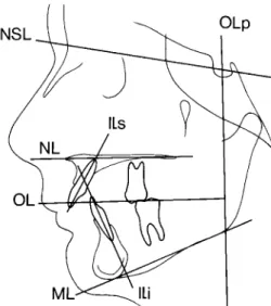
(3) 273. U S E O F T H E JA S P E R J U M P E R. Profile cephalograms were made at the insertion and removal of the Jumpers as well as at the end of the observation period, median time 7 months, range 4–12.5 months, after the removal of the Jumper. At the end of the observation period 11 cases were still in retention while in the remaining 12 cases the retention had been discontinued 1–9 months (median 4 months) before the final recording.. The cephalograms were made with the mandible in the retruded contact position. To maintain this position a wax index, made with the technique of Ingervall (1968), was inserted between the jaws during the exposure. The reference points and lines shown in Figures 2 and 3 were used for the cephalometric analysis. The cephalograms were traced on matte acetate paper using a 0.3-mm lead pencil. The computerized cephalometric analysis system of Gebauer (1977) was used. Distances were reduced to zero magnification. The analysis of antero-posterior linear changes was undertaken using the method of Pancherz (1982). A co-ordinate system, consisting of the occlusal line (OL) and a perpendicular to this line through the point sella (OLp) was drawn on the tracing of the pretreatment cephalogram. The coordinate system was transferred to the subsequent cephalograms of the series by superimposing on structures of the anterior cranial base as described by Björk (1968). The overjet was recorded as the difference between the variables Is–OLp and Ii–OLp (Table 1). The overbite was recorded as the vertical distance between the points Is and Ii, measured perpendicular to a constructed horizontal line, angulated 6 degrees to the nasion–sella line (NSL). The molar relationship was recorded as the difference between the. Figure 2 analysis.. Figure 3 analysis.. remove the main arch and that both lower premolars can be bonded. The Jumper was selected according to the instructions of the manufacturer, i.e. with an activation of 4 mm. In cases with a need for reactivation a new Jumper was inserted or the lower acrylic ball stop of the appliance was moved back with the aid of a crimpable stop on the sectional arch. The Jumpers were used bilaterally until correction of the posterior teeth into a Class I relationship was obtained. The median treatment time with the Jumper was 5 months (range 2–11 months). After removal of the Jumpers 18 cases were retained with Class II elastics, five cases with an activator, while three cases were left without retention. The median retention time was 5 months (range 2–9 months). Recordings. Reference points used in the cephalometric. Reference lines used in the cephalometric.
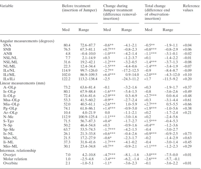
(4) 274. N. S T U C K I A N D B. I N G E RVA L L. Table 1 Median and range of the cephalometric variables at the insertion of the Jumper and of the differences between the variables at the removal of the Jumper and between the end of observation and the insertion of the Jumper. Variable. Angular measurements (degrees) SNA SNB ANB NSL/NL NSL/ML NL/ML ILs/NL ILi/ML ILs/ILi Linear measurements (mm) A–OLp Is–OLp Ii–OLp Msa–OLp Mia–OLp Pg–OLp Ar–OLp N–Me S–Tgo N–Sp Sp–Me Is–NL Mso–NL Ii–ML Mio–ML Intermax. relationship Overjet Molar relation Overbite. Before treatment (insertion of Jumper). Change during Jumper treatment (difference removalinsertion). Total change (difference end of observationinsertion). Med. Range. Med. Range. Med. Range. 80.4 76.5 4.8 7.7 31.6 22.3 114.9 102.0 122.2. 72.6–87.7 67.3–81.1 –0.4–10.0 2.1–14.9 19.2–42.1 12.4–34.4 99.7–126.0 86.9–109.5 113.2–138.4. –0.6** +0.7*** –1.0*** +0.3 –1.2*** –1.5*** –2.7** +6.4*** –2.5. –4.1–2.1 –0.8–2.3 –4.2–1.4 –1.2–3.7 –3.2–0.5 –4.4–0.6 –17.2–12.5 0.9–14.0 –24.3–11.2. –0.5** +0.8*** –1.1*** +0.1 –1.4*** –1.4*** –0.9 +2.0*** +1.7. –1.9–1.1 –0.8–2.9 –3.1–0.1 –1.4–2.2 –3.7–1.3 –3.4–1.9 –8.4–8.7 –4.3–12.0 –11.5–9.2. +0.04 +0.06 –0.02 –0.01 –0.08 –0.07 –0.09 +0.10 +0.20. 73.2 80.1 72.4 53.3 52.0 74.1 10.4 112.9 71.5 50.2 63.7 26.1 21.5 37.3 30.1. 63.6–81.4 67.9–88.4 63.6–81.6 41.5–60.2 40.5–61.1 61.8–86.1 4.0–21.9 100.9–125.4 56.7–87.3 46.4–54.8 53.5–74.5 21.3–33.8 17.2–27.9 31.8–41.6 23.4–34.8. –0.1 –1.6*** +2.9*** –0.9** +2.6*** +1.4*** 0.0 –1.1*** +0.4* +0.3 –1.7*** +0.6*** –0.6* –1.7*** +0.7**. –3.2–1.6 –5.4–1.5 0.3–6.9 –2.7–2.4 1.0–5.9 –0.9–5.0 –1.1–2.1 –3.0–1.6 –1.7–2.7 –0.9–1.6 –4.2–1.5 –0.4–2.6 –2.3–1.7 –4.1–0.2 –0.9–2.1. +0.3 –0.8 +2.7*** +0.3 +2.7*** +1.9*** +0.2 +0.2 +1.5*** +0.4** –0.4 +0.9*** –0.2 –0.4 +1.1***. –1.9–1.7 –3.6–2.6 0.0–6.4 –3.1–4.4 0.5–5.5 –1.0–5.6 –1.7–2.4 –2.4–5.6 –0.4–5.3 –1.2–3.6 –3.0–2.7 –0.9–2.5 –1.4–2.9 –3.0–1.4 –1.2–2.3. +0.37 +0.49 +0.48 +0.61 +0.66 +0.38 +0.21. 4.2–10.8 –2.5–4.8 –1.0–5.1. –4.7*** –3.4*** –1.1*. –8.1, –1.6 –6.2, –1.4 –3.6–2.3. –3.0*** –2.6*** –0.1. –7.5–0.0 +0.01 –5.7, –0.1 –3.6–2.2 +0.01. 7.0 1.0 2.1. Reference values. +0.73 +0.63 +0.45 +0.29. *0.01 < P < 0.05; **0.001 < P < 0.01; ***P < 0.001.. variables Msa–OLp and Mia–OLp. Thus, a positive sign denotes a distal and a negative sign a neutral molar relation. Errors of the method and statistical methods used The errors of the method were calculated from double measurements of 15 randomly selected radiograms that were retraced. Systematic differences between the double determinations were tested with Wilcoxon’s matched pairs, signed. ranks test. Seven distances had small, significant differences between the two measurements (mean 0.25–0.54 mm). The same was true for the angle ILs/NL (mean difference 0.50 degrees). The accidental errors of the method (si) were calculated with the formula si =. ∑d 2. ! 2n. where d is the difference between the two measurements and n is the number of recordings. The.
(5) 275. U S E O F T H E JA S P E R J U M P E R. accidental errors for distances varied between 0.31 and 0.61 mm, and for angles between 0.23 and 0.72 degrees with the exception of the angles ILi/ML and ILs/ILi, which had errors of 1.4 and 1.8 degrees, respectively. Differences between distributions were tested with Mann–Whitney’s U-test and between paired observations with Wilcoxon’s matched pairs, signed ranks test. Relationships between variables were analysed with Spearman rank correlation. Results Use of Jumpers All of the subjects adjusted rapidly to the Jumpers. These were well tolerated. The number of Jumpers used per side varied between 1 and 6 (median two on each side). Ten patients had only one Jumper on the right and 12 patients one on the left side. Nine and 10 patients had two Jumpers on the right and left sides, respectively. Thus, 19 and 22 patients had 1–2 Jumpers on the right and left sides, respectively. Only one patient had six Jumpers. Altogether, 100 Jumpers were used. Nine Jumpers fractured and had to be replaced. The fracture rate was thus 9 per cent.. nor was there any significant difference between the sexes in any of the variables at the start of the treatment. In the analysis all subjects were therefore included in one group. The values of the cephalometric variables at the insertion of the Jumpers and the changes during the active treatment (removal – insertion) as well as the final effect of the treatment (end of observation – insertion) are given in Table 1. The table also gives reference values for the change of the variables during a period of 6 months in children with untreated Class II malocclusions. These reference values were calculated and used in a previous study (Weiland et al., 1997). They were collected from many sources in the literature. In all, 15 studies were used. They mainly refer to children younger than the subjects of this study. Partly different reference points and measurements were used, especially for linear variables. No correction for linear enlargement was made as many studies lacked these data. The observation periods varied from 6 months to 6 years. As the growth rate during a longer period is not constant, the growth rates for an observation period of 6 months, as given in Table 1, are just indicative. Antero-posterior changes. Effect of treatment The cephalometric variables at the insertion of the Jumpers were tested for differences between the 10 cases where the Jumper was planned at the start of the treatment and the 16 subjects where this was not the case. Only two variables differed significantly between the two types of subjects. Thus, the molar relationship, (Msa–OLp) – (Mia–OLp), was more distal in the 10 cases where a Jumper had been planned as part of the treatment than in the other 16 cases (0.001 < P < 0.01). The median of the molar relationship in the 10 cases was 2.7 mm, compared with 1.0 mm in the other 16 cases. In the 10 cases where the Jumper was part of the original treatment plan the upper molar was in median 3.7 mm more anterior (var. Msa–OLp, 0.01 < P < 0.05) than in the other cases. There was, however, no significant difference between the two types of patients in the treatment time with the Jumper,. The maxillary prognathism (var. SNA) decreased slightly during treatment, but the mandibular prognathism (var. SNB) increased. There was consequently a decrease of the antero-posterior inter-maxillary relationship (angle ANB) during treatment. This decrease was obtained during the period of use of the Jumper and remained stable at the end of observation. In contrast to the decrease of the angle SNA, the distance A–OLp remained constant during treatment. The distance Pg–OLp, on the other hand, increased during the period of use of the Jumper, in analogy with the increase of the SNB angle. After removal of the Jumpers, the distance Pg–OLp continued to increase somewhat until the end of the observation period. The effect of the Jumpers on the maxillary dentition was a moderate retrusion (decrease of the distances Is–OLp and Msa–OLp), but this effect relapsed after removal of the Jumpers so.
(6) 276 that at the end of the observation period there was no difference relative to the start of the treatment. The two distances Ii–OLp and Mia–OLp increased markedly with the use of the Jumpers and remained stable at the end of observation. There was a considerable decrease of the overjet and improvement of the molar relationship during treatment with the Jumpers. For the overjet, much of this effect relapsed after removal of the Jumpers so that only 64 per cent remained at the end of observation. The stability of the improvement of the molar relationship was better, with 76 per cent remaining at the end of observation.. N. S T U C K I A N D B. I N G E RVA L L. the active phase of treatment. The decrease of anterior face height occurred in the lower part of the face (distance Sp–Me) as the upper anterior face height (distance N–Sp) was unchanged during the period of observation. The decrease of the anterior face height took place during the use of the Jumper and relapsed in the following period so that no difference relative to the original face height was found at the end of observation. Contributing to the change in mandibular inclination was the slight increase in posterior face height (distance S–Tgo) observed at the end of the Jumper phase and at the end of observation. Vertical dento-alveolar changes. Axial inclination of the incisors In general, the maxillary incisors (var. ILs/NL) retroclined moderately when the Jumpers were used. After this phase of treatment, however, they uprighted again so that at the end of observation no significant difference relative to their original axial inclination was found. The mandibular incisors (var. Ii/ML) proclined markedly when the Jumpers were used but uprighted largely when the Jumpers were removed. Only about 30 per cent (median) of the proclination that had taken place during the phase of Jumper therapy remained at the end of the observation period. There was a remarkably large range of variation both for the change of the upper incisor inclination, where both retroclination and proclination were found, and for the proclination of the lower incisors during the treatment. The maximum value for lower incisor proclination at the end of observation was 12 degrees, but in one case the lower incisors had retroclined 4 degrees.. The maxillary incisors were slightly elongated (distance Is–NL) during the active treatment and remained so at the end of observation. The mandibular incisors, on the other hand, intruded during the use of the Jumpers (distance Ii–ML). The intrusion relapsed, however, during the second period of observation. The overbite developed in analogy with the vertical skeletal changes and with the changes in the vertical positions of the incisors. Thus, the overbite decreased during the active phase of treatment, but was not significantly different from its original value at the end of observation. The upper first molars (distance Mso–NL) intruded slightly when the Jumpers were used. After the active phase of treatment, however, they extruded again so that no significant difference relative to the original value was found at the end of observation. The lower first molars (distance Mio–ML) extruded slightly during treatment with the Jumper and remained extruded at the end of observation.. Vertical skeletal changes The inclination of the maxilla (var. NSL/NL) remained stable during treatment. In contrast, the inclination of the mandible (var. NSL/ML) and the vertical jaw relationship (var. NL/ML) decreased during treatment. This decrease took place during the use of the Jumpers and remained at the end of observation. In analogy with the change in mandibular inclination, the anterior face height (distance N–Me) decreased during. Effect of age on changes during the period of observation The age of the subjects varied between 13 and 24 years. In fact, however, the oldest subject was an extreme case. The next oldest subject was 16 years, 8 months. Any effect of age on the outcome of the treatment was evaluated by assigning the subjects to one of two groups, i.e. (1) younger than the median age or (2) older than the median.
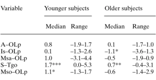
(7) 277. U S E O F T H E JA S P E R J U M P E R. age. The group of younger subjects contained seven boys and six girls with an age below 14 years, 8 months at the start of treatment. The group of older subjects included three boys and 10 girls older than 14 years, 8 months at the start of treatment. Only one variable differed between the two subgroups. This was the anterior face height (distance N–Me) which was larger in the younger group than in the older group at all three recordings, i.e. before and after active treatment and at the end of observation (0.01 < P < 0.05). There was no significant difference between the two groups in the change of the variables during the active treatment. During the whole period of observation five variables changed differently in the two subgroups (0.01 < P < 0.05). Three of these variables concerned the antero-posterior position of maxillary structures (Table 2). In the group of older subjects there was less increase of the maxillary protrusion (distance A–OLp) and the maxillary incisors and molars moved backwards (distances Is–OLp, Msa–OLp) which was not the case in the group of young subjects. The posterior face height (distance S–Tgo) increased and the upper molars extruded (distance Mso–NL) more in the group of younger subjects. There were no differences between the groups in antero-posterior changes of the mandibular structures.. Table 2 Median and range (in mm) of total change (end of observation—insertion) for variables with significantly different change in younger and older subjects. Variable. A–OLp Is–OLp Msa–OLp S–Tgo Mso–OLp. Younger subjects. Older subjects. Median Range. Median. Range. 0.8 0.1 1.0 1.7*** 1.1*. 0.1 –1.1* –0.5 0.7** –0.6. –1.7–1.0 –3.6–1.3 –1.9–0.9 –0.4–3.1 –1.4–2.9. –1.9–1.7 –1.3–2.6 –3.1–4.4 0.0–5.3 –1.3–1.7. *0.01 < P < 0.05; **0.001 < P < 0.01; ***P < 0.001.. values of the changes with treatment at the removal of the Jumpers and at the end of observation, are presented for the overjet in Figure 4 and for the molar relationship in Figure 5. During the use of the Jumpers all components changed in a direction contributing to the reduction of the overjet and to the improvement of the molar. Components of Class II correction The changes in overjet and in molar relationship were brought about by skeletal and dento-alveolar changes. These can be differentiated with the method of Pancherz (1982). The skeletal component of the changes in overjet and in molar relationship can be calculated as the difference in the relationship between the variables A–OLp and Pg–OLp during treatment. The dento-alveolar changes, i.e. movement of the incisors within the jaws, can be calculated as the change of the difference Is–OLp minus A–OLp and Ii–OLp minus Pg–OLp, respectively, during the treatment. In a similar way, the movement of the molars within the jaws can be calculated as the change of the difference Msa–OLp minus A–OLp and Mia–OLp minus Pg–OLp during treatment. The results of this analysis, based on the mean. Figure 4 Components contributing to the reduction of the overjet at removal of the Jumpers (a) and at the end of observation (b). + denotes an anterior and – a posterior movement of the point. Mean values in mm..
(8) 278. N. S T U C K I A N D B. I N G E RVA L L. Discussion. Figure 5 Components contributing to the improvement of the molar relationship at removal of the Jumpers (a) and at the end of observation (b). + denotes an anterior and – a posterior movement of the point. Mean values in mm.. relationship. In the period thereafter the maxillary relapse and the partial relapse of the dentoalveolar mandibular changes mean that the effects of the treatment at the end of observation were mainly accomplished by a skeletal mandibular reaction. The increase in mandibular prognathism during treatment was correlated to sex, age, duration of treatment, number of Jumpers used, and to the cephalometric variables at the start of the treatment. No significant correlation was found. In order to further evaluate any possible effect of age on the components of Class II correction these variables were compared between the subgroups of younger and older subjects (Table 3). Only one significant difference was found, namely the already described greater total skeletal effect on the maxilla (distance A–OLp) in the group of older subjects. As stated previously, the skeletal mandibular effect was the same in the groups of younger and older subjects, and there were no significant differences between the groups in intra-maxillary and intra-mandibular dental movements.. The subjects of this investigation are the consecutive cases treated with the Jasper Jumper in the Department of Orthodontics, University of Bern, during the period covered by the investigation. Most of the cases were treated by the same orthodontist (N.S.), but the sample also includes cases treated by other orthodontists. All treatments were, however, performed according to the same principles and were supervised by the first author. There are, however, some discrepancies with regard to dimension and material of the edgewise arches, as well as type and duration of the retention. These inconsistencies are due to the needs of the individual patients and the preferences of the orthodontists. Homogeneity of the sample would have been desirable but, on the other hand, the present sample represents clinical reality. The cephalograms were taken with the mandible in the retruded contact position. This position was obtained by the guidance of the examiner with the patient as passive as possible. In order to stabilize the mandible in the retruded position, a wax bite was made which was used during exposure of the cephalogram. The wax bite was taken to initial (first) contact in the retruded position. The use of a wax bite, and the recording in the retruded position, implies a source of error, especially in the vertical dimension, because it cannot be guaranteed that the bite is taken to the exact first contact on different occasions. This disadvantage is, however, greatly outweighed by the certainty that the intermaxillary relationship is not falsified by protrusion of the mandible into the intercuspal position (a large slide in centric). Such a protrusion could easily have taken place in cases with an unstable occlusion at the start of treatment and could also have occurred as a result of the patient being used to protruding the mandible in order to reduce an uncomfortable force from the Jumper. The Jumper works with an anteriorly directed force on the mandible and a posterior force on the maxilla. Simultaneously, the Jumper exerts intrusive forces on the anterior part of the mandibular and on the posterior part of the maxillary dentitions. The intrusive forces resulted in.
(9) 279. U S E O F T H E JA S P E R J U M P E R. Table 3 Median and range (in mm) of components of Class II correction in groups of younger and older subjects.. Maxilla Skeletal (A–OLp) Incisor (intramax.) Molar (intramax.) Mandible Skeletal (Pg–OLp) Incisor (intramand.) Molar (intramand.). Change during Jasper treatment. Total change. Younger. Younger. Older. Older. Med. Range. Med. Range. Med. Range. Med. Range. –0.1 –0.9 –0.4. –3.2–1.5 –3.7–1.5 –2.0–4.7. –0.3 –1.7 –0.7. –1.5–1.6 –5.4–1.4 –2.5–0.7. 0.8 –0.2 0.6. –1.9–1.7 –2.1–1.4 –3.9–3.7. 0.1 –1.2 –0.4. –1.7–1.0 –3.4–1.9 –1.6–1.1. 1.6 1.5 1.2. –0.9–5.0 –0.8–1.9 0.0–2.0. 1.4 1.2 1.1. 1.0–2.6 0.1–2.7 0.0–2.4. 1.8 1.3 1.2. 0.9–5.6 –2.2–2.8 –1.6–2.0. 2.1 0.3 0.4. –1.0–4.4 –1.6–2.4 –0.3–1.8. intrusion of the mandibular incisors and of the maxillary first molars. These effects were expected and in agreement with the results of previous studies (Cope et al., 1994; Weiland et al., 1997). The intrusion of the maxillary molars enabled the mandible to autorotate in the direction of bite closure. The lower and total anterior face heights thus decreased when the Jumper was used. These effects were, however, to some extent transient. When the use of the Jumper was discontinued the maxillary molars erupted so that at the end of observation there was no difference in lower face height relative to the start of treatment. The inclination of the mandible, on the other hand, was somewhat less at the end of observation than at the start. This may, however, like the increase of the posterior face height, be an effect of normal growth because in most cases the mandible rotates anteriorly (Björk, 1963). Another effect of the intrusion of the maxillary molars during the use of Jumpers was an increased eruption of the mandibular molars. This is evident by comparison with the reference values and was also reported by Cope et al. (1994) and Weiland et al. (1997). The lower molars erupted much more than would have been expected from normal growth and development. This is logical because an intrusion or retardation of the eruption of the maxillary molars gives way to an increased eruption of the mandibular molars. This effect is in line with the. change in inclination of the occlusal plane reported by Weiland and Bantleon (1995). The overbite decreased during the active phase of treatment, but increased during the period of retention and observation. Again, this is an effect of a transient intrusion of the mandibular incisors together with transient changes of the inclination of the maxillary and mandibular incisors. In summary, the Jasper Jumper induces slight vertical changes which, however, are largely transient in nature. The maxillary incisors retroclined and the mandibular incisors proclined markedly during the use of the Jumpers. This was expected from previous studies (Weiland and Bantleon, 1995; Weiland et al., 1997). Our follow-up observations after the Jumper therapy showed, however, that the incisors uprighted to a large extent when the use of Jumpers was discontinued. In general, however, a moderate increase of the lower incisor proclination is a lasting effect of the Jumper treatment. In this study, the treatment with Jumpers had little or no effect on the maxillary prognathism. The angle SNA decreased somewhat but the distance A–OLp remained constant. In contrast, the effect on the mandibular prognathism was evident, with an increase both of the SNB angle and of the mandibular length (distance Pg–OLp). The increase in mandibular prognathism is in line.
(10) 280 with the results of Weiland and Bantleon (1995), and Weiland et al. (1997) after treatment in the mixed dentition and of May et al. (1993) in adolescents. Cope et al. (1994), on the other hand, found no skeletal mandibular effect in adolescents but a maxillary posterior displacement. The latter effect was limited in this study and in the investigations by May et al. (1993), Weiland and Bantleon (1995), and Weiland et al. (1997). Like this investigation, all previous studies have shown a retrusion of the maxillary incisors and molars and a protrusion of the mandibular teeth through bone after the Jumper therapy. The inclusion of a follow-up period in our study showed, however, that these effects were totally or largely transient. Thus, at the end of observation, the retrusion of the maxillary molar had relapsed completely and that of the maxillary incisors by 50 per cent. The large anterior movements of the mandibular incisors and molars had relapsed by about 50 per cent at the end of observation. The most important remaining component contributing to the successful reduction of the overjet and the correction of the molar relationship was the increase in mandibular prognathism. That a skeletal mandibular reaction, evident by comparison with the reference values, is possible in individuals of this age is somewhat surprising. The median age of the subjects at the start of treatment approached 15 years. Obviously, enough mandibular growth potential remained in these individuals to enable a skeletal mandibular reaction to occur. A considerable mandibular skeletal effect has also been reported in pubertal post-peak patients treated with the Herbst appliance (Pancherz and Hägg, 1985). In this study, at the removal of the Jumpers, 50 per cent of the correction of the molar relation and 40 per cent of the reduction of the overjet was due to skeletal components. For the overjet correction, this percentage compares favourably with the results of most previous studies, which reported a skeletal component of 38–48 per cent (Rankin, 1991; Weiland and Bantleon, 1995; Weiland et al., 1997). Only the study by May et al. (1993) had as low a skeletal component as 8 per cent for the overjet correction and 10 per cent for the molar correction. As. N. S T U C K I A N D B. I N G E RVA L L. mentioned, we found 50 per cent skeletal component in the molar correction, compared with about 40 per cent in the studies by Weiland and Bantleon (1995), and Weiland et al. (1997). At the recording at the end of observation the proportions between skeletal and dental components were quite different. Due to the relapse of some of the dental effects, the remaining overjet reduction was then to 64 per cent and the remaining molar correction to 78 per cent due to skeletal components (increase of the length of the mandible). Conclusions The study shows that the Jasper Jumper is an effective appliance for the correction of Class II division 1 malocclusions in the young permanent dentition. Partial relapse of the dental effects of the therapy after use of the Jumpers makes it clear that moderate over-correction is indicated. The appliance is easy to use and can be integrated in a normal edgewise set-up without the need of laboratory assistance. It also needs little chair-side time and is therefore inexpensive. It is well tolerated by the patients and the incidence of breakage with subsequent need for replacement is not very high. Address for correspondence Professor Bengt Ingervall Klinik für Kieferorthopädie Freiburgstrasse 7 CH-3010 Bern Switzerland References Bantleon H-P 1994 Die kieferorthopädische Behandlung beim jugendlichen und erwachsenen Patienten mit Anomalien der Klasse II/2. In: Ketterl W (ed.) Deutscher Zahnärztekalender, Hanser, Wien München, pp. 169–188 Björk A 1963 Variations in the growth pattern of the human mandible: Longitudinal radiographic study by the implant method. Journal of Dental Research 42: 400–411 Björk A 1968 The use of metallic implants in the study of facial growth in children: Method and application. American Journal of Physical Anthropology 29: 243–254 Blackwood H O 1991 Clinical management of the Jasper Jumper. Journal of Clinical Orthodontics 25: 755–760.
(11) U S E O F T H E JA S P E R J U M P E R. Cash R G 1991 Adult nonextraction treatment with a Jasper Jumper. Journal of Clinical Orthodontics 25: 43–47 Cope J B, Buschang P H, Cope D D, Parker J, Blackwood H O 1994 Quantitative evaluation of craniofacial changes with Jasper Jumper therapy. Angle Orthodontist 64: 113–122 Gebauer U 1977 Elektronische Mess- und Rechenanlage zur arcogrammetrischen Modelldiagnostik und zum Auswerten von Fernröntgenbildern. Schweizerische Monatsschrift für Zahnheilkunde 87: 1170–1180 Ingervall B 1968 Studies of mandibular positions in children. Odontologisk Revy 19, suppl. 15 May P W, Chada J, Ledoux W R, Weinberg R, McMinn R W 1993 A cephalometric examination of skeletal and dental changes in Class II, division 1 malocclusion treated with a fixed functional appliance. Certificate Thesis, Louisiana State University School of Dentistry, New Orleans Pancherz H 1982 The mechanism of Class II correction in Herbst appliance treatment. A cephalometric investigation. American Journal of Orthodontics 82: 104–113. 281 Pancherz H, Hägg U 1985 Dentofacial orthopedics in relation to somatic maturation. An analysis of 70 consecutive cases treated with the Herbst appliance. American Journal of Orthodontics 88: 273–287 Rankin T H 1991 Correction of Class II malocclusions with a fixed functional appliance. American Journal of Orthodontics and Dentofacial Orthopedics 100: 390 (Abstract) Weiland F J, Bantleon H-P 1995 Treatment of Class II malocclusions with the Jasper Jumper appliance—a preliminary report. American Journal of Orthodontics and Dentofacial Orthopedics 108: 341–350 Weiland F J, Droschl H 1996 Treatment of a Class II, division 1 malocclusion with the Jasper Jumper: a case report. American Journal of Orthodontics and Dentofacial Orthopedics 109: 1–7 Weiland F J, Ingervall B, Bantleon H-P, Droschl H 1997 Initial effects of treatment of Class II malocclusion with the Herren activator, activator-headgear combination and with the Jasper Jumper. American Journal of Orthodontics and Dentofacial Orthopedics 112: 19–27.
(12)
Figure
Documents relatifs