JON 3112 Urs Fischer Irene Ledermann Krassen Nedeltchev Niklaus Meier Jan Gralla Matthias Sturzenegger Heinrich P. Mattle Marcel Arnold
Quality of life in survivors
after cervical artery dissection
Introduction
Spontaneous cervical artery dissection (sCAD) is a well-recognized cause of stroke, especially in young patients, with a wide spectrum of clinical presentations [1, 2]. Pa-tients may present with isolated local manifestations, ischemic signs or both [3]. However, the most
appropri-ate end point in follow-up studies remains unclear: sev-eral studies used functional outcome, measured with the modified Rankin Scale score (mRS) or the recurrence rate. But the recurrence rate is low and an excellent out-come measured with the mRS is seen in the majority of these patients in hospital- and population-based studies [4, 5]. But since the mRS captures only functional dis-ability, it may not be sensitive enough to assess the se-Received: 7 April 2008
Received in revised form: 13 September 2008 Accepted: 24 September 2008 Published online: 18 March 2009
U. Fischer, MD · I. Ledermann, MS · K. Nedeltchev, MD · N. Meier, MD · M. Sturzenegger, MD · H. P. Mattle, MD · M. Arnold, MD (쾷)
Dept. of Neurology, Inselspital University Hospital Bern and University of Bern Freiburgstrasse 4 3010 Bern, Switzerland Tel.: +41-31/632-1072 Fax: +41-31/632-0321 E-Mail: [email protected] J. Gralla, MD, MSc
Dept. of Neuroradiology, Inselspital University Hospital Bern and University of Bern
Bern, Switzerland
■ Abstract Background and pur-pose Little data exists about
long-term outcome, quality of life (QOL) and its predictors after spontane-ous cervical artery dissections (sCAD). Methods Clinical and
ra-diological data of 114 patients with sCAD were collected prospectively. Six patients died within 3 months, the remaining 108 were contacted after a mean of 1498 days (range: 379–3455), 99 survivors (92 %) replied. QOL, assessed with the stroke-specific QOL scale (SS-QOL), and functional abilities, measured with modified Rankin Scale (mRS) were compared, and predictors of QOL were analyzed. Subgroup analyses were performed for patients with ischemic stroke, those with isolated local symptoms or transient ischemic symptoms and those without significant dis-abilities (mRS 0–1) at follow-up.
Results Seventy-one of 99 patients
(72 %) had no significant disability, but only 53 (54 %) reported a good QOL (SS-QOL ≥ 4). Compared to the self-rated premorbid QOL of all
patients, SS-QOL was impaired after sCAD (p < 0.001); impairment of QOL was observed in patients with ischemic stroke (p < 0.001), in patients with isolated local or transient ischemic symptoms (p < 0.038) and those without sig-nificant disabilities at follow-up (p = 0.013). Nevertheless, low mRS was associated with better overall QOL (Kendall’s tau > 0.5). High National Institute of Health Stroke Scale score on admission and higher age were independent pre-dictors of impaired QOL (p < 0.05).
Conclusion QOL is impaired in
almost half of long-term survivors after sCAD, even in patients with local or transient symptoms or without functional disability. Im-pairment of QOL is a surprisingly frequent long-term sequela after sCAD and deserves attention as an outcome measure in these patients. ■ Key words cervical artery dis-section · quality of life · outcome research
quelae in patients with sCAD, especially those with tran-sient or local symptoms and patients with no functional disability at follow-up.
QOL scales measuring patient centered outcome are sensitive for minimal changes and do not focus on gross physical aspects of disability only, but include psycho-logical and cognitive functions like memory, emotion, thinking, communication and social role, which may be impaired in patients who have suffered a potentially life-threatening event such as sCAD.
The aim of the present study was therefore to deter-mine QOL, functional dependency and social status in survivors after sCAD. We analyzed whether QOL was impaired compared to the premorbid condition in all patients and in subgroups (patients with ischemic stroke, those with isolated local symptoms or transient is chemic symptoms and those without significant disabilities (mRS 0–1) at follow-up). In addition, we analyzed whether baseline data on hospital admission predict QOL at long-term follow-up.
Methods
■ Investigations
We prospectively collected data at our stroke center on consecutive patients, presenting with first-ever sCAD from January 1997 to Sep-tember 2005. Patients with a cervical artery dissection due to major trauma were excluded from this study. All patients underwent a neu-rological examination, routine blood examination, electrocardiogra-phy, magnetic resonance imaging (MRI) of the brain, cervical MRI with T1 fat suppression and magnetic resonance angiography (MRA) or digital subtraction angiography of the four cervical arteries. Inter-nal carotid (ICAD) and/or vertebral artery (VAD) dissection was con-sidered proven if the affected vessel showed an intramural hematoma on axial cervical MRI cuts, or a string sign, intimal flap or pseudoan-eurysm on angiography [6–8].
■ Classification and risk factors
A sCAD was classified as spontaneous when occurring spontaneously or secondary to minimal trauma [9]. Dissections occurring after ob-vious head or neck trauma were classified as traumatic and were ex-cluded from the study. sCAD were differentiated into those with and without focal ischemic deficits of the brain, retina, or both. Ischemic deficits were classified according to their duration as stroke (> 24 hours), transient ischemic attack (TIA; ≤ 24 hours). The following lo-cal signs and symptoms were assessed: headache, neck pain, Horner’s syndrome, pulsatile tinnitus and cranial nerve palsy located on the side of dissection. Risk factors for ischemic stroke and sCAD were assessed as reported previously [10]. Arterial hypertension was de-fined as a positive history of treated or untreated hypertension. A history of migraine with or without aura was diagnosed by a neurolo-gist based on the International Headache Society’s criteria [11, 12]. The neurological deficit on admission was graded in all patients using the National Institute of Health Stroke Scale (NIHSS) score [13]. ■ Treatment
Patients with extracranial sCAD were treated with intravenous hepa-rin, followed by oral warfarin with an international target normalized
ratio of 2.5 (range 2.0–3.0) for 3–6 months, unless the patient suffered a hemispheric infarction with a high risk of secondary hemorrhage. Patients with large infarcts or with intracranial extension of a spon-taneous VAD received aspirin 100–300 mg/d for 3–6 months, if no subarachnoid hemorrhage was present on computed tomography or MRI.
■ QOL and functional outcome measurement
QOL was assessed with the stroke specific quality of life scale (SS-QOL) [14]. The SS-QOL is a disease-specific QOL scale, assessing 12 domains (energy, family roles, language, mobility, mood, personality, self-care, social roles, thinking, upper extremity function, vision and work) (Link to the questionnaire: http://www.strokecenter.org/trials/ scales/ssqol.html). Each item is ranked on a 5-point Likert scale, with higher scores indicating better function. Domain scores are un-weighted averages of the items compromising that domain, and the summary SS-QOL is an unweighted average of the 12 domain scores. A summary SS-QOL score of ≥ 4 was considered to indicate good QOL, < 4 an impaired QOL. Functional outcome was measured using the mRS [15]. Favorable outcome was defined as mRS 0–1, unfavor-able outcome as mRS 2 to 6.
■ Follow-up
Functional outcome and QOL were assessed from February 2006 to March 2007. If a patient was not able to communicate via phone, e. g., because of aphasia, a caregiver was contacted. In a structured tele-phone interview mRS, residential status, occupation and marital sta-tus were assessed [16]. QOL was assessed using the SS-QOL question-naire, which was sent to patients after the phone contact. They were asked to rate their QOL in the month before their dissection and in the month prior to follow-up. In addition, we asked whether the pa-tient needed help from their proxies to answer the questions. ■ Statistical analysis
Statistical analysis was performed using SPSS 13 for Macintosh statis-tical software (SPSS Inc.©, 2001). Demographic data are given as mean values. The NIHSS score is given as a median value. Correlation between functional outcome and QOL was measured with non-para-metric correlations (Kendall’s tau). Continuous variables were com-pared with the t-test for normal or the Mann-Whitney U test for ab-normal distributed variables. Categorical variables were compared with χ² and Fisher’s exact test as appropriate. QOL of all patients and subgroups (patients with ICAD and VAD; survivors with the follow-ing presentfollow-ing symptoms: ischemic stroke, those with isolated local symptoms or transient ischemic symptoms, those with no or minimal disabilities [mRS 0 or 1] and those with severe disabilities [mRS 2 to 5] at follow-up), were compared with the self-rated QOL before dis-section in all patients. Before performing the multivariate regression analysis, a univariate analysis was performed. Variables available at hospital admission were tested one by one against the dependent vari-able, and variables without association (p > 0.2) were removed from the model. The dependent variable was QOL, dichotomized as men-tioned above. Then logistic regression analysis with the forward step-wise method was performed to determine the independent associa-tion of QOL with other clinical factors. A two sided p-value < 0.05 was considered statistically significant.
Results
■ Baseline data
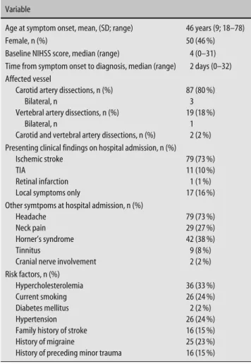
From January 1997 to September 2005, 114 consecutive patients with a sCAD were hospitalized in our stroke center. Six patients died within the first 3 months. Base-line characteristics of 108 survivors (50 women, 58 men; mean age 46 years) are shown in Table 1. Eighty-seven patients had an ICAD, 19 a VAD and 2 a dissection of both vessels. Twenty-eight patients presented with tran-sient or local symptoms, and 80 patients had an ischemic stroke or a retinal infarction. The 108 survivors were contacted after a mean of 1498 days (range 379–3455). Ninety-eight of the 108 survivors (91 %) were contacted by phone, 9 patients were lost to follow-up and one pa-tient returned the questionnaire, but could not be reached by phone. Outcome at follow-up, measured with the mRS, is given in Table 2.
■ QOL before and after sCAD
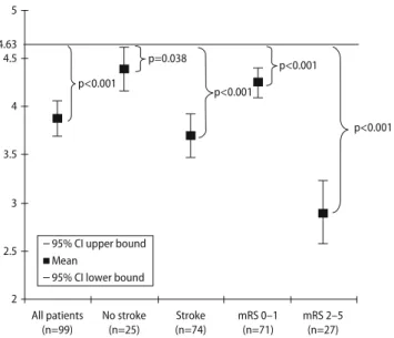
The SS-QOL questionnaire was completed by 99 of the 108 survivors (92 %). Seventy-four survivors (75 %) were able to complete the questionnaires without help. SS-QOL before dissection was rated as good in 92 of 99 sur-vivors (93 %), after dissection in 53 patients (54 %). Compared to the self-rated QOL of all patients before dissection, SS-QOL was significantly lower after sCAD (p < 0.001): mean SS-QOL before dissection was 4.63 (95 % confidence interval (CI) 4.55–4.71) and 3.87 (95 % CI 3.69–4.05) after dissection (Fig. 1).
■ QOL and functional outcome (mRS)
There was an overall correlation of high mRS and im-paired QOL (p < 0.001): patients with a high mRS score were more likely to suffer from an impaired QOL (Ken-dall’s tau > 0.5). Nevertheless, 21 of 71 patients (30 %) with a favorable outcome (mRS 0–1) reported an im-paired QOL. Only 2 of 27 patients (7 %) with an unfavor-able outcome (mRS 2–5) experienced a good QOL.
■ Subgroup analysis
SS-QOL varied among subgroups (Fig. 1). QOL was best in survivors with transient or local symptoms and worst in disabled patients due to stroke (mRS 2–5). A signifi-cantly impaired QQL compared to the overall QOL be-fore dissection was not only observed in patients with an ischemic stroke (p < 0.001), but also in patients with iso-lated local or transient ischemic symptoms (p < 0.038) and in patients without a disability (mRS = 0–1) at
fol-low-up (p = 0.013). SS-QOL was similar in patients with VAD (mean: 3.74, SD: 1.13) and ICAD (mean: 3.92, SD: 0.85) (p > 0.05). Patients with stroke and with local or Table 1 Baseline characteristics of survivors (n = 108)
Variable
Age at symptom onset, mean, (SD; range) 46 years (9; 18–78)
Female, n (%) 50 (46 %)
Baseline NIHSS score, median (range) 4 (0–31) Time from symptom onset to diagnosis, median (range) 2 days (0–32) Affected vessel
Carotid artery dissections, n (%) 87 (80 %)
Bilateral, n 3
Vertebral artery dissections, n (%) 19 (18 %)
Bilateral, n 1
Carotid and vertebral artery dissections, n (%) 2 (2 %) Presenting clinical findings on hospital admission, n (%)
Ischemic stroke 79 (73 %)
TIA 11 (10 %)
Retinal infarction 1 (1 %) Local symptoms only 17 (16 %) Other symtpoms at hospital admission, n (%)
Headache 79 (73 %)
Neck pain 29 (27 %)
Horner’s syndrome 42 (38 %)
Tinnitus 9 (8 %)
Cranial nerve involvement 2 (2 %) Risk factors, n (%)
Hypercholesterolemia 36 (33 %)
Current smoking 26 (24 %)
Diabetes mellitus 2 (2 %)
Hypertension 26 (24 %)
Family history of stroke 16 (15 %) History of migraine 25 (23 %) History of preceding minor trauma 16 (15 %)
Table 2 Outcome Modified Rankin Scale All (CAD and VAD), n = 108
mRS 0–1, n (%) 71 (66 %)
mRS 2–5, n (%) 27 (25 %)
Lost to follow-up, n (%) 9 (8 %)
Missing data, n (%) 1 (1 %)
Carotid artery dissection, n = 87
mRS 0–1, n (%) 56 (65 %)
mRS 2–5, n (%) 22 (25 %)
Missing data, n (%) 1 (1 %)
Lost to follow-up, n (%) 8 (9 %) Vertebral artery dissection, n = 19
mRS 0–1, n (%) 14 (74 %)
mRS 2–5, n (%) 5 (26 %)
Carotid and vertebral artery dissection, n = 2
transient symptoms did not differ regarding sex, vascu-lar risk factors and time to follow-up (p > 0.05).
■ Subitems of QOL
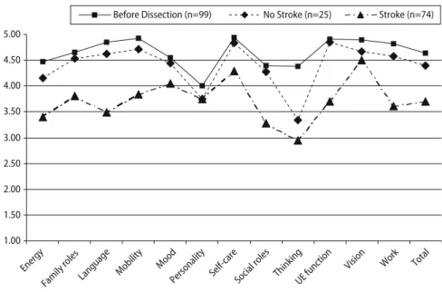
Figs. 2 and 3 show the different items of the SS-QOL in patients before and after dissection. The biggest change in QOL in survivors without significant disabilities at
follow-up (mRS 0–1) could be observed in the items “thinking”, “energy” and “language”, whereas in the do-mains “self care”, “personality” and “mood” the smallest change could be assessed. In patients with local or tran-sient symptoms QOL decreased most in the domains “thinking”, “energy” and “personality”, whereas “mood”, “family roles”, “self-care”, “social roles” and “upper ex-tremity function” were hardly impaired.
■ Residential status, occupation and marital status
At follow-up, 86 patients (88 %) were living in their home without help, 10 (10 %) with help and 2 (2 %) in a nurs-ing home. 81 survivors were able to communicate via phone. Before sCAD, 90 patients had been working full time, 8 part-time. After sCAD, 45 survivors had resumed full-time employment, 24 part-time, 2 were retired, 2 were unemployed and 24 were no longer able to work because of their handicap. Marital status did not change significantly before and after sCAD: one patient married after her sCAD.
■ Predictors of QOL
In a multivariate logistic regression analysis, higher NIHSS score on admission (odds ratio (OR): 0.853; 95 % confidence interval (CI): 0.788–0.922; p < 0.001) and higher age (OR: 0.914; CI: 0.860–0.971; p = 0.003) were independent predictors of an impaired QOL. Time inter-val from symptom onset to diagnosis (p = 0.249), minor trauma before dissection (p = 0.435), cephalgia (p = 0.360), cervicalgia (p = 0.188), tinnitus (p = 0.260) and Horner’s syndrome (p = 0.114) were not
indepen-5 p<0.001 p=0.038 p<0.001 p<0.001 p<0.001 4.63 4.5 4 3.5 3 2.5 2 All patients (n=99) No stroke (n=25) Stroke (n=74) mRS 0–1 (n=71) mRS 2–5 (n=27) 95% CI upper bound Mean 95% CI lower bound
Fig. 1 SS-QOL in survivors after spontaneous cervical artery dissection (sCAD). SS-QOL ranges from 1 to 5 with higher scores indicating better function. The black bar (4.63) shows the mean value of SS-QOL in all patients before their sCAD. CI con-fidence interval; p p value (A two sided p-value < 0.05 was considered statistically significant); mRS modified Rankin Scale; Stroke all patients with ischemic stroke and or retinal ischemia; No Stroke all patients with a transient ischemic attack or local symptoms 1.00 1.50 2.00 2.50 3.00 3.50 4.00 4.50 5.00 Energy
Family roles Language
Mobility Mood
Personality Self-careSocial roles ThinkingUE function
Vision Work Total Before Dissection (n=99) mRS 0–1 (n=71) mRS 2–5 (n=27) Fig. 2 SS-QOL in patients before spontaneous
cervical artery dissection (sCAD) and in survivors with a favorable (mRS 0–1) and an unfavorable (mRS 2–5) outcome. SS-QOL ranges from 1 to 5 with higher scores indicating better function. mRS modi-fied Rankin Scale; Stroke all patients with ischemic stroke and or retinal ischemia; No Stroke all patients with a transient ischemic attack or local symptoms; UE function upper extremity function
dently associated with QOL in the multivariate analy-sis.
Discussion
In this study we analyzed QOL and long-term functional outcome in survivors after sCAD. After a mean follow-up of 4 years, 72 % of the patients had no significant func-tional disabilities (mRS 0–1). Patients’ opinion of out-come differed significantly from the functional outout-come assessed with the mRS. When using QOL measures only 54 % of all survivors reported a good QOL (SS-QOL ≥ 4). This is the main finding of our study analyzing func-tional outcome and QOL in all survivors after sCAD.
In general, QOL was worse in handicapped patients than in those without significant disabilities, a fact al-ready previously shown in other patients with stroke [17]. However, when compared to the premorbid condi-tion of all patients, QOL was not only impaired in the disabled, but also in those without significant disabili-ties at follow-up and in survivors without an ischemic stroke. This interesting finding reflects our clinical ob-servation that some of these patients are complaining of discomfort even years after their dissection despite no obvious physical abnormalities. sCAD affects mostly young and previously healthy patients, who are not ex-pecting such an incident, which is often experienced as a life-threatening event. Survivors are concerned about suffering a second dissection, even when aware of the low recurrence rate and the generally favorable outcome [5, 18]. The lack of a convincing scientific explanation for the pathogenesis and etiology of sCAD worries some patients. In contrast to a stroke due to atherosclerosis, the chance of having a sCAD is not associated with vas-cular risk factors and hence is not predictable.
There-fore, primary and secondary prevention measures are scarce and patients cannot influence their destiny.
Domains of the SS-QOL that were mostly affected in survivors without significant disabilities at follow-up or without a stroke were nonspecific items such as “energy” and “thinking”. This may reflect the patient’s general concern and represent a psychological phenomenon, because physical function-related items such as “self-care”, “social roles”, “mobility” and “upper extremity function” were barely affected. The item “mood” was not affected. Therefore, the deterioration in QOL is unlikely to be due to major depression, a common important contributing factor of an impaired QOL.
The most appropriate end-point in follow-up studies after sCAD is still under debate: recurrence rate is low; lack of complete morphological recovery of the affected vessel does not seem to be an appropriate outcome mea-sure unless it is accompanied by an ischemic stroke, and traditional functional outcome measurements such as mRS show ceiling effects [19]. Half of our patients had a mRS of 0 and 72 % a mRS of 0 or 1. QOL scales on the other hand measure not only functional outcomes but also assess psychological and cognitive functions like memory, emotion, thinking, communication and social role. In the present study, QOL was more sensitive than traditional outcome measures and was able to assess se-quelae in patients with local symptoms only and in those without functional deficits at follow-up. Previous stud-ies of QOL in patients after sCAD are scarce: to our knowledge, QOL has never been assessed in patients af-ter carotid araf-tery dissections and in patients with sCAD without concomitant ischemic events. To date, there is only one retrospective study of 30 patients with a stroke due to VAD: Czechowsky et al. found a poorer outcome on QOL measures than on standard stroke scales, which is in line with our results [20].
1.00 1.50 2.00 2.50 3.00 3.50 4.00 4.50 5.00
Before Dissection (n=99) No Stroke (n=25) Stroke (n=74)
Energy
Family roles Language
Mobility Mood
Personality Self-careSocial roles ThinkingUE function
Vision Work Total Fig. 3 SS-QOL in patients before spontaneous
cervical artery dissection (sCAD) and in survivors with and without stroke. SS-QOL ranges from 1 to 5 with higher scores indicating better function. mRS modified Rankin Scale; Stroke all patients with ischemic stroke and or retinal ischemia; No Stroke all patients with a transient ischemic attack or local symptoms; UE function upper extremity function
In a multivariate regression analysis, high NIHSS score on admission and higher age were independent predictors of impaired QOL. In a previous study of 195 survivors after VAD low baseline NIHSS and younger age were independent predictors of a favorable func-tional outcome (mRS 0–1) [21]. Because of the correla-tion of mRS and QOL our results are in line with these data.
Our study has several limitations. A total of 25 % of survivors were unable to complete the questionnaire without help. Patients’ perception of QOL and caregiv-ers’ perception may vary [22, 23]. However only 8 % without a stroke and 8 % without significant disability needed help by their proxies. Therefore, it seems unlikely that these results might have substantially influenced our results.
Another limitation is the wide range of time to fol-low-up inquiry. However all patients had at least one year of follow-up. Because most of the changes after a stroke occur within a few months and changes after years are usually small, it is unlikely that this might have biased our results. Thomassen et al. showed in an analy-sis, assessing mRS 3, 6 and 12 months after intravenous thrombolysis that major improvement does not occur between 3 to 12 months [24]. Outcome in 173 patients after intraarterial thrombolysis of our own cohort was sustained two years after thrombolysis and major im-provement between 3 months and 2 years was not ob-served [25]. Potentially, new and/or other diseases or life conditions might have influenced QOL in our patients because of the wide range of time to follow-up. However, time to follow-up did not differ significantly in patients with and without stroke as well as in patients with mRS 0–1 and 2–5. Further analyses such as stratification ac-cording to time to follow-up, were not sensible because of the rather small sample size of our study. Further-more, QOL was related to the functional status, which should have decreased in the presence of a severe dis-abling disease. Moreover marital status – an important
factor that can influence QOL – did not change in our patients.
Survivors had to rate their QOL before dissection: in retrospect, the past might have been perceived more pleasantly than the present, as unpleasant conditions may have been forgotten. Patients with major disabili-ties might rate their QOL before their sCAD significantly higher and therefore may overestimate the impact of the sCAD on their QOL, whereas patients with little disabil-ities might rate their QOL before dissection too low. However, even if patients with no significant functional disabilities had underestimated their QOL before their sCAD in the present analysis, QOL was still significantly lower than before the sCAD. Assessing QOL before dis-section is impossible and population-based values of SS-QOL to compare our patients’ condition with healthy controls are not available. Therefore, mean SS-QOL in all patients before their dissection was assumed as best available measure for a good QOL. There was no differ-ence in rating premorbid QOL by proxies and patients and 95 % CI in SS-QOL before dissection was very low.
This study may provide a basis for further QOL re-search in patients with sCAD. QOL should be assessed in all prospective studies, analyzing outcome in patients with sCAD and should be compared with healthy con-trols. Mediator variables such as coping behavior, re-sponse shift, benefit finding and aspects of individual QOL assessment should be focused in further studies.
In conclusion QOL is significantly impaired in long-term survivors after sCAD, even in patients with local or transient symptoms only and in patients without func-tional disability. QOL is more sensitive than tradifunc-tional outcome scales to assess sequelae after sCAD and de-serves attention as an outcome measure in these pa-tients.
■ Conflict of interest The authors declare no conflict of interest. ■ Acknowledgments We thank Pietro Ballinari for statistical advice.
References
1. Leys D, Bandu L, Hénon H, Mounier-Vehier F, Rondepierre P, Godefroy O (2002) Clinical outcome in 287 con-secutive young adults (15 to 45 years) with ischemic stroke. Neurology 59:26–33
2. Biousse V, D’Anglejan-Chatillon J, Touboul PJ, Amarenco P, Bousser MG (1995) Time course of symptoms in extracranial carotid artery dissections. A series of 80 patients. Stroke 26: 235–239
3. Arnold M, Cumurciuc R, Stapf C, Favrole P, Berthet K, Bousser MG (2006) Pain as the only symptom of cervical artery dissection. J Neurol Neurosurg Psychiatry 77:1021–1024 4. Arauz A, Hoyos L, Espinoza C, Cantu C,
Barinagarrementeria F, Roman G (2006) Dissection of cervical arteries: long-term follow-up study of 130 consecutive cases. Cerebrovasc Dis 22:150–154
5. Lee VH, Brown RD, Mandrekar JN, Mokri B (2006) Incidence and outcome of cervical artery dissection: A popula-tion-based study. Neurology 67: 1809–1812
6. Kasner SE, Hankins LL, Bratina P, Morgenstern LB (1997) Magnetic reso-nance angiography demonstrates vas-cular healing of carotid and vertebral artery dissections. Stroke 28:1993–1997 7. Auer A, Felber S, Schmidauer C,
Waldenberger P, Aichner F (1998) Magnetic resonance angiographic and clinical features of extracranial verte-bral artery dissection. J Neurol Neuro-surg Psychiatry 64:474–481
8. Provenzale JM, Morgenlander JC, Gress D (1996) Spontaneous vertebral dis-section: clinical, conventional angio-graphic, CT, and MRI findings. J Com-put Assist Tomogr 20:185–193
9. Mokri B (1990) Traumatic and sponta-neous extracranial internal carotid artery dissections. J Neurol 237: 356–361
10. Baumgartner RW, Arnold M, Baum-gartner I, Mosso M, Gönner F, Studer A, Schroth G, Schuknecht B, Sturzeneg-ger M (2001) Carotid dissection with and without ischemic events. Neurol-ogy 57:827–832
11. Headache Classification Committee of the International Headache Society (1988) Classification and diagnostic criteria for headache disorders, cranial neuralgias and facial pain. Cephalalgia 8(Suppl 7):1–96
12. International Classification of Head-ache Disorders, 2nd edition (2004)
Cephalalgia24(Suppl 1):1–160 13. Brott T, Adams HP Jr, Olinger CP,
Marler JR, Barsan WG, Biller J, Spilker J, Holleran R, Eberle R, Hertzberg V, Roorick M, Moomaw CJ, Walker M (1989) Measurements of acute cerebral infarction: a clinical examination scale. Stroke 20:846–870
14. Williams LS, Weinberger M, Harris LE, Clark DO, Biller J (1999) Development of a Stroke-Specific Quality of Life Scale. Stroke 30:1362–1369
15. Van Swieten JC, Koudstaal PJ, Visser MC, Schouten HJ, van Gijn J (1988) Interobserver agreement for the assessment of handicap in stroke patients. Stroke 19:604–607 16. Merino JG, Lattimore SU, Warach S
(2005) Telephone assessment of stroke outcome is reliable. Stroke 36:232–233 17. Fischer U, Anca D, Arnold M,
Nedelt-chev K, Kappeler L, Ballinari P, Schroth G, Mattle HP (2008) Quality of life in stroke survivors after local intraarte-rial thrombolysis. Cerebrovasc Dis 25(5):438–444
18. Bassetti C, Caruzzo A, Sturzenegger M, Tuncdogan E (1996) Recurrence of cervical artery dissection. A prospec-tive study of 81 patients. Stroke 27: 1804–1807
19. Leys D, Debette S (2006) Long-term outcome in patients with cervical- artery dissections: there is still a lot to know. Cerebrovasc Dis 22:215 20. Czechowsky D, Hill MD (2002)
Neuro-logical outcome and quality of life after stroke due to vertebral artery dissection. Cerebrovasc Dis 13:192–197
21. Arnold M, Bousser MG, Fahrni G, Fischer U, Georgiadis D, Gandjour J, Benninger D, Sturzenegger M, Mattle HP, Baumgartner RW (2006) Vertebral artery dissection. Presenting findings and predictors of outcome. Stroke 37: 2499–2503
22. Dorman PJ, Waddell F, Slattery J, Dennis M, Sandercock P (1997) Are Proxy assessments of health status after stroke with the EuroQol ques-tionnaire feasible, accurate, and un-biased? Stroke 28:1883–1887 23. Williams LS, Bakas T, Brizendine E,
Plue L, Tu W, Hendrie H, Kroenke K (2006) How valid are family proxy assessments of stroke patients’ health-related quality of life? Stroke 37: 2081–2085
24. Thomassen L, Waje-Andreassen U, Naess H, Elvik MK, Russell D (2005) Long-term effect of intravenous thrombolytic therapy in acute stroke: responder analysis versus uniform analysis of excellent outcome. Cerebro-vasc Dis 20:470–474
25. Nedeltchev K, Fischer U, Arnold M, Ballinari P, Haefeli T, Kappeler L, Brekenfeld C, Remonda L, Schroth G, Heinrich PM (2006) Long-term effect of intraarterial thrombolysis in stroke. Stroke 37:3002–3007