© 1994 Oxford University Press Human Molecular Genetics, 1994, Vol. 3, No. 4 663-665
Exon skipping associated with A—G transition at +4 of the IVS33 splice
donor site of the neurofibromatosis type 1 (NF1) gene
Pierre Hutter*, Styllanos E.Antonarakls, Cella D.Delozler-Blanchet and Michael A.Morris
Division of Medical Genetics, University Medical Centre and Cantonal Hospital, 1211 Geneva, Switzerland Received January 4, 1994, Revised and Accepted February 11, 1994
Von Recklinghausen neurofibromatosis (neurofibromatosis type 1, NF1, McKusick no. 162200) is one of the most common human genetic disorders, with an incidence of approximately 1 in 3500 in all ethnic groups (1). The mutation rate is close to 1 in 104 meioses, and about half the cases result from new mutations (2). The clinical hallmarks of the disease are cutaneous or subcutaneous neurofibromas and hyperpigmented cafe"-au-lait skin patches, but the disorder presents a remarkable phenotypic variability, with many other clinical signs such as axillary freckling, Lisch nodules, bone deformities, or learning disabilities. In addition, NF1 patients show an increased frequency of nervous system tumors, such as malignant neurofibrosarcomas, optic nerve gliomas, and pheochromo-cytomas. The NF1 locus was mapped by linkage analysis to chromosome 17qll.2 ( 3 - 5 ) , and subsequently the NF1 gene was identified by positional cloning ( 6 - 8 ) . The gene, which consists of 51 exons extending over a genomic distance of approximately 350 kilobases, produces a ubiquitously-expressed mRNA of 13 kilobases (kb) (6-8). The gene codes for the protein neurofibromin, which contains a domain related to GAP (GTPase-activating protein) (9), and is capable of down-regulating
ras by stimulating its intrinsic GTPase activity (10, 11). Many
germline mutations have been identified in patients with NF1 (see ref. 12 for review). The observation of loss of heterozygosity in at least one patient (13) supports a tumor suppressor gene hypothesis for NF1.
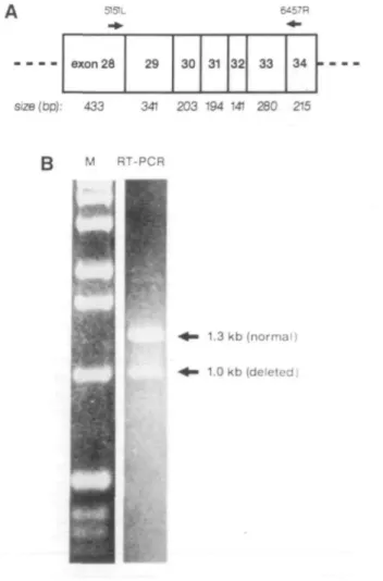
In screening for mutations in 42 NF1 patients by RT-PCR, we used a set of 7 primer pairs to amplify the coding region of the NF1 cDNA. We found a 59 year-old Swiss patient (C34-1) with NF1 where RT-PCR revealed one abnormally small amplification product, of about 1 kb instead of the expected 1.3 kb (Figure 1). The amplification product extends from the 3' end of exon 28 to the 5' end of exon 34. Although no formal quantification was performed, a consistent difference in the intensity of the two bands suggested that slightly less mutant than normal mRNA was present.
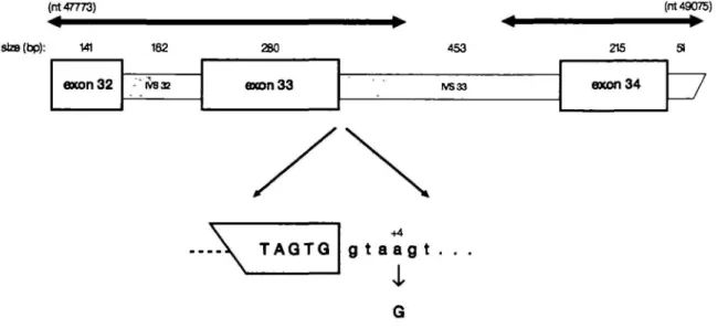
Sequence analysis revealed that the abnormal cDNA lacked the 280 nucleotides of exon 33 and had a perfect juxtaposition of exons 32 and 34. Genomic DNA sequences from nucleotide 47773 (exon 32) to nucleotide 49075 (TVS 34) were determined from uncloned PCR products (see Figure 2). A single nucleotide change was found, an A—G transition at nucleotide +4 of the IVS33 splice donor site (GTG/gtaagt to GTG/gtaggt; Figure 2). The relative frequencies of the four nucleotides at the +4 position of mammalian splice donor sites are: A — 0.71; G — 0.12; C - 0.08; T - 0.09 (14).
Although G occurs at position +4 in 12% of splice donor sites, we hypothesized that, in the context of this intron, the IVS33 + 4 A - G transition was responsible for the splicing error and
was disease-causing. In order to exclude the possibility that this alteration was merely a polymorphism, we used a mismatch PCR assay to detect the A—G transition. By introducing a single mismatch in the sequence of one primer, the presence of G (mutant) rather than A (normal) at the mutation site creates a
Sau96l restriction site in the amplification product, easily detected
by subsequent digestion (Figure 3). The +4 A—G transition was not present in 170 non-NFl chromosomes from unrelated Swiss individuals (data not shown), excluding the possibility of it being
51SIL 6457R exon 26 29 30 31 32 33 34 Size(bp): 433 341 203 194 141 280 215 M RT-PCR 1.3 kb (normal) 1.0 kb (deleted)
Figure 1. RT-PCR of NF1 cDNA exons 28 to 34. A. The specific primers for this PCR were 5151L (5'-GGTATTCCACAATGCTCTCAAG-3') and 6457R (5'-CTGACTTGACTTTGCTAATGCC-3'), numbered according to EMBL database, accession no. M89914. B. The lower band of 1.0 kb corresponds to the abnormal RT-PCR product which lacks exon 33.
664 Human Molecular Genetics, 1994, Vol. 3, No. 4 (nt 47773) staB(bp): (nt 49075) — • 141 exon32 162 - IV832 280 axon33 453 IVS33 215 exon34 TAQTQ +4 g t a ag t .
i
Figure 2. Structure of the genomic region surrounding the IVS33 splice donor site. The mutation is shown at the bottom. The heavy lines above represent the areas for which the'genomic sequence was determined. Nucleotide numbers are according to the EMBL database, accession nos L05367 and LO3723,
a common polymorphism. In addition, the aberrant splicing of exon 33 is predicted to result in a change of the reading frame and a loss of 777 of the 2811 amino acids of the mature protein. Finally, the mutation correlated with the disease in the patient's family: the mildly-affected son but not the asymptomatic daughter (28 year-old dizygotic twins), had inherited the mutation (Figure 3). We are thus led to conclude that mutations at nucleotide +4 of the splice donor site may lead to abnormal splicing and human disease.
The mutation reported here is the second independent case of NF1 with skipping of exon 33, the first being due to the insertion of an Alu retrotransposon in the 3' end of IVS32 (15). The 31 year-old patient of ref. 15 had no cafe'-au-lait spots but one cutaneous neurofibroma, axillary freckling, Lisch nodules, cervical root tumors and macrocephaly. Our patient (C34-1) had typical but relatively mild dermatological features (multiple small neurofibromas, and three cafe'-au-lait spots), and from the age of 48 showed progressive sensorimotor impairment due to multiple radicular neurofibromas, mostly in the intervertebral foramen. His affected son, who has five cafe'-au-lait spots and three neurofibromas, had a history of excision of minor cysts in his childhood.
Three other +4 mutations at splice donor sites have been reported in human disorders. The first (A —G) was detected by genomic sequencing in the IVS19 of the CFTR gene in a patient with CF (16); however, the possibility of exon skipping was not investigated. The second +4 mutation (also A —G) was in the IVS33 of the COL1A2 gene, and led to the use of an alternative splice site within the intron in a patient with a non-lethal form of osteogenesis imperfecta (17). Finally, an IVS4 + 4 A —T transversion has been reported in the FACC gene in Ashkenazi Jewish patients with Fanconi anemia, where the mutation apparently results in either complete or partial excision of exon 4 during splicing (18).
ACKNOWLEDGMENTS
This work was supported by a grant from the Carlos et Elsie de Reuter Foundation to P.H. and a grant from the Fondation Suisse pour 1'Encouragement de la
A.
Sau96\ (constant) 188 bp 1?9bp 188 bp 91 bp Sau961 (mutation)B.
OrE
+4 A ,G 188 91Figure 3. Diagnostic mismatch PCR analysis for the +4 IVS33 splice donor site mutation in the NF1 gene. The sequence of the two primers were NFx33L 5'-TAATTGGGAGTGATTTCGAATTCACAAGACATGCTTATC-3' and NFi33Sau96R 5 '-AATACGACTCACTATAGCACAAATTCCTTTCCTAG-GAC-3' (the mismatched base is underlined). PCR amplification from mutant DNA produces a novel Sau961 restriction site (GGNCC). A. The expected fragments from normal and mutant DNAs after Sou96I digestion. B. The proband and his son are heterozygous for the +4 A—G mutation as shown by the presence of the 91 bp fragment.
Human Molecular Genetics, 1994, Vol. 3, No. 4 665 Recherche Scientifique sur l'Arrieration mentale (FERAM) to M.M. We thank
Dr P.Cirafici for his clinical contribution and Dr A.Schonbomer for technical help.
REFERENCES
1. Riccardi,V.M. and EichnerJ.E. (1986) Johns Hopkins University Press, Baltimore, MD.
2. Ponder,B.A.J. (1990) Nature 346, 703.
3 Barker,D., Wright,E., Nguyen.K., Cannon.L., Fain,P., Goldgar.D., Bishop.D.T., CareyJ., Baty3-, KilvinJ., W01ard,H., WayeJ.S., Greig.G., Leinwand.L., Nakamura,Y., O'Connell,P., Leppert.M., Lalouel,J.-M.,White,R. and Skolnkk.M. (1987) Science 236, 1100-1102. 4. Seizinger.B.R., Rouleau,G.A., Ozelius,L.J., Lane.A.H., Faryniarz.A.G.,
Chao.M.V., Huson.S., Korf.B.R., Parry.D.M., Pericak-Vance.M.A., ColUns.F.S., Hobbs,W.J., Falcone.B.G., lannuzziJ.A., Roy.J.C, St Georges-Hyslop.P.H., Tanzi.R.E., Bochwell,M.A., Upadhyaya,M., Harper.P., Goldstein.A.E., Hoover.D.L., BaderJ.L., SpeDce.M.A., Mulvihill,J.J., Aylsworth.A.S., Vance J.M., Rossenwasser,G.O.D., Gaskwell.P.C, Roses,A.D., Martuza,R.L., Breakefield,X.O. and GuseUa,J.F. (1987) Cell 49, 589-594.
5. Goldgar.D E.,Green,P., Parry.D.M. and MulvihilU.J. (1989) Am. J. Hum. Genet. 44, 6 - 1 2 .
6. Cawthon.R.M., Weiss,R., Xu,G., Viskochil,D., Culver.M., Stevens,!., Robertson,M., Dunn,D., Gesteland.R., O'Connell.P. and White.R. (1990) Cell 62, 193-201.
7. Viskochil.D., Buchberg,A.M., Xu,G., Cawthon.R.M., Stevens.J., Wolff.R.K., Culver,M., CareyJ.C, Copeland.N.G., Jenkins,N.A., White.R. and O'Connell.P. Cell 62, 187-192.
8. Wallace.M.R., Marchuk.D.A., Andersen,L.B., Letcher.R., Odeh.H.M., Saulino.A.M., Fountain,.!. W., Breeton.A., Nicholson J., Mitchell.A.L., Brownstein.B.H. and Collins.F.S. (1990) Science 249, 181-186. 9. Xu,G., O'Connell.P., Viskochil.D., Cawthon.R., Robertson.M., Culver.M.,
Dunn.D., Stevens.J., Gesteland.R., Whhe,R. and Weiss.R. (1990) Cell62, 599-608.
10. Trahey.M. and McCormick.F. (1987) Science 238, 542-545. 11. Vogel,U., Dixon,R., Schaber.M., Diehl,R., Marshall,M. Scolnick.E.,
Sigal.I. and GibbsJ. (1988) Nature 335, 9 0 - 9 3 .
12. Viskochil.D., White.R. and Cawthon.R. (1993) Annu. Rev. Neurosci. 16, 183-205.
13. Ugius.E., Marchuk.D.A., CoUins,F.S. and Glover.T.W. (1993) Nature Genet. 3, 122-126.
14. In: D.N.Cooper and M.Krawczak (1993) Bios Scientific publishers, Oxford. 15. Wallace.M.R., Andersen.L.B., Saulino.A.M., Gregory.P.E., Glover.T.W.
and Collins.F.S. (1991) Nature 353, 864-866.
16. Ronchetto.P., Telleria Orriols.J.J., Fanen.P., Cremonesi.L., Ferrari,M., Magnani.C, Seia.M., Goossens.M., Romeo.G. and Devoto,M. (1992) Genomes 12, 417-418.
17. Wenstrup,R.J., Uver.L.W., Phillips.C.L. and Quaries.L.D. (1993) Mm. J. Med. Genet. 45, 228-232.
18. Whitney,M.A., Saito,H., Jakobs,P.M., Gibson.R.A., Moses,R.E. and Grompe.M. (1993) Nature Genet. 4, 202-205.