New insights regarding the incidence, presentation and treatment
options of aorto-oesophageal
fistulation after thoracic endovascular
aortic repair: the European Registry of Endovascular Aortic
Repair Complications
Martin Czerny
a,*, Holger Eggebrecht
b, Gottfried Sodeck
c, Ernst Weigang
d, Ugolino Livi
e, Fabio Verzini
f,
Jürg Schmidli
a, Roberto Chiesa
g, Germano Melissano
g, Andrea Kahlberg
g, Philippe Amabile
h,
Wolfgang Harringer
i, Michael Horacek
j, Raimund Erbel
j, Kay-Hyun Park
k, Friedhelm Beyersdorf
l,
Bartosz Rylski
l, Philipp Blanke
m, Ludovic Canaud
n, Ali Khoynezhad
o, Lars Lonn
p, Hervè Rousseau
q,
Santi Trimarchi
r, Jan Brunkwall
s, Michael Gawenda
s, Zhihui Dong
t, Weiguo Fu
t, Ingrid Schuster
uand Michael Grimm
vaDepartment of Cardiovascular Surgery, Inselspital, University Hospital Berne, Berne, Switzerland
b
Cardioangiological Center Bethanien, Frankfurt, Germany
cDepartment of Emergency Medicine, Medical University of Vienna, Vienna, Austria
d
Department of Cardio-Thoracic and Vascular Surgery, University Medical Center Mainz, Mainz, Germany
eDepartment of Cardiopulmonary Science, S. Maria della Misericordia Hospital, Udine, Italy
f
Vascular and Endovascular Surgery Unit, Hospital S. Maria Misericordia, Perugia, Italy
gDepartment of Vascular Surgery, Scientific Institute H San Raffaele, Milan, Italy
hDepartment of Interventional Radiology, AP-HM–Hôpital Nord, Marseille, France
i Department of Cardiovascular and Thoracic Surgery, Braunschweig, Germany
j Department of Cardiology, West-German Heart Center Essen, Essen, Germany
k
Seoul National University Bundang Hospital, Bundang-gu, South Korea
l Heart Centre Freiburg University, Freiburg, Germany
m
Department of Radiology, Albert-Ludwigs-University Freiburg, Freiburg, Germany
nDepartment of Vascular and Thoracic Surgery, Arnaud de Villeneuve Hospital, Montpellier, France
o
Division of Cardiothoracic Surgery, Cedars Sinai Medical Center, Los Angeles, CA, USA
pDepartment of Vascular Surgery and Cardiovascular Radiology, University of Copenhagen, Copenhagen, Denmark
q
Department of Interventional Radiology, CHU Rangueil, Toulouse, France
r IRCCS Policlinico San Donato, Thoracic Aortic Research Center, Milan, Italy
sDepartment of Vascular Surgery, University of Cologne, Cologne, Germany
tDepartment of Vascular Surgery, Zhongshan Hospital, Fudan University, Shanghai, China
uDepartment of Cardiac Surgery, University of Kosice, Kosice, Slowakia
v
Department of Cardiac Surgery, Medical University of Innsbruck, Innsbruck, Austria
* Corresponding author. Department of Cardiovascular Surgery, Inselspital, University Hospital Berne, Freiburgstrasse 18, 3010 Berne, Switzerland. Tel: +41-31-6322376; fax: +41-31-6322919; e-mail: martin.czerny@insel.ch (M. Czerny).
Received 25 April 2013; received in revised form 17 June 2013; accepted 24 June 2013
Abstract
OBJECTIVES: To review the incidence, clinical presentation, definite management and 1-year outcome in patients with aorto-oesophageal fistulation (AOF) following thoracic endovascular aortic repair (TEVAR).
METHODS: International multicentre registry (European Registry of Endovascular Aortic Repair Complications) between 2001 and 2011 with a total caseload of 2387 TEVAR procedures (17 centres).
RESULTS: Thirty-six patients with a median age of 69 years (IQR 56–75), 25% females and 9 patients (19%) following previous aortic surgery were identified. The incidence of AOF in the entire cohort after TEVAR in the study period was 1.5%. The primary underlying aortic path-ology for TEVAR was atherosclerotic aneurysm formation in 53% of patients and the median time to development of AOF was 90 days (IQR 30–150). Leading clinical symptoms were fever of unknown origin in 29 (81%), haematemesis in 19 (53%) and shock in 8 (22%) patients. Diagnosis could be confirmed via computed tomography in 92% of the cases with the leading sign of a new mediastinal mass in 28 (78%) patients. A conservative approach resulted in a 100% 1-year mortality, and 1-year survival for an oesophageal stenting-only approach was 17%. Survival after isolated oesophagectomy was 43%. The highest 1-year survival rate (46%) could be achieved via an aggressive treatment including radical oesophagectomy and aortic replacement [relative risk increase 1.73 95% confidence interval (CI) 1.03–2.92]. The survival advantage of this aggressive treatment modality could be confirmed in bootstrap analysis (95% CI 1.11–3.33).
CONCLUSIONS: The development of AOF is a rare but lethal complication after TEVAR, being associated with the need for emergency TEVAR as well as mediastinal haematoma formation. The only durable and successful approach to cure the disease is radical oesophagect-omy and extensive aortic reconstruction. These findings may serve as a decision-making tool for physicians treating these complex patients.
Keywords:Thoracic endovascular aortic repair• Aorto-oesophageal fistulation • Complications • Treatment
INTRODUCTION
Thoracic endovascular aortic repair (TEVAR) has been rapidly embraced by many physicians from different specialities as a means to treat a broader cohort of patients with acute and chronic thoracic aortic pathology [1–3]. Since the introduction of TEVAR in the mid-90s, much has been learnt on the appropriate application of the method as well as on pitfalls and their avoidance [4,5]. However, there remains a black box regarding the occurrence of orphan serious adverse events. In order to learn about these mechanisms and their prevention in the future, the European Registry of Endovascular Aortic Repair Complications (EuREC) was founded. Thefirst two projects focused on the issue of retrograde type A aortic dissection and the issue of symptomatic spinal cord injury after TEVAR [6,7].
The aim of the third EuREC project was to review the incidence, clinical presentation, definite management and 1-year outcome in patients with aorto-oesophagealfistulation (AOF) following TEVAR.
METHODS
Patients
The records of 36 patients who developed AOF after TEVAR with a median age of 69 years (IQR 56–75) between 2001 and 2011 were analysed. The cumulative caseload of all 17 participating centres in this time period was 2387. Twenty-five percent of patients were female and 19% had already undergone various kinds of previous open aortic surgery in different segments. The primary underlying aortic pathology for TEVAR was thoracic aortic aneurysm in 53% of patients, and the median time to the development of AOF was 90 days (IQR 30–150).
Definition of aorto-oesophageal fistulation
AOF was defined as any communication between the thoracic aorta and the oesophagus post-TEVAR. Patients in whom AOF could have been already present at the time of TEVAR or patients having native AOF, either due to the underlying aortic pathology or due to any reasons, were excluded.
Parameters
We collected clinical data, including any kind of previous aortic surgery, underlying aortic pathology and extension of aortic disease regarding the indication for TEVAR. Procedural data included the time of TEVAR, elective or emergent intervention, number of prostheses, covered length and landing zone according to current definitions. Furthermore, the presence or absence of mediastinal haematoma at the time of TEVAR was recorded. We recorded the time between TEVAR and the diagnosis of AOF as well as leading clinical symptoms. The diagnostic modalities of
AOF were recorded as was the strategy to treat AOF. Finally, 1-year survival and reasons of death were recorded.
Statistical methods
Continuous data are presented as the median and IQR (range from the 25th to 75th percentile). Discrete data are given as counts and percentages. Comparisons of continuous data between the patient cohorts were performed using the Mann– WhitneyU-test, and categorical data were compared using the Fisher exact test. In the sub-analysis, we sought to determine the optimal mode of treatment of oesophagealfistulization following TEVAR, and primary results are expressed as relative risk increase (RRI) with its 95% confidence interval (CI); the conservative treat-ment option served as reference standard since 100% of patients in this group died. A resampling model with 2000 replications was then used to confirm CIs. A two-sided P-value of <0.05 was con-sidered statistically significant. All calculations were performed with SPSS 20.0 for Mac OsX (IBM SPSS, Inc., NY, USA).
RESULTS
Patient demographics
The incidence of AOF in the entire cohort after TEVAR in the study period was 1.5%. The descriptive characteristics of the patient cohort are depicted in Table 1. Coronary artery disease was
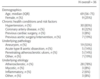
Table 1: Descriptive characteristics of the cohort
N overall = 36 Demographics
Age, median (IQR) 69 (56–75)
Female,n (%) 9 (25%)
Chronic health conditions and risk factors
Hypertension,n (%) 30 (83%)
Coronary artery disease,n (%) 8 (22%)
Previous cardiac surgery,n (%) 3 (8%)
Previous aortic surgery/intervention,n (%) 7 (19%)
Underlying pathology
Aneurysm,n (%) 19 (53%)
Acute type B aortic dissection,n (%) 5 (14%)
Penetrating atherosclerotic ulcers,n (%) 5 (14%)
Other,n (%) 7 (19%) Underlying etiology Atherosclerotic,n (%) 28 (78%) Mycotic,n (%) 3 (8%) Inflammatory,n (%) 2 (6%) Other,n (%) 3 (8%)
Unless otherwise indicated, data are number (percentage). IQR: interquartile range. A O RT IC S U RGE R Y
present in 22% of patients, 19% had already undergone various kinds of previous aortic surgeries and 8% had already undergone previous cardiac surgery. The indications for TEVAR were athero-sclerotic aneurysms in 53%, acute type B aortic dissections in 14% and penetrating atherosclerotic ulcers in 14% (Table1).
Index TEVAR procedure
Initial interventional characteristics of the cohort are given in Table 2. Forty-four percent of patients underwent emergency TEVAR and 36% had a detectable mediastinal haematoma at the time of TEVAR. Severe intraoperative hypotension defined as a period of ≥ 5 min with a systolic pressure of <60mmHg was observed in 3%. Details with regard to prior supra-aortic rerouting procedures as well as the landing zones are given in Table2. The median covered length was 17 cm (IQR 15–22) and the median oversizing factor of prostheses was 15% (IQR 0–30).
Presentation of aorto-oesophageal
fistulation
The median time interval between TEVAR and the diagnosis of AOF was 90 days (IQR 30–150). Clinical symptoms were fever of unknown origin in 81%, haematemesis in various extents in 53% and shock in 22% (septic and/or haemorrhagic) (Table3). Imaging was preferably done by computed tomography (CT)—92% fol-lowed by confirmation of the suspected diagnosis by endoscopy in 50% (Figs1and2). Median serum CRP levels at the time of diag-nosis were 18 mg/dl (IQR 10–27).Management of aorto-oesophageal
fistulation
In 28%, a conservative strategy was chosen, and an oesophageal stenting-only approach in 17%. Nineteen percent underwent iso-lated oesophagectomy andfinally, 36% of patients underwent a radical surgical approach of both oesophagectomy and aortic
replacement of various kinds (Table4). In these patients, stent-grafts were removedin toto. Figure3shows the intraoperative situs of a patient undergoing such a radical surgical approach. Figure4A shows the situs post-oesophagectomy and after orthotopic recon-struction of the thoracic segment with a bovine pericardial neoaorta.
Follow-up
Two patients who initially underwent an oesophagectomy-only approach, subsequently underwent extension TEVAR procedures proximally and distally to the initially repaired segment due to continuing mycotic aneurismal formation. One patient underwent revision of the gastric pull-up anastomosis due to dehiscence.
One-year survival was 28% (n = 10). Thirteen patients died due to bleeding complications as a consequence of the continuing in-fective process. Fatal bleeding complications occurred via arrosion of the adjacent anatomical structures. Mediastinitis and multior-gan failure accounted for 4 deaths each and have also to be seen in context with the active infective process. One patient died due to respiratory failure after surgery and another 4 deaths were clas-sified as sudden cardiac and therefore not related to AOF.
A conservative approach resulted in a 100% 1-year mortality, and 1-year survival for an oesophageal- stenting-only approach was 17%. Survival after isolated oesophagectomy was 43%. The highest 1-year survival rate (46%) could be achieved via an aggres-sive treatment including radical oesophagectomy and aortic re-placement [RRI 1.73 95% CI 1.03–2.92]. The survival advantage of this aggressive treatment modality could be confirmed in boot-strap analysis (95% CI 1.11–3.33). Figure4B shows thefinal result in patients after orthotopic aortic reconstruction and gastric pull-up.
COMMENT
The development of AOF is a rare but lethal complication after TEVAR, being associated with the need for emergency TEVAR as well as mediastinal haematoma formation. The only durable and successful approach to cure the disease is radical oesophagectomy and extensive aortic reconstruction. Thesefindings may serve as a Table 2: Initial interventional characteristics of the cohort
N overall = 36 Initial procedure
Emergency, (%) 16 (44%)
Prior supra-aortic rerouting,n (%) 3 (8%)
Overstenting of arch vessels,n (%) 5 (14%)
Proximal bare metal springs,n (%) 14 (39%)
Criado zone 0,n (%) 1 (3%)
Criado zone 1,n (%) 6 (17%)
Criado zone 2,n (%) 8 (22%)
Criado zone 3,n (%) 19 (53%)
Stent coverage
Number of protheses, median (range) 1 (1–4)
Coverage in cm, median (IQR) 17 (15–22)
Oversizing factor in percentages, median (range) 15 (0–30)
Previous aortic surgery/ intervention,n (%) 7 (19%)
Mediastinal haematoma at diagnosis,n (%) 13 (36%)
Intraoperative hypotension,n (%) 1 (3%)
Unless otherwise indicated, data are number (percentage). IQR: interquartile range.
Table 3: Presentation of aorto-oesophageal fistulation N overall = 36 Timing
Days since initial TEVAR procedure, median (IQR) 90 (30–150)
Clinical presentation
Fever of unknown origin,n (%) 29 (81%)
Haematemesis,n (%) 19 (53%) Shock,n (%) 8 (22%) Dyspnoea,n (%) 2 (6%) Others,n (%) 3 (8%) Diagnostics Computed tomography,n (%) 33 (92%) Partial thrombosis,n (%) 7 (19%)
Confirmation via endoscopy,n (%) 18 (50%)
Acute phase protein, mg/dl, median (IQR) 18 (10–27)
Unless otherwise indicated, data are number (percentage). IQR: interquartile range.
decision-making tool for physicians treating these complex patients.
It was interesting to realize that a substantial number of patients had undergone emergency TEVAR and according to the active underlying aortic disease progress had mediastinal haematoma for-mation at the time of TEVAR. We feel that this is potentially the most importantfinding of our study. Years ago, when we had our first case of AOF, which was a ‘simple’ monosegmental rupture of the descending aorta, we hypothesized that elevated pressure in the posterior mediastinum could well be responsible for secondary oesophageal ischaemia and consequently AOF formation also due to the inflammation of the resorption haematoma [8]. This mechan-ism has also been postulated by others and may therefore now be seen as one proven way to develop AOF [9,10]. In our setting, we try to attenuate this problem by elective left-sided mini-thoracotomy on the second day after TEVAR with broad opening of the mediastinal pleura in order to decompress the posterior medi-astinum. It is clear that this approach might not be applicable to all patients, but it might serve as a technical tip to avoid this dreadful complication. Another radiographicfinding which should increase alertness constitutes right-sided haemothorax as a surrogate for ex-cessive pressure formation in the posterior mediastinum leading to secondary right-sided pleural rupture.
Furthermore, the size of the aneurysm may play another leading role in the development of AOF. It is known that AOF and
also aortobronchialfistulization may occur as a natural course of huge thoracic aneurysms without any intervention [11]. The mech-anism here may be seen in mechanical compression and second-ary erosion. The geometry change in the aortic arch and the descending aorta by TEVAR as well as the radial force of the graft against the native aortic wall may lead to the samefinal pathway of ischaemia and secondaryfistula formation.
Obviously, AOF formation is a gradual process as the median time to developing AOF in our study was 90 days. Interestingly, we did not observe a single case of primary fatal free rupture of AOF, indicating that the natural course of the disease is stepwise. However, clinical and laboratory sings of inflammation are very common [9,10]. Eighty-one percent of our patients had fever as the leading clinical sign in combination with haematemesis to various extents in 50% of patients. This combination should alert the treating physician to the problem and enhance the establish-ment of an appropriate diagnosis and an adequate treatestablish-ment strat-egy.
Diagnosis was—as expected—mainly established by CT imaging. A new mediastinal mass was present in 19% of patients, suggesting contained rupture. However, the leading radiographic sign remains air surrounding the prosthesis and the posterior mediasti-num. Diagnosis was confirmed by additional endoscopy in 50% of patients and it has been used as primary diagnostic means in casuistics with the leading clinical sign of haematemesis.
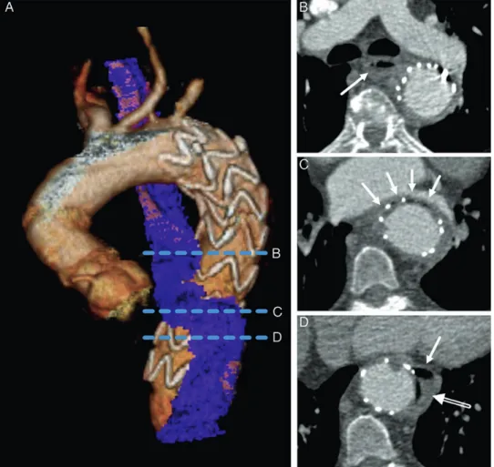
Figure 1:CT of a 58-year old female with AOF after TEVAR for acute complicated type B aortic dissection. (A), volume rendered image displaying the thoracic aorta
and the close spatial relation of the stent graft and the oesophagus (blue). Dotted blue lines indicate the position of the transverse views in (B–D): (B) at the level below the tracheal bifurcation with the oesophagus (arrow) located posteriorly to the left main bronchus and medial to the aorta; (C) at the level of the left atrium with the oesophagus (arrows) intersecting anteriorly to the aorta with slit-shaped compression between aorta and left atrium. (D) Further below, the oesophagus is located left to the aorta (arrow) with air adjacent to the stent graft (open arrow), corresponding to the AOF.
A O RT IC S U RGE R Y
Due to the associated high degree of systemic inflammation in patients with AOF and the fact that they underwent primary TEVAR instead of open surgery, these patients were selected for a higher degree of frailty and consecutively for a lower probability of being suitable for open repair [4]. This is reflected by the fact that in this
study, a conservative approach had to be chosen in 28% of patients. By analysing the different treatment strategies, it became obvious that any trade-off regarding debridement of infected structures, irrespective if oesophagus or stent-graft is prone to failure. The highest success rates were achieved in patients for whom a radical strategy with oesophagectomy and removal of the stent-graft with orthotopic aortic reconstruction was chosen. It seems wise to delay oesophageal reconstruction due to the higher probability of primary anastomotic healing of a gastric pull-up or a colonic interposition [8]. From our database, we cannot draw
conclusions regarding whether the entire aortic wall was removed during surgery. However, as there was no recurrence of infection in the radically treated group, it is highly likely that all patients Table 4: The management and outcome of
aorto-oesophageal fistulation
N overall = 36 Management
Conservative,n (%) 10 (28%)
Oesophageal stenting only,n (%) 6 (17%)
Isolated oesophagectomy,n (%) 7 (19%)
Aortic replacement plus oesophagectomy,n (%) 13 (36%)
Outcome
1-year survival,n (%) 10 (28%)
Unless otherwise indicated, data are number (percentage).
Figure 3:Intraoperative view of a patient with AOF showing the aneurismal sac
open, as well as the spatial relation of the stent-graft to the defect in the oe-sophageal wall.
Figure 4:(A). Intraoperative view after oesophagectomy and orthotopic
recon-struction with a bovine pericardial neoaorta. (B). CT 3 months after orthotopic aortic reconstruction and gastric pull-up.
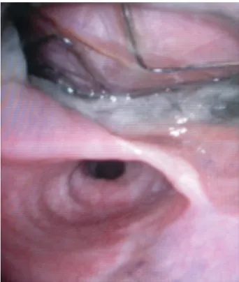
Figure 2:Endoscopic view of AOF showing the stents and fabric of the
were treated properly without knowing the details regarding whether all remnants of the aortic wall were removed. In case of remaining dead space, the use of Dacron grafts or in case of direct oesophageal suture, omentopexy is highly useful to enhance the healing process in a fragile area [12].
Regarding the material of choice for aortic replacement, several options are available where the implantation of a Dacron graft with or without antibiotic treatment might be at higher risk for infection. However, several other groups have reported very fa-vourable results using Dacron grafts with antibiotic treatment [12,13]. We have developed a new means of orthotopic aortic re-construction by using a bovine pericardial patch as a neoaorta with excellent success rates with regard to freedom from reinfec-tion [14]. Additionally, calcification of this graft after a median
follow-up of 24 months is not an issue, which might prove this ap-proach to be superior to homograft replacement in the long-term. Regarding the reasons for death, bleeding complications had a leading role due to the continuing infective process. Fatal bleeding complications occurred via arrosion of the adjacent ana-tomical structures. Despite 1-year survival in patients with an oesophagectomy-only approach being only slightly less than in patients with a radical approach to both aorta and esophagus, a risk of reinfection remains as shown by 2 cases who had already undergone extension TEVAR procedures proximally and distally to the initially repaired segment due to continuing mycotic aneur-ismal formation. Furthermore, these patients have to remain under life-long antibiotic therapy, which is usually not the case in patients undergoing a radical approach.
Strengths and limitations of this study
This study represents a systematic approach to learning about the incidence, mechanisms and treatment strategies of AOF after TEVAR. However, it is clear that not every patient undergoing emergency TEVAR having mediastinal haematoma will develop AOF. As such, this report is not the answer to all questions.
Summarizing, the development of AOF is a rare but lethal complication after TEVAR, being associated with the need for emergency TEVAR as well as mediastinal haematoma formation. The only durable and successful approach to cure the disease is radical oesophagectomy and extensive aortic reconstruction. Thesefindings may serve as a decision-making tool for physicians treating these complex patients.
Conflict of interest: none declared.
REFERENCES
[1] Eggebrecht H, Nienaber C A, Neuhäuser M, Baumgart D, Kische S,
Schmermund Aet al. Endovascular stent-graft placement in aortic
dissec-tion: a meta-analysis. Eur Heart J 2006;27:489–98.
[2] Patel H J, Sood V, Williams D M, Dasika N L, Diener A C, Deeb G M. Late outcomes with repair of penetrating thoracic aortic ulcers: the merits of an endovascular approach. Ann Thorac Surg 2012;94:516–22.
[3] Grabenwöger M, Alfonso F, Bachet J, Bonser R, Czerny M, Eggebrecht H et al. European Association for Cardio-Thoracic Surgery (EACTS); European Society of Cardiology (ESC); European Association of Percutaneous Cardiovascular Interventions (EAPCI). Thoracic endovascular aortic repair (TEVAR) for the treatment of aortic diseases: a position statement from the European Association for Cardio-Thoracic surgery (EACTS) and the European Society of Cardiology (ESC), in collaboration with the European Association of Percutaneous Cardiovascular Interventions (EAPCI). Eur J Cardiothorac Surg 2012;42:17–24.
[4] Dumfarth J, Michel M, Schmidli J, Sodeck G, Ehrlich M, Grimm Met al.
Mechanisms of failure and outcome of secondary surgical interventions after thoracic endovascular aortic repair (TEVAR). Ann Thorac Surg 2011;
91:1141–6.
[5] Desai N D, Burtch K, Moser W, Moeller P, Szeto W Y, Pochettino Aet al.
Long-term comparison of thoracic endovascular aortic repair (TEVAR) to open surgery for the treatment of thoracic aortic aneurysms. J Thorac
Cardiovasc Surg 2012;144:604–9.
[6] Eggebrecht H, Thompson M, Rousseau H, Czerny M, Lönn L, Mehta R H et al. European Registry on Endovascular Aortic Repair Complications. Retrograde ascending aortic dissection during or after thoracic aortic stent graft placement: insight from the European registry on endovascular aortic
repair complications. Circulation 2009;120(11 Suppl):S276–81.
[7] Czerny M, Eggebrecht H, Sodeck G, Verzini F, Cao P, Maritati Get al.
Mechanisms of symptomatic spinal cord ischemia after TEVAR: insights from the European Registry of Endovascular Aortic Repair Complications
(EuREC). J Endovasc Ther 2012;19:37–43.
[8] Czerny M, Zimpfer D, Fleck T, Gottardi R, Cejna M, Schoder M et al.
Successful treatment of an aortoesophagealfistula after emergency
endo-vascular thoracic aortic stent-graft placement. Ann Thorac Surg 2005;80: 1117–20.
[9] Chiesa R, Melissano G, Marone E M, Kahlberg A, Marrocco-Trischitta M M, Tshomba Y. Endovascular treatment of aortoesophageal and
aortobron-chialfistulae. J Vasc Surg 2010;51:1195–202.
[10] Chiesa R, Melissano G, Marone E M, Marrocco-Trischitta M M, Kahlberg
A. Aorto-oesophageal and aortobronchial fistulae following thoracic
endovascular aortic repair: a national survey. Eur J Vasc Endovasc Surg
2010;39:273–9.
[11] Coselli J, Crawford E S. Primary aorto-esophageal fistula from aortic
aneurysm: successful surgical treatment by use of omental pedicle graft. J Vasc Surg 1990;12:269–77.
[12] Munakata H, Yamanaka K, Okada K, Okita Y. Successful surgical treatment
of aortoesophagealfistula after emergency thoracic endovascular aortic
repair: aggressive debridement including esophageal resection and
extended aortic replacement. J Thorac Cardiovasc Surg 2013;146:235–7.
[13] Kirkwood M L, Pochettino A, Fairman R M, Jackson B M, Woo E Y, Wang G J. Thoracic aortic endograft explant: a single center experience. Vasc Endovasc Surg 2010;44:440–5.
[14] Czerny M, von Allmen R, Opfermann P, Sodeck G, Dick F, Stellmes Aet al.
Self-made pericardial tube graft: a new surgical concept for treatment of graft infections after thoracic and abdominal aortic procedures. Ann Thorac Surg 2011;92:1657–62. A O RT IC S U RGE R Y