HAL Id: hal-03087700
https://hal.archives-ouvertes.fr/hal-03087700
Submitted on 29 Dec 2020HAL is a multi-disciplinary open access archive for the deposit and dissemination of sci-entific research documents, whether they are pub-lished or not. The documents may come from teaching and research institutions in France or abroad, or from public or private research centers.
L’archive ouverte pluridisciplinaire HAL, est destinée au dépôt et à la diffusion de documents scientifiques de niveau recherche, publiés ou non, émanant des établissements d’enseignement et de recherche français ou étrangers, des laboratoires publics ou privés.
Impact of A2T and D23N Mutations on Tetrameric
Aβ42 Barrel within a Dipalmitoylphosphatidylcholine
Lipid Bilayer Membrane by Replica Exchange Molecular
Dynamics
Son Tung Ngo, Phuong Nguyen, Philippe Derreumaux
To cite this version:
Son Tung Ngo, Phuong Nguyen, Philippe Derreumaux. Impact of A2T and D23N Mutations on Tetrameric Aβ42 Barrel within a Dipalmitoylphosphatidylcholine Lipid Bilayer Membrane by Replica Exchange Molecular Dynamics. Journal of Physical Chemistry B, American Chemical Society, 2020, 124 (7), pp.1175-1182. �10.1021/acs.jpcb.9b11881�. �hal-03087700�
Impact of A2T and D23N Mutations on Tetrameric Aβ42 Barrel within a
DPPC Lipid Bilayer Membrane by Replica Exchange Molecular Dynamics
Son Tung Ngo,ab Phuong H. Nguyen,cd and Philippe Derreumauxcdef*
aLaboratory of Theoretical and Computational Biophysics, Ton Duc Thang University, Ho Chi Minh City, Vietnam bFaculty of Applied Sciences, Ton Duc Thang University, Ho Chi Minh City, Vietnam cCNRS, Université de Paris, UPR 9080, Laboratoire de Biochimie Théorique, 13 rue Pierre et Marie Curie, F-75005, Paris, France
dInstitut de Biologie Physico-Chimique, Fondation Edmond de Rothschild, PSL Research
University, Paris, France
e Laboratory of Theoretical Chemistry, Ton Duc Thang University, Ho Chi Minh City,
Vietnam
fFaculty of Pharmacy, Ton Duc Thang University, Ho Chi Minh City, Vietnam
Abstract: In Alzheimer’s disease (AD), many experimental and computational studies support the amyloid pore hypothesis of the Aβ42 peptide. We recently designed a β-barrel tetramer in a membrane-mimicking environment consistent with some low-resolution experimental data. In this earlier study by using extensive replica exchange molecular dynamics simulations we found that the wild-type (WT) Aβ42 peptides have a high propensity to form β-barrels, while the WT Aβ40 peptides do not. In this work, we have investigated the effect of the mutations D23N and A2T on the Aβ42 barrel tetramer by using the same enhanced conformational sampling technique. It is known that the D23N mutation leads to early-onset AD, while the A2T mutation protects from AD. This computational study in a DPPC lipid bilayer membrane shows that the WT sequence and its A2T variant have similar β-barrel populations, and the 3D model is slightly destabilized for D23N compared to its WT sequence. These extensive modelling calculations indicate the lower and higher induced-toxicity of these two mutations in AD cannot be correlated to their β-barrel tetramer stabilities in a DPPC lipid bilayer membrane.
Introduction
In Alzheimer’s disease (AD), the amyloid cascade, based on the deleterious effect of Aβ oligomers, is the most accepted hypothesis.1,2 Although all clinical attempts targeting these oligomers have failed to deliver drugs,3,4 negative Alzheimer's trials have not killed the leading theory of disease.5 It is known that Aβ oligomers are formed during the lag-phase of the aggregation kinetics (primary nucleation) or from the fragmentation of fibrils (secondary nucleation).6 In vitro interactions of these Aβ oligomers with the
prion protein,7 the tau protein,8 the NMDA receptor,9 and many other cellular proteins have been characterized.
Amyloid oligomers interactions with model and cellular membranes are documented by a large number of experimental techniques, and Aβ oligomers, but not monomers, induce toxicity.10-15 Among the different types of amyloid protein-membrane
interactions, there is increasing evidence that Aβ42 can form β-barrels in various membrane-mimicking environments; micelles composed of dodecylmaltoside (DDM, 2 ×
CMC), 200 mM ammonium acetate and PC12 lipids,16
micelles composed of dodecylphos-phocholine (DPC) and 1,2-dimyristoyl-sn-glycero-3-phosphocholine (DMPC) lipids,17 and
cellular membranes,18 based on electrical currents using patch-clamp18,19 or biochemical
and biophysical experiments.16,17 Overall these annular pores can span various oligomer
sizes as small as tetramers and hexamers and up to 24-mers.16,17,20
AD is sporadic in 95% of the cases, but several familial AD (FAD) mutations provoke early-onset disease: H6R (English), D7H (Taiwanese), D7N (Tottori), A21G
(Flemish), E22Q (Dutch), E22G (Artic), E22Δ (Osaka), and D23N (Iowa).8 These mutations
are located in the N-terminus of Aβ (residues 1-11), the central hydrophobic core (residues 17-21) and in the loop region (residues 23-28). All these FAD mutations impact the aggregation kinetics in aqueous solution,6,8,21 change the ratio of Aβ40 and Aβ42 peptides released by the β- and γ-secretases, and notably augment the production of the most toxic Aβ42 peptide.22 The D23N mutation also led to the discovery to a new amyloid fibril structure with antiparallel β-strands rather than parallel β-strands within
the sheets as observed for the WT Aβ40 and Aβ42 peptides under various experimental conditions, and for the α-synuclein and tau fibrils.23 This transition from antiparallel to parallel β-strands in the sheets is not unique in the amyloid world, as it was observed for the Aβ16-22 variant with E22Q mutation.24
Due to the intrinsic disorder of all amyloid proteins and the transient character of all oligomers, experimentally-derived atomic structures are lacking, and computer simulations have been employed to determine the dominant microstates of the system in aqueous solution,8,25,26 on material surface,27 and interacting with membrane models.
28,29 The influence of D23N has motivated computational studies aimed at determining
the free energy landscapes of Aβ21-30,30,31 and Aβ40/Aβ42 monomers,32,33 Aβ40/42
dimers,34 Aβ16-35 6-mers,35 and the thermodynamics of fibril elongation,36 all
simulations performed in aqueous solution with various force fields and protein representations.
Along with disease-causing mutations, the A2T (homozygous) and A2V (heterozygous) mutations protect from AD and increase the lag-phase of aggregation.8,37
These mutations are particularly intriguing as the amino acid Ala is not located in the core of Aβ40/42 amyloid fibril structures.23 Atomistic simulations have investigated the
impact of these substitutions on the equilibrium ensemble of Aβ28, Aβ40 and Aβ42 in aqueous solution, revealing a significant change in the intrinsic disorder of the monomers,37,38 and the higher content of intrapeptide-stabilized conformations in the
mutant dimers compared to its WT counterpart dimer. 39,40
While the impact of mutations (E22Δ, F19P, F20C) and N-terminal truncations (Aβ17-42, Aβp3-42) has been investigated on the pore ability of double layers of β-sheets in a lipid bilayer membrane by MD simulations,29 the influence of D23N and A2T on Aβ42 pore ability has not been studied by experimental and computational means. Recently, we proposed a 3D model for Aβ42 β-barrel tetramer that remains formed for 2 microseconds molecular dynamics (MD) simulation in a bilayer consisting of POPC/POPS/cholesterol/sphingomyelin with a 40/10/25/25 molar ratio.41 Extensive
replica exchange molecular dynamics (REMD) simulations of this 3D model in a DPPC lipid bilayer membrane showed that Aβ42 forms barrels with a very high probability, while Aβ40 does not.41 In this study, our aim is to determine whether the A2T and D23N mutations change the probability of β-barrel formation of the Aβ42 peptides in the DPPC lipid bilayer membrane and if the predicted barrel stabilities can be correlated to the reduced and increased toxicity of these two variants in AD.
Materials and Methods
A2T and D23N barrels were centred in rectangular box of 7.12×7.15×6.60 and 7.12×7.15×7.60 nm, respectively, with (x, y, z) lipid bilayer dimensions of 7.12×7.15×
4.00 nm solvated by the SPC water model.42 We used the united-atom force field
GROMOS 53a643 for the peptides and the Berger force field44 for the DPPC lipid bilayer.
The peptides at pH 7 have NH3+ and CO2- termini, deprotonated Glu and Asp, protonated
Arg and Lys, and neutral His with a protonated Nε atom. In total, the transmembrane
D23N Aβ42 system consists of 123 DPPC lipid molecules, 4908 water molecules, 8 Na+
atoms (22,526 atoms), while the transmembrane A2T Aβ42 system consists of 123 DPPC
lipid molecules, 3282 water molecules, 12 Na+ atoms (17,656 atoms).
The WT MD-refined Aβ42 structure obtained in Ref. 41 was mutated by using
PyMOL45 and further refined by a 5 ns MD simulation with decreasing restraints on the
backbone and all side-chain atoms of all Aβ residues, except at positions 2 and 23.
REMD simulations were performed using GROMACS version 5.1.346 and all parameters described in Ref. 41. As for the WT Aβ42 barrel, the D23N and A2T barrels were sampled by REMD with 28 replicas at 310.0, 312.8, 315.6, 318.4, 321.3, 324.1, 327.0, 329.9, 332.8, 335.8, 338.8, 341.7, 344.7, 347.8, 350.8, 353.9, 357.0, 360.1, 363.2, 366.4, 369.5, 372.7, 375.9, 379.2, 382.4, 385.7, 389.0, and 392.3 K. The D23N and A2T barrels were simulated by 500 ns of REMD, the first 50 ns were removed, and the comparison between the three sequences was monitored over the time interval 50-500 ns at 310 K. The data were recorded every 10 ps and 45,000 snapshots per system were used to determine the physical impact of the two mutations.
The time-averaged secondary structure compositions were predicted using DSSP tool,47 and the REMD-averaged collision cross sections (CCS) were determined using the trajectory method of the IMPACT software.48 The free energy landscape of each system was projected on the RMSD using all heavy backbone atoms of residues 9-40 with respected to its MD-refined structure, and the mean number of intermolecular side chain
contacts using residues 9-40. Residues 1-8 are not included because they are disordered, and residues 41-42 are excluded to allow comparison with our earlier study on Aβ40 peptide.41 An intermolecular side-chain (SC) contact 𝑛
!"
!" between residue 𝑖 of chain 𝑙 and
𝑗 of chain 𝑘 is counted when the minimum distance 𝑟!"!" between two residues is smaller than 0.45 nm, the indexes 𝑖, 𝑗 are in the range of [9,40] and the indexes 𝑙, 𝑘 are in the range of [1,4] with 𝑙 ≠ 𝑘. The total SC contact between two chains is the sum of all available SC between residues of two chains 𝑁!"!"= 𝑛
!" !"
!" , and the mean value of intermolecular SC contacts per pair is computed by 𝑁!" = !!"
!" !!!
! .
Because of the high dimensionality of the conformational space, the trajectories were also analysed by computing the distribution of the tilt angle of the β-strands relative to the axis of the barrel, the distribution of the inner pore diameter, and the distributions of intra-molecular and intermolecular H-bonds. A H-bond was considered formed if the acceptor-donor-H angle is less than 30 degrees and the acceptor – donor distance is less than 0.35 nm.41
Results and Discussion
In our earlier study, the convergence of the WT Aβ42 REMD simulation was assessed by the average of exchange rates between consecutive replicas (18%), the propagation of the 1st and 26th replicas over the temperature index during the
simulation time, and the high similarity of the free energy landscapes projected on the
RMSD and number of intermolecular contacts using the two time interval 50-500 ns and 150-500 ns.41The same mean exchange ratio between consecutive replicas was obtained for the two variants (18-20%) and the other two controls for convergence are also satisfied for A2T and D23N mutants (data not shown).
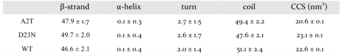
The secondary structure remains constant upon mutation within statistical errors (2%): 48% of β-strand, 48% of coil, 2% of turn and almost 0% of α-helix (Table 1). The β-strand content along the sequence, see Fig. 1, remains identical between the WT and A2T sequences with the residues 12-22 and the residues 29-40 having each more than 50% of β-character. There is an averaged β-strand increase of 10% for residues 13-16, 21-24, 28-29 and 35-40 from WT to D23N. Upon D23N mutation, the turn content of residues 23-26 increases from 8 to 16% and the coil content augments moderately in the N-terminus. The coil and turn profiles of A2T and WT are nearly identical. In all species, the α-helix signal is less than 5% except in the region 2-12 for WT and D23N and the region 23-27 in A2T. The averaged CCS for the WT system (22.6 ± 1.4 nm2) is in between
that for D23N (23.1 ± 1.0 nm2) and A2T (20.6 ± 0.8 nm2).
Figure 2 shows the free energy landscapes (FEL) of the D23N and A2T Aβ42 tetramers projected on the backbone RMSD with respect to the MD-refined structure and the mean value of intermolecular SC contacts per pair (Nc). The six free energy minima on the A2T FEL (Fig. 2A) are denoted by A0, B0, C0, D0, E0, and F0 with (Nc, population) values of (27.0, 34%), (32.6, 14%), (29.0, 17%), (25.0, 4%), (26.2, 5%), and (20.2, 7%), respectively. The seven free energy minima on the D23N FEL (Fig. 2B) are designated by A1, B1, C1, D1, E1, F1, and G1 with (Nc, population) values of (33.0, 29%), (36.2, 17%), (31.0, 9%), (28.2, 12%), (28.2, 13%), (28.2, 7%) and (29.0, 5%), respectively.
Our earlier study on the WT FEL revealed six free energy minima A-F (see Fig. 4A in Ref. 41) characterized by (Nc, population) values of (A: 24.8, 48%), (B: 22.4, 30%), (C: 25.4, 4%), (D: 28.6, 5%), (E: 27.0, 2%), and (F: 36.5, 5%). The minimum A is a perfect close barrel with an inner pore diameter of 0.75 nm and a tilt angle of 37.5 degrees, the minima B-D and F representing 48% of the ensemble are barrel-like and the minimum E is non-barrel.
Analysis of the three FELs reveals that both A2T and D23N mutations explore a larger conformational space than the WT sequence, RMSD varying between 0.57 and 0.78 nm (Fig. 2) vs. 0.42 and 0.67 nm for WT. The mean Nc value is 27 for both WT and
A2T and increases to 31 for D23N, indicating an additional difference upon D23N mutation compared to A2T.
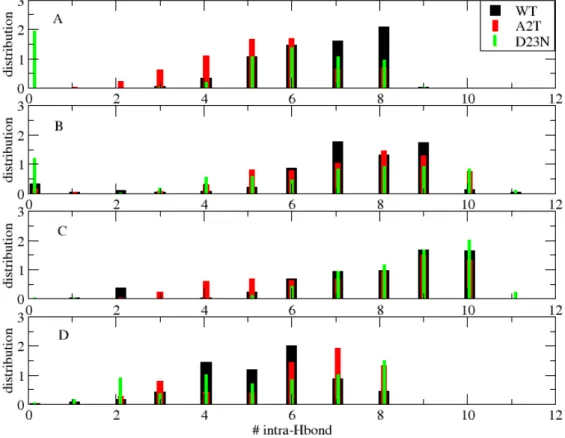
Figure 3 shows the distributions of the numbers of intra-molecular H-bonds formed between the residues 12-22 and the residues 29-40 in the four consecutive chains (hairpins). These selections of amino acids is consistent with the β-strand secondary profile, see Fig. 1. It is not surprising that the distributions of the chains 1 and 4, that close the barrel, have similar profiles (many states with 5, 6, 7 and 8 H-bonds, see panels 3A and 3D) and differ from the two similar profiles of chains 2 and 3 (many states with 7, 8, 9 and 10 H-bonds, see panels 3B and 3C). Looking at the effect of the mutations on each profile, they both reduce the population of 8 H-bonds in chain 1 (panel 3A), the population of 7 and 9 H-bonds in chain 2 (panel 3B), and increase the populations of 7 and 8 H-bonds in chain 4 (panel 3D). Interestingly, D23N is able to destabilize the first hairpin (0 H-bonds with a population of 2%, see panel 3A).
Figure 4 shows the distributions of the numbers of intermolecular H-bonds formed between the residues 12-22 and 12-22 and between the residues 29-40 and 29-40 of the four interfaces. The four distributions differ substantially from each other, the profile of the first interface varying between 0 and 9 H-bonds (panel 4A), for the third interface it is from 0 to 12 H-bonds (panel 4C) and for the fourth interface it is between 1 and 10 H-bonds (panel 4D). Looking at the effect of the mutations on each profile, we see that D23N either increases the population of the maximum number of H-bonds (e.g., peaks for 7 and 8 H-bonds in interface 1 or peaks for 7, 8 and 9 H-bonds in interface 2) or decreases the populations of the same interface at other locations (e.g., peaks for 4, 5 and 6 H-bonds in interface 2). For A2T, its long-range effect is reflected for instance by the population of the maximum number of H-bonds stabilizing the interface 4 (almost 2% for 9 and 10 H-bonds) or destabilizing the same interface 4 (peaks for 2 and 3 H-bonds, panel 4D). Upon mutation, there are some variations in the intermolecular side-chain--side-chain contact map of each individual interface, indicating structural and packing
reorganization (data not shown). Averaged over the four interfaces, however, the intermolecular contact maps of the WT and A2T sequences are very similar and have identical probabilities for the region 12-22, but the A2T map shows more intermolecular contacts in the C-terminus region (Figures 5A-B). The intermolecular pattern changes for D23N where both the hydrophobic core region and the C-terminus region display different and more contacts than their WT and A2T counterparts (Fig. 5C).
Figure 6 shows the distribution of the tilt angle and the inner pore diameter for the three species using all frames between 50 and 500 ns. The tilt angle profiles for D23N and A2T are very similar with two peaks of identical probabilities at 37-38 and 35.5 degrees, respectively. In contrast, the tilt angle profile for A2T displays a dominant peak at 36.2 degrees with a shoulder at 37.2 degrees, see Fig. 6A. The distribution of the inner pore diameter is rather uniform for the WT sequence varying between 0.42 and 0.8 nm; those for A2T and D23 display a more pronounced peak at 0.5 nm and smaller fluctuations than the WT sequence (Fig. 6B).
Based on a minimal number of 5 residues with a β Ramachandran state in each β-strand and a minimal number of x H-bonds within all hairpins and connecting the four consecutive hairpins, we can quantify the population of perfect close eight-stranded β-sheets or β-barrels for the two variants. Using all frames between 50 and 500 ns, i.e., 45000 for each species, we find 75% for WT, 71% for A2T and 44% of barrels for D23N if x=2 (loose condition); and 48% for WT (identical to previous estimations),41 45% for A2T
and 32% of barrels for D23N if x =3 (more stringent condition).
Irrespective of the values of minimal number of H-bonds, the β-barrel tetramer has the same probability for WT and A2T and is slightly destabilized for D23N. There is therefore no correlation between the predicted β-barrel populations and the experimentally increased toxicity upon D23N and decreased toxicity upon A2T. The predicted stability of D23N barrel is consistent with MD simulations demonstrating that the Osaka (E22Δ) Aβ42 barrel, i.e. with a deletion of E22 before the D23 amino acid, and the WT Aβ42 barrel made of annular double layers of β-sheets have similar morphologies
and dimensions.29 Its lower stability compared to WT results from the absence of the D23-K28 salt-bridge that leads to subtle change of short-range and long-range interactions between the different hairpins and interfaces (see Figures 4 and 5) leading to more flexibility, although the loop region (residues 23 to 28) remains exposed to solvent as for WT, see Figure 7 reporting the time-averaged number of water molecules interacting with specific Aβ residues.
It is not surprising that A2T does not impact the global barrel population compared to its WT counterpart, as this mutation does not change significantly the conformational space of the N-terminal region mainly coil (98%) compared to the WT sequence, and this N-terminus resides in the bulk water region (see Fig.7), with transient interactions with the phosphate atoms of the lipid membrane bilayer. Our finding for A2T is also in agreement with the small variation in the experimental pore conductance of the Aβ42 and Aβp3-42 peptides, where in the latter peptide, the two first residues are cleaved and the glutamate is replaced by a pyroglutamate, although MD simulations revealed that the N-termini of Aβp3-42 peptides tend to be located in the hydrophobic lipid core.49
We find that both mutations do not change the lipid order parameters of the bilayer membrane within our simulation time, and much longer timescales might be necessary to see distinct detergent-like effects on lipid membrane bilayers upon mutation.13 It is possible that toxicity decrease upon A2T mutation may result from
distinct metal site structure and metal exchange of the Cu2+-Aβ complex, as it has been
found for the A2V mutant.50 Toxicity variation upon specific mutations might also result from distinct early oligomer-membrane interactions,51,52 as the N-terminal residues
modulate these interactions,53 from larger amyloid β-barrel pores,16,20 or from variation in the lipid composition of membranes upon aging.54
Conclusion
In summary, based on REMD simulations of the WT, A2T and D23N Aβ42 β-barrel tetramers, we have shown that the probability of a perfect close β-barrel remains the
same between WT and A2T, approximately of 45%, and only slightly decreases to 32% in a DPPC lipid membrane bilayer. The Aβ42 barrel conformation is dynamic in all species and its heterogeneity varies with the amino acid sequence.
Overall we do not find an enhancement of barrel stability for D23N known to be more toxic, and a reduction of barrel stability for A2T known to be protective. Whether our results on D23N mutation may be extrapolated to the FAD mutations located at positions 21 and 22 remain to be explored.55 At the present time, we go beyond the DPPC membrane model and explore other compositions of cellular membranes, as they change in aging and AD.54,56 AUTHOR INFORMATION Corresponding Author *E-mail: philippe.derreumaux@tdtu.edu.vn. ORCID Son Tung Ngo: 0000-0003-1034-1768 Phuong H. Nguyen: 0000-0003-1284-967X Philippe Derreumaux: 0000-0001-9110-5585 Notes The authors declare no competing financial interest. ACKNOWLEDGMENTS We thank the support of the Vietnam National Foundation for Science & Technology Development (NAFOSTED) under the grant number 104.99-2019.57, University of Paris, and the French State (Grant “DYNAMO”, ANR-11-LABX-0011-01, and “CACSICE”, ANR-11-EQPX-0008). PhD also thanks the support of CNRS. References 1. Selkoe, D. J.; Hardy, J. The Amyloid hypothesis of Alzheimer's disease at 25 years. EMBO Mol. Med. 2016, 8, 595-608. 2. Walsh, D. M.; Selkoe, D. J. Aβ Oligomers – a Decade of Discovery. J. Neurochem. 2007, 101, 1172-1184. 3. Jarvis, L. M. Clinical Trial Failures. Chem. Eng. News 2012, 90, 8. 4. Doig, A. J.; del Castillo-Frias, M. P.; Berthoumieu, O.; Tarus, B.; Nasica-Labouze, J.; Sterpone, F.; Nguyen, P. H.; Hooper, N. M.; Faller, P.; Derreumaux, P. Why Is Research on Amyloid-β Failing to Give New Drugs for Alzheimer’s Disease? ACS
Chem Neurosci 2017, 8, 1435-1437.
5. Abbott, A.; Dolgin, E. Failed Alzheimer's Trial Does not Kill Leading Theory of Disease. Nature 2016, 540, 15-16.
6. Yang, X.; Meisl, G.; Frohm, B.; Thulin, E.; Knowles, T. P. J.; Linse, S. On the Role of Sidechain Size and Charge in the Aggregation of Abeta42 with Familial Mutations.
Proc Natl Acad Sci U S A 2018, 115, E5849-E5858.
7. Wallin, C. ; Hiruma, Y. ; Wärmländer, S.K.T.S. ; Huvent, I. ; Jarvet, J. ; Abrahams, J.P. ; Gräslund, A. ; Lippens, G. ; Luo, J. The Neuronal Tau Protein Blocks in Vitro Fibrillation of the Amyloid-β (Aβ) Peptide at the Oligomeric Stage. J Am Chem Soc.
2018, 140, 8138-8146.
8. Nasica-Labouze, J.; Nguyen, P. H.; Sterpone, F.; Berthoumieu, O.; Buchete, N.-V.; Coté, S.; De Simone, A.; Doig, A. J.; Faller, P.; Garcia et al. Amyloid β Protein and Alzheimer’s Disease: When Computer Simulations Complement Experimental Studies. Chem. Rev. 2015, 115, 3518-3563.
9. Foster, T.C. ; Kyritsopoulos, C. ; Kumar, A. Central Role for NMDA Receptors in Redox Mediated Impairment of Synaptic Function During Aging and Alzheimer's Disease. Behav Brain Res. 2017, 322(Pt B), 223-232.
10. Kayed, R. ; Sokolov, Y. ; Edmonds, B. ; McIntire, T.M. ; Milton, S.C. ; Hall, J.E. ; Glabe, C.G. Permeabilization of Lipid Bilayers is a Common Conformation-Dependent Activity of Soluble Amyloid Oligomers in Protein Misfolding Diseases. J
Biol Chem. 2004, 279, 46363-46366.
11. Arispe, N. ; Pollard, H.B. ; Rojas, E. Giant Multivelel Cation Channels Formed by Alzheimer Disease Amyloid beta-protein (Abeta P-(1-40)) in Bilayer Membranes.
Proc Natl Acad Sci U S A. 1993, 90, 10573-10577.
12. Berthelot, K. ; Cullin, C. ; Lecomte, S. What Does Make an Amyloid Toxic: Morphology, Structure or Interaction with Membrane? Biochimie 2013, 95, 12-19. 13. Bode, D.C. ; Freeley, M. ; Nield, J. ; Palma, M. ; Viles, J.H. Amyloid-β Oligomers Have a Profound Detergent-like Effect on Lipid Membrane Bilayers, Imaged by Atomic Force and Electron Microscopy. J Biol Chem. 2019, 294, 7566-7572. 14. Sarkar, B. ; Das, A.K. ; Maiti. S. Thermodynamically Stable Amyloid-β Monomers
Have Much Lower Membrane Affinity Than the Small Oligomers. Front Physiol.
2013, 4, 84-95.
15. Malishev, R. ; Nandi, S. ; Kolusheva, S. ; Levi-Kalisman, Y. ; Klärner, F.G. ; Schrader, T., Bitan, G. ; Jelinek, R. Toxicity Inhibitors Protect Lipid Membranes from Disruption by Aβ42. ACS Chem Neurosci. 2015, 6, 1860-1869.
16. Österlund, N.; Moons, R.; Ilag, L.L.; Sobott, F. ; Gräslund, A. Native Ion Mobility-Mass Spectrometry Reveals the Formation of β-Barrel Shaped Amyloid-β Hexamers in a Membrane-Mimicking Environment. J Am Chem Soc. 2019, 141, 10440-10450.
17. Serra-Batiste, M.; Ninot-Pedrosa, M.; Bayoumi, M.; Gairí, M.; Maglia, G.; Carulla, N. Aβ42 Assembles into Specific β-Barrel Pore-Forming Oligomers in Membrane-Mimicking Environments. Proc. Natl. Acad. Sci. U. S. A. 2016, 113, 10866–10871.
18. Bode, D.C. ; Baker, M.D. ; Viles, J.H. Ion Channel Formation by Amyloid-β42 Oligomers but Not Amyloid-β40 in Cellular Membranes. J Biol Chem. 2017, 292, 1404-1413.
19. Arispe, N. ; Pollard, H.B. ; Rojas, E. Zn2+ Interaction with Alzheimer amyloid beta Protein Calcium Channels. Proc Natl Acad Sci U S A. 1996, 93, 1710-1715.
20. Lashuel, H.A.; Hartley, D.; Petre, B.M.; Walz, T.; Lansbury, P.T. Jr. Neurodegenerative Disease: Amyloid Pores from Pathogenic Mutations. Nature
2002, 418, 291.
21. Kirkitadze, M.D.; Condron, M.M.; Teplow, D.B. Identification and Characterization of Key Kinetic Intermediates in Amyloid beta-Protein Fibrillogenesis. J Mol Biol
2001, 312, 1103-1119. 22. Kuperstein, I; Broersen, K.; Benilova, I.; Rozenski, J.; Jonckheere, W.; Debulpaep, M.: Vandersteen, A.; Segers-Nolten, I.; Van Der Werf, K.; Subramaniam, V. et al. Neurotoxicity of Alzheimer’s Disease Abeta Peptides is Induced by Small Changes in the Abeta42 to Abeta40 Ratio. EMBO J. 2010, 29, 3408-3420. 23. Kreutzer, A.G.; Nowick, J.S. Elucidating the Structures of Amyloid Oligomers with Macrocyclic beta-Hairpin peptides: Insights into Alzheimer’s Disease and Other Amyloid Diseases. Acc. Chem. Res. 2018, 51, 706-718.
24. Li, X. ; Lei, J. ; Qi, R. ; Xie, L. ; Wei, G. Mechanistic Insight into
E22Q-Mutation-Induced Antiparallel-to-Parallel β-sheet Transition of Aβ16-22 Fibrils: an All-atom
Simulation Study. Phys Chem Chem Phys. 2019, 21, 15686-15694.
25. Tran, T.T. ; Nguyen, P.H. ; Derreumaux P. Lattice Model for Amyloid Peptides: OPEP Force Field Parametrization and Applications to the Nucleus Size of Alzheimer's Peptides. J Chem Phys. 2016, 144, 205103.
26. Tuffery, P.; Derreumaux, P. Flexibility and Binding Affinity in Protein-Ligand, Protein-Protein and Multi-Component Protein Interactions: Limitations of Current Computational Approaches. J R Soc Interface. 2012, 9, 20-33.
27. Fu, Z.; Luo, Y.; Derreumaux, P.; Wei, G. Induced Beta-Barrel Formation of the Alzheimer's Abeta25-35 Oligomers on Carbon Nanotube Surfaces: Implication for Amyloid Fibril Inhibition. Biophys J. 2009, 97, 1795-803.
28. Zhang, M.; Ren, B.; Chen, H.; Sun, Y.; Ma, J.; Jiang, B.; Zheng, J. Molecular Simulations of Amyloid Structures, Toxicity, and Inhibition. Isr. J. Chem. 2017, 7-8, 586-601. 29. Jang, H.; Connelly, L.; Arce, F.T.; Ramachandran, S.; Kagan, B.L.; Lal, R. ; Nussinov, R. Mechanisms for the Insertion of Toxic, Fibril-like β-Amyloid Oligomers into the Membrane. J Chem Theory Comput. 2013, 9, 822-833. 30. Tarus, B.; Straub, J. E.; Thirumalai, D. Structures and Free-Energy Landscapes of the Wild Type and Mutants of the Aβ21–30 Peptide Are Determined by an Interplay between Intrapeptide Electrostatic and Hydrophobic Interactions. J.
Mol. Biol. 2008, 379, 815-829.
31. Murray, M.M.; Krone, M.G.; Bernstein, S.L.; Baumketner, A.; Condron, M.M.;
Lazo, N.D. ; Teplow, D.B.; Wyttenbach, T. ; Shea, J.E.; Bowers, M.T. Amyloid beta-Protein: Experiment and Theory on the 21-30 Fragment. J Phys Chem B. 2009, 113, 6041-6046. 32. Rosenman, D.J.; Wang, C.; García, A.E. Characterization of Aβ Monomers through the Convergence of Ensemble Properties among Simulations with Multiple Force Fields. J Phys Chem B. 2016, 120, 259-77. 33. Côté, S.; Derreumaux, P.; Mousseau, N. Distinct Morphologies for Amyloid Beta Protein Monomer: Aβ1–40, Aβ1–42, and Aβ1–40(D23N). J. Chem. Theory Comput. 2011, 7, 2584-2592. 34. Sterpone, F.; Melchionna, S.; Tuffery, P.; Pasquali, S.; Mousseau, N.; Cragnolini, T.; Chebaro, Y.; St-Pierre, J.; Kalimeri, M.; Barducci, A. et al. The OPEP Protein Model: From Single Molecules, Amyloid Formation, Crowding and Hydrodynamics to DNA/RNA Systems. Chem. Soc. Rev. 2014, 43, 4871-4893.
35. Han, W.; Wu, Y.-D., Molecular Dynamics Studies of Hexamers of Amyloid-β Peptide (16–35) and its Mutants: Influence of Charge States on Amyloid Formation. Proteins 2007, 66, 575-587.
36. Rodriguez, R.A.; Chen, L.Y.; Plascencia-Villa, G. ; Perry, G. Thermodynamics of Amyloid-β Fibril Elongation: Atomistic Details of the Transition State. ACS Chem
Neurosci. 2018, 9, 783-789.
37. Sharma, B. ; Ranganathan, S.V. ; Belfort, G. Weaker N-Terminal Interactions for the Protective over the Causative Aβ Peptide Dimer Mutants. ACS Chem Neurosci.
2018, 9, 1247-1253.
38. Nguyen, P.H. ; Tarus, B. ; Derreumaux, P. Familial Alzheimer A2V Mutation Reduces the Intrinsic Disorder and Completely Changes the Free Energy Landscape of the Aβ1-28 Monomer. J Phys Chem B. 2014, 118, 501-510.
39. Nguyen, P.H. ; Sterpone, F.; Campanera, J.M. ; Nasica-Labouze, J. ; Derreumaux, P. Impact of the A2V Mutation on the Heterozygous and Homozygous Aβ1-40 Dimer Structures from Atomistic Simulations. ACS Chem Neurosci. 2016, 7, 823-832.
40. Nguyen, P.H. ; Sterpone, F. ; Pouplana, R. ; Derreumaux, P. ; Campanera, J.M.
Dimerization Mechanism of Alzheimer Aβ40 Peptides: The High Content of
Intrapeptide-Stabilized Conformations in A2V and A2T Heterozygous Dimers Retards Amyloid Fibril Formation. J Phys Chem B. 2016, 120, 12111-12126.
41. Nguyen, P.H. ; Campanera, J.M. ; Ngo, S.T. ; Loquet, A. ; Derreumaux, P. Tetrameric Aβ40 and Aβ42 β-Barrel Structures by Extensive Atomistic Simulations. I. In a Bilayer Mimicking a Neuronal Membrane. J Phys Chem B. 2019,
123, 3643-3648.
42. Berendsen, H. J. C.; Postma, J. P. M.; van Gunsteren, W. F.; Hermans, A. J.
Intermolecular Forces. Reidel, Dordrecht: Jerusalem, Israel, 1981.
43. Oostenbrink, C.; Villa, A.; Mark, A. E.; Van Gunsteren, W. F., A Biomolecular Force Field Based on the Free Enthalpy of Hydration and Solvation: The GROMOS Force-Field Parameter Sets 53A5 and 53A6. J. Comput. Chem. 2004, 25, 1656-1676.
44. Berger, O. ; Edholm, O ; Jahnig, F. Molecular Dynamics Simulations of a Fluid Bilayer of Dipalmitoylphosphatidylcholine at Full Hydration, Constant Pressure and Constant Temperature. Biophys J. 1997, 72, 2002-2013.
45. Schrödinger LLC, P. The PyMOL molecular graphics system, Version 1.3r1; August, 2010.
46. Abraham, M. J.; Murtola, T.; Schulz, R.; Páll, S.; Smith, J. C.; Hess, B.; Lindahl, E. GROMACS: High Performance Molecular Simulations through Multi-Level Parallelism from Laptops to Supercomputers. SoftwareX 2015, 1–2, 19-25.
47. Touw, W. G.; Baakman, C.; Black, J.; te Beek, T. A. H.; Krieger, E.; Joosten, R. P.; Vriend, G. A Series of PDB-Related Databanks for Everyday Needs. Nucleic Acids
Res 2015, 43, D364-D368.
48. Marklund, E.G.; Degiacomi, M.T.; Robinson, C.V.; Baldwin, A.J.; Benesch, J.L., Collision Cross Sections for Structural Proteomics. Structure 2015, 23, 791-799. 49. Gillman, A.L.; Jang, H. ; Lee, J. ; Ramachandran, S. ; Kagan, B.L. ; Nussinov ; R.
Teran Arce, F. Activity and Architecture of Pyroglutamate-Modified Amyloid-β (AβpE3-42) pores. J Phys Chem B. 2014, 118, 7335-44.
50. Somavarapu, A.K.; Shen, F. ; Teilum, K.; Zhang, J.; Mossin, S.; Thulstrup, P.W. ; Bjerrum, M.J.; Tiwari, M.K.; Szunyogh, D.; Søtofte, P.M. et al. The Pathogenic A2V Mutant Exhibits Distinct Aggregation Kinetics, Metal Site Structure, and Metal Exchange of the Cu2+ -Aβ Complex. Chemistry. 2017, 23, 13591-13595.
51. Colombo, L. ; Gamba, A. ; Cantù, L. ; Salmona, M. ; Tagliavini, F. ; Rondelli, V. ; Del Favero, E. ; Brocca, P. Pathogenic Aβ A2V versus Protective Aβ A2T Mutation: Early Stage Aggregation and Membrane Interaction. Biophys Chem. 2017, 229, 11-18.
52. Habchi, J. ; Chia, S. Galvagnion, C. ; Michaels, T.C.T. ; Bellaiche, M.M.J. ; Ruggeri F.S.; Sanguanini, M. ; Idini, I. ; Kumita, J.R.; Sparr, E. et al. Cholesterol Catalyses Aβ42 Aggregation Through a Heterogeneous Nucleation Pathway in the Presence of Lipid Membranes. Nat Chem. 2018, 10, 673-683.
53. Morris, C.; Cupples, S.; Kent, T.W.; Elbassal, E.A. ; Wojcikiewicz, E.P. ; Yi, P. ; Du, D. N-Terminal Charged Residues of Amyloid-β Peptide Modulate Amyloidogenesis and Interaction with Lipid Membrane. Chemistry. 2018, 24, 9494-9498.
54. Drolle, E. ; Negoda, A. ; Hammond, K. ; Pavlov, E. ; Leonenko, Z. Changes in Lipid Membranes May Trigger Amyloid Toxicity in Alzheimer's Disease. PLoS One.
2017,12, e0182194.
55. Huet, A. ; Derreumaux, P. Impact of the Mutation A21G (Flemish variant) on Alzheimer’s beta-amyloid Dimers by Molecular Dynamics Simulations. Biophys J
2006, 91, 3829-3840.
56. Lu, Y. ; Shi, X.F. ; Nguyen, P.H. ; Sterpone, F. ; Salsbury, F.R. Jr ; Derreumaux, P. Amyloid-β(29-42) Dimeric Conformations in Membranes Rich in Omega-3 and Omega-6 Polyunsaturated Fatty Acids. J Phys Chem B. 2019, 123, 2687-2696.
Table 1. Secondary structures and cross-collision sections of A2T, D23N and WT systems
at 310 K using the time interval from 50 to 500 ns of REMD simulations.
β-strand α-helix turn coil CCS (nm2)
A2T 47.9 ± 1.7 0.1 ± 0.3 2.7 ± 1.5 49.4 ± 2.2 20.6 ± 0.1
D23N 49.7 ± 2.0 0.1 ± 0.4 2.6 ± 1.7 47.6 ± 2.1 23.1 ± 0.1
Figure 1.Secondary structure compositions along the sequence. The results are obtained using the interval 50-500 ns of REMD at 310 K
(A) (B)
Figure 2. Free energy landscapes of the transmembrane full-length A2T (A) and D23N (B)
Figure 3. Distributions of the number of intra-molecular H-bonds between the amino acid regions 12-22 and 29-40 for each individual chain (hairpin) of the WT (black), A2T (red) and D23N (green) systems at 310 K. Panels A, B, C and D refer to chain 1, chain 2, chain 3 and chain 4.
Figure 4. Distributions of the number of intermolecular H-bonds between the β-strand regions in the four interfaces of the WT (black), A2T (red) and D23N (green) systems at 310 K. Panels A and B show interactions 12-22:12-22, and panels C and D refer to interactions 29-40:29-40.
(A) (B) (C)
Figure 5. The intermolecular side chain probability contact maps averaged over the four interfaces of the WT (panel A), A2T (panel B) and D23N (panel C) systems at 310 K.
Figure 6. The distributions of the tilt angles (panel A) and the inner pore diameters (panel B) in the WT (black), A2T (red) and D23N (green) systems at 310 K.
Figure 7. Water molecules interacting with the residues 1-9 and 22-28 using a cut-off distance of 0.3 nm in the WT (black), A2T (red) and D23N (green) systems at 310 K averaged over the four peptides. Note that each chain shows very similar profile.