HAL Id: hal-01657312
https://hal.archives-ouvertes.fr/hal-01657312
Submitted on 15 Nov 2018
HAL is a multi-disciplinary open access
archive for the deposit and dissemination of
sci-entific research documents, whether they are
pub-lished or not. The documents may come from
teaching and research institutions in France or
abroad, or from public or private research centers.
L’archive ouverte pluridisciplinaire HAL, est
destinée au dépôt et à la diffusion de documents
scientifiques de niveau recherche, publiés ou non,
émanant des établissements d’enseignement et de
recherche français ou étrangers, des laboratoires
publics ou privés.
Distributed under a Creative Commons Attribution - NonCommercial| 4.0 International
License
An original technique of venous autoplasty after
duodenopancreatectomy for tumors involving the
infrarenal inferior vena cava
Bertrand Le Roy, Emmanuel Buc, Constance Hordonneau, Julie Veziant,
Denis Pezet, Johan Gagnière
To cite this version:
Bertrand Le Roy, Emmanuel Buc, Constance Hordonneau, Julie Veziant, Denis Pezet, et al.. An
orig-inal technique of venous autoplasty after duodenopancreatectomy for tumors involving the infrarenal
inferior vena cava. Journal of Surgical Case Reports, Oxford University Press, 2017, 2017 (2), pp.1
-3. �10.1093/jscr/rjx011�. �hal-01657312�
Journal of Surgical Case Reports, 2017;2, 1–3
doi: 10.1093/jscr/rjx011 Case Report
C A S E R E P O R T
An original technique of venous autoplasty after
duodenopancreatectomy for tumors involving
the infrarenal inferior vena cava
Bertrand Le Roy
1
, Emmanuel Buc
1,2
, Constance Hordonneau
3
, Julie Veziant
1
,
Denis Pezet
1,2
, and Johan Gagnière
1,2,
*
1
Department of Digestive and Hepatobiliary Surgery, Liver Transplantation, Estaing University Hospital,
Clermont-Ferrand 63000, France,
2UMR 1071 Inserm, University of Auvergne, Clermont-Ferrand 63000, France,
and
3Department of Radiology, Estaing University Hospital, Clermont-Ferrand 63000, France
*Correspondence address. Department of Digestive and Hepatobiliary Surgery, Liver Transplantation, Estaing University Hospital, 1, Place Lucie & Raymond Aubrac, Clermont-Ferrand 63000, France. Tel:+33-4-73-75-04-94; Fax: +33-4-73-75-04-59; E-mail: jgagniere@chu-clermontferrand.fr
Abstract
Tumor involvement of the inferior vena cava (IVC) by hepatobiliary, pancreatic or duodenal malignancies can compromise adequate resection. However, radical resection with negative histological margins remains the only chance of cure. Various techniques are used for venous reconstruction, using a prosthetic graft interposition in most of the cases. However, in case of associated digestive resections, such as pancreaticoduodenectomy, postoperative complications can be responsible for prosthesis infection and related vascular complications. In this setting, the use of biological material for venous reconstruc-tion appears to be preferable. We present an original, easy and useful technique of a venous autoplasty after pancreatico-duodenectomy for tumors involving the anterior wall of the infrarenal IVC, using a patch from the posterior wall of the IVC.
INTRODUCTION
Although rare, tumor involvement of the inferior vena cava (IVC) by hepatobiliary, pancreatic or duodenal malignancies can be difficult to assess preoperatively, and can compromise an adequate oncological resection, especially when found inci-dentally at laparotomy. However, radical resection with nega-tive histological margins remains the only chance of cure, especially for duodenal tumors which have better prognosis.
Lessons learned from liver transplantation have improved technical considerations and popularized partial or complete resection of the IVC, with acceptable morbidity. Various techni-ques are used for venous reconstruction, using a prosthetic graft interposition in most of the cases. However, in case of
associated digestive resections, postoperative complications can be responsible for prosthesis infection and related vascular complications. In this setting, the use of biological material for venous reconstruction appears to be preferable.
We present an original technique of a venous autoplasty in a woman who underwent pancreaticoduodenectomy for a duodenal adenocarcinoma involving the anterior part of the infrarenal IVC.
CASE REPORT
A 74-year-old woman was referred in our tertiary center for the exploration of an anemia. She had no previous remarkable med-ical history. Gastroduodenal endoscopy with biopsies identified an adenocarcinoma of the second part of the duodenum.
Received: October 12, 2016. Accepted: January 24, 2017
Published by Oxford University Press and JSCR Publishing Ltd. All rights reserved. © The Author 2017.
This is an Open Access article distributed under the terms of the Creative Commons Attribution Non-Commercial License (http://creativecommons.org/ licenses/by-nc/4.0/), which permits non-commercial re-use, distribution, and reproduction in any medium, provided the original work is properly cited. For commercial re-use, please contact journals.permissions@oup.com
1
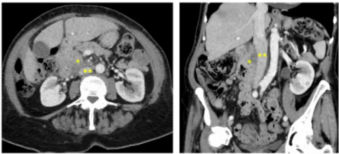
Abdominal computed tomography (CT) scan showed a huge duodenal mass of 7 cm in size developed from the second part of the duodenum, close to the anterior wall of the IVC, without extension to other major vessels (e.g. coeliac, hepatic or superior mesenteric artery) or metastases (Fig.1). After discussion in a multidisciplinary digestive cancer board meeting at our institu-tion, she was planned for a pancreaticoduodenectomy.
The patient underwent a bi-subcostal laparotomy. A Kocher maneuver was not possible because of strong adhesions between the tumor and the anterior wall of the IVC. In order to ensure adequate resection margins, we performed a pancreati-coduodenectomy with en-bloc resection of a 3 cm² area of the anterior wall of the IVC (Fig.2A), after total clamping of the right and the left renal veins, and of the IVC from either side of the involved area. Direct suture of the IVC was not possible. We decided not to use a prosthetic graft for the venous reconstruc-tion because of the risk of infecreconstruc-tion regarding the high rate of infectious complications after pancreaticoduodenectomy, par-ticularly in case of soft pancreas texture. Thus, since the poster-ior wall of the IVC is usually morefloppy than the anterior one because of its higher distance from the renal veins, we harvested a transversal patch of 20× 30 mm in size from the posterior wall of the IVC, below the confluence with the renal veins (Fig.2B). We did not encounter any vertebral veins. Reconstruction of the posterior wall of the IVC was achieved by a direct hand-sewn
hemi-circumferential anastomosis using a single-layer non-absorbable 4/0 polypropylene (Fig. 2C). Thereafter, the venous patch was disposed longitudinally in place of the resected area at the anterior wall of the IVC, and was sutured using a single-layer non-absorbable 4/0 polypropylene (Figs.2C, D and F). Total duration of the clamping procedure was 90 minutes. The digest-ive reconstruction was then performed according to the Child procedure with a pancreaticogastrostomy.
The patient received a prophylactic anticoagulation. Postoperative course was marked by a bile leakage on post-operative day 2 which required surgery to repair the hepaticojeju-nal anastomosis. Postoperative CT-scan showed good permeability of the IVC with no symptoms related to the reconstruction. She was discharged on postoperative day 39.
Pathological examination confirmed the R0 resection of a T4 N1 M0 duodenal adenocarcinoma involving the IVC. She received a 6 months adjuvant FOLFOX chemotherapy regimen and was followed up every 3 months with no recurrence at time.
DISCUSSION
Resection of the IVC is widely performed during surgery for vari-ous abdominal and retroperitoneal malignancies, since a radical resection is the only chance of cure. Suprarenal IVC resection without venous replacement has been proposed because the ven-ous collateral pathways can supply the cavalflow through the lumbar and epigastric pathways, the renal veins, and the vertebral venous system, which are often well developed in case of a IVC chronic occlusion [1]. However, large dissection of a retroperiton-eal tumor and/or thoracoabdominal incisions can both interrupt the ascending collateral venous pathways, resulting in a high risk of major venous insufficiency [2]. In these cases, reconstruction of the IVC is mandatory and can be performed through a direct end-to-end anastomosis, or after the interposition of a venous/pros-thetic patch. Nardo et al. [3] reported that resection≤2 cm of the IVC could be reconstructed by a direct suture while resection >2 cm required the interposition of a graft. In the same way, Tsuji et al. [4] performed a direct suture when the wall defect was less than one-third of the circumference of the IVC. In our case, the venous resection was too large to perform a direct suture of the
Figure 1: Preoperative CT-scan showing a huge duodenal mass (*) of 7 cm in size developed from the second part of the duodenum, close to the anterior wall of the infrarenal IVC (**).
Figure 2: (A) We performed a pancreaticoduodenectomy with en-bloc resection of a 3 cm² area of the anterior wall of the IVC (*). Direct suture of the IVC was not pos-sible and venous reconstruction required the interposition of a graft. (B) Since the posterior wall of the IVC is usually morefloppy than the anterior one because of its longer distance from the renal veins, we harvested a transversal patch of 20× 30 mm in size from the posterior wall of the IVC, below the confluence with the renal veins. (C) Reconstruction of the posterior wall of the IVC was achieved by a direct hand-sewn hemi-circumferential anastomosis using a single-layer non-absorbable 4/0 polypropylene. (D and E) The transversal venous patch was placed longitudinally on the resected area at the anterior wall of the IVC (**), and was sutured using a single-layer non-absorbable 4/0 polypropylene. (F) Intraoperative view showing the venous autoplasty of the IVC. SRIVC, suprarenal inferior vena cava; IRIVC, infra-renal inferior vena cava; RRV, right infra-renal vein; LRV, left infra-renal vein.
2
|
B. Le Roy et al.IVC, either longitudinal or transversal. Therefore, the technique of choice regarding this large venous defect appeared to be a pros-thetic reconstruction. However, considering the high risk of infec-tious complications after duodenopancreatectomy, especially when performed in the settings of a duodenal tumor with a soft pancreatic gland texture and without dilatation of both the pan-creatic and the main bile duct, we chose a reconstruction tech-nique that required a large venous patch. Thus, we decided to harvest the posterior wall of the IVC because it was veryfloppy, that allowed its direct suture. Indeed, these elastic properties of the IVC have already been investigated. Wigmore et al. [5] reported the possibility to easily harvest a venous patch from the anterior wall of the IVC, above the confluence of the renal veins, with a low risk to narrow the IVC because its diameter is larger at this point. Other large veins, such as the portal vein, can also be resected. Our original technique of venous autoplasty could be thus extrapo-lated to these veins, in order to prevent the use of prosthetic patches and to facilitate reconstruction.
CONFLICT OF INTEREST STATEMENT
None declared.
REFERENCES
1. Duckett JW Jr, Lifland JH, Peters PC. Resection of the inferior vena cava for adjacent malignant diseases. Surg Gynecol Obstet 1973;136:711–6.
2. Huguet C, Ferri M, Gavelli A. Resection of the suprarenal inferior vena cava. The role of prosthetic replacement. Arch Surg 1995;130:793–7.
3. Nardo B, Ercolani G, Montalti R, Bertelli R, Gardini A, Beltempo P, et al. Hepatic resection for primary or second-ary malignancies with involvement of the inferior vena cava: is this operation safe or hazardous? J Am Coll Surg 2005;201:671–9.
4. Tsuji Y, Goto A, Hara I, Ataka K, Yamashita C, Okita Y, et al. Renal cell carcinoma with extension of tumor thrombus into the vena cava: surgical strategy and prognosis. J Vasc Surg 2001;33:789–96.
5. Wigmore S. A quick and easy technique for patching small holes in the portal vein.http://profstevewigmorewordpress com/2012/10/24/a-quick-and-easy-technique-for-patching-s mall-holes-in-the-portal-vein/. 2012.
An original technique of venous autoplasty after duodenopancreatectomy for tumors