HAL Id: tel-01343867
https://tel.archives-ouvertes.fr/tel-01343867
Submitted on 10 Jul 2016HAL is a multi-disciplinary open access
archive for the deposit and dissemination of sci-entific research documents, whether they are pub-lished or not. The documents may come from teaching and research institutions in France or abroad, or from public or private research centers.
L’archive ouverte pluridisciplinaire HAL, est destinée au dépôt et à la diffusion de documents scientifiques de niveau recherche, publiés ou non, émanant des établissements d’enseignement et de recherche français ou étrangers, des laboratoires publics ou privés.
Pneumococcus morphogenesis and resistance to
beta-lactams
Jules Philippe
To cite this version:
Jules Philippe. Pneumococcus morphogenesis and resistance to beta-lactams. Virology. Université de Grenoble, 2014. English. �NNT : 2014GRENV018�. �tel-01343867�
Université Joseph Fourier / Université Pierre Mendès France / Université Stendhal / Université de Savoie / Grenoble INP
Thesis
to obtain the grade of
PhD for the UNIVERSITY OF GRENOBLE
Specialty: Virology – Microbiology – Immunology
Presented by
Jules PHILIPPE
Thesis directed by André ZAPUN
prepared within the Pneumococcus Group – Institute for Structural Biology – UMR 5075
at the Doctoral School of Chemistry and Life Sciences
Pneumococcus Morphogenesis
and resistance to beta-lactams
Thesis defended in public on September 29th, 2014to the following jury:
Dr. Rut Carballido-López Research director at INRA, Reporter Pr. Leiv Sigve Håvarstein
Professor at the Norwegian University of Life Sciences, Reporter Pr. Christelle Breton
Professor at the University Joseph Fourier, Examiner Dr. Christophe Grangeasse
Research director at CNRS, Examiner Pr. Malcolm G. P. Page
Head of Biology at Basilea Pharmaceutica International Ltd, Examiner Dr. André Zapun
Université Joseph Fourier / Université Pierre Mendès France / Université Stendhal / Université de Savoie / Grenoble INP
THÈSE
Pour obtenir le grade de
DOCTEUR DE L’UNIVERSITÉ DE GRENOBLE
Spécialité : Virologie – Microbiologie – Immunologie
Arrêté ministériel : 7 août 2006
Présentée par
Jules PHILIPPE
Thèse dirigée par André ZAPUN
préparée au sein du Groupe Pneumocoque – Institut de Biologie Structurale – UMR 5075
dans l'École Doctorale Chimie et Sciences du Vivant
Morphogénèse du
pneumocoque et résistance aux
bêta-lactamines
Thèse soutenue publiquement le 29 septembre 2014, devant le jury composé de :
Dr. Rut Carballido-López
Directrice de recherches à l’INRA, Rapporteur Pr. Leiv Sigve Håvarstein
Professeur à l’Université de Norvège, Rapporteur Pr. Christelle Breton
Professeur à l’Université Joseph Fourier, Examinatrice Dr. Christophe Grangeasse
Directeur de recherches au CNRS, Examinateur Pr. Malcolm G. P. Page
Directeur Biologie chez Basilea Pharmaceutica International Ltd, Examinateur Dr. André Zapun
1
First description of the pneumococcus by Louis Pasteur.
Extract of the French ‘Bulletin de l’Académie de médecine’, 1881.
3
Acknowledgements
First, I would like to thank Dr. Rut Rut Carballido-López and Pr. Leiv Sigve Håvarstein to have accepted to review this manuscript, as reporters of the jury. I am also grateful to Pr. Christelle Breton, Dr. Christophe Grangeasse and Pr. Malcolm G. P. Page to participate in the evaluation as examinators.
I would like to acknowledge Thierry VERNET to have welcomed me in the Pneumococcus Group from 2009 to 2014. Thierry always made sure to provide me with a pleasant and fruitful environment. He has always been of very good advice and appreciable company in scientific discussion, but also speaking about travelling and mountain outdoor activities. Thank you Thierry, I guess I have been lucky to spend these years in your lab.
Many thanks go to André Zapun, a great PhD director that knew how to deal with a wild PhD student. André, when I met you the first time, you had a cast to recover from the thumb you broke in climbing. During the “interview” and later on, you told me about your backpacking experiences around the world, your mountaineering expeditions, the incredible variety of birds and if I remember well, about a bug I think you called “pneumococcus”. How could I miss it, I could not refuse! Thank you for the supervision you finely adapted to my personality. Thank you for being so logical that any unexpected result can make sense. Thank you for keeping the energy to transmit your great knowledge of Biochemistry to a student that always privileged Microbiology classes at the expense of Biochemistry at the University. You taught me how to plan experiments, how to avoid useless ones, how to interpret results and (I don’t know if it is appropriate here, but) how to write. Thank you again.
I would like to thank the other researchers of the lab for their help and precious advices to make not-working experiments work, and explaining me various incredible molecular mechanisms of the pneumococcus. Claire, thank you for the rhubarb, Cécile thank you for the white wine at each occasion we could celebrate something, Marjolaine, thank you for your smile and showing me how to subjugate membrane proteins with detergents, Anne-Marie D.G., thank you for your advices. Also Anne-Marie V, thank you for your pleasant company.
I would like to thank Laure Roux, the best lab technician ever, always happy, taking care of the people, easy going, accommodating, I think my English vocabulary becomes limiting. Thank you for making things easy in the lab, and for your great company outside the lab. Greetings to your family!
My acknowledgements also go to the Eefjan Breukink that hosted me for three weeks in his laboratory of the University of Utrecht. Thank you for the supervision in lipid II synthesis. I also thank Martijn Koorengevel that also helped me in the experiments in Utrecht. I also thank Régine
4
Hakenbeck and Dalia Denapaite for the generous gift of the GFP:PBP2x encoding strain of pneumococcus.
I would like to acknowledge Nordine Helassa, who built many constructs I used and Fany Couppey that initiated the work on piperacillin prior to my arrival at the laboratory. This work would not have been the same without the tools and knowledge you generated.
Thanks to Yann Huon de Kermadec tried to convert me to the world of nanodiscs. Thank you for the introduction to the “nanos”, but also for the enjoyable time we have outside the lab.
Gravier, thanks for introducing me to the world of the good beer. OK, I’ll never be a fine Belgium beer taster as you, but it has always been a pleasure to share a drink with you. Also at the lab, my office-mate, always here to give me precious advices and transmitting your wide knowledge on PBPs, thank you.
Max, Rémi and Julie, the other PhD students of the lab. Many thanks for the nice environment you brought at work, for the meals and beers we had together to forget about our heavy student duty…
Louise, Widade, Yann H2K, Papa Jimmy, Yann Fichou, Gianluca, Mathieu (with 1 or 2 “t”, I never remember), Hicham, Michel (not PhD, but it’s the same) and Didier, thank you for being PhD students at the same time as me, it’s always good to know we are not alone. I’ll miss the Wednesday H2 meals!
Of course, many thanks to anybody that worked at the lab and brought a nice atmosphere as Ana, Guillaume, Benjamin, Flo, Justine, Lucie, Violaine, Iza it was great to “work” with you!
Thank you Lorraine, for the support, for the best meals when I had only chineese noodles to survive, for the soft words that made me keeping a good mood even when printing spoiled the whole formatting!
Thank you Jérôme and Steph, the roomates without whom I’d be 5kg lighter… Thanks to all the potes grenoblois, these studies where rich in people and mountains!
Thank you papa and maman for all the support and for allowing me to complete these studies and to have let me travelling for several years as far and as much as I could!
This work used the platforms of the Grenoble Instruct center (ISBG ; UMS3518 CNRS-CEA-UJF-EMBL) with support from FRISBI (ANR-10-INSB-05-02) and GRAL (ANR-10-LABX-49-01) within the Grenoble Partnership for Structural Biology (PSB).
I would like to thank Daphna Fenel and Dr Guy Schoehn from the Electron Microscopy platform, I thank Aline Leroy and Christine Ebel from the PAOL platform and Lionel Imbert from the cell free platform. I thank Anne Marie Villard and Dr. Marjolaine Noirclerc-Savoye, for the detergent
5
screening for membrane protein solubilization and purification; and Céline Charavay and Stéphane Segard (GIPSE, CEA, Grenoble) for the RoBioLIMS database development.
Finally, I would like to thank the Région Rhône Alpes and the ARC1 Santé for financing my PhD. I also thank the CEA, the UJF and the CNRS that are the three institutes of the UMR 5075.
6
Contents
Acknowledgements ... 3
Contents ... 6
Abbreviations ... 10
I- Introduction
... 13
Le pneumocoque – résumé ... 14
The pneumococcus ... 16
Discovery ... 16
General description and characteristics ... 16
Bacteriology ... 16
Pneumococcus cell surface ... 17
Serotypes ... 19
Natural environment ... 19
Competence ... 21
Autolysis ... 22
Pathogenicity ... 23
Vaccines ... 25
Antibiotics ... 27
Epidemiology ... 28
Concluding remarks ... 30
Le peptidoglycane du pneumocoque – résumé ... 32
Pneumococcus peptidoglycan ... 34
Pneumococcus peptidoglycan composition ... 34
Architecture ... 37
Peptidoglycan Biosynthesis ... 38
Cytoplasmic steps: synthesis of the lipid II ... 40
Flipping the lipid II across the plasma membrane: the flippases ... 41
Extra-cellular steps: elongation and cross-linking of the glycan chains ... 41
Peptidoglycan Maturation ... 44
Concluding remarks ... 47
Morphogenèse du pneumocoque – résumé ... 48
Morphogenesis of the pneumococcus ... 50
7
Elongation proteins ... 52
Division proteins ... 56
Proteins involved in the organization of the division ring ... 57
Peptidoglycan assembly-related proteins ... 59
Proteins involved in the separation of daughter cells ... 61
Relationship between the two morphogenesis machineries ... 63
Concluding remarks ... 67
Pneumocoque et β-lactamines ... 69
Pneumococcus and β-lactams ... 70
β-Lactam antibiotics ... 71
Resistance of the pneumococcus against the β-lactams ... 73
Alterations in the PBPs... 73
Mosaic genes ... 75
Concluding remarks ... 76
Objectives ... 78
II- Material & Methods
... 79
Reconstitution of pneumococcus membrane protein complexes ... 80
Strains, plasmids and growth conditions ... 80
Bacterial transformation... 81
Plasmid preparation and sequencing ... 81
Construction of pET30-RA2bSMCHMD and pETduet-RA2bSMCHMD ... 82
Pneumococcus growth ... 82
Escherichia coli expression system ... 83
Protein expression ... 83
Membranes isolation ... 83
Protein purification ... 84
Buffers ... 84
Solubilization of the membrane samples ... 84
Solubilization of mixed membrane samples ... 84
Ni-NTA ... 84
Strep-Tactine
®... 85
Successive purifications ... 85
Analysis of the protein samples ... 85
8
Western blot ... 86
PAOL and EM sample preparation ... 86
Cell free expression system ... 87
Plasmids construction and preparation ... 87
Plasmids ... 87
Construction of pIVEX-HMC, pIVEX-HMD and pIVEX-SMCHMD ... 87
DNA preparation for cell free expression reaction ... 88
Protein expression ... 88
Protein purification (RoBioMol platform) ... 89
In vitro reconstitution of the activities of pneumococcus PBPs ... 91
Lipid II synthesis ... 91
Preparation of Micrococcus flavus membranes ... 91
Lipid II synthesis... 91
Lipid II purification ... 92
Dansylation of the lipid II ... 93
Activity tests ... 94
Expression and purification of the PBPs ... 94
In vitro reconstitution of the activities of PBP1a ... 94
Analysis of the peptidoglycan synthesized in vitro ... 95
SDS-PAGE, ... 95
Densitometry ... 95
III- Results
... 97
Vers la reconstitution de l’élongasome du pneumocoque – résumé ... 98
Towards the reconstitution of the pneumococcus elongasome in vitro ... 100
Expression in E. coli membranes ... 101
Previously characterized complexes: PBP2b-RodA and PBP2x-FtsW ... 101
MreC/MreD ... 101
MreC/MreD/PBP2b/RodA ... 104
MreC/MreD/PBP2x/FtsW ... 109
MreC/MreD and DivIB/DivIC/FtsL ... 111
Concluding remarks ... 112
Cell-free expression ... 113
pIVEX vectors ... 114
9
Purification of MreC and MreD after cell free expression ... 115
Concluding remarks and perspectives ... 116
Synthèse de peptidoglycane de pneumocoque in vitro – résumé ... 118
Synthesis of pneumococcus peptidoglycan in vitro ... 120
Lipid II synthesis ... 121
In vitro peptidoglycan synthesis activity of PBP1a ... 123
Concluding remarks ... 125
Mécanismes d’action des β-lactamines sur S. pneumoniae : le paradoxe de la
pipéracilline - Résumé ... 126
Mechanisms of β-lactam action in Streptococcus pneumoniae: the piperacillin paradox
... 129
IV- Discussion
... 171
Discussion - Résumé ... 172
Discussion ... 174
Organization of the morphogenesis machineries in the pneumococcus ... 174
What can we learn from the activity of the PBPs? ... 178
Redundancy of the class A PBPs ... 178
A preponderant role for PBP2b compared to PBP2x ... 179
Compensation of impaired class B PBP activity ... 180
Conclusion générale - Résumé ... 182
General conclusion ... 182
References ... 183
Annexes... 200
Summary - Résumé
10
Abbreviations
3D-SIM: three-dimensional structured illumination microscopy aLII: amidated lipid II
AT: amidotransferase ATP: adenosin triphosphate AU: arbitrary units
CDC: center for disease control CMC: critical micelle concentration CSP: competence-simulating peptide CV: column volume
DDM: n-dodecyl-β-D-maltopyranoside
DEAE: Diethylaminoethyl
DIC: differential interference contrast microscopy DMSO: dimethyl sulfoxide
DNA: deoxyribonucleic acid EM: electron microscopy FosC12: fos-choline-12
g: gram
GlcNAc: N-acetylglucosamine
GT: glycosyltransferase
h: hour
HPLC: high-performance liquid chromatography HRP: horseradish peroxidase
IPD: invasive pneumococcal diseases IPTG: isopropyl β-D-1-thiogalactopyranoside
k: kilo
Da: dalton
L: liter
LII: lipid II
LII-DNS: dansylated lipid II LTA: lipoteichoic acids
m: meter
m…: milli…
µ: micro
M: molar (mole / liter)
MALLS: multi-angle laser light scattering MCS: multiple cloning site
MIC: minimum inhibitory concentration min: minute
MurNAc: N-acetylmuramic acid
n: nano
OD: optical density (in nanometer) PBP: penicillin-binding protein PBS: phosphate buffer saline
11
PCR: polymerase chain reactionPCV: pneumococcal conjugate vaccine PEG: polyethylene glycol
PLP: protéine liant la pénicilline psi: pound per square inch QELS: quasi-elastic light scattering
qs: quantity sufficient RI: refractive index RNA: ribonucleic acid
ROS: reactive oxygen species rpm: rotation per minute
SEC: size exclusion chromatography
SDS-PAGE: sodium dodecyl sulfate – polyacrylamide gel electrophoresis SEDS: shape, elongation, division and sporulation proteins
TLC: thin layer chromatography
TM: transmembrane
TP: transpeptidase tRNA: transfer RNA UDP: uridine diphosphate UV: ultra-violet
V: volt
WHO: world health organization WTA: wall teichoic acids
13
I- Introduction
Louis Pasteur (left) and George Miller Sternberg (right) simultaneously
discovered
Streptococcus pneumoniae in the blood of rabbits
inoculated with human saliva in 1881.
14
Le pneumocoque – résumé
Streptococcus pneumoniae, communément appelé le pneumocoque, est une bactérie à Gram
positif souvent rencontrée dans la flore commensale du nasopharynx de l’humain. Dans certaines conditions, elle peut engendrer des maladies, tout particulièrement chez les personnes au système immunitaire faible (nourrissons, personnes âgées ou immunodéprimées). Les infections à pneumocoques peuvent être diverses selon le site de propagation de la bactérie dans le corps humain. Les plus communes sont les otites chez les enfants, mais on rencontre aussi des sinusites, pneumonies ou septicémies qui entraînent dans les cas les plus graves des méningites. Dans le monde, plus d’un million et demi de personnes meurent chaque année d’infections à pneumocoques. Les enfants des pays en voie de développement sont les plus touchés.
Le pneumocoque adopte une forme caractéristique en diplocoque et peut former des chainettes lorsqu’elle est cultivée en milieu liquide en laboratoire. A sa surface, il possède une membrane cytoplasmique entourée d’une paroi et éventuellement d’une capsule. La paroi (qui fait l’objet du chapitre suivant) est une structure essentielle qui protège la bactérie contre la pression osmotique. Elle donne sa forme à la bactérie et permet l’ancrage de diverses molécules de surface, telles que les polysaccharides qui constituent la capsule du pneumocoque. En tant que structure de surface (la plus à l’extérieure de cette bactérie), la capsule est un facteur de virulence majeur qui gêne la reconnaissance par le système immunitaire de l’hôte.
Le pneumocoque est capable de s’adapter à diverses conditions, ce qui lui permet d’évoluer au travers d’environnements très différents au cours de l’infection. En temps qu’anaérobe aéro-tolérant, il peut passer du nasopharynx au sang où les concentrations d’oxygène sont très différentes. Aussi, la génération d’une forte concentration de peroxyde d’hydrogène lui permet d’évoluer lors d’infections, en particulier dans les pneumonies ou septicémies. Le pneumocoque est aussi naturellement résistant au lysozyme, une des défenses majeures de l’hôte contre les bactéries.
L’importante capacité d’adaptation du pneumocoque est renforcée par sa facilité à intégrer les caractères génétiques des bactéries de son environnement. Deux caractéristiques du pneumocoque y jouent un rôle clé. Premièrement, il est naturellement compétant, ce qui lui permet d’intégrer dans son génome des fragments d’ADN contenant des informations génétiques pouvant être sélectionnées si elles lui sont avantageuses. De plus, le pneumocoque autolyse, c’est-à-dire que lorsque la densité de pneumocoques est trop importante, une enzyme entraîne la lyse des bactéries, relâchant du matériel génétique dans le milieu extérieur, disponible pour les pneumocoques restants. Le phénomène de compétence est impliqué dans la résistance aux antibiotiques et l’évasion des vaccins.
15
A ce jour, plus de 90 sérotypes de pneumocoques ont été identifiés qui diffèrent par la composition de la capsule. Les vaccins disponibles étant basé sur l’immunisation par injection de polysaccharides de surface, seuls certains sérotypes sont pris en compte. Le Pneumovax®23 (Merck) protège les adultes contre 23 sérotypes de pneumocoque mais est faiblement immunogénique chez l’enfant. Deux vaccins utilisés chez l’enfant, le Prevenar®7 et le Prevenar®13 (Pfizer) sont plus efficaces dans ce contexte, grâce à la conjugaison de 7 ou 13 polysaccharides de surface à une protéine porteuse. Ces vaccins, développés aux Etats-Unis, ciblent les sérotypes qui étaient les plus présents et résistants dans les isolats cliniques de ce pays au moment de leur confection. Leur utilisation a permis de faire baisser la proportion des sérotypes concernés dans les infections à pneumocoques recensées dans les pays occidentaux. Cependant, ils sont moins adaptés aux sérotypes présents en Asie et en Amérique du Sud, particulièrement.
Le développement précoce des bêta-lactamines a procuré un traitement de choix pour les infections à pneumocoques grâce à sa forte sensibilité pour ces antibiotiques. Aussi, leur utilisation de manière intensive a-t-elle engendré l’émergence de souches résistantes dès la fin des années 70. En France, la fréquence des souches cliniques de pneumocoques résistantes à la pénicilline a cessé d’augmenter en 2002 grâce à la mise en place d’un plan national pour l’efficacité des antibiotiques et l’augmentation de la vaccination des nourrissons.
16
The pneumococcus
The pneumococcus is only 1 µm long. Behind its tiny appearance, this bacterium hides many properties and characteristics that make it of great scientific and medical interest. First, it causes major health problems around the world, from the poorest to the richest country and treatments must be continuously developed to limit infections. Then, many mechanisms are used by this opportunistic pathogen to cope with its environment and the strategies used by humans to fight it. This part intends to give a general overview of Streptococcus pneumoniae that shall help the reader to understand why the pneumococcus is of interest.
Discovery
Streptococcus pneumoniae – the pneumococcus – was first described in 1881 from two
independent accidental observations. George Miller Sternberg, one of the first scientists studying bacteria in the United States was intending to identify the cause of malaria. He injected rabbits with different samples collected in swamps of the suburbs of New Orleans. As a “sterile” control, he used his own saliva. The “control” rabbit died in forty eight hours and Sternberg observed in its blood some micro-organisms he called micrococci (Sternberg, 1881). Almost eight thousand kilometers away, the same year, Louis Pasteur could isolate the same organism from the blood of two rabbits that died in thirty six hours after inoculation with the saliva of a dead patient with rabies (Pasteur, 1881). Because of its capacity to cause pulmonary diseases, this bacterium was soon renamed pneumococcus, and Diplococcus pneumoniae for its shape. It was eventually called S. pneumoniae in 1974 for its propensity to form chains of cocci in liquid cultures (Watson, et al., 1993).
General description and characteristics
Bacteriology
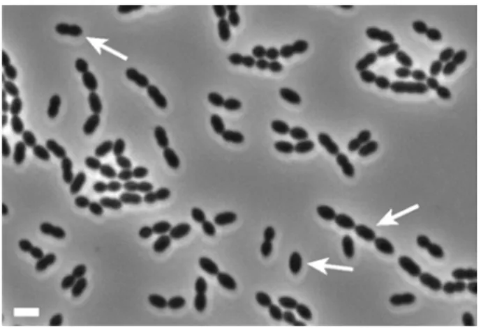
S. pneumoniae is a 1 µm Gram-positive bacterium that has an ovoid shape (slightly elongated
coccus with an “American football” shape). In liquid cultures, it is often observed as diplococci (a pair of cocci), but it can also be seen as isolated cells or in chains (Figure 1). A pneumococcus cell is not motile and does not form spores. Its density must be close to that of water as liquid cultures in exponential phase without stirring appear turbid from surface to bottom. In these conditions, the generation time is typically forty minutes. S. pneumoniae is an aero-tolerant anaerobic bacterium that prefers an environment enriched in carbon dioxide (typically, an atmosphere containing 5% of carbon dioxide is used for cultivation), where it ferments glucose to lactic acid for growth. The
17
pneumococcus does not produce catalase,which is often added to the cultivation media (by adding blood, for example) in order to protect it against the hydrogen peroxide it produces as a byproduct of its metabolism. One convenient property of S.
pneumoniae is that it is α-hemolytic: the
hydrogen peroxide it produces oxidizes hemoglobin (red, with a Fe2+ ferrous ion in
the heme) to methemoglobin (green, with a Fe3+ ferric ion in the heme). Thus, colonies
of pneumococci grown on blood agar medium are characterized by the dark green halo surrounding them, which enables an easy identification. The pneumococcus can be differentiated from other α-hemolytic streptococci by its unique sensitivity to optochin (a quinine derivative).
S. pneumoniae belongs to the phylum Firmicutes, the class Bacilli, the order Lactobacillales,
the family Streptococcaceae and the genus Streptococcus. This genus includes six major clusters, as defined by 16S rRNA sequencing: pyogenic, anginosus, mitis (including S. pneumoniae), salivarius, bovis and mutans (Kawamura, et al., 1995).
Pneumococcus cell surface
The surface of the pneumococcus is comprised of two major layers surrounding the plasma membrane: the cell wall and the capsule (Figure 2).
The cell wall contains peptidoglycan, a single macromolecule made of glycan chains reticulated by peptide bridges. This molecule is described in details in the next chapter. Cell wall also comprises the teichoic acids (40 - 50 % of
the cell wall dry weight (Bui, et al., 2012)). Teichoic acids are polysaccharides that contain glycerol-phosphate or ribitol-phosphate subunits, found in Gram-positive bacteria. They can be anchored to the plasma membrane (lipoteichoic acids, or LTA) or to the peptidoglycan (wall teichoic acids, or WTA). In
S. pneumoniae, the teichoic acids are unusual
Figure 1: Phase contrast image of pneumococcus R6 cells. Arrows indicate a single cell, a diplococcus and a chain of pneumococci. Scale bar: 2µm.
Figure 2: Schematic representation of S.
pneumoniae cell surface. Grey: capsule, black: cell
18
for several reasons. Contrary tomost bacteria, the repeating unit of WTA is identical to that of LTA (Fischer, 1997). It is composed of the rare amino sugar 2-acetamido-4-amino-2,4,6-trideoxy-galactose, glucose, ribitol-phosphate,
N-acetylgalactosamine, and phosphocholine, repeated 6 to 7
times in one WTA molecule (Figure 3, (Bui, et al., 2012)). The phosphocholines are particular to the pneumococcus. They enable the binding of several surface proteins to the cell wall through non-covalent linkage (the choline-binding proteins, or CBPs). Also, choline is essential for the growth of pneumococcus, which is the only bacterium known with this requirement. The phosphocholines play a role in multiple mechanisms involved in the formation of chains, competence, autolysis (Tomasz, 1968) and penicillin-induced lysis (Tomasz, et al., 1970). This is explained by the fact that some of the CBPs are peptidoglycan hydrolases required for all these mechanisms.
The polysaccharide capsule overlays the peptidoglycan layer and its composition determines the serotype of a given strain. Generally, the capsule is covalently attached to the cell wall of pneumococcus, with some exceptions (serotype 3, for example (Sorensen, et al., 1990)), and is typically 200 to 400 nm thick. The capsule increases the virulence of this pathogen (Kelly, et al., 1994). Of note though, two cases of conjunctivitis have been reported to be caused by non-encapsulated isolates (Martin, et al., 2003), (Crum,
et al., 2004), indicating that this structure is not
essential for the development of pneumococcal conjunctivitis, at least. The capsule was shown to help pneumococci evade the host immune system (Avery & Dubos, 1931). Globally, the capsule reduces the interaction with phagocytes by charge at physiological pH, it impairs phagocyte recognition of immunoglobulins and complement molecules bound to surface molecules (teichoic acids or surface proteins) because the latter are shorter than the capsule thickness, it reduces the complement deposition at the pneumococcus
Figure 4: Composition of pneumococcal teichoic acids, modified from (Bui, et al., 2012). WTA are anchored to peptidoglycan and LTA to the plasma membrane. AATGal: 2-acetamido-4-amino-2,4,6-trideoxy-galactose, Glc: glucose, P: phosphate, GalNAc: N-acetylgalactosamine, P-Cho: phosphocholine, PG: peptidoglycan.
Figure 3: Scanning electron microscopy picture of A66 encapsulated pneumococci after 3 h of incubation with HEp-2 larynx cells. The arrow indicates a pneumococcus in close interaction with the cell, with a thinner capsule than the other pneumococci of the chain. Adapted from (Hammerschmidt, et al., 2005).
19
surface and the trapping by neutrophil extracellular traps (reviewed in (Kadioglu, et al., 2008)).
During colonization of the host, the capsule layer becomes thinner upon adhesion to epithelial tissues to favor the interaction (Figure 4, (Hammerschmidt, et al., 2005)). Also, S.
pneumoniae can switch the composition of their capsule (hence serotype) by transformation to
evade immunity, for example.
Serotypes
In 1910, a typing method was developed by Franz Neufeld and Ludwig Händel providing evidence, for the first time, that pneumococci can belong to different serotypes. This method consisted in injecting clinical pneumococcal strains to mice previously immunized with different types of pneumococci, and observing death or survival of the animal (Neufeld & Haendel, 1910). A series of additional serotypes were later identified in the United States by Dochez and Gillespie and in South Africa by Lister, present in the latter case only in South Africa (Watson, et al., 1993). Nowadays, serotyping is performed by coagglutination with antibodies, and recently by multiplex PCR or the use of DNA microarrays.
The serotype of an organism is determined by its antigenic constitution, which differs from other organisms of the same species. To date, at least 93 distinct pneumococcus serogroups with distinct capsular poly-saccharides structures have been described (Henriques-Normark & Tuomanen, 2013). The different serotypes are associated with distinct characteristics in terms of virulence or antibiotic resistance (Song, et al., 2012).
Note that the R6 laboratory strain, used in the present study, is not encapsulated and therefore avirulent.
Natural environment
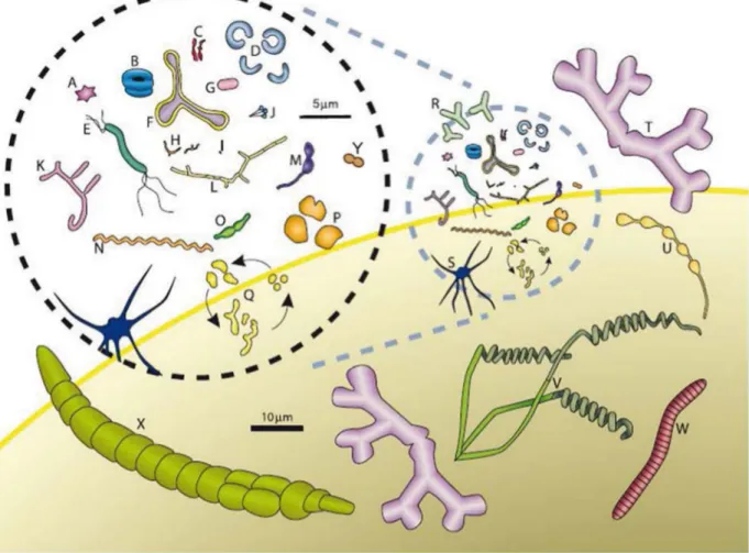
The pneumococcus naturally inhabits as a commensal organism the nasopharynx of humans. In healthy persons, the nasopharyngeal microbiota comprises several bacterial species that cohabit or compete with each other. This microbiota was recently investigated in healthy children and compared with that of children with pneumonia from three Swiss hospitals (Sakwinska, et al., 2014). It showed that three genera are dominant in the nasopharynx of both healthy children and pneumonia patients: Moraxella, Streptococcus and Haemophilus (Figure 5). As one could expect, the abundance of the Streptococcus and the Haemophilus genera is increased in patients with pneumonia. Of note is that the nasopharyngeal microbiota strongly differs from one person to another.
20
In the nasopharynx, the pneumococcus can be exposed to high oxygen concentrations (likely up to 20 % at the surface of the mucus layer) whereas it deals with low oxygen concentrations in deeper layers (5 %) (Yesilkaya, et al., 2013). In some instances, the pneumococcus can lead to infection by colonizing the lower respiratory tract, or even the blood and the cerebrospinal fluid. In those latter cases, the pneumococcus copes with nearly anaerobic conditions.
S. pneumoniae encounters several reactive oxygen species (ROS) in the different
environments it occupies. Hydrogen peroxide (H2O2) is one of them that is produced as a byproduct
of its own metabolism at a concentration that can reach 2 mM. This compound contributes to the virulence of the pneumococcus and is required for the colonization of the nasopharynx and the development of pneumonia and sepsis (Spellerberg, et al., 1996). A drawback of hydrogen peroxide is that it is detrimental for S. pneumoniae cells, as the other ROS. Indeed, it damages proteins by oxidation, and this is used by the immune system to fight infections. In inflammation, neutrophils and macrophages release several ROS including H2O2, superoxide anion (O2•–) and hydroxyl radicals
(OH•) that also damage other molecules such as DNA. The lactic acid bacteria of the nasopharynx can
also be a source of ROS.
Figure 5: Relative abundance of the 12 most common micro-organisms of the nasopharyngeal microbiota of 50 healthy (H) and 50 pneumonia (P) children (2 months to 16 years old) from 3 Swiss hospitals. OTUs stands for operational taxonomic unit. Adpated from (Sakwinska, et al., 2014).
21
The pneumococcus lacks catalase, which is utilized by most bacteria to eliminate H2O2. It
also lacks the main Gram-negative proteins known to confer resistance to oxidative stress. A set of proteins has been described to date that allows the pneumococcus to live in the presence of ROS (reviewed in (Yesilkaya, et al., 2013)), but the mechanisms are not completely understood yet.
One of the primary defense of the host is the use of lysozyme to disrupt the peptidoglycan of the pathogens (Nonomura, et al., 1991), (Bercovici, et al., 1975). The pneumococcus is naturally resistant to this enzyme thanks to its largely de-acetylated peptidoglycan.
Competence
An interesting property of the pneumococcus is its intrinsic competence. The competence is the capacity of a cell to integrate DNA from the environment. This mechanism enables the cell to beneficiate of advantageous characteristics possibly encoded by the fragment of DNA incorporated. Competence occurs naturally, and it can be artificially induced under specific conditions . The analysis of competence genes revealed that it is conserved among the genus Streptococcus (Berg, et
al., 2012).
In 1928, a British bacteriologist, Frederick Griffith, described competence for the first time by using S. pneumoniae cells. First, he injected mice with an avirulent strain of pneumococcus with no effect on the survival of the rodents. However, the co-injection of this avirulent strain and a heat-killed virulent strain of pneumococcus caused the injected mice to die (Griffith, 1928). In fact, the avirulent strain of pneumococcus underwent transformation, which had never been described before. In 1931 in New York, Dawson and Sia could perform pneumococcus transformation in vitro,
i.e. with no passage in an animal: the dead “donor” pneumococci where added in the culture
medium for transformation (Dawson & Sia, 1931). Twelve years later, Avery et al identified the factor containing the transferred information: the deoxyribonucleic acid (DNA) (Avery, et al., 1944). At this time, DNA was shown to provide transformants with their new property, but the molecular mechanisms responsible for competence activation remained unclear.
Half a century later, the pheromone-like signal that induces competence was identified in the pneumococcus (Havarstein, et al., 1995). It was called CSP for competence-simulating peptide. CSP is a 17 amino-acids peptide, ribosomally synthesized as a precursor peptide that is then exported and cleaved by the ComA ABC transporter (Havarstein, et al., 1995). Subsequently, the pneumococcus
comCDE operon was described, encoding for the three core-proteins of the competence: comC
encodes for CSP, comD and comE encode for a two-component signal transduction system, including the CSP-specific receptor (ComD) and its cognate response regulator (ComE, (Pestova, et al., 1996)).
22
CSP and ComD are species-specific (Johnsborg & Havarstein, 2009), which enables species-dependent competence signaling. This can be useful in an environment where multiple species cohabit or compete with each other.
The expression of more than 180 genes is influenced by the competence state of the pneumococcus, from which only 10 to 20% were shown to be required for natural transformation. Further details on those genes are in (Johnsborg & Havarstein, 2009) and (Berg, et al., 2012).
The natural competence ability provides an important advantage to S. pneumoniae in terms of resistance. Indeed, β-lactam resistance genes are exchanged from strain to strain, and even between different species co-habiting the same niche that have developped resistance due to in-adequate antibiotic exposure (Dowson, et al., 1989), (Laible, et al., 1991). Other types of antibiotic resistance has also been associated with recombinantion events, such as the macrolides, the fluoroquinolones and the rifampicin (Croucher, et al., 2011). Also, the combination of genetic transformation and recombination events has allowed serotype switching, enabling pneumococci to evade vaccine immunization (Croucher, et al., 2011).
Autolysis
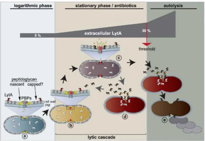
Another property of S. pneumoniae is the autolysis. The growth of this organism is characterized by a short stationary phase followed by a strong decrease of the optical density (OD) in laboratory conditions. The major autolysin responsible for autolysis in the pneumococcus is LytA. Its depletion restores the stationary phase (Figure 6). This property is related to competence in that cells in the competent state are more prone to autolysis (Seto & Tomasz, 1975). Also, it was shown in 2004 that the release of DNA in the medium upon competence induction was mainly due to the action of two proteins in the pneumococcus: the major autolysin LytA and LytC, two peptidoglycan hydrolases (Moscoso & Claverys,
2004).
LytA has an N-acetylmuramoyl L-alanine amidase activity (Howard & Gooder, 1974), (Mellroth, et al., 2012). In other words, it separates the glycan chains of peptidoglycan from their peptide crosslinks. This protein
includes two domains: a N- Figure 6: Growth curves of S. pneumoniae R6 and its ΔlytA derivative in laboratory conditions.
23
acetylmuramoyl L-alanine amidase domain and a choline-binding domain. The latter allows to localize it to the cell wall via its interaction with the phosphocholine residues of the teichoic acids, confirming an early observation (Briese & Hakenbeck, 1985). LytA is a cytoplasmic protein. It is thought to be released in the extracellular medium through bacterial lysis, resulting in a cascade lysis event, which in turn kills the whole population of pneumococci in laboratory conditions (Mellroth, et
al., 2012).
The exact role of LytA in vivo is not fully understood, although several suggestions have been made. First, LytA deficient mutants have no morphological deffects (Tomasz, et al., 1988), (Berg, et
al., 2013), but only a higher propensity for the formation of chains in liquid culture. Thus, its role in
cell wall maturation or in the division process is probably limited. Rather, LytA is involved in the virulence of the pneumococcus, as demonstrated in a rat model of meningitis (Hirst, et al., 2008). Also, it was shown to reduce the secretion of some cytokines that activate phagocytosis, in addition of preventing phagocytosis by unclear mechanisms (Martner, et al., 2009). The authors suggest that the fragments of autolyzed bacteria could constitute a diversion, in turn diminishing phagocytosis of living bacteria.
Under penicillin treatment, pneumococcus lysis is mainly due to the action of LytA. Indeed, in a LytA-negative mutant, penicillin addition at the minimum inhibitory concentration (MIC) inhibits growth, but does not result in lysis contrary to the same treatment in a LytA-positive background (Tomasz & Waks, 1975). Addition of exogeneous LytA restores lysis in β-lactam treated cells but it does not induce lysis of pneumococci treated with antibiotics that do not target peptidoglycan synthesis (chloramphenicol) (Tomasz & Waks, 1975). In exponential growth phase, it seems that pneumococci are protected from LytA activity, which is not the case in stationary phase or under peptidoglycan-directed antibiotic treatment (Mellroth, et al., 2012). The mechanisms of activation of LytA are not fully understood to date, but Mellroth et al proposed it to be through the recognition of its peptidoglycan substrate (Figure 7).
Pathogenicity
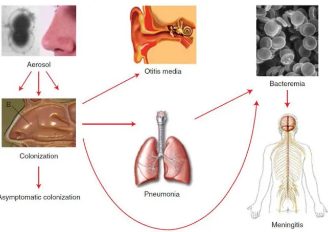
Before the beginning of the twentieth century, the pneumococcus was already shown to cause meningitis, otitis media, arthritis and endocarditis (Netter, 1887), (Zaufal, 1887). Nowadays, it has been described in many infections that also include pneumonia, sinusitis and septicemia.
24
The pneumococcus is a commensal of the human nasopharyngeal flora. However, it can initiate a variety of non-invasive diseases (sinusitis and otitis media), or more severe invasive diseases (pneumonia, septicemia and meningitis). According to a 2005 estimate of the world health organization (WHO), 1.6 million people die of pneumococcal diseases in the world each year. The most at risk populations are people with a weak immune system. The elderly and the youth are especially vulnerable. 700,000 to 1 million children under five years old die of pneumococcal infections every year worldwide: (WHO – http://www.who.int/ith/diseases/pneumococcal/en/). Immuno-compromised patients are also at risk (with AIDS, for example).
S. pneumoniae cells present in the upper respiratory tract are transmitted to another person
in droplets of respiratory secretions (eg: saliva or mucus). They will first establish in the nasopharynx from which they are usually cleared. In some cases, they will move to the ear and cause otitis, which is common in children under 5 years old. Invasive diseases result from the propagation of pneumococci to the lungs, the blood stream and the cerebrospinal fluid, the latter case being the most life-threatening (Figure 8).
Figure 7: Model of the mode of action of LytA during pneumococcus growth, from (Mellroth, et al., 2012). Cytoplasmic LytA is released through lysis and can access peptidoglycan when its synthesis is stopped (in stationary phase or in the presence of peptidoglycan-targeting antibiotics). More than 30 % of LytA in the medium results in extensive lysis of the bacteria contained in the culture.
25
Prophylaxis and treatmentVaccines
The capsular polysaccharides protect the pneumococcus from phagocytosis. Nevertheless, opsonization of S. pneumoniae cells by antibodies directed against the capsule results in recognition by the host phagocytes and elimination of the pathogen. Several vaccines against pneumococcal infections have been developed based on this principle.
The longest lasting pneumococcus vaccine is on the market since 1983. The Pneumovax®23 (Merck) consists of a mixture of purified polysaccharides of 23 different serotypes. The use of this vaccine is limited to the adults (mainly the elderly and immunocompromised patients) because of its low immunogenicity in children under 2 years old. Also, it does not prevent pneumococcus carriage without symptoms.
In the year 2000, a vaccine comprised of pneumococcal polysaccharides from 7 serotypes conjugated to a protein carrier was licensed in the United States (PCV7, Prevenar®, Pfizer), and adopted one year later by several European countries. The protein carrier, CRM197™, allows better immunization of the children under 2 years old. Furthermore, the 7 serotypes covered by this
26
vaccine were dominant in the United States at the time it was developed. As a consequence, the rate of pediatric invasive pneumococcal diseases (IPD) and nasopharyngeal carriage was reduced for the vaccine-targeted serotypes (Pilishvili, et al., 2010). Furthermore, the proportion of IPD in the non-vaccinated population (children too young for vaccination and people above 50 years old) diminished, indicating that the global immunization programs consistently impaired the spread of targeted serotypes.
Unfortunately, the reduced prevalence of PCV7 serogroups left a free ecological niche for others, and they became more and more common. Also, some emerging serotypes, such as 19A, have been associated with increased virulence and antibiotic resistance (McGee, 2007). Another drawback of the PCV7 is that it is not as efficient in preventing pneumonia or acute otitis media as bacteremia.
Therefore, additional conjugated vaccines were developed, covering more serotypes: 10- and 13-valent vaccines for example. The later, PCV13, Prevenar® (Pfizer) has replaced the PCV7 in the French vaccination program in 2010. It led to a decreased prevalence of the serogroups 19A, 7F and 1 (Varon, et al., 2013).
PCVs are expensive due to complex development and production that limit their use in developing countries where the burden of pneumococcal infections is the most substantial. Also, as polysaccharide vaccines, PCVs target limited sets of serotypes and replacement has been observed. Moreover, serotype distribution is area dependent, and the efficacy of the existing vaccines depends on the country. For example, the PCV7 covered 70 to 88 % of the serogroups found in children from the United States, Canada, Africa and Europe at the time of its development, while it covered less than 65 % of the serotypes causing IPD in Asia and South America (Hausdorff, et al., 2000).
An affordable vaccine with a target independent of the capsular layer would be ideal, but several approaches are considered (Moffitt & Malley, 2011). Briefly, the adaptation of conjugate vaccines to serotypes prevalent in low-income countries is envisaged in China. Also, the development of polysaccharides associated to a conserved pneumococcus protein carrier could have a better efficacy. Another promising approach would be to develop a vaccine that targets proteins conserved among the different pneumococcal serogroups. Several of those proteins have been studied and tested in clinical trials, some of them giving encouraging results. A combination of those proteins may provide robust and serotype-independent protection to pneumococcal infections. Finally, a very low-cost and promising strategy is the use of whole pneumococcal killed cells as a vaccine, with the advantage of presenting many antigens at once. Recently, this strategy was shown
27
to protect mice from pneumococcal infections to a similar extent as currently used polysaccharide vaccines, in a serotype-independent manner (Xu, et al., 2014).
Antibiotics
The first antimicrobial used to treat pneumococcus infections appeared in the early forties with the development of sulfonamide drugs (Whitby, 1938). However, a pneumococcus resistant to sulfonamide was rapidly found in a patient (Tillett, et al., 1943). This isolate, however, was sensitive to another antimicrobial compound first described in 1929: the penicillin (Fleming, 1929). Penicillin was then intensively and successfully used to fight various bacterial infections (Keefer, 1943), (Tillett,
et al., 1944). However, the extensive use of antibiotics was followed by the emergence of resistant
pneumococcus strains, first reported in 1967 (Hansman, 1967). Multi-resistant strains were reported half a century after the introduction of antibiotics in healthcare (Appelbaum, et al., 1977). Recently, extensively drug-resistant pneumococci have emerged (Kang, et al., 2012). These strains resist to at least one antibiotic in all classes, except vancomycin and linezolid.
Nowadays, pneumococcal infections are treated with antibiotics. In the case of invasive diseases, the treatment should be initiated quickly to limit the progression of the infection. In France, if a bacterial infection is suspected in the case of otitis media, meningitis or pneumonia, the first antibiotic treatment is usually assuming pneumococcal infections as for those diseases, it is the most probable etiologic agent. Guidelines are provided by the heath products and medicine safety national agency (ANSM, in French) for the choice of treatment of the different diseases. The following paragraphs sum up the major treatments utilized in France against infections that can be cause by S. pneumoniae (from (Brisou, et al., 2004)).
Community-acquired pneumopathies are mostly treated with amoxicillin, a bacteriolytic β-lactam with good absorption properties (oral administration). If there is no sign of improvement after 2 – 3 days, recommendation should be asked to a specialist or at the hospital. A better-adapted treatment can be another β-lactam or antibiotics of a different class in the case of β-lactam-resistant pneumococcus strain, or β-lactam allergy.
In the case of meningitis, the patient should be immediately hospitalized and a lumbar puncture (cerebrospinal fluid sampling) will be performed to determine the etiology of the disease. Depending on the age, the gravity of the infection and the probability that the meningitis is caused by the pneumococcus, different antibiotic treatments are recommended, including cefotaxime, ceftriaxone, amoxicillin (β-lactams) and / or vancomycin (a glycopeptide). The treatment is then adapted by considering the results of the lumbar puncture. No codified recommendations exist and
28
the best-suited treatment is usually determined by consultation of the medical doctor, the pharmacist and the microbiologist.
Acute otitis media are also treated on a different manner depending on the age. Before 2 years old, antibiotic therapies are systematic while it is recommended only in the case of strong symptoms after 2 years old. The antibiotics utilized in acute otitis media treatment are β-lactam including amoxicillin, cefuroxime and cefpodoxime, or macrolide (erythromycin) in the case of β-lactam allergies. β-Lactamase inhibitors are associated to these β-β-lactam antibiotics because
Haemophilus influenzae is common in young children. This organism produces β-lactamases that
inactivate the β-lactams. Again, the treatment should be evaluated 2 – 3 days after initiation. If it is not efficient, the etiology of the infection should be determined and the treatment adapted.
Epidemiology
The world-wide spread of pneumococcus strains is facilitated by two properties of the organism. First, most people naturally carry this pathogen at least once in their lifetime (Center for Disease Control, CDC – http://www.cdc.gov/pneumococcal/about/risk-transmission.html). Second, transmission by direct contact with respiratory secretions of carrier persons allows it to quickly propagate through the population. Despite its global repartition, the mortality rate does not correlate with the incidence rate in several countries, due to distinct policies in terms of prevention or accessibility to the treatment (Figure 9).
In developing countries, the young children constitute the most at-risk population because of low access to adapted treatments and bad living conditions. By contrast in developed countries, the elderly and immunocompromised persons are most at-risk. However, pneumococcus diseases also occur in persons recovering from influenza, cigarette smokers or alcohol abusers, for example (Grau,
et al., 2014).
In France, the Pneumococcus Reference National Center (CNRP, in French) is in charge of the surveillance of pneumococcus infections and pneumococcus antibiotics resistance. It relies on 23 Regional Pneumococcus Observatories (ORP, in French) that collect data in about 500 healthcare centers. A yearly activity report summarizes the epidemiology of the pneumococcus of the previous year (Varon, et al., 2013).
In 2012, 993 strains were collected by the CNRP and serotyped. The prevalence of each serotype was determined and compared to that defined in previous years since 2001. The prevalence differs in distinct age groups and sampling types. We will focus herein on the global results of IPDs in the population (meningitis and bacteremia). In 2012, the serotype 12F was predominant, followed by the serotypes 19A, 3, 7F and 1 (Varon, et al., 2013). In 2001, the most prevalent serogroups were the 14, 19A, 23F, 6B, 19F and 3 (1968 isolates). From those, the PCV7
29
Figure 9: Top 2 panels. Pneumococcus incidence and mortality rates in children (< 5 years old) in 2000. Legend is in number of case per 100,000 children. Adapted from the WHO slides available on the web: http://www.who.int/immunization/monitoring_surveillance/burden/estimates/Pneumo_hib_2000/en/index2.html). Lower panel: Countries that include pneumococcal conjugate vaccines in their national immunization programs, by incoming status. Adapted from (CDC, 2013).
30
vaccine (which was introduced in 2003 in the French immunization program) covers the serotypes 14, 23F, 6B and 19F, which were not the most prevalent strains anymore in 2012. The replacement of PCV7 by PCV13 in June 2010 in the French vaccination program did not result yet in the removal serogroups 19A, 3, 7F and 1 from the most prevalent serotypes in 2012. However, this is probably a matter of time, as they all showed a drastic prevalence decrease since the records of 2009, except for serotype 3 (Varon, et al., 2013).
The proportion of antibiotic resistance in pneumococcal invasive diseases dramatically increased in the eighties and nineties in France. However, the efforts of the health ministry in the prevention of abusive use of antibiotics and the introduction of the PCV7 and the PCV13 in the French immunization program have inverted the tendency. At its maximum, in 2002, the proportion of pneumococci with diminished susceptibility to penicillin reached the frightening value of 53 % of the investigated invasive disease pneumococcal clinical isolates. In 2012, it had diminished to 22 % and continuous efforts will hopefully bring resistance proportion back to reasonable level (Figure 10).
Concluding remarks
S. pneumoniae is of significant scientific and medical importance. It is an ideal Gram-positive
model organism for several reasons. First, the pneumococcus has allowed to identify DNA as the molecule encoding the characteristics of living organisms. Many physiological processes have been discovered on the pneumococcus, comprising competence, autolysis and quorum sensing. Furthermore, the first Gram-positive genome sequence to be determined was that of the pneumococcus. Concerning medical interests, it allowed the development of polysaccharide vaccines, and revealed many aspects of the mechanisms of resistance against antibiotics. Also, Figure 10: Evolution of penicillin resistance in pneumococcal clinical isolates in France. Adapted from (Varon, et al., 2013).
31
general characteristics of the Gram-positive bacterial pathogenesis have been understood thanks to studies on the pneumococcus.
For these reasons, one can reasonably claim that further investigations on this organism will answer many of the lasting questions on the microbial physiology and the pathogenesis of Gram-positive bacteria. Also, pneumococcal research efforts will participate in the fight against bacterial infections that are in constant evolution, as evidenced in S. pneumoniae.
32
Le peptidoglycane du pneumocoque – résumé
Le peptidoglycane est le constituant majeur de la paroi et est une structure essentielle et très conservée dans le règne des bactéries. Cette molécule géante forme un sacculus qui entoure complètement la cellule. Cette structure permet à la bactérie de conserver son intégralité face à la pression osmotique et sert d’ancrage à plusieurs molécules de surface. Enfin, le peptidoglycane procure sa forme à la bactérie, ce qui a son importance car la forme d’une bactérie lui confère des avantages spécifiques à certains environnements. L’essentialité et la position du peptidoglycane à la surface de la cellule bactérienne en font la cible d’antibiotiques.
Le peptidoglycane est composé de chaines oligosaccharidiques réticulées par des ponts peptidiques. Ces chaînes glycanes sont formées d’une succession d’unités disaccharidiques constituées d’un résidu N-acetylglucosamine (NAG) liés à un acide N-acetylmuramique (NAM) par une liaison osidique β1→4. Dans le peptidoglycane naissant, les résidus NAM sont décorés du pentapeptide L-Ala-γ-D-iso-Gln-L-Lys-D-Ala-D-Ala. Ces pentapeptides sont utilisés pour la formation de liaisons entre les chaînes glycanes, constituant à terme le sacculus. Chez le pneumocoque, il est admis que les chaînes glycanes sont parallèles à la membrane plasmique, insérées de manière perpendiculaire à l’axe le plus long de la cellule.
La composition du peptidoglycane diffère entre espèces de bactéries, ce qui leur donne des propriétés différentes. Chez le pneumocoque, une caractéristique intéressante est la présence de peptides branchés dans le peptidoglycane. Il s’agit de dipeptides (L-Ser-L-Ala ou L-Ala-L-Ala) liés à la lysine en position 3 du pentapeptide, qui peuvent aussi être impliqués dans les ponts peptidiques entre chaînes glycanes. Ces peptides branchés sont plus abondants chez les souches cliniques résistantes aux antibiotiques, bien qu’on ne comprenne pas encore clairement pourquoi. D’autres modifications rencontrées chez le pneumocoque sont la dé-acétylation des NAG ou la O-acétylation des NAM, qui procurent la résistance au lysozyme.
La synthèse du peptidoglycane est une suite de réactions enzymatiques commençant dans le cytoplasme et finissant à l’extérieur de la cellule après transfert du précurseur à travers la membrane plasmique. Le lipide II est le précurseur du peptidoglycane synthétisé dans le cytoplasme de la cellule. Il s’agit de la sous-unité dissaccharide-pentapeptide NAG-NAM-L-Ala-D-(γ)Gln-L-Lys-D-Ala-D-Ala ancrée à la membrane plasmique par un undécaprényl-pyrophosphate lié au sucre NAM. La synthèse du lipide II est catalysée à partir d’un résidu NAG par l’intermédiaire de 8 enzymes Mur (MurA, B, D, D, E, F, G et T), MraY et GatD. Ensuite, le lipide II est transféré à travers la membrane par une « flippase » et la sous-unité dissaccharide-pentapeptide est utilisée pour l’assemblage du sacculus. Les protéines liant la pénicilline (PLPs, PBPs en anglais) sont responsables de l’activité glycosyl-transférase permettant l’élongation des chaînes glycanes et de l’activité transpeptidase
33
permettant leur réticulation. Ces activités sont complétées d’une activité « hydrolase », qui permet l’« ouverture » du sacculus requise pour l’insertion de nouveau matériel. Les modifications observées dans la composition du peptidoglycane sont effectuées à l’intérieur (ajout de dipeptides au pentapeptides par MurM et MurN) ou à l’extérieur de la cellule (dé-acétylation des résidus NAG par PgdA, O-acétylation des résidus NAM par Adr, raccourcissement du pentapeptide par PBP3 et LdcB).
Les différentes protéines impliquées dans la synthèse et les modifications du peptidoglycane chez le pneumocoque sont détaillées dans cette partie.
34
Pneumococcus peptidoglycan
The peptidoglycan is the major component of the cell wall, and is extensively conserved in the bacterial kingdom (Liechti, et al., 2014). It consists of a giant single molecule, the sacculus, which encases the plasma membrane of the bacterium. In Gram-negative bacteria the peptidoglycan is found in the periplasm between the plasma membrane and the outer membrane. The peptidoglycan is required by bacterial cells to maintain their integrity under the turgor pressure. This stress is imposed by the osmotic flow of water from the external environment to the cytoplasm induced by the difference of concentration of solutes in these compartments. The peptidoglycan also serves as a scaffold for several surface molecules, including proteins (eg. the pneumococcus pilus), wall teichoic acids and the capsular polysaccharides. The peptidoglycan is also involved in the interaction with the host in that peptidoglycan fragments are recognized by the immune system (Boneca, 2005). Finally, the peptidoglycan architecture determines the bacterial shape, and isolated sacculi retain the cell shape, even after elimination of all the other components of the cell. This feature is important because the shape confers specific advantages to the cell that can be essential in some environments (Young, 2006).
Given its essentiality and its localization at the surface of bacterial cells, peptidoglycan is the target of some antibiotics, reviewed in the last part of the introduction. In the present part, I intend to describe the peptidoglycan of the pneumococcus, in terms of chemical composition, architecture, and how it is synthesized and modified.
Pneumococcus peptidoglycan composition
The peptidoglycan is a mesh of glycan strands cross-linked by short peptides (Vollmer, et al., 2008). The glycan strands are comprised of a succession of alternating N-acetylmuramic acid (MurNAc) and N-acetylglucosamine (GlcNAc) residues linked by β1 → 4 bonds. The MurNac residues are decorated with a peptide in substitution of the D-lactoyl group. In newly-synthesized pneumococcus peptidoglycan, this pentapeptide has the following composition: L-Ala-γ-D-iso-Gln-L
-Lys-D-Ala-D-Ala (Bui, et al., 2012). During peptidoglycan synthesis, the pentapeptides are used by DD
-transpeptidases to covalently link adjacent glycan chains, resulting mostly in dimeric, but also in trimeric or tetrameric structures that attach 2, 3 or 4 glycan strands together, respectively. As a consequence, the peptidoglycan constitutes a giant mesh-like molecule that surrounds the entire cell. The presence of D-amino-acids in the composition of the peptidoglycan is probably not a
35
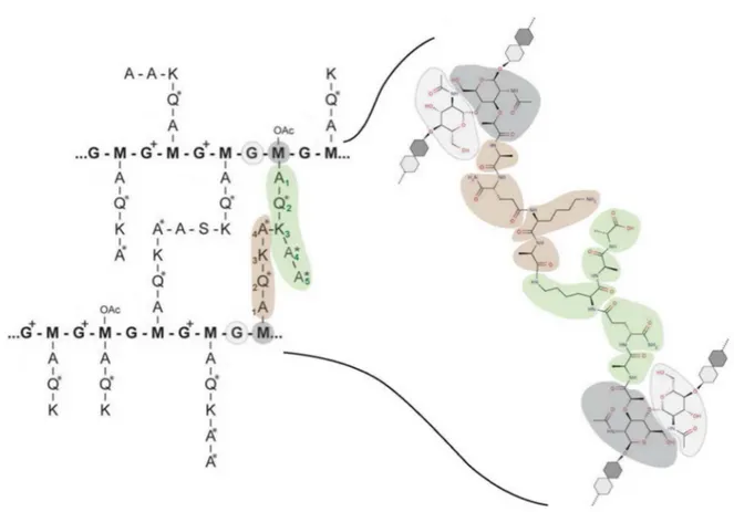
In S. pneumoniae, the peptidoglycan contains numerous variations (Figure 11). First, the monomers (term used to designate the peptides that are not part of cross-links) are mostly tripeptides, cumulated tetra- and pentapeptides representing only 20% of the total monomers (Bui,
et al., 2012), (Table I).
The glutamate in second position of the peptide is mostly amidated in the pneumococcus and in other Gram-positives species (eg. Staphylococcus aureus). This was recently shown to be required for the transpeptidation activity (Zapun, et al., 2013). Furthermore, it is linked to the resistance against both β-lactam antibiotics and lysozyme digestion of the peptidoglycan (Figueiredo,
et al., 2014).
The peptide bridges (Figure 11) can be found with the following combinations: tetra-tri-, tetra-tetra- or tetra-pentapeptides, as the linkage is between the L-Lys and the first D-Ala residues of
the peptides requiring at list a tetrapeptide and a tripeptide.
Figure 11: Representation of the chemical variations found in the peptidoglycan of pneumococcus, and detail of a tetra-pentapeptide cross-link. M and G stand for MurNac (dark grey) and GlcNac (light grey), respectively. A, Q and K represent alanine, isoglutamine and lysine residues, respectively (the amino-acids in D- configuration are designed by an asterisk *). G+ stand for GlcN, OAc-M are O-acetylated MurNac residues. Adapted from (Bui, et al., 2012) and (Zapun, et al., 2013).
36
Another common variation found in pneumococcal peptidoglycan is the presence of “branched peptides”, which are peptides containing two amino-acids (L-Ser-L-Ala or L-Ala-L-Ala)
bound to the ε-amino group of the L-Lys in position 3. Interestingly, these branched peptides are
more abundant in penicillin-resistant clinical isolates. In the study of 20 pneumococcal clinical isolates, Garcia-Bustos and Tomasz found < 30% of branched peptides in penicillin-susceptible pneumococci, while penicillin-resistant isolates had > 70 % of branched peptides (Garcia-Bustos & Tomasz, 1990). Of note is that the R6 laboratory strain has a slightly higher proportion of branched peptides than the clinical isolates studied by Garcia-Bustos and Tomasz. Globally, 45% of the peptides are branched in the R6 strain, and the cross-links between glycan chains are comprised of > 90% of branched peptides (Bui, et al., 2012). This suggests that globally, branched peptides are
Table I: The muropeptides of S. pneumoniae (R6 strain). n.d.: not detected, a: percentage in the mono-, di- or
trimmers. (Bui, et al., 2012)
37
preferred than linear peptides for transpeptidation.The glycan chains also undergo secondary modifications. In the pneumococcus, 80% of the GlcNac and 10% of the MurNAc residues are de-acetylated (Vollmer & Tomasz, 2000). Also, some MurNAc residues are O-acetylated.
The peptidoglycan composition of the R6 strain was investigated in-depth (Bui, et al., 2012). First, specific digestion of purified peptidoglycan by cellosyl generated the muropeptides, the subunits of the peptidoglycan (GlcNAc-MurNac-peptide). Thereafter, the muropeptides of different composition were separated by high-performance liquid chromatography (HPLC) and analyzed on-line by electrospray mass spectrometry, revealing a big heterogeneity in the composition of the peptidoglycan of S. pneumoniae (Table I). The variations observed in muropeptides were different in monomers, dimers and trimmers. Indeed, glutamate was virtually exclusively found in monomers, suggesting the fact that a glutamine is required for transpeptidation (Zapun, et al., 2013). Also, GlcNAc residues were often deacetylated in dimers (45 %) whereas deacetylation was found in less than 20 % of monomers and trimers. Finally, the branched peptides were most abundant in dimers and trimers, suggesting that they are preferred for transpeptidation.
Architecture
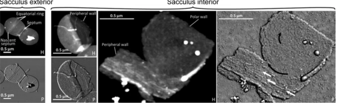
In the early 2000s, cell wall architecture was investigated using computational methods. In Gram-positive bacteria with a thick cell wall, a scaffold model was proposed in which glycan strands are directed perpendicularly from the plasma membrane. The scaffold model fitted the experimentally available data of that time, including the length of glycan chains and the proportion of peptide cross-links (Dmitriev, et al., 2003). This contrasted with the “classical” model of peptidoglycan architecture in which glycan strands are parallel to the plasma membrane (Holtje, 1998). With the advance of methodology, peptidoglycan architecture has been recently investigated experimentally in ovoid bacteria (Lactococcus lactis, Enterococcus faecalis and S. pneumoniae) by atomic force microscopy. It now appears that glycan strands are organized along the short axis of the cell, parallel to the plasma membrane (Andre, et al., 2010), (Wheeler, et al., 2011) (Figure 12).
The orientation is the same at the periphery and at the septum, suggesting that peptidoglycan insertion is organized similarly in both zones. Interestingly, the glycan chains of the three ovoid species have similar length to those of Bacillus subtilis, longer than in S. aureus. Indeed half of the glycan strands are longer than 50 disaccharides whereas most are 5- to 10-unit long in S.