HAL Id: inserm-00319531
https://www.hal.inserm.fr/inserm-00319531
Submitted on 8 Sep 2008HAL is a multi-disciplinary open access archive for the deposit and dissemination of sci-entific research documents, whether they are pub-lished or not. The documents may come from teaching and research institutions in France or abroad, or from public or private research centers.
L’archive ouverte pluridisciplinaire HAL, est destinée au dépôt et à la diffusion de documents scientifiques de niveau recherche, publiés ou non, émanant des établissements d’enseignement et de recherche français ou étrangers, des laboratoires publics ou privés.
Role of estrogens and their receptors in adhesion and
invasiveness of breast cancer cells.
Marie Maynadier, Philippe Nirdé, Jean-Marie Ramirez, Anne Marie Cathiard,
Nadine Platet, Monique Chambon, Marcel Garcia
To cite this version:
Marie Maynadier, Philippe Nirdé, Jean-Marie Ramirez, Anne Marie Cathiard, Nadine Platet, et al.. Role of estrogens and their receptors in adhesion and invasiveness of breast cancer cells.. Advances in Experimental Medicine and Biology, Kluwer, 2008, 617, pp.485-91. �inserm-00319531�
Marie Maynadier, Philippe Nirdé, Jean-Marie Ramirez, Anne Marie Cathiard, Nadine Platet, Monique Chambon, Marcel Garcia.
Institut National de la Santé et de la Recherche Médicale, Unité 540, Molecular and Cellular Endocrinology of Cancers and University Montpellier I, 60 rue de Navacelles, 34090 Montpellier Cedex, France
Role of estrogens and their receptors in adhesion and invasiveness of breast cancer
cells
Marie Maynadier, Philippe Nirdé, Jean-Marie Ramirez, Anne Marie Cathiard, Nadine Platet, Monique Chambon, Marcel Garcia
Summary
Estrogen receptors are overexpressed in human breast cancers and associated with differentiated tumors with a more favorable prognosis. Paradoxically, these receptors mediated the mitogenic action of estrogens in human breast cancer cells and the efficacy of antiestrogens in adjuvant therapy of primary tumors. The exact mechanism underlying the ER protection against cancer progression to metastasis, remains to be investigated. Herein, we show that estrogen receptors decrease invasiveness of breast cancer cells. Detailed studies revealed that the unliganded and the estradiol-activated estrogen receptors decrease cancer cell invasion in vitro through two distinct mechanisms. In the presence of ligand, ERα inhibits invasion through a mechanism requiring the functional ERα domains involved in the transcriptional activation of target genes. Moreover, we found using different approaches that cell-cell contacts were markedly increased by estradiol treatment and decreased by the pure antiestrogen, ICI 182,780. This cell-cell adhesion was associated with an increase of the major intercellular junctions, desmosomes. Conversely, in the absence of ligand, ERα also inhibits invasion through a distinct mechanism involving protein-protein interaction with the region of the first zinc finger of ERα. The relationship of these data with clinical studies and their potential therapeutic consequences will be discussed.
Introduction
Breast cancer is the most frequent malignancy diagnosed in women in western countries and estrogen receptor (ER) is overexpressed in about two thirds of breast tumors. Estrogens are the major sex steroid hormones involved in the development of normal mammary gland and in the etiology of human breast cancer. The promoter role of estrogens in breast cancer has been evidenced by epidemiological studies indicating an increased incidence in women with prolonged exposure to estrogens and a drastic decrease of incidence in women having non functional ovaries (1). Most of the estrogen effects are mediated by two specific receptors ERα and ERβ that have a structure characteristic of members of the large nuclear receptor superfamily (2-3). These receptors are subdivided into several functional domains (Figure 1).
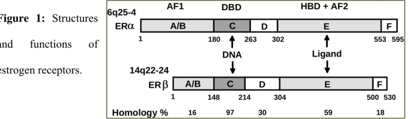
Figure 1: Structures
and functions of estrogen receptors.
Functional domains were defined as a DNA binding domain (DBD, C) containing two zinc fingers involved in DNA binding and receptor dimerization, a ligand binding domain (LBD, E) important for ligand binding, receptor dimerization and interaction with transcriptional coactivators and corepressors (4). The ligand binding domain contains a ligand activable transactivation function AF2 whereas the N-terminal A/B region contains a ligand independent transactivating function AF1 (5). The hinge domain (D) contains the nuclear localization signal of the receptor and contributes to the flexibility of the two moieties containing the DNA and the ligand binding. The F domain participates to the transactivation capacity.
F 180 263 302 553 595 E D C A/B DBD F 148 214 304 500 530 E D C A/B α AF1 ERβ Homology % 16 97 30 59 18 HBD + AF2 1 1 6q25-4 14q22-24 DNA Ligand ER
In malignant mammary cells, the effects of estrogens are more complex since estrogens affect directly the growth, motility and invasiveness of cancer cells but, could also influence some biological responses of the host such as neo-angiogenesis and immune response. The presence of ERα is associated with more differentiated and less invasive tumors and, several clinical studies have demonstrated the favorable prognostic value of ER in primary breast tumors (6). The routine practice of ERα assay in primary tumors has been established to predict the efficacy of antiestrogens, widely used as first-line adjuvant therapy (7). The second receptor, ERα, has likewise been detected in primary breast cancers by immunohistochemistry but its own clinical relevance in prognosis and tumor progression remains to be established (8, 9).
In this report, we will focus on the effects of estrogens and ERα on the invasiveness of breast cancer cells. Estrogens are classically known to favor growth of cancer cells and, estrogen antagonists are clinically important in the treatment of hormone-dependent breast cancers. However, a series of evidences have demonstrated that ER could protect against cancer cell invasion of basement membranes, an important step of the metastatic process required for cancer dissemination. This last observation proposes a plausible explanation for the good prognosis value associated with the presence of ERα in primary tumors.
Results
Estrogen receptors inhibit invasion through two distinct mechanisms in the presence or absence of hormone.
Estrogens inhibit invasion via ERE-regulated genes. The effects of estrogens on cell
invasiveness have been studied in vitro using a two-chamber culture model and Matrigel, a reconstituted basement membrane. The initial studies indicated that the
invasiveness of MCF7 breast cancer cells was increased by antiestrogens (10, 11). More recent studies have demonstrated that estradiol significantly reduces invasiveness which is reversed by antiestrogens (12-16). This conclusion was noted in several ERα-positive cancer cell lines established from breast (12) or ovary (14), and in different ERα-negative cancer cells constitutively expressing ERα after stable transfection (13, 16, 17). Similar results were also obtained on the migration of normal cells from vascular smooth muscle (15). These in vitro data were confirmed in nude mice, since the formation of experimental lung metastases from metastatic ERα-negative MDA-MB-231 breast cancer cells was inhibited by estradiol after ERα expression by transfection (16, 17).
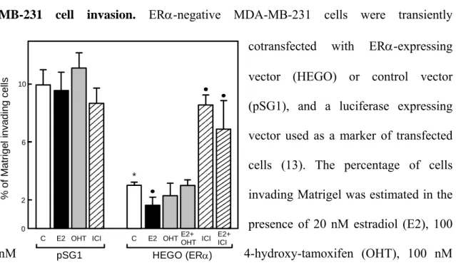
The mechanism by which estradiol inhibits invasion was studied using a new invasion assay based on the transient expression of ERα in the ERα-negative MDA-MB-231 cell line (18, 19). Estradiol treatment decreased by 2-fold the invasiveness in ERα-transfectant (Figure 2). The inhibitory effect of estradiol is reversed by both types of antiestrogens, OH-tamoxifen (4-hydroxytamoxifen, the active metabolite of tamoxifen) and ICI 164,384, a pure antiestrogen.
By contrast, estradiol or antiestrogen treatments did not significantly affect invasion of control ER-negative cells. Moreover, the analysis of different ERα deletion mutants strongly suggested that some estrogen-regulated genes negatively control invasion since the integrity of the hormone-binding domain, the DNA-binding domain and activating function 2 (AF2) of ERα was required (19). In contrast, the N-terminal domain containing the AF1 function is not involved since a deletion ΔAB mutant was as efficient as the wild-type receptor. As possible candidates among estrogen-regulated proteins, those that increase cell-cell adhesion, such as E-cadherin, or that decrease
% of M a tr ig e l in v adi n g c e lls pSG1 HEGO (ERα) 10 *
C E2 OHT ICI C E2 OHT ICI 2 6 0 E2+ OHT E2+ ICI
matrix degradation, such as α1-antichymotrypsin, should be considered (reviewed in 12, 20)
Figure 2: Effect of ERα transient transfection and estradiol treatment on MDA-MB-231 cell invasion. ERα-negative MDA-MB-231 cells were transiently
cotransfected with ERα-expressing vector (HEGO) or control vector (pSG1), and a luciferase expressing vector used as a marker of transfected cells (13). The percentage of cells invading Matrigel was estimated in the presence of 20 nM estradiol (E2), 100
nM 4-hydroxy-tamoxifen (OHT), 100 nM
ICI 164,384 (ICI) or ethanol alone (C). * p<0.01 versus pSG1 control; • p<0.05 versus HEGO control. Reproduced from (19) with permission.
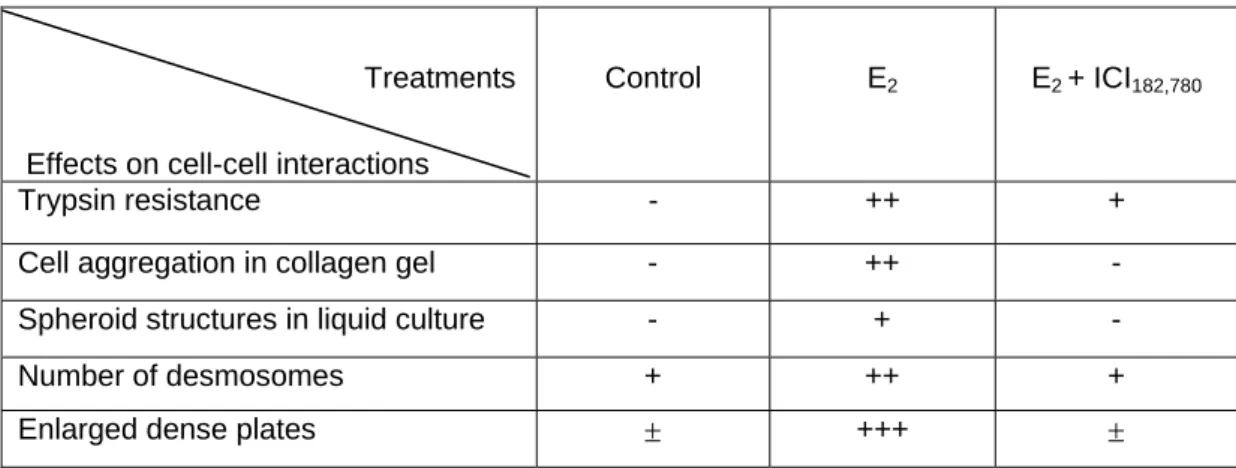
More recently we studied the effects of estrogens on the adhesiveness of breast cancer cells. Using different approaches, we found that cell-cell contacts were markedly increased by estradiol treatment and decreased by the pure antiestrogen, ICI 182,780 (Table 1). Estradiol-treated cells formed aggregates in standard culture conditions or on a collagen layer and, were more resistant to trypsin treatment. In suspension culture conditions, only estrogen-treated cells constituted large spherical multilayered cell structures. Using electron microscope analysis, the increase in cell-cell adhesion was associated with the formation of the major intercellular junctions, desmosomes. Estradiol increased both the number and the shape of desmosomes with enlarged dense plates in MCF7 cancer cells but also in normal mammary cells isolated from reduction
mammoplasties. The estrogen-induced proteins involved in desmosome production are currently investigated.
Table I. Effects of estradiol and ICI182,780 on cell-cell interactions. Steroid-stripped
MCF7 cells were treated for 5 days with 1nM estradiol (E2), 1nM E2 + 1µM ICI 182,780
or treated with vehicle alone (control) and the cell-cell interactions studied in the indicated cultures conditions.
Overall, these data indicated that the up-regulation of cell-cell adhesion by estrogen found in normal mammary cells is a characteristic maintained in ERα -positive breast cancer cells, and could participate to the low invasive potential of ERα -positive tumors.
Estrogen receptors inhibit invasion independently of ligand binding: involvement
of protein-protein interactions Using the transfection/invasion method described
above, we also demonstrated that expression of unliganded ERα and several mutants deleted in the hormone-binding domain drastically reduced MDA-MB-231 cell invasiveness in Matrigel tests. As shown in Figure 2, in estrogen-deprived conditions, transient wild type ERα expression induced a 3-fold decrease in the invasiveness of transfected cells and estradiol treatment reinforced the ligand independent effect by an additional 2-fold reduction. The strong inhibition due to the unliganded ERα is reversed by the pure antiestrogen ICI 164,384 which is known to decrease receptor concentration
Treatments
Effects on cell-cell interactions
Control E2 E2 + ICI182,780
Trypsin resistance - ++ +
Cell aggregation in collagen gel - ++ - Spheroid structures in liquid culture - + -
Number of desmosomes + ++ +
but not by the partial antiestrogen, OH-tamoxifen. In breast cancer cells, OH-tamoxifen and other tamoxifen derivatives were shown to up-regulate the receptor by decreasing its degradation (21). This increase of ERα concentration could explain the anti-invasive properties of tamoxifen in certain models in vitro (22) and could participate to its beneficial effects in vivo.
The domain involved in ligand-independent inhibition of invasion has been further characterized by progressive deletions in the ERα sequence.
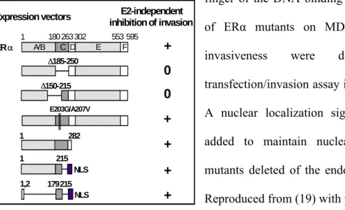
As shown in Figure 3, the first zinc finger of the DNA-binding domain (i.e., amino acids 179-215) is responsible for the anti-invasive activity. This activity is independent of the two key aminoacids which are essential for ERE binding and the estrogen specificity of the responses (23). Among the different nuclear receptors, invasion was
Figure 3: E2-independent inhibition of cancer cell invasion requires the first zinc
finger of the DNA binding domain. Effects of ERα mutants on MDA-MB-231 cell invasiveness were determined in transfection/invasion assay in Matrigel (13). A nuclear localization signal (NLS) was added to maintain nuclear targeting of mutants deleted of the endogenous signals. Reproduced from (19) with permission. specifically decreased by the expression of ERα (3-fold) and to a lesser extent by ERα (2-fold), but was not affected by thyroid hormone receptor α1, vitamin D receptor, retinoid acid receptor α, or glucocorticoid and androgen receptors. On the basis of these
NLS Δ185-250 E2-independent inhibition of invasion + + + 0 + 0 + E203G/A207V 1 215 1,2 179215 NLS Expression vectors 1 282 Δ150-215 ER α A/B C D E F 1 180263302 553 595
data, it was proposed that unliganded ER decreases invasiveness via interaction of the first zinc finger region with an unknown nuclear factor.
Moreover, immunocytochemical studies of ERα-positive breast cancer cell lines (MCF7, ZR75.1, T47D) indicated that in hormone-deprived conditions, ERα expression was inversely correlated with cell motility (19). Migrating cells had lower ER levels than non-migrating cells. Finally, treatments such as phorbol ester or pure antiestrogen, known to decrease ERα levels in MCF7 breast cancer cells, significantly increased in
vitro invasiveness (19, 24). Taken together, these in vitro data indicate a protective role
of ERα against the invasiveness of breast cancer cells.
These data obtained on cell cultures cannot be extrapolated to the in vivo situation, where the endocrine and paracrine effects of estrogens may have major consequences on the invasiveness of cancer cells. However, their possible implications in the monitoring of breast cancer should be discussed. Particularly, negative effects should be anticipated in the clinical use of pure antiestrogens such as ICI 164,384 or ICI 182,780, since these drugs increased in vitro cancer cell invasiveness by inhibiting the protective effect of both estrogens and ERα by decreasing its content. ERα expression should perhaps also be preserved in cancer cells during adjuvant therapies in order to maintain differentiation and a low invasive potential. In addition, these results suggest new therapeutic strategies based on ERα re-expression to prevent the proliferation, invasiveness and metastatic potency of ERα-negative breast cancer cells (20).
Discussion
Our data demonstrated that estradiol and ERα expression inhibit cancer cell invasiveness in breast cancer cells through different mechanisms. Inhibition by estrogens is dependent to transcriptional activation of specific target genes that are probably involved in an increase of cell-cell adhesion. The unliganded inhibits ERα
invasion via protein-protein interactions within the first zinc finger region of the receptor. These data could explain the protective role of ERα against tumor invasion and metastasis previously found in these cell lines. ERα expression has been associated with a low invasiveness and low motility in culture tests (24, 25). Moreover, when ERα-positive cells were implanted in nude mice, tumors appeared only in the presence of estrogens and are poorly metastatic as compared to those developed from ERα-negative breast cancer cell lines (20).
Clinical data supporting that estrogens prevent invasion. There is a great deal of
evidence to support the hypothesis that estrogens are important because they are potent mitogens for normal breast epithelial cells, and it is believed that the duration of breast epithelium exposure to estrogens is a significant risk factor for breast cancer development. However, in mammary carcinogenesis, even though the mitogenic effect of estrogens is well demonstrated, the presence of ERα is associated with more differentiated and less invasive tumors and a more favorable prognosis. Moreover, there is some clinical evidences indicating that estrogens and their receptors protect against invasion. Epidemiological studies have evaluated the breast cancer risk in women using hormone replacement therapy (HRT) where 80% were taking preparations containing estrogen alone (26, 27). Among the women using HRT, the risk of breast cancer slightly increased, but the tumors under estradiol treatment were confined to localized disease with more favorable prognosis. Tumors in HRT-users were less invasive to axillary lymph nodes and to more distant sites. Other studies of tamoxifen therapy of primary breast cancer suggest that tamoxifen increased the spreading of ERα-positive primary tumor cells to contralateral sites. Tamoxifen use decreased (0.8 fold) the risk of ERα-positive tumors, but it appeared to increase (4.9-fold) the risk of ERα-negative
contralateral tumors (28). All together, these clinical data are in agreement with an anti-invasive effect of estrogens.
Role of estrogen receptor variants in cancer progression? Numerous studies have
identified ERα and ERα variants from differential splicing of their mRNAs in both neoplastic breast tissue and cell lines (29, 30). These mRNA variants lack one or several exons and are usually coexpressed with the wild-type ER message. However, their pathophysiological significance is unclear. Several studies using transient transfection have shown that individual ERα variant proteins can have positive or negative effects on the wild-type ERα activity (29, 30). The presence of one or more variant proteins in variable levels in normal breast epithelium and neoplastic tissue could infuence the wild-type receptor (31). The variants ERα Δ3 (lacking the second zinc finger) and ERα Δ4 (deleted in the hormone-binding domain) are overexpressed in normal cells rather than in breast cancer cells. These variants, which contain the first zinc finger domain, could have an invasion-suppression activity independent of the hormone action. This was verified by expression of the ERα Δ3 variant (31). A more detailed clinical evaluation of the ER variants is required to determine their influence in mammary carcinogenesis and the response to therapy.
Missense mutants of the ERα gene with point mutation have been found in only 1 % of primary breast tumors (32). Even the functionality of only few mutations has been studied; initial data indicate that these mutations could affect normal ERα function and alter the evolution of individual tumors. This suggests that these somatic mutations, although infrequent, may significantly alter the evolution of individual tumors.
In conclusion, we present evidences that estrogens inhibit invasiveness of breast cancer cells via a classical activation of ERE-regulated genes leading to an increase in cell-cell adhesion. In the absence of ligand, the receptor could also prevent invasion through
interaction with an unknown protein. Non-classical mechanisms of action, in which the receptor may bind to other transcription factors instead of DNA or to the proteins involved in pathways such as motility and invasion, requires further investigation. The identification of the factors that inhibit the invasiveness of ERα positive cells would be a useful step in the development of new therapeutic targets to cure the most aggressive ERα -negative tumors.
Acknowledgements
We thank Michel Gleizes for excellent technical assistance and Jean-Yves Cance for artwork. This work was supported by the "Institut National de la Santé et de la Recherche Médicale", the "Association pour la Recherche sur le Cancer, and the “Ligue contre le cancer, Comité de l’Hérault” (fellowship for MM).
References
1. Pike MC, Krailo MD, Henderson BE, et al (1983) Hormonal risk factors, breast tissue age and the age-incidence of breast cancer. Nature 303:767-770.
2. Mangelsdorf, D.J., Thummel, C., Beato, M., et al (1996) The nuclear receptor superfamily: the second decade. Cell 86:835-839.
3. Gustafsson JA, (1999) Estrogen receptor ß—a new dimension in estrogen mechanism of action. J. Endocrinol 163:379-383.
4. Kumar V, Green S, Stack G, et al (1987) Functional domains of the estrogen receptor. Cell 51:941-951.
5. Moras D, Gronemeyer H, (1998) The nuclear receptor ligand-binding domain: structure and function. Curr Opin Cell Biol 10:384-391.
6. Mc Guire WL, (1978) Hormone receptor: their role in predicting prognosis and response to endocrine therapy. Semi Oncol 5:2428-2433.
7. Fisher B, Jeong JH, Dignam J, et al (2001) Findings from recent National Surgical Adjuvant Breast and Bowel Project adjuvant studies in stage I breast cancer. J Natl Cancer Inst Monogr 30:62-66.
8. Hayashi SI, Eguchi H, Tanimoto K, et al (2003) The expression and function of estrogen receptor alpha and beta in human breast cancer and its clinical application. Endocr Relat Cancer 10:193-202.
9. Fuqua SA, Schiff R, Parra I, et al (2003) Estrogen receptor beta protein in human breast cancer: correlation with clinical tumor parameters. Cancer Res 63:2434-2439.
10. Albini A, Graf J, Kitten GT, et al (1986) 17β-Estradiol regulates and v-Ha-ras transfection constitutively enhances MCF7 breast cancer cell interactions with basement membrane. Proc Natl Acad Sci USA 83:8182-8186.
11. Thompson EW, Reich R, Shima TB, et al (1998) Differential regulation of growth and invasiveness of MCF-7 breast cancer cells by antiestrogens. Cancer Res 48:6764-6768.
12. Rochefort H, Platet N, Hayashido Y, et al (1998) Estrogen receptor mediated inhibition of cancer cell invasion and motility: an overview. J Steroid Biochem Molec Biol 65:163-168.
13. Garcia M, Derocq D, Platet N, et al (1997) Both estradiol and tamoxifen decrease proliferation and invasiveness of cancer cells transfected with a mutated estrogen receptor. J Steroid Biochem Molec Biol 61:11-17.
14. Hayashido Y, Lucas A, Rougeot C, et al (1998) Estradiol and fibulin-1 inhibit motility of human ovarian- and breast-cancer cells induced by fibronectin. Int J Cancer 75:654-658
15. Kolodgie FD, Jacob A, Wilson PS, et al (1996) Estradiol attenuates directed migration of vascular smooth muscle cells in vitro. Am J Pathol 148:969-976. 16. Long BJ, Rose DP. (1996) Invasive capacity and regulation of urokinase-type
plasminogen activator in estrogen receptor (ER)-negative MDA-MB-231 human breast cancer cells, and a transfectant (S30) stably expressing ER. Cancer Lett 99:209-215.
17. Garcia, M, Derocq D, Freiss G, et al (1992) Activation of estrogen receptor into a receptor-negative breast cancer cell line decreases the metastatic and invasive potential of the cells. Proc Natl Acad Sci USA 89:1538-11542.
18. Platet N, Garcia M, (1999) A new bioassay using transient transfection for invasion-related gene analysis. Invasion Metastasis 18:198-206.
19. Platet N, Cunat S, Chalbos D, et al (2000) Unliganded and liganded estrogen receptors protect against cancer invasion via different mechanisms. Mol Endocrinol 14:999-1009.
20. Garcia M, Rochefort H, (2000) Estrogen receptor targeted therapies of breast cancer. Cur Opin Oncol Endocr Metab Invest Drugs 2:60-67.
21. Laios I, Journe F, Laurent G, et al (2003) Mechanisms governing the accumulation of estrogen receptor alpha in MCF-7 breast cancer cells treated with hydroxytamoxifen and related antiestrogens. J Steroid Biochem Mol Biol 87:207-221.
22. Bracke ME, Charlier C, Bruyneel EA, et al (1994) Tamoxifen restores the E-cadherin function in human breast cancer MCF-7/6 cells and suppresses their invasive phenotype. Cancer Res 54:4607-4609.
23. Green S, Kumar V, Theulaz I, et al (1988) The N-terminal DNA-binding 'zinc finger' of the oestrogen and glucocorticoid receptors determines target gene specificity. EMBO J 7:3037-44.
24. Platet N, Prevostel C, Derocq D, et al (1998) Breast cancer cell invasiveness: correlation with protein kinase C activity and differential regulation by phorbol ester in estrogen receptor-positive and -negative cells. Int J Cancer 75:750-756. 25. Thompson EW, Paik S, Brünner N, et al (1992) Association of increased
basement membrane-invasiveness with absence of estrogen receptor and expression of vimentin in human breast cancer cell lines. J Cell Physiol 150:534–544.
26. Marsden J, Backs NPM, (1996) Hormone replacement therapy and breast cancer. Endocr Relat Cancer 3:81–97.
27. Collaborative group on hormonal factors in breast cancer (1997) Lancet 350:1047-1059.
28. Li CI, Malone KE, Weiss NS, et al (2001) Tamoxifen therapy for primary breast cancer and risk of contralateral breast cancer. J Natl Cancer Inst 13:1008-1013. 29. Fuqua SA, Wolf DM, (1995) Molecular aspects of estrogen receptor variants in
breast cancer. Breast Cancer Res Treat 35:233-241.
30. Murphy LC, Leygue E, Dotzlaw H, et al (1997) Oestrogen receptor variants and mutations in human breast cancer. Ann Med 29:221-234.
31. Erenburg I, Schachter B, Mira y Lopez R, et al (1997) Loss of an estrogen receptor isoform (ER alpha delta 3) in breast cancer and the consequences of its reexpression: interference with estrogen-stimulated properties of malignant transformation. Mol Endocrinol 11:2004-2015.
32. Roodi N, Bailey LR, Kao WY, et al (1995) Estrogen receptor gene analysis in estrogen receptor-positive and receptor-negative primary breast cancer. J Natl Cancer Inst 87:446-451.