HAL Id: hal-01766862
https://hal-amu.archives-ouvertes.fr/hal-01766862
Submitted on 27 Aug 2018
HAL is a multi-disciplinary open access
archive for the deposit and dissemination of
sci-entific research documents, whether they are
pub-lished or not. The documents may come from
teaching and research institutions in France or
abroad, or from public or private research centers.
L’archive ouverte pluridisciplinaire HAL, est
destinée au dépôt et à la diffusion de documents
scientifiques de niveau recherche, publiés ou non,
émanant des établissements d’enseignement et de
recherche français ou étrangers, des laboratoires
publics ou privés.
Information processing in the axon
Dominique Debanne
To cite this version:
Dominique Debanne. Information processing in the axon. Nature Reviews Neuroscience, Nature
Publishing Group, 2004, 5 (4), pp.304-316. �10.1038/nrn1397�. �hal-01766862�
Since the pioneering work of Santiago Ramón y Cajal, the axon has been defined as a long neuronal process that insures the conduction of information from the cell body to the nerve terminal1. Generally, axons are highly ramified and contact several hundred target neurons locally or distally. However, the function of the axon is not limited to conduction of the action potential from its site of initiation near the cell body to the terminal. Recent experimental findings have shed new light on the functional and computational capabilities of single axons, indicating that several different, complex opera-tions are specifically achieved along the axon. Decreased conduction or backward propagation (reflection) might occur at specific axonal branch points under a defined regime of activity. Axonal geometry and the biophysical properties of axonal voltage-gated channels determine the timing of propagation of the output message in different axonal branches. In addition, axons link central neurons through GAP JUNCTIONSthat allow ultra-fast
net-work synchrony. Local shaping of the axonal action potential might subsequently determine synaptic efficacy during repetitive stimulation. These operations have been primarily described from observation of in vitro preparations of brain tissue, and evidence for these processes is scarce in the mammalian brain in vivo. In this article, I review the different ways in which the properties of axons can modify the transmission of electrical signals. I begin with a brief discussion of the basic characteristics
of propagation and how intrinsic channels that are present in the axon shape the action potential. I then con-sider two structural specializations that affect the way in which signals are propagated down the axon (branch points and varicosities), and review three ways in which these features can affect propagation — by introducing conduction delays, by causing propagation failures and by causing the action potential to be reflected.
K+and Na+channels in the axon
Propagation of an action potential is insured by local activation of sodium channels. Evidence for this was first provided by Hodgkin and Huxley in the giant squid axon (reviewed in REF. 2). Reduction of the sodium gradient or selective blockade of sodium chan-nels by TETRODOTOXIN(TTX) prevents conduction. So,
sodium channel activation can be considered as the motor for action potential conduction along axons. The molecular nature of sodium channels in myelinated and unmyelinated axons has been reviewed elsewhere3.
If the sodium channel is the motor for active propaga-tion along axons, potassium channels provide funcpropaga-tional opposition to action potential conduction. For example, demyelination of sciatic nerves exposes voltage-gated potassium channels that normally lie behind the myelin to the extracellular space4. The resulting outward current is responsible for propagation failures along the nerve because the voltage threshold of the action potential
INFORMATION PROCESSING IN
THE AXON
Dominique Debanne
Axons link distant brain regions and are generally regarded as reliable transmission cables in
which stable propagation
occurs once an action potential has been generated. However, recent
experimental and theoretical data indicate that the
functional capabilities of axons are much more diverse than traditionally thought. Beyond axonal propagation, intrinsic
voltage-gated
conductances together with the intrinsic geometrical properties of the axon determine complex phenomena
such as branch-point failures and reflected propagation. This review considers
recent evidence for the role of these forms of
axonal computation in the short-term dynamics of
neural communication.
GAP JUNCTIONS Morphological equivalent of electrical synapses. They are composed of two pairs of six connexins that form two apposed hemichannels constituting a pore between two neurons.
TETRODOTOXIN
A neurotoxin derived from the
Fugu, or puffer fish, which
specifically and reversibly blocks voltage-gated sodium channels.
Equipe INSERM AVENIR, Plasticité de l’excitabilité neuronale, Neurobiologie des Canaux Ioniques INSERM U641, Institut Fédératif de Recherche Jean Roche, Université de la Méditerranée, Boulevard Pierre Dramard, 13916 Marseille, France. e-mail: debanne.d@ jean-roche.univ-mrs.fr
and at the calyx of Held28. I
his an inward cationic current that is slowly activated by hyperpolarization. The molecular basis of Ih was revealed by the recent cloning of the hyperpolarization-activated, cyclic-nucleotide-gated, cationic non-selective channel subunits 1–4 (HCN1–4). In the axon, HCN1–4 have an important regulatory role and dampen shifts in membrane potentials. Another cationic channel that is activated by G protein-dependent receptors — the
heteromeric TRPC1/TRPC5 channel — has been
reported in axons of cultured hippocampal neurons29. These channels generate an inward current and might regulate the growth of axons in developing neurons30, but their precise function in mature axons remains largely unknown.
Functional computation in the axon
The shape of the presynaptic action potential is of fundamental importance in determining the strength of synapses by modulating transmitter release. The waveform of the depolarization dictates the calcium signal that is available to trigger vesicle fusion by con-trolling the opening of voltage-gated calcium channels and the driving force for calcium influx. Two types of modification of the presynaptic action potential have been reported experimentally — modification of width and/or modification of amplitude.
Activity-dependent broadening of presynaptic action potentials. The duration of the presynaptic spike is not fixed and activity-dependent short-term broadening of the spike has been observed in en passant mossy fibre boutons31. The mossy fibre–CA3 pyramidal cell synapse exhibits fast and synchronized transmitter release from several active zones and dynamic changes in synaptic strength over a more than tenfold range. The exception-ally large synaptic facilitation is in clear contrast to the weak facilitation (~150%) that is observed at most central synapses. Granule cell axons have several voltage-gated potassium channels including Kv1.1 (REF. 32), Kv1.2 (REF. 7) and two A-type potassium channels, Kv1.4 (REFS 6,10,11) and Kv3.4 (REF. 10). Geiger and Jonas have shown that the action potential at the mossy fibre terminal is half as wide as that at the soma. During repetitive stimulation, the action potential gets broader in the axon terminal but not in the soma31(FIG. 1). Using simultaneous recordings from the granule cell terminal and the corresponding postsynaptic apical dendrite of a CA3 neuron, Geiger and Jonas showed that action potential broadening enhanced presynaptic calcium influx and doubled the amplitude of the excitatory postsynaptic current (FIG. 1). This broadening results from the inactivation of A-type potassium channels that are located in the membrane of the terminal. Consequently, the pronounced short-term facilitation probably results from the conjugated action of spike widening and the classical accumulation of residual calcium in the presynaptic terminal. Because ultrastructural analysis reveals A-type channel immuno-reactivity in the terminal and in the axonal membrane11, activity-dependent spike broadening might also occur in the axon.
is locally raised as the result of an additional increase in membrane conductance. The block of conduction can be overcome by pharmacological blockade of voltage-gated potassium channels4,5. Several subtypes of voltage-gated potassium channels have been identified in myelinated and unmyelinated axons.Kv1.1,Kv1.2,Kv1.4,Kv3.1 and Kv3.4have been identified in mammalian axons6–14. In addition, large-conductance calcium-dependent potas-sium channels (BK, also called Maxi-K or Slo1 channels) are present in axons and presynaptic terminals15–19. Finally, large conductance sodium-dependent potassium channels (KNa, also called Slack channels or Slo2.2) are found in myelinated axons20,21. All of these potassium channels usually accelerate the repolarization of the action potential, but they might also prevent repetitive discharge and reduce the width of the action potential.
More recently, other types of voltage-gated channels have been discovered in axons. The hyperpolarization-activated non-selective cationic current (Ih) is present in spinal root axons22, leech neurons23, optic nerve fibres24, crayfish axons25,26, cerebellar basket cell axons27
100 mV 100 mV 200 pA 1 nA 100 mV 100 ms 1 ms 1 ms 50-Hz train of APs a b c 100th AP 50th AP 1st AP 20 µm MFB
Mossy fibre axon
Voltage command
Ca2+ current
Synaptic current
Figure 1 | Shaping of the action potential in the axon. a | A mossy fibre bouton (MFB) is recorded in the whole-cell configuration and activated at a frequency of 50 Hz. b | During repetitive stimulation of the axon, the action potential (AP) becomes wider. Every fiftieth AP is compared with the first AP in the train. c | AP broadening potentiates transmitter release. The mossy fibre terminal (green) and the corresponding CA3 cell (blue) were recorded simultaneously. AP waveforms were imposed at the presynaptic terminal. The increased duration of the waveform incremented the presynaptic calcium current and potentiated the amplitude of the synaptic current. Adapted, with permission, from REF. 31 (2000) Cell Press.
(FIG. 2a; S. Boudkkazi, E. Carlier & D.D., unpublished observations). In addition, depolarization of the presynaptic terminal by raising the external potassium concentration increases paired-pulse depression at autap-tic contacts of cultured hippocampal cells38and decreases
PAIRED-PULSE FACILITATION at Schaffer collateral–CA1
synapses38. In this case, depolarization of the presynaptic axons probably enhances presynaptic spike attenuation. Importantly, inactivation of sodium channels by high external concentrations of potassium increases the proportion of conduction failures during repetitive stimulation of Schaffer collateral axons39. Alternatively, paired-pulse depression can be reduced by increasing the external concentration of sodium, perhaps acting to suppress presynaptic spike attenuation38(FIG. 2b). These data indicate that slow recovery of sodium channels from inactivation might have an important role in shaping the short-term dynamics of synaptic transmission.
Interestingly, the manipulations of the sodium current that are mentioned above have no or little effect on GABA (γ-aminobutyric acid)-containing axons37–39.RILUZOLE, TTX or external potassium do not affect GABA-mediated synaptic transmission or short-term GABA-mediated plasticity. This difference between glutamatergic and GABA-containing axons might result from several factors. Sodium currents in interneurons are less sensitive to inactivation, and slow recovery from inactivation has been observed for pyra-midal cells but not for inhibitory interneurons40. Moreover, the density of sodium current is greater in interneurons than in pyramidal neurons41. So, axons of GABA-containinginterneurons could be better cables for propagation than those of pyramidal cells42,43. This unusual property could be functionally important — safe propagation along inhibitory axons could protect the brain from sporadic hyperactivity.
Axonal arborization: branch points and varicosities. In addition to affecting conductances, axonal morphology influences information processing in the axon. Axonal morphology is highly variable. At one extreme, axons of cerebellar granule cells possess a single T-shaped branch point that gives rise to the parallel fibres (FIG. 3a). At the other extreme, many axons typically form an elaborate tree. For instance, the terminal arbor of thalamocortical axons in layer IV of the cat visual cortex contains 150 to 275 branch points44(FIG. 3b). The complexity of axonal arborization is also extensive in cortical pyramidal neu-rons45(FIG. 3c). Axons of hippocampal CA3 pyramidal cells have at least 100 to 200 branch points in a total axonal length of 150 to 300 mm, and a single cell might contact 30,000 to 60,000 neurons46–48.But the champions of axonal complexity might be GABA-containing inter-neurons of different brain regions (FIG. 3d). Hippocampal and cortical inhibitory interneurons emit an axon with dense and highly branched arborization49. One obvious function of axonal divergence is to allow synchronous transmission to a wide population of target neurons within a given brain area. But branching axons might also link brain territories that are involved in complementary functional tasks, such as perception and action50. Inactivation of sodium channels. Reduction of the
amplitude of the presynaptic action potential has been reported following repetitive stimulation of invertebrate33 and mammalian axons31,34. This decline results from sodium channel inactivation and can be amplified by low concentrations of TTX35,36. The consequences of sodium channel inactivation for synaptic transmission have been studied at various central synapses. Interestingly, reduction of the sodium current by application of TTX in the nanomolar range decreases glutamatergic trans-mission and enhances short-term depression36–38
PAIRED-PULSE FACILITATION If two stimuli are delivered in close succession to an axon, the postsynaptic response to the second stimulus is often larger than to the first one. This phenomenon is referred to as paired-pulse facilitation, and is thought to depend on the accumulation of Ca2+ that
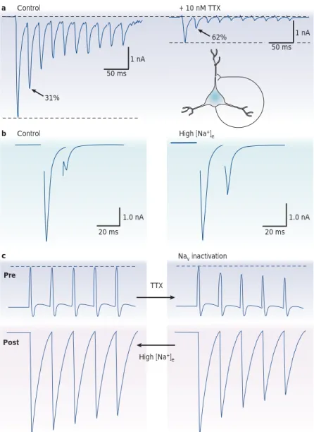
ensues after successive stimuli. c b Nav inactivation TTX Pre Post High [Na+] e High [Na+] e Control a Control + 10 nM TTX 31% 62% 1.0 nA 20 ms 1.0 nA 1 nA 20 ms 50 ms 1 nA 50 ms
Figure 2 | The role of sodium channel inactivation in short-term synaptic depression.
a | Repetitive stimulation of an autaptic contact produces short-term depression. The application
of a low concentration of the channel blocker tetrodotoxin (TTX) reduces synaptic transmission and enhances short-term depression. Adapted, with permission, from REF. 36(2000) Society for Neuroscience. b | Rescue of paired-pulse transmission by elevation of the extracellular sodium concentration ([Na+]
e). The large paired-pulse depression that is observed at an autaptic contact
of a cultured hippocampal cell is dramatically reduced when [Na+]
eis increased by 40 mM. Adapted,
with permission, from REF. 38(2002) The American Physiological Society. c | Hypothetical regulation of short-term depression by the modulation of activity-dependent attenuation of presynaptic spike amplitude. Sodium channel inactivation (TTX, right column) attenuates the spike train and enhances depression. Reduced inactivation (high [Na+]
e, left column) opposes
Axonal propagation and spike timing
The timing of action potentials is thought to determine the coding of neuronal information in the brain. Axonal conduction introduces a delay into the propa-gation of neuronal output, and axonal arborization might transform a temporal pattern of activity in the main axon into spatial patterns in the terminals58. Axonal delay initially depends on the velocity of the action potential in axons (generally between 0.1 m s–1in unmyelinated axons and 100 m s–1in large myelinated axons), which is a direct function of the diameter of the axon and the presence of a myelin sheath. Axonal delays might have crucial functional consequences for the integration of sensory information. In the first relay of the auditory system of the barn owl, differences in the delay of axonal conduction from each ear (which in this case is a function of differences in axonal length) produce sharp temporal tuning of the binaural infor-mation that is essential for acute sound localization59–61. In addition to this initial delay, local changes in the geometry of the axon produce an extra delay.
The presence of axonal irregularities such as vari-cosities and branch points reduces the conduction velocity (FIG. 4a). This reduction in conduction velocity occurs as a result of a high geometrical ratio (GR) (BOX 1). The degree of temporal dispersion has been simulated for an axon from the somatosensory cortex of the cat62. The delay introduced by high GR branch points could account for a delay of between 0.5 and 1 ms (REF. 62). But this extra delay seems rather small compared with the delay that is imposed by conduc-tion in axon branches with variable lengths (in the range of 2–4 ms).
A third source of delays in conduction is repetitive stimulation or activation of specific ion channels. The magnitude of this type of delay is usually variable, and it has been measured in a few cases. In lobster axons, the conduction velocity of the axon was decreased by ~30% following repetitive stimulation33. In dorsal root ganglion neurons, the latency of conducted spikes was enhanced by about 1 ms following antidromic paired-pulse stimulation of the axon63. Computational studies indicate that this delay might also result from a local distortion of the shape of the action potential. Extra activity-dependent delays might have important consequences for synaptic transmission. For instance, the synaptic delay was extended by 1–2 ms during repetitive stimulation of crayfish motor neurons64. Monosynaptic connections to motor neurons show an increase in synaptic latency concomitant with the synaptic depression that is induced by repetitive stimu-lation at 5–10 Hz and that induced near-propagation failures65. Similarly, a longer synaptic delay has been measured between connected hippocampal cells when conduction nearly fails owing to reactivation of A-type potassium channels66(FIG. 4b). So, axonal conduction might introduce some noise into the temporal pattern of action potentials that is produced at the initial segment. At the scale of a nerve, delays in individual axons introduce a temporal dispersion of conduction, indicating a model of stuttering propagation67. The second morphological feature of axons is the
presence of many varicosities (synaptic boutons) that are commonly distributed in an en passant, ‘beads-on-a-string’ manner along thin axon branches (FIG. 3e). A single axon can contain several thousands of bou-tons48,49,51. Bouton size varies between ~1 µm for thin unmyelinated axons52,53, to between 3 and 5 µm for large mossy fibre terminals of the hippocampus53,54. Their density varies among axons and the spacing between varicosities ranges from ~4 µm to ~6 µm in unmyelinated axons53,55.
Length and diameter also contribute to the variability of axons. Some axons extend locally (about 1 mm for inhibitory interneurons) whereas others can be as long as 1 metre or more. The diameter of axons also varies considerably. The largest axon (the squid giant axon) is close to 1 mm in diameter56whereas the diameter of unmyelinated cortical axons varies between 0.08 and 0.4 µm (REFS 52,57).
RILUZOLE
2-amino-6-trifluoromethoxy-benzothiazole). A voltage-dependent sodium channel blocker that is used as an anticonvulsant. PC GC PF MF 100 µm 25 µm Alveus str. ori. str. pyr. str. rad. str. I.-m. a b c e PF MF CA1 d
Figure 3 | Branch points and varicosities. a | Drawing of the cerebellar afferent circuit. Mossy fibre (MF) activity excites granule cells (GC) whose axons project towards the surface of the cortex. The GC axon bifurcates into two branches, which form the parallel fibres (PFs). Adapted from REF. 1.
b | Reconstruction of the terminal bouquet of a thalamocortical axon in area 17 of the cat. Horizontal
arrowheads indicate the limits of layer IV. Adapted, with permission, from REF. 44 (1998) Society for Neuroscience. c | Dendritic (red) and axonal (blue) arborizations of a layer II pyramidal neuron in the barrel cortex. Adapted, with permission, from REF. 45 (2003) Society for Neuroscience. d | Camera lucida reconstruction of a hippocampal GABA (γ-aminobutyric acid)-containing interneuron. The dendritic arbor is represented in black and the dense axonal arborization in red. Adapted, with permission, from REF. 41(2000) American Association for the Advancement of Science. Str. l.-m.; stratum lacunosum-moleculare; str. ori.; stratum oriens; str. pyr.; stratum pyramidale; str. rad.; stratum radiatum. e | Varicosities on stratum oriens axons in the CA1 area (upper), on MF axons in area CA3 (middle) and on cerebellar PFs (bottom). Adapted, with permission, from REF. 53 (2002) The National Academy of Sciences.
specifically at the branch point because the parent axon and one of the daughter branches continued to conduct action potentials. Failures appeared first in the thicker daughter branch, but they were also observed in the thin branch at a higher stimulus frequency. In the leech, con-duction block occurs at central branch points where fine axons from the periphery meet thicker axons73. Branch point failures have been observed or are suspected to occur in a number of mammalian neurons66,77,78.
Propagation failures also occur when the action potential enters a zone wherein the diameter changes abruptly. This occurs at en passant boutons84–86but also when impulses propagating along the axon enter the soma63. For instance, in the megacerebral cell of the snail, propagation failures have been observed when a spike enters the cell body87(FIG. 5b). These failures occur because the electrical load on the arriving action poten-tial is significantly higher and the current generated by the parent axon is not sufficient to allow propagation to proceed (see BOX 1).
Frequency-dependent propagation failures. In most cases, failures occur following moderate or high frequency stimulation (10–50 Hz) of the axon. For instance, a fre-quency of 20–30 Hz is sufficient to produce conduction failures at the neuromuscular terminal arborization69or at the branch point of spiny lobster motor neurons33. These failures are often observed as partial spikes or spikelets that are electrotonic residues of full action potentials. The functional consequences of conduction failures might be important in vivo. For example, in the leech, propagation failures produce an effect that is similar to sensory adaptation. Propagation failures are a non-synaptic mechanism that temporarily disconnects the neuron from one defined set of postsynaptic neurons and specifically routes sensory information in the gang-lion72–74,88. Propagation failure in the leech is also a means by which activity alters electrical synaptic transmission, which, in contrast to chemical synaptic transmission, is resistant to changes that are induced by activity89.
What are the mechanisms of frequency-dependent conduction failure? As mentioned above, the presence of a low safety conduction point such as a branch point, a bottleneck (that is, an axon entering the soma) or an axonal swelling determines the success or failure of con-duction (see also BOX 1). However, these geometrical constraints are not sufficient to account fully for all conduction failures and additional factors should be considered. The mechanisms of propagation failure can be grouped in two main categories.
First, propagation can fail during repetitive axon stimulation as a result of a slight depolarization of the membrane (FIG. 6a). At spiny lobster axons, propagation failures were associated with a 10–15% reduction of the action potential amplitude in the main axon and a membrane depolarization of 1–3 mV (REF. 33). These observations are consistent with potassium efflux into the periaxonal space that is induced by repetitive activa-tion. In most cases, the membrane depolarization that is produced by external accumulation of potassium ions around the axon probably contributes to the inactivation
Propagation failures
One of the more unusual operations of axons is selective conduction failure. When the action potential fails to propagate along the axon, no signal can reach the output of the cell. Conduction failure filters communication with postsynaptic neurons, and has been observed exper-imentally in various axons including vertebrate spinal axons68,69, spiny lobster or crayfish motor neurons33,64,70,71, leech mechanosensory neurons72–76, thalamocortical axons77, rabbit nodose ganglion neurons78, rat dorsal root ganglion neurons63,79, neurohypophysial axons17,80and hippocampal pyramidal cells39,66,81. Several factors deter-mine whether propagation along axons fails or succeeds. Geometrical factors: branch points and swellings. Although the possibility that propagation might fail at branch points was previously discussed by Krnjevic and Miledi69, the first clear indication that propagation is perturbed by axonal branch points came from the early studies on spiny lobster, crayfish and leech axons33,70–73,82,83. The large size of invertebrate axons allowed multi-electrode recordings upstream and downstream of the branch point to be performed. For example, in lobster axons, conduction across the branch point was found to fail at frequencies above 30 Hz (REF. 33) (FIG. 5a). The block of conduction occurred
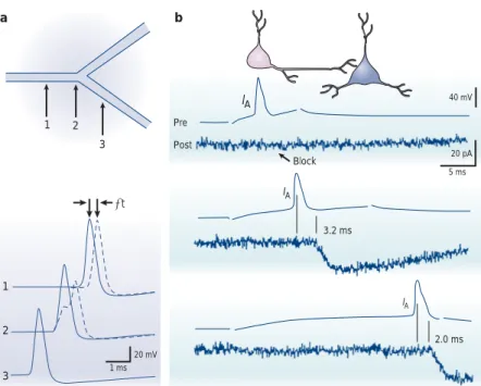
IA IA IA 40 mV 20 mV 20 pA 5 ms 1 ms Pre Post Block 3.2 ms 2.0 ms a b 1 1 2 3 2 3 ∆t
Figure 4 | Axonal propagation and spike timing. a | Comparison of the delay of propagation introduced by a branch point with a geometrical ratio (GR) > 1 (dashed traces) versus a branch point with perfect impedance matching (GR = 1, continuous traces). Upper, schematic drawing of a branched axon with three points of recording. At the branch point with GR = 8, the shape of the action potential is distorted and the propagation displays a small latency (∆t). Adapted, with permission, from REF. 62 (1991) The Biophysical Society. b | Propagation failures in hippocampal cell axons are associated with conduction delays. The presynaptic neuron (Pre) was slightly hyperpolarized with constant current to remove inactivation of the A-current. A presynaptic action potential induced with a short delay after onset of the depolarizing pulse did not elicit excitatory postsynaptic current in the postsynaptic cell (Post) because of the large activation of IA. Increasing the delay permitted action potential propagation because IAwas reduced during the action potential. For complete inactivation of IA(lower pair of traces), latency decreased. Adapted, with permission, from REF. 66 (1997) Macmillan Magazines Ltd.
the axon (FIG. 6b). Hyperpolarization-induced conduction block has been observed in leech72,73,88, locust92and mam-malian axons17,78.Axonal hyperpolarization opposes spike generation. Activity-dependent hyperpolarization of the axon usually results from activation of the Na+/K+ATPase and/or activation of calcium-dependent potassium channels. Unmyelinated axons in the PNS (for example, vagal C-fibres) hyperpolarize in response to repeated action potentials93,94as a result of the intracellular accu-mulation of sodium ions and the subsequent activation of the electrogenic Na+/K+pump26,93,94. In crayfish axons, this hyperpolarization can have a magnitude of 5–10 mV (REF. 26). Blockade of the Na+/K+ATPase with ouabain results in axon depolarization, probably as a consequence of post-tetanic changes in the concentration of extra-cellular potassium.In the leech, hyperpolarization-dependent conduction block occurs at central branch points in all three types of mechanosensory neurons in the ganglion — touch (T), pressure (P) and nociceptive (N) neurons. In these neurons, hyperpolarization is induced by the Na+/K+ATPase and by cumulative activa-tion of a calcium-activated potassium conductance. It is interesting to note that the conduction state can be changed by neuromodulatory processes. 5-HT (5-hydroxytryptamine, serotonin) decreases the prob-ability of conduction block of P and T cells, probably by limiting hyperpolarization95.
of sodium channels. In fact, hyperpolarization of the axon membrane or local application of physiological saline with a low concentration of potassium in the vicinity of a block can restore propagation in crayfish axons71. Elevation of the extracellular potassium con-centration produced conduction block in spiny lobster axons82. However, this manipulation did not reproduce the differential block that is induced by repetitive stimu-lation, as failures occurred simultaneously in both branches82. Interestingly, conduction could also be restored by increasing the concentration of intracellular calcium. Failures were also induced with a lower thresh-old when the electrogenic Na+/K+pump was blocked with OUABAIN. So, differential conduction block could be
explained as follows. During high frequency activation, potassium initially accumulates at the same rate around the parent axon and the daughter branches. Sodium and calcium accumulate more rapidly in the thin branch than in the thick branch because of the higher surface/volume ratio. The Na+/K+pump is activated and the concentration of extracellular potassium decreases at a greater rate around the thin branch82. Accumulation of extracellular potassium has also been observed in the olfactory nerve90and in hippocampal axons91, and could be the origin of unreliable conduction.
Propagation failures that are induced by repetitive stimulation might also result from hyperpolarization of
OUABAIN
Extracted from the seed of the
Strophantus, a tropical creeper,
ouabain is a cardiotonic that blocks sodium channel electrogenic pumps.
Box 1 | Theory of geometrical constraints on axonal propagation
The functional consequences of geometrical irregularities for axonal propagation have been addressed using numerical models (reviewed in REF. 128). Simulations show that at geometrical irregularities, the amplitude and width of the propagating action potential are usually distorted, and the local conduction velocity can change. For instance, an abrupt increase in axon diameter causes a decrease in both velocity and peak amplitude of the action potential, whereas a sudden reduction of diameter has the opposite local effects on these two parameters62,86,105,135–138. In fact,
the interplay between the total longitudinal current that is produced by the action potential and the input impedance of the axon segments ahead of the action potential determines the fate of the propagating action potential.
The case of the branch point has been studied in detail105,139,140.
The so-called 3/2 power law that was developed by Rall describes an ideal relationship between the geometry of mother and daughter branches105,141,142. A geometrical parameter (the geometrical ratio, GR) is defined as follows:
GR = (d3/2 daughter 1+ d
3/2 daughter 2)/d
3/2
mother, where ddaughter 1and ddaughter 2are the diameters of the daughter branches and dmother
is the diameter of the parent axon. For GR = 1, impedances match perfectly and spikes propagate in both branches (upper panel of figure). If GR > 1, the combined electrical load of the daughter branches exceeds the load of the main branch (lower panel of the figure). In other words, the active membrane of the mother branch might not provide enough current to activate both daughter branches. For 1 < GR < 10 — the most common situation — propagation beyond the branch point is delayed. All of these conclusions hold only if the characteristics of the membrane are identical. Any change in ion channel density might positively or negatively change the probability of sucessful propagation at a given branch point.
GR has been experimentally evaluated in a limited number of axon branch points. In lobster axons that innervate the deep abdominal muscles, GR at the branch point between medial and lateral bundles is close to the ideal value, allowing perfect impedance matching (0.97;REF. 33). In thalamic and cortical axons of the cat, GR at branch points varies between 0.78 and 1.98 (REF. 77). In axons of the crayfish, GR varies between 0.95 and 1.25 (REF. 83). Interestingly, in crayfish and lobster axons, branch points with a high GR were more susceptible to branch-point failures during repetitive stimulation. In axons of leech pressure neurons, the branch point formed by the thin peripheral axon (mean diameter 2.1 µm) with the principal axon (mean diameter 9.8 µm) has a GR near 20 (REF. 75). Branch points also have a very high GR in the giant metacerebral cell from the cerebral ganglia of Helix. Propagation might fail at low frequency when an active thin axon enters a large diameter axon87, indicating that, exceptionally, failures result uniquely from geometrical factors.
GR = 1 GR > 1 Parent Daughter Parent Daughter Daughter Daughter
Several recent studies indicate that the hyper-polarization that is produced by repetitive stimulation could be dampened by hyperpolarization-induced cationic current (Ih)26,81. This inward current is activated at resting membrane potential and produces a tonic depolarization of the axonal membrane26. So, reduction of this current induces hyperpolarization and perturbs propagation. The pharmacological blockade of Ihby ZD7288 or by external caesium can produce more failures in Schaffer collateral axons81. The peculiar bio-physical properties of Ihindicate that it might limit large hyperpolarizations or depolarizations that are produced by external and internal accumulation of ions. In fact, hyperpolarization of the axon will activate Ih, which in turn produces an inward current that compensates for the hyperpolarization26(FIG. 6b). Reciprocally, this com-pensatory mechanism is also valid for depolarization by removing basal activation of Ih.
Frequency-independent propagation failures. Action potential propagation in some axon collaterals of CA3 pyramidal neurons can be gated by activation of a presynaptic A-type potassium current66. Synaptic trans-mission between monosynaptically coupled pairs of CA3–CA3 or CA3–CA1 pyramidal cells can be blocked if a brief hyperpolarizing current pulse is applied a few milliseconds before induction of the action potential in the presynaptic neuron (FIG. 7; see also FIG. 4b). This regulation is observed in synaptic connections that have no transmission failures, indicating that the lack of postsynaptic response is a consequence of a conduction failure along the presynaptic axon. Interestingly, failures can also be induced when the presynaptic hyperpolarizing current pulse is replaced by a somatic inhibitory postsynaptic potential66,96. When presynaptic cells are recorded with a microelectrode containing 4-aminopyridine (4-AP), a blocker of IA-like conduc-tances, failures are abolished, which indicates that IA gates action potential propagation (see also REF. 97). Because A-channels are partially inactivated at the resting membrane potential, their contribution during an action potential that is elicited from the resting membrane potential is minimal, and the action poten-tial propagates successfully from the cell body to the nerve terminal. By contrast, A-channels recover from inactivation with a transient hyperpolarization and impede successful propagation to the terminal.
Propagation failures have been induced in only 30% of cases66, showing that propagation is generally reliable in hippocampal axons98–100. I
A-dependent conduction failures occur at some axon collaterals but not at others66. Using a theoretical approach, it has been shown that failures occur at branch points when A-type potassium channels are distributed in clusters near the bifurcation96. Perhaps because these conditions do not prevail in layer II/III neocortical neurons101,102or in dissociated hippocampal neurons99, this form of gating has not been reported in these cell types. It would be interesting to explore the actual distribution of potassium channel clusters near branch points using immunofluorescence methods.
Hyperpolarization-dependent failures have also been reported in axons of hypothalamic neurons (from paraventricular and supraoptic nuclei) that run into the neurohypophysis. The morphology of their boutons is unusual in that their diameter varies between 5 and 15 µm (REF. 85). In single axons, propagation failures are observed at stimulation rates that are greater than 12 Hz and they are concomitant with a hyperpolarization of 4 mV (REF. 17). These failures might account for the non-linear decline in hormone release from the pituitary and the activity-dependent fatigue of neuro-secretion. The induced hyperpolarization of the neuron might result from activation of the calcium-dependent BK potassium channel. In fact, action potential failures were more frequent when BK channels were indirectly activated by adding the L-type calcium channel agonist Bay K 8644 to the external medium. By contrast, no failures were observed in the presence of the voltage-gated calcium channel blocker cadmium, indicating that propagation failure might result from accumula-tion of intracellular calcium and activaaccumula-tion of BK17. Subsequently, activation of BK channels would decrease axonal excitability and promote failures of incoming action potentials. The slow inactivation of the channel is compatible with the critical frequency for propagation failures. Axon Lateral Medial Stimulus EPSP 250 µm Br3 Br4 Br5 Soma Axon Br1 250 µm a b
Figure 5 | Propagation failures. a | Propagation failure at a branch point in a lobster axon33. b | Propagation failure at the
junction between an axonal branch and the soma of a snail neuron (metacerebral cell). The propagation in the axonal arborization was analysed by the local fluorescence transients owing to the action potential. The recording region is indicated by an outline of a subset of individual detectors, superimposed over the fluorescence image of the neuron in situ. When the action potential was evoked by direct stimulation of the soma, it propagated actively in all axonal branches (green traces). By contrast, when the action potential was evoked by the synaptic stimulation (EPSP) of the right axonal branch (Br1), the amplitude of the fluorescent transient declined when approaching the cell body, indicating a propagation failure (blue traces). Adapted, with permission, from REF. 87 (2000) The Physiological Society.
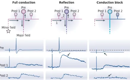
frequencies. But another way that a neuron’s branching pattern can affect impulse propagation is by reflecting the impulse105–107. Reflection (or reverse propagation) occurs when an action potential is near failure105. This form of axonal computation has been well described in leech mechanosensory neurons75,76(FIG. 8)in which an un-expected event occurs when conduction nearly becomes blocked — the action potential that has nearly failed to invade the thick branch of the principal axon sets up a local potential that propagates backwards. Reflection occurs because impulses are sufficiently delayed as they travel through the branch point. So, when the delay exceeds the refractory period of the afferent axon, the impulse will propagate backwards as well as forwards, creating a reflection. This phenomenon can be identified electrophysiologically at the cell body of the P neuron because action potentials that reflect have a longer initial rising phase (or ‘foot’), indicating a delay in conduction through the branch point. This fast double firing in the thin branch of mechanosensory neurons has important functional consequences. It facilitates synaptic trans-mission at synapses that are formed by this axon and postsynaptic neurons by a mechanism of paired-pulse facilitation with the orthodromic spike and the antidromic action potential that reflected at the branch point (FIG. 8). Reflection also occurs in T cells75. Interestingly, the facilitation of synaptic transmission also affects the chemical synapse between the P cell and the S neuron, a neuron that has an essential role in sensitiza-tion, a non-associative form of learning76. Reflected prop-agation is not restricted to mechanosensory neurons of the leech but has also been observed in the axon of an identified snail neuron87. Reflection has not yet been definitively reported in mammalian axons (REF. 108)but it has been demonstrated in dendrites (BOX 2).
Axo-axonal coupling and fast synchronization
Ephaptic interactions. Interactions between neighbouring axons were first studied by Katz and Schmitt109,110in the crab. The passage of an impulse through one axonal fibre produced a subthreshold change in excitability in the adjacent fibre. As the action potential approached in the active axon, the excitability of the resting fibre was first reduced, then quickly enhanced. This effect results from depolarization of the resting axon by the active axon, which locally generates an extracellular potential of a few mV. Interactions of this type are called ephaptic (from the Greek for ‘touching onto’111) and have also been observed at frog sciatic nerve112.
One of the most interesting features of ephaptic inter-action between adjacent axons is that the conduction velocity in neighbouring fibres might be unified, thereby synchronizing activity in a bundle of axons. If one action potential precedes the other by a few milliseconds, it accelerates the conduction rate of the lagging action potential in the other axon110. However, perfectly syn-chronized action potentials decrease the conduction velocity in both branches. Synchronization can only occur if the individual velocities differ only slightly and are significant for a sufficient axonal length110. Does such synchronization also occur in mammalian axons? Functionally, this form of gating might determine part
of the short-term synaptic facilitation that is observed during repetitive presynaptic stimulation. Apparent paired-pulse facilitation is observed because the first, but not the second, action potential fails to propagate owing to inactivation of the A-type potassium current103. Another voltage-gated potassium channel (ID) has been recently proposed to control synaptic transmission between individual CA3 cells104. However, additional investigation will be required to determine whether this current also gates action potential propagation.
Reflection of action potential propagation. Branch points are usually regarded as frequency filters, allowing separate branches of an axon to activate their synapses at different
Failure Failure a b Na+ K+ Nav Nav Cav KCa Inactivated Nav Kv Depolarization Hyperpolarization Repolarization Na+ Na+ Ca2+ K+ HCN
Figure 6 | Mechanisms of propagation failures induced by repetitive stimulation.
a | Activity-dependent depolarization of the axon. Following high frequency stimulation of the axon
(burst on the left) activation of voltage-gated potassium channels (Kv) produces a large efflux of potassium ions (K+) that accumulate in the periphery of the axon. The resulting axonal depolarization
together with the slow recovery of sodium channels (Nav) from inactivation produce conduction failures seen as partial spikes (burst on the right). b | Activity-dependent hyperpolarization of the axon. High frequency stimulation of the axon produces an accumulation of intracellular sodium and intracellular calcium through voltage-gated sodium (Nav) and calcium (Cav) channels. Activation of
the electrogenic Na+/K+pump by internal sodium ions and calcium-dependent potassium channels
(KCa) by internal calcium ions hyperpolarizes the axon membrane and produces conduction failures. The hyperpolarization can be partially compensated by activation of the IH current through hyperpolarization-activated, non-selective cationic (HCN) channels.
40 mV 2 mV 1 nA Pre Post a Inactivated IA b Recovered IA
Figure 7 | Gating of action-potential propagation by the potassium current IA.
a | At resting membrane potential, presynaptic IAwas inactivated and the action potential evoked in the presynaptic cell propagated and elicited an excitatory postsynaptic potential (EPSP) in the postsynaptic cell. b | Following a brief hyperpolarizing pre-pulse, presynaptic IArecovered from inactivation and blocked propagation. Consequently, no EPSP was evoked by the presynaptic action potential. Adapted, with permission, from REF. 66 (1997) Macmillan Magazines Ltd.
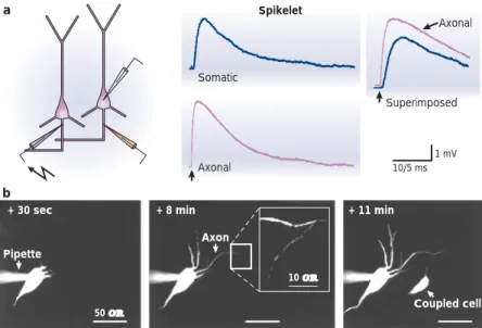
Electrical coupling of axons. Another type of axo-axonal communication has recently been demon-strated in hippocampal neurons. In the hippocampus, one type of high frequency oscillation (100–200 Hz) called ‘ripple’ arises from the high-frequency firing of inhibitory interneurons and phase-locked firing of many CA1 neurons116. Some of the properties of ripple oscillation are, however, difficult to explain. First, the oscillations are so fast (near 200 Hz) that synchrony across many cells would be difficult to achieve through chemical synaptic transmission. In addition, ripples persist during pharmacological blockade of chemical transmission in vitro117. While some inhibitory interneurons might synchronize a large number of pyramidal cells during the ripple118, a significant part of the synchronous activity could be mediated by axo-axonal electrical synaptic contacts through gap junctions119(FIG. 9a). Antidromic stimulation of a neigh-bouring axon elicits a spikelet that has a fast rate of rise (near 180 mV ms–1) and an amplitude between 1 and 10 mV. Spikelets can be evoked at the rate of a ripple (200 Hz) and are blocked by TTX or by the gap junc-tion blocker carbenoxolone. Simultaneous recording from the axon and cell body showed that the spikelet traversed the axon prior to invading the soma and the dendrites. Finally, labelling of pyramidal neurons with rhodamine, a small fluorescent molecule, showed dye coupling in adjacent neurons that was initiated through the axon119(FIG. 9b). Models indicate that the density of gap junctions might be very low, accounting for the fact that they have not yet been visualized in axons. But pannexins, a recently identified family of gap-junction proteins that are found throughout the brain, could be the molecular substrate of gap-junctions in axons120. So, the function of the axon is not limited to conduction of impulses to the terminal, and information might process between adjacent pyramidal neurons through ELECTRICAL SYNAPSES that are located
close to their axon hillock. There is no evidence for this yet, but modelling studies
indicate that the relative location of nodes of Ranvier on two adjacent myelinated axons might also determine the degree of temporal synchrony between fibres113,114. On small unmyelinated axons, ephaptic interaction between axons is predicted to be minimal115but future research might reveal a powerful means to thoroughly synchro-nize neuronal activity downstream of the site of action potential initiation.
Box 2 | Reflected propagation in dendrites
Whether ‘ping-pong’ propagation occurs in mammalian axons is still debated108. However, in mitral cells of the
mammalian olfactory bulb, both conduction failures143
and reflection121have been observed for impulses that are
initiated in dendrites. Propagation in dendrites of mitral cells is unusual. Like propagation in axons, it is highly active and no decrement in the amplitude of the action potential is observed between the soma and the dendrite144. In addition, mitral cell dendrites are both
pre- and postsynaptic elements. Ping-pong propagation
has been observed following near failure of dendritic action potentials that are evoked in distal primary dendrites121.
Forward propagation of an action potential (1) in a dendrite (d) can be evoked by an excitatory postysynaptic potential that is elicited by strong stimulation of the glomerulus. This particular form of propagation might fail near the cell body when the soma (s) is slightly hyperpolarized (asterisk, dashed line in the right panel of the figure). For an intermediate range of membrane potentials, the action potential invades the soma and might trigger a back-propagating action potential (2), which is observed as a dendritic double spike in the primary dendrite (thick trace in the right panel of the figure). The function of reflected propagation has not been definitively established, but when axonal output is prevented by inhibition of the soma, the primary dendrite of the mitral cell can function as a local interneuron affecting its immediate environment. Reflection of fast action potentials has also been observed in dendrites of retinal ganglion cells145. Figure
adapted, with permission, from REF. 121 (2002) The American Physiological Society.
On d d s s 2 2 1 1
*
ELECTRICAL SYNAPSE Specialized sites where gap-junction channels bridge the membrane of adjacent neurons and provide a low-resistance pathway for ions and small molecules, thereby permitting direct transmission of electrical signals. Pre Post 1 Post 2 Minor field Major field Pre Post 1 Post 2 Pre Post 1 Post 2 1 2Full conduction Reflection Conduction block
Pre
Post 1
Post 2
Figure 8 | Reflection of action potentials. Reflection and conduction block produce multilevel synaptic transmission in mechanosensory neurons of the leech. Left, an action potential that is initiated by anterior minor field stimulation invades the whole axonal arborization (pink) and evokes an excitatory postsynaptic potential in all postsynaptic cells. Middle, following repetitive stimulation, the cell body is slightly hyperpolarized (purple) and the same stimulation induces a reflected action potential at the branch point between the left branch and the principal axon. The reflected action potential (pink arrow 2) stimulates the presynaptic terminal on postsynaptic cell 1 twice, thereby enhancing synaptic transmission (arrow in lower panel). Right, when the cell body is further hyperpolarized (turquoise), the stimulation of the minor field now produces an action potential that fails to propagate at the branch point. The failed spike is seen as a spikelet at the cell body (upward arrow). No postsynaptic response is evoked in postsynaptic cell 2 (downward arrow). Adapted, with permission, from REF. 75(1998) The National Academy of Sciences.
axons to build more realistic models. In particular, the comparison of these parameters in reliable and unreli-able cortical axons might reveal unexpected differences. Another fundamental challenge in the near future will be to manipulate local axonal architecture to produce branch points with defined GRs. Local modification of the cytoskeleton in simple models of axons could be a powerful tool to address this question.
The subcellular localization of ion channels also has a crucial role in propagation. In many axons, GR might be greater than 1 and propagation still possible and reliable. A reasonable explanation for the reliability of conduction is that axonal structures are endowed with a heteroge-neous distribution of ion channels. For example, a high density of sodium channels near branch points or at boutons could account for reliable conduction. Detailed quantitative immunostaining of sodium channels on single axonal fibres will be needed to test this hypothesis. In models of axonal conduction, the density and bio-physical properties of ion channels can be easily tested96,122. Attaining a detailed understanding of axonal function will also require manipulation of the expression of specific ion channels at precise locations in the axon. The use of recently developed molecular tools to target defined channel subunits to specific axonal compart-ments could help to determine their role in axonal prop-agation123,124. For instance, the controlled expression of sodium or potassium channels at a high density at branch points would help us understand branch point failures. Moreover, chromophore-assisted laser inactiva-tion of proteins could be used to modify ion channel density along axons in a spatially controlled manner125,126.
Fine temporal tuning can be achieved by axons. Differences in axonal length in the terminal axonal tuft introduce delays of several milliseconds. Is temporal scaling of action potential propagation in the axonal arborization relevant to the coding of neuronal infor-mation? Differential conduction delays in axonal branches contribute to precise temporal coding in the barn owl auditory system59–61. But a role for axonal delays in synchronizing mammalian networks127is yet to be demonstrated. Local axonal interactions such as ephaptic coupling and gap-junction coupling allow fast synchronization of activity in neighbouring neurons. Surprisingly, little experimental effort has been devoted to ephaptic interactions, which are a powerful means of precisely synchronizing the outputs of neighbouring neurons. Perhaps ephaptic interactions between parallel axons could compensate for the ‘stuttering conduction’ that results from axonal varicosities and branch points67. The role of these mechanisms in synchronizing activity will have to be determined in axons that have a geometri-cal arrangement that is favourable for ephaptic coupling (that is, fasciculation over a sufficient axonal length). Callosal axons, mossy fibres and Schaffer collaterals are possible experimental subjects. In the case of gap-junction coupling, it will be important to determine whether electrical coupling also favours synchronization of neocortical neurons. The molecular substrates of electrical coupling in axons will need to be identified — pannexins have already been proposed120.
Conclusion
Increased computational capabilities. Axons achieve sev-eral fundamental operations that go beyond classical propagation. The output message can be routed along selected axonal pathways at a defined regime of activity. The consequences of this in mammalian axons are not yet well understood, but branch point failures might contribute to the elaboration of sensory processing in invertebrate neurons74. Axonal propagation might also ‘bounce back’ at a branch point or at the cell body. However, at present, only a handful of examples of reflected propagation have been observed75,76,87,108,121. Reflected impulses might limit the spread of the neuronal message and enhance synaptic transmission. Theoretical and experimental studies indicate that reflection of action potentials could occur in axons that have large swellings or a branch point with a high GR. Finally, axonal coupling through ephaptic interactions or gap junctions might precisely synchronize network activity119. All of these operations increase the computational capabilities of axons and affect the dynamics of synaptic coupling. Many pieces of the puzzle are, however, still missing.
Future directions and missing pieces. Axonal morphology has a crucial role in conduction, and propagation failures or reflected propagation might result from the presence of axonal irregularities such as varicosities and branch points. However, detailed quantitative analysis of the morphology of single axons is relatively scarce. It will be of great interest to determine the precise number of branch points and GR in several types of mammalian
Somatic Axonal Axonal Superimposed 10/5 ms 1 mV a b Spikelet
+ 30 sec + 8 min + 11 min
Pipette
Axon
Coupled cell
50 µm
10 µm
Figure 9 | Axo-axonic coupling of hippocampal pyramidal neurons. a | Spikelets propagate antidromically. Whole-cell recordings were obtained from the soma (blue traces) and from the axon hillock (pink traces). Spikelets were evoked by the stimulation of the axon of a neighbouring neuron. Superimposed traces show a delay between the axonal recording and the somatic recording, indicating that spikelets traverse the axon prior to invading the soma.
b | Dye coupling between hippocampal pyramidal neurons. A neuron was recorded in
whole-cell configuration with a pipette containing a fluorescent dye (+ 30 sec). Axon and dendrites were labelled about 8 min after establishing whole-cell configuration. The boxed region shows an axon of a second pyramidal neuron containing dye from the axon of the first cell. The cell body of the dye-coupled pyramidal cell appears a few minutes later (+ 11 min). Adapted, with permission, from REF. 119(2001) Cell Press.
microscopy42,101,102or voltage-sensitive dyes67,87have enor-mous potential. But it will also be extremely important to use other probes that do not perturb the rather fragile equilibrium that underlies propagation. Non-invasive recording techniques such as extracellular recording from single axons will be extremely helpful39,81,100,131.
Dendrites have recently received a great deal of attention and substantial progress has been made in understanding their function132–134. Axons deserve com-parable attention so that their complex properties can be fully explored. We predict that future investigation will reveal that fundamental neuronal operations are not only achieved by the cell body, the dendrites and the synapse. Axonal operations will also be shown to be important determinants of information processing in the brain.
New methods of investigation. Most of our knowledge about axonal computation is derived from experiments on invertebrate neurons or from computer simu-lations128. Our understanding of propagation in mam-malian axons is still fragmentary and direct evidence for propagation failures is still controversial99,101,129,130. Moreover, evidence for an important role of propagation failures and axonal reflection in information processing
in vivo is still scarce77,108. A main difficulty in studying propagation along thin unmyelinated axons is their rela-tive complexity and their small size, which make direct electrophysiological recordings almost impossible. New tools and experimental techniques will need to be developed if the mechanisms of axonal computation in mammalian CNS neurons are to be dissected. High reso-lution imaging techniques like multiphoton confocal
1. Ramón y Cajal, S. Histologie du Système Nerveux (Maloine, Paris, 1911)
2. Huxley, A. From overshoot to voltage clamp. Trends
Neurosci. 25, 553–558 (2002).
3. Poliak, S. & Peles, E. The local differentiation of myelinated axons at nodes of ranvier. Nature Rev. Neurosci. 4, 968–980 (2003).
4. Bostock, H., Sherrat, R. M. & Sears, T. A. Overcoming conduction failure in demyelinated nerve fibres by proloning action potentials. Nature 274, 385–387 (1978). 5. Bostock, H., Sears, T. A. & Sherratt, R. M. The effects of
4-aminopyridine and tetraethylammonium ions on normal and demyelinated mammalian nerve fibres. J. Physiol. (Lond.)
313, 301–315 (1981).
6. Sheng, M. Tsaur, M. L., Jan, Y. N. & Jan, L. Y. Subcellular segregation of two A-type K+channel protein in rat central neurons. Neuron 9, 243–259 (1992).
7. Sheng, M., Liao, Y. J., Jan, Y. N. & Jan, L. Y. Presynaptic A-current based on heteromultimeric K+channels detercted in
vivo. Nature 365, 72–75 (1993).
8. Wang, H., Kunkel, D. D., Martin, T. M., Schwarztkroin, P. A. & Tempel, B. L. Heteromultimeric K+channels in terminals and juxtaparanodal regions of neurons. Nature 365, 75–79 (1993).
9. Wang, H., Kundel, D. D., Schwartzkroin, P. A. & Tempel, B. L. Localization of Kv1. 1 and Kv1. 2, two K channel proteins, to synaptic terminals, somata, and dendrites in the mouse brain. J. Neurosci. 14, 4588–4599 (1994).
10. Veh, R. W. et al. Immunohistochemical localization of five members of the Kv1 channel subunits: contrasting subcellular locations and neuron-specific co-localizations in rat brain. Eur. J. Neurosci. 7, 2189–2205 (1995) 11. Cooper, E. C., Milroy, A., Jan, Y. N., Jan, L. Y. & Lowenstein,
D. H. Presynaptic localization of Kv1.4-containing A-type potassium channels near excitatory synapses in the hippocampus. J. Neurosci. 18, 965–974 (1998). 12. Devaux, J. et al. Kv3.1b is an novel component of CNS
nodes. J. Neurosci. 23, 4509–4518 (2003). 13. Dodson, P. D. et al. Presynaptic rat Kv1. 2 channels
suppress synaptic terminal hyperexcitability following action potential invasion. J. Physiol. (Lond.) 550, 27–33 (2003). 14. Ishikawa, T. et al. Distinct roles of Kv1 and Kv3 potassium
channels at the Calyx of Held presynaptic terminal.
J. Neurosci. 23, 10445–10453 (2003).
15. Jonas, P., Koh, D. S., Kampe, K., Hermsteiner, M. & Vogel, W. ATP-sensitive and Ca-activated K channels in vertebrate as novel links between metabolism and excitability. Pflugers
Arch. 418, 68–73 (1991).
16. Kraus, H. G. et al. Distribution of high-conductance Ca2+ -activated K+channels in rat brain: targeting to axons and nerve terminals. J. Neurosci. 16, 955–963 (1996). 17. Bielefeldt, K. & Jackson, M. B. A calcium-activated
potassium channel causes frequency-dependent action-potential failures in a mammalian nerve terminal.
J. Neurophysiol. 70, 284–298 (1993).
18. Hu, H. et al. Presynaptic Ca2+-activated K+channels in glutamatergic hippocampal terminals and their role in spike repolarization and regulation of transmitter release.
J. Neurosci. 21, 9585–9597 (2001).
19. Roncarati, R., Di Chio, M., Sava, A., Terstappen, G. C. & Fumagalli, G. Presynaptic localization of the small conductance calcium-activated potassium channel SK3 at the
neuromuscular junction. Neuroscience 104, 253–262 (2001).
20. Koh, D. S., Jonas, P. & Vogel, W. Na+-activated K+channels localized in the nodal region of myelinated axons of
Xenopus. J. Physiol. (Lond.) 479, 183–197 (1994)
21. Bhattacharjee, A., Gan, L. & Kaczmarek, L. K. Localization of the Slack potassium channel in the rat central nervous system. J. Comp. Neurol. 454, 241–254 (2002). 22. Baker, M., Bostock, P., Grafe, P. & Martins, P. Function and
distribution of three types of rectifying channel in rat spinal root myelinated axons. J. Physiol. (Lond.) 383, 45–87 (1987).
23. Angstadt, J. D. & Calabrese, R. L. A hyperpolarization-activated inward current in heart interneurons of the medicinal leech. J. Neurosci. 9, 2846–2857 (1989). 24. Eng, D. L., Gordon, T. R., Kocsis, J. D. & Waxman, S. G.
Current-clamp analysis of a time-dependent rectification in rat optic nerve. J. Physiol. (Lond.) 421, 185–202 (1990). 25. Beaumont, V. & Zucker, R. S. Enhancement of synaptic transmission by cyclic AMP modulation of presynaptic Ih channels. Nature Neurosci. 3, 133–141 (2000). 26. Beaumont, V., Zhong, N., Froemke, R. C., Ball, R. W. &
Zucker, R. S. Temporal synaptic tagging by Ihactivation and actin: involvement in long-term facilitation and cAMP-induced synaptic enhancement. Neuron, 33, 601–613 (2002). 27. Southan, A. P., Morris, N. P., Stephens, G. J. & Robertson, B.
Hyperpolarization-activated currents in presynaptic terminals of mouse cerebellar bascket cells. J. Physiol.
(Lond.) 526, 91–97 (2000).
28. Cuttle, M. F., Rusznak, Z., Wong, A. Y., Owens, S. & Forsythe, I. Modulation of a presynaptic hyperpolarization-activated cationic current (Ih) at an excitatory synaptic terminal in the rat auditory brainstem. J. Physiol. (Lond.)
534, 733–744 (2001).
29. Strübing, C., Krapivinsky, G., Krapivinsky, L. & Clapham, D. E. TRPC1 and TRPC5 form a novel cation channel in mammalian brain. Neuron 29, 645–655 (2001). 30. Greka, A., Navarro, B., Oancea, E., Duggan, A. & Clapham,
D. E. TRPC5 is a regulator of hippocampal neurite length and growth cone morphology. Nature Neurosci. 6, 837–845 (2003).
31. Geiger, J. R. P. & Jonas, P. Dynamic control of presynaptic Ca2+ inflow by fast-inactivating K+channels in hippocampal mossy fiber boutons. Neuron 28, 927–939 (2000).
With elegant recording techniques this paper shows that repetitive axon stimulation inactivates A-type potassium channels, broadens presynaptic action potentials and facilitates synaptic transmission.
32. Rhodes, K. J. et al. Association and colocalization of the Kvβ1 and Kvβ2 β-subunits with Kv1 α-subunits in mammalian brain K+channel complexes. J. Neurosci. 17, 8246–8258 (1997).
33. Grossman, Y., Parnas, I. & Spira, M. E. Differential conduction block in branches of a bifurcating axon.
J. Physiol. (Lond.) 295, 283–305 (1979).
Demonstration and analysis of differential propagation block at the branch point of a lobster peripheral axon.
34. Wang, L. Y. & Kaczmarek, L. K. High-frequency firing help replenish the readily releasable pool of synaptic vesicles.
Nature 394, 384–388 (1998).
35. Madeja, M. Do neurons have a reserve of sodium channels for the generation of action potentials? A study on acutely isolated CA1 neurons from the guinea-pig hippocampus.
Eur. J. Neurosci. 12, 1–7 (2000).
36. Brody, D. L. & Yue, D. T. Release-independent short-term synaptic depression in cultured hippocampal neurons.
J. Neurosci. 20, 2480–2494 (2000).
Evidence that sodium channel inactivation might participate in short-term synaptic depression at autaptic contacts.
37. Prakriya, M. & Mennerick, S. Selective depression of low-release probability excitatory synapses by sodium channel blockers. Neuron 26, 671–682 (2000).
Differential sensitivity of glutamatergic and GABA-mediated axons to low concentrations of sodium channel blockers.
38. He, Y., Zorumski, C. F. & Mennerick, S. Contribution of presynaptic Na+channel inactivation to paired-pulse synaptic depression in cultured hippocampal neurons.
J. Neurophysiol. 87, 925–936 (2002).
39. Meeks, J. P. & Mennerick, S. The selective effects of potassium elevation on glutamate signaling and action potential conduction in hippocampus. J. Neurosci. 24, 197–206 (2004).
40. Martina, M. & Jonas, P. Functional differences in Na+channel gating between fast-spiking interneurones and principal neurons of rat hippocampus. J. Physiol. (Lond) 505, 593–603 (1997).
41. Martina, M., Vida, I. & Jonas, P. Distal initiation and active propagation of action potentials in interneurons dendrites.
Science 287, 295–300 (2000).
42. Forti, L., Pouzat, C. & Llano, I. Action potential-evoked Ca2+ signals and calcium channels in axons of developing rat cerebellar interneurones. J. Physiol. (Lond). 527, 33–48 (2000). 43. Tan, Y. P. & Llano, I. Modulation by K+channels of action
potential-evoked intracellular Ca2+concentration rises in rat cerebellar basket cell axons. J. Physiol. (Lond.) 520, 65–78 (2000).
44. Antonini, A., Gillespie, D. C., Crair, M. C. & Stryker, M. P. Morphology of single geniculocortical afferents and functional recovery of the visual cortex after reverse monocular deprivation in the kitten. J. Neurosci. 18, 9896–9909 (1998). 45. Petersen, C., Grinvald, A. & Sakmann, B. Spatiotemporal
dynamics of sensory responses in layer 2/3 of rat barrel cortex measured in vivo by voltage-sensitive dye imaging combined with whole-cell voltage recordings and neuron
reconstructions. J. Neurosci. 23, 1298–1309 (2003). 46. Ishizuka, N., Weber, J. & Amaral, D. G. Organization of
intrahippocampal projections originating from CA3 pyramidal cells in the rat. J. Comp. Neurol., 295, 580–623 (1990). 47. Major, G., Larkman, A. U., Jonas, P., Sakmann, B. &
Jack, J. J. B. Detailed passive cable models of whole-cell recorded CA3 pyramidal neurons in rat hippocampal slices.
J. Neurosci. 14, 4613–4638 (1994).
48. Li, X., Somogyi, P., Ylinen, A. & Buzsaki, G. The hippocampal CA3 network: an in vivo intracellular labeling study. J. Comp.
Neurol. 339, 181–208 (1994).
49. Gulyas, A. I., Miles, R., Hajos, N. & Freund, T. Precision and variability in postsynaptic target selection of inhibitory cells in the hippocampus CA3 region. Eur. J. Neurosci. 5, 1729–1751 (1993).
50. Guillery, R. W. Branching thalamic afferents link action and perception. J. Neurophysiol. 90, 539–548 (2003). 51. Pinault, D. & Deschênes, M. Projection and innervation
patterns of individual thalamic reticular axons in the thalamus of the adult rat: a three-dimensional, graphic, and morphometric analysis. J. Comp. Neurol. 391, 180–203 (1998).