HAL Id: inserm-01369186
https://www.hal.inserm.fr/inserm-01369186
Submitted on 20 Sep 2016HAL is a multi-disciplinary open access archive for the deposit and dissemination of sci-entific research documents, whether they are pub-lished or not. The documents may come from teaching and research institutions in France or abroad, or from public or private research centers.
L’archive ouverte pluridisciplinaire HAL, est destinée au dépôt et à la diffusion de documents scientifiques de niveau recherche, publiés ou non, émanant des établissements d’enseignement et de recherche français ou étrangers, des laboratoires publics ou privés.
Targeting the ROS-HIF-1-endothelin axis as a
therapeutic approach for the treatment of obstructive
sleep apnea-related cardiovascular complications
Elise Belaidi, Jessica Morand, Emmanuelle Gras, Jean-Louis Pépin, Diane
Godin-Ribuot
To cite this version:
Elise Belaidi, Jessica Morand, Emmanuelle Gras, Jean-Louis Pépin, Diane Godin-Ribuot. Targeting the ROS-HIF-1-endothelin axis as a therapeutic approach for the treatment of obstructive sleep apnea-related cardiovascular complications. Pharmacology and Therapeutics, Elsevier, 2016, [Epub ahead of print]. �inserm-01369186�
Targeting the ROS-‐HIF-‐1-‐endothelin axis as a therapeutic approach for the
treatment of obstructive sleep apnea-‐related cardiovascular complications
Elise Belaidi,
a,bJessica Morand,
a,bEmmanuelle Gras,
a,bJean-‐Louis Pépin
a,b,cand Diane Godin-‐Ribuot
a,b*
a
Université Grenoble Alpes -‐Laboratoire HP2 -‐ Grenoble -‐ F-‐38042 France
b
INSERM -‐ U1042 -‐ Grenoble -‐ F-‐38042 France
c
CHU de Grenoble -‐ Grenoble -‐ F-‐38042 France
*
Corresponding author at:Laboratoire HP2, Inserm U1042
Faculté de Médecine-‐Pharmacie, Université Grenoble Alpes, BP 170, 38042 Grenoble Cedex 9, France.
Tel: +33 476 63 75 56; fax: +33 476 63 71 78
Abstract
Obstructive sleep apnea (OSA) is now recognized as an independent and important risk factor for cardiovascular diseases such as hypertension, coronary heart disease, heart failure and stroke. Clinical and experimental data have confirmed that intermittent hypoxia is a major contributor to these deleterious consequences. The repetitive occurrence of hypoxia-‐ reoxygenation sequences generates significant amounts of free radicals, particularly in moderate to severe OSA patients. Moreover, in addition to hypoxia, reactive oxygen species (ROS) are potential inducers of the hypoxia inducible transcription factor-‐1 (HIF-‐1) that promotes the transcription of numerous adaptive genes some of which being deleterious for the cardiovascular system, such as the endothelin-‐1 gene. This review will focus on the involvement of the ROS-‐HIF-‐1-‐endotelin signaling pathway in OSA and intermittent hypoxia and discuss current and potential therapeutic approaches targeting this pathway to treat or prevent cardiovascular disease in moderate to severe OSA patients.
Keywords (6 max)
Obstructive sleep apnea -‐ Cardiovascular disease -‐ Intermittent hypoxia -‐ Oxidative stress-‐ Hypoxia inducible factor-‐1 -‐ Endothelin-‐1
Abbreviations
AHI, apnea-‐hypopnea index; ARBs, angiotensin II receptor blockers; BP, blood pressure; CAD, coronary artery disease; CamK, calcium-‐calmodulin kinase; CPAP, continuous positive airway pressure; ET-‐1, endothelin-‐1; ET-‐A, type A endothelin receptor; FIH, factor inhibiting HIF-‐1; HF, heart failure; HIF-‐1, hypoxia inducible factor-‐1; IH, intermittent hypoxia; LV, left ventricle; mTOR, mammalian target of rapamycin; miRNA, micro-‐ribonucleic acid; NADPH, nicotinamide adenine dinucleotide phosphate; NO, nitric oxide; NOX, NADPH oxidase; OSA, obstructive sleep apnea syndrome; PHDs, HIF-‐1 prolyl-‐hydroxylases; PKC, protein kinase C; PLC, phospholipase C; ROS, reactive oxygen species.
Contents
1. Introduction ... 5
2. Cardiovascular impact of OSA and intermittent hypoxia ... 7
2.1. Systemic hypertension ... 7
2.2. Vascular dysfunction and remodeling, atherosclerosis ... 7
2.3. Myocardial infarction and stroke ... 7
2.4. Cardiac dysfunction and heart failure ... 8
3. The ROS-‐HIF-‐1-‐endothelin pathway in OSA and intermittent hypoxia and its role in the cardiovascular alterations ... 9
3.1. Why the ROS-‐HIF-‐1-‐ET-‐1 pathway? ... 9
3.2. Role in systemic hypertension ... 13
3.3. Role in vascular dysfunction and remodeling ... 14
3.4. Role in myocardial infarction and stroke ... 14
3.5. Role in myocardial remodeling and dysfunction ... 15
4. Targeting the ROS-‐HIF-‐1-‐ET-‐1 axis as a novel therapeutic approach in OSA ... 16
4.1. Antioxidant treatment ... 16
4.2. Endothelin system antagonism ... 19
4.3. HIF-‐1 inhibition ... 20
4.3.1. Genetic strategies to decrease HIF-‐1α protein levels ... 21
4.3.2. Chemical inhibitors ... 22 4.3.3. Natural inhibitors ... 23 5. Conclusion ... 23 6. References ... 24
1. Introduction
Obstructive sleep apnea (OSA) is characterized by repetitive upper airway occlusions during sleep leading to intermittent hypoxia. Factors promoting OSA include functional or anatomical anomalies of the upper airways, obesity, age over 60 years, smoking and alcohol consumption (Young et al., 2002). According to recent estimates, the prevalence of the disease has more than doubled in the last 20 years and is now estimated to be between 9 and 17% in women and men, respectively, aged 50 to 70 years (Peppard et al., 2013). It is a major public health concern worldwide and in Europe since it is associated with high cardiovascular morbidity and mortality (Baguet et al., 2012; Lévy et al., 2015). Intermittent hypoxia (IH), the landmark of OSA, induces oxidative stress and consequently promotes low-‐ grade inflammation, endothelial dysfunction and cardiometabolic co-‐morbidities. Thus, effective treatment of OSA is expected to represent an important target for improving cardiometabolic risk.
Continuous positive airway pressure (CPAP) is the first line therapy for OSA. While CPAP therapy clearly improves vigilance and cognitive function, it is still debated whether CPAP alone may substantially reduce the rate of new cardiovascular events (Barbe et al., 2012). One of the major barriers to CPAP efficacy is patient adherence. Indeed, 15% of OSA patients initially refuse the treatment and 20% of those treated discontinue or use it irregularly or suboptimally. Moreover, the response to CPAP therapy, in terms of cardiovascular and metabolic outcomes, is obtained only in CPAP compliers (Bratton et al., 2015) and differs in non-‐obese and obese OSA patients. Thus, in obese OSA patients, CPAP alone has a limited effect, compared to weight loss, on blood pressure, insulin sensitivity, lipid profile and inflammation (Chirinos et al., 2014). The failure of CPAP to improve cardiometabolic and inflammatory markers in obese OSA patients (Jullian-‐Desayes et al., 2015) emphasizes the
need for combined therapeutic strategies (Pepin et al., 2012). Hence, angiotensin II receptor blockers (ARBs) are more effective than CPAP to lower blood pressure (BP), but there is an added benefit of combining both treatments especially on nighttime BP (Pepin et al., 2010; Thunstrom et al., 2016). Therefore, it is important to identify the best pharmacological approach to counteract the key intermediary mechanisms responsible for OSA-‐related cardiovascular alterations, namely oxidative stress, sympathetic activation and inflammation.
As previously described (Dematteis et al., 2009), IH is a major consequence of OSA in terms of impact on the cardiovascular system, in particular through the generation of reactive oxygen species (ROS) (Lévy et al., 2015). Indeed, ROS enhance the stabilization and activity of the hypoxia inducible factor-‐1 (HIF-‐1) transcription factor well known to promote adaptive and maladaptive responses to hypoxia (Semenza, 2009). Amongst the various HIF-‐1 target gene products, some have beneficial effects on the cardiovascular system, such as nitric oxide and carbon monoxide (Andreadou et al., 2015), while others, like endothelin-‐1 (ET-‐1), are involved in several pathophysiological pathways associated with cardiovascular disease (Kaoukis et al., 2013). Differential modulation of these gene pathways could account for the dual aspect of IH, which has been shown to be protective or deleterious according to the duration and severity of the hypoxia-‐reoxygenation cycles (Verges et al., 2015).
In the present review, we will focus on the involvement of the ROS-‐HIF-‐1-‐endothelin signaling pathway in the deleterious cardiovascular consequences induced by the rapid IH cycles and low desaturation levels encountered in moderate to severe OSA patients (Fig. 1). We will propose new therapeutic avenues targeting this pathway to treat or prevent the OSA-‐related cardiovascular complications. We will discuss the rationale for innovative clinical trials based on the mechanistic insights presented in this review.
2. Cardiovascular impact of OSA and intermittent hypoxia 2.1. Systemic hypertension
It is now well established that OSA and intermittent hypoxia causes arterial hypertension (Pepin et al., 2014; Torres et al., 2015). There is indeed a linear relationship between the severity of sleep-‐disordered breathing, determined by the apnea hypopnea index (AHI), and blood pressure elevation, independently of confounding factors (Young et al., 1997).
Fletcher et al. were the first to demonstrate that intermittent hypoxia was one of the landmark features of OSA, showing its role in the development of hypertension (Fletcher et al., 1992). This has been confirmed in several rodent models of IH (Dematteis et al., 2009) as well in healthy human volunteers exposed exclusively to intermittent hypoxia (Tamisier et al., 2011).
2.2. Vascular dysfunction and remodeling, atherosclerosis
OSA promotes endothelial dysfunction (Hoyos et al., 2015), vascular remodeling (Baguet et al., 2005) and atherosclerosis (Drager et al., 2011; Stanke-‐Labesque et al., 2014; Trzepizur et al., 2014). Similarly, IH exposure results in endothelial dysfunction in various vascular territories (Krause et al., 2015; S. A. Phillips et al., 2004; Totoson et al., 2013), in inflammatory and fibrotic vascular remodeling (Arnaud et al., 2011a; Castro-‐Grattoni et al., 2016; Gras et al., 2016; Yang et al., 2011) and in accelerated atherosclerosis (Arnaud et al., 2011b; Drager et al., 2013; Savransky et al., 2008).
2.3. Myocardial infarction and stroke
Moderate to severe OSA is associated by an enhanced risk of coronary heart disease (CAD) (Arzt et al., 2015) and stroke (Arzt et al., 2005; Dong et al., 2013). OSA patients with acute myocardial infarction have more severe CAD, prolonged myocardial ischemia and less
salvaged myocardium than non-‐apneic patients (Arzt et al., 2015). Thus, a high AHI is independently associated with a larger infarct size 3 months after acute myocardial infarction (S. Buchner et al., 2014). IH appears to be a major contributor since several experimental studies have demonstrated an increase in myocardial infarct size following IH exposure (Belaidi et al., 2009; Belaidi et al., 2016; Bourdier et al., 2016; Joyeux-‐Faure et al., 2005; Ramond et al., 2013; Yeung et al., 2015). Cerebral infarct size and neuronal excitotoxicity (an index of stroke vulnerability) are also increased by IH (Jackman et al., 2014) (Jagadapillai et al., 2014).
2.4. Cardiac dysfunction and heart failure
Left ventricular dysfunction and remodeling have been reported in OSA patients prior to the development of hypertension or other cardiovascular diseases (Aslan et al., 2013; Baguet et al., 2012). Following acute myocardial infarction, cardiac dysfunction and remodeling is worsened in OSA compared to non-‐apneic patients (Arzt et al., 2015). Thus, not only is OSA a significant risk factor for the development of incident heart failure (Gottlieb et al., 2010), but it also enhances mortality in heart failure patients (Khayat et al., 2015; H. Wang et al., 2007). In rodent models, prolonged IH exposure results in left ventricular dysfunction (Chen et al., 2008), fibrotic ventricular remodeling (Hayashi et al., 2008) (Ding et al., 2014) and cardiac hypertrophy (Chen et al., 2010).
Regarding heart failure (HF), a recent study in rats has shown that a 4-‐week IH exposure exacerbated myocardial remodeling and cardiac dysfunction in a pressure overload-‐induced HF model (S. Li et al., 2015).
3. The ROS-‐HIF-‐1-‐endothelin pathway in OSA and intermittent hypoxia and its role in the cardiovascular alterations
3.1. Why the ROS-‐HIF-‐1-‐ET-‐1 pathway?
OSA is widely recognized as an oxidative stress disorder (Badran et al., 2014; Eisele et al., 2015; Lavie, 2015; Lavie & Lavie, 2009). As recently reviewed by Eisele et al., OSA patients present with increased superoxide anion release by circulating leucocytes associated with an upregulation of NADPH oxidase (NOX), reduced nitric oxide bioavailability reflected by a decrease in total nitrate and nitrite, increased lipid peroxidation and reduced antioxidant status characterized by lower plasma levels of antioxidant substances, such as vitamin A and E, or antioxidant enzymes, such as superoxide dismutase and catalase (Eisele et al., 2015). Accordingly, lipid peroxidation products, such as thiobarbituric reactive substances (Barcelo et al., 2000; Lavie et al., 2004) or isoprostanes (Biltagi et al., 2008; Carpagnano et al., 2003; Del Ben et al., 2012; Monneret et al., 2010) are increased in OSA patients and their levels are correlated with sleep apnea severity (Biltagi et al., 2008; Lavie et al., 2004; Monneret et al., 2010). Few studies have investigated the correlation of OSA-‐related oxidative stress with cardiovascular disease markers but we have demonstrated a direct relationship between urinary 15-‐F2t-‐isoprostane levels and carotid intima-‐media thickness in non-‐obese OSA patients, in support for a role of oxidative stress in the early atherosclerotic process (Monneret et al., 2010). Indeed, there is a linear relationship between oxidative stress and sleep apnea severity (Franco et al., 2012). Thus the lowest antioxidant status and the highest ROS production by circulating leucocytes are observed in the most severe OSA patients (Mancuso et al., 2012; Pilkauskaite et al., 2013). In accordance, the severity of the cardiometabolic alterations is correlated to that of hypoxemia, with alterations most significant in patients with severe nocturnal hypoxia (Cakmak et al., 2015; Gami et al., 2013;
He et al., 2010). In a recent retrospective follow-‐up study on 1,068 men originally referred to night polysomnography due to suspected OSA, Muraja-‐Murro et al. conclude that adjusting AHI with severity of obstruction events enhances the detection of patients with the highest risk of cardiovascular morbidity or mortality (Muraja-‐Murro et al., 2014).
Animal models of chronic short-‐cycling IH exposure closely reproduce the desaturation pattern and cardiovascular alterations seen in moderate to severe OSA patients (Dematteis et al., 2009). Chronic cyclic IH exposure also induces an oxidative stress in the brain, lung and liver, in proportion to hypoxia intensity (Quintero et al., 2013) while a 3-‐day exposure is sufficient to produce oxidative-‐stress related kidney damage (Wu et al., 2015). Likewise, others and we have shown that IH induces an important oxidative stress in cardiovascular tissues (Chen et al., 2005; Friedman et al., 2014; Ramond et al., 2013; Troncoso Brindeiro et al., 2007). Moreover, animals exposed to IH experience the same alterations in oxidant status than OSA patients, namely increased NOX expression and decreased catalase activity (Totoson et al., 2013).
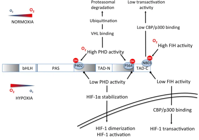
The hypoxia inducible factor-‐1 (HIF-‐1) is a fundamental transcription factor in the molecular physiology of O2 homeostasis. HIF-‐1 is a complex protein composed of 2 subunits, the cytosolic HIF-‐1α (O2-‐sensitive) subunit and the nuclear HIF-‐1β subunit, that dimerize in the nucleus to form the functional HIF-‐1 transcription factor and activate gene transcription. The N-‐terminal portion of HIF-‐1α and HIF-‐1β contains bHLH (basic helix-‐loop-‐helix) and PAS (Per-‐ARNT-‐Sim homology) domains necessary for heterodimerization and DNA binding (Xia et al., 2012). The C-‐terminal portion contains two transactivation domains (TAD), called TAD-‐ N and TAD-‐C separated by an inhibitory domain. Activation of transcription by HIF-‐1 requires various co activators such as the CREB Binding Protein (CBP)/p300 co-‐activator. The O2-‐
sensitive region of HIF-‐1α is called the oxygen degradation domain and is pivotal for the stabilization of this subunit under hypoxic conditions.
In well-‐oxygenated cells, proline hydroxylation of the HIF-‐1α subunit by prolyl-‐hydroxylase enzymes (PHDs) leads to binding of the von Hippel-‐Lindau (VHL) protein, recruitment of an E3-‐ubiquitin protein ligase complex with subsequent ubiquitination and proteasomal degradation. Moreover, the factor inhibiting HIF-‐1 (FIH) negatively regulates HIF-‐1 transactivation by asparagine hydroxylation, which blocks HIF-‐1 binding on its co-‐activator CBP/p300 (Fig. 2).
In sustained hypoxia, there is a general consensus that oxygen deprivation is the main mechanism behind HIF-‐1 activation with an on-‐going debate on a potential role of mitochondrial ROS (Hagen, 2012; Movafagh et al., 2015). In contrast, ROS production by the repetitive desaturation-‐reoxygenation sequences induced by IH appears to play a major role in up-‐regulating HIF-‐1 activity through complex mechanisms involving prolyl-‐hydroxylase inactivation and HIF-‐1 transcription and transactivation (Fig. 3). Briefly, repetitive apneas and intermittent hypoxia trigger nicotinamide adenine dinucleotide phosphate (NADPH) oxidase-‐dependent production of ROS, mostly superoxide anions, which in turn activate phospholipase C (PLC) leading to calcium-‐calmodulin kinase (CamK) and protein kinase C (PKC) activation. PKC stimulates mammalian target of rapamycin (mTOR)-‐dependent synthesis of HIF-‐1α and inhibits PHD-‐dependent HIF-‐1α degradation (Prabhakar & Semenza, 2012). CamK also promotes the interaction between HIF-‐1α and its coactivator p300, thereby leading to increased transcriptional activation upon dimerization with HIF-‐1β. While HIF-‐1α is rapidly degraded (t1/2 < 5 min) upon reoxygenation following sustained hypoxia, HIF-‐1α levels remain elevated following intermittent hypoxia due to the persistent activation of mTOR (Semenza, 2009).
Although in vitro studies established that the pro-‐inflammatory transcription factor nuclear factor-‐κB (NF-‐κB) plays a more important role than HIF-‐1 in the response to IH (Taylor et al., 2014) (Polotsky et al., 2010; Ryan et al., 2005), we and others have observed that chronic in
vivo exposure to IH leads to a strong and sustained activation of HIF-‐1 in the heart (Belaidi et
al., 2008; Belaidi et al., 2009; Belaidi et al., 2016), aorta (Gras et al., 2016), brain (Chou et al., 2013; Z. F. Wang et al., 2009) and liver (da Rosa et al., 2012). Nevertheless, the important role of pro-‐inflammatory pathways is in accordance with these observations since there is a strong crosstalk and synergistic activity between NF-‐κB and HIF-‐1 (Bruning et al., 2012; Gorlach & Bonello, 2008) (Fig. 1). An interesting aspect of HIF-‐1 activation by IH is that it persists when the animals are back in normoxic conditions. This is in agreement with in vitro observations showing that HIF-‐1 activity remains lastingly elevated after termination of IH (Yuan et al., 2008) and can explain the long-‐lasting HIF-‐1 levels and activity observed hours after termination of IH in experimental animals. Although data in OSA patients are scarce, Kazmareck et al. have confirmed that HIF-‐1α and target genes mRNA are increased in skin biopsies from severe OSA patients and concluded that HIF-‐1 expression profile may correlate with disease severity and could possibly be used to predict cardiovascular risk (Kaczmarek et al., 2013).
Among the various HIF-‐1 target gene products, some encode for proteins that have an impact on the cardiovascular system, such as erythropoietin, inducible/endothelial nitric oxide synthases, vascular endothelial growth factor and ET-‐1. We have identified ET-‐1 as a major gene product of interest based on the fact that:
- Through its vasoconstrictive, growth promoting and proinflammatory properties, ET-‐1 has been implicated in several pathophysiological pathways associated with cardiovascular disease (Kaoukis et al., 2013; Ohkita et al., 2012).
- Plasma big ET-‐1 and ET-‐1 are increased in OSA patients (Gjorup et al., 2007; Karkoulias et al., 2010; B. G. Phillips et al., 1999; Saarelainen et al., 1997; Zamarron et al., 2011), in correlation with BP values (Anunciato et al., 2013; B. G. Phillips et al., 1999), and in plasma and tissues of animals exposed to IH (Belaidi et al., 2009; Capone et al., 2012; Kanagy et al., 2001; Peng et al., 2012).
- Endothelin receptors, in particular ET-‐A receptors, are upregulated by IH and the vascular response to ET-‐1 is increased (Allahdadi et al., 2005; Belaidi et al., 2009; Friedman et al., 2014; Lefebvre et al., 2006; Pawar et al., 2009; Rey et al., 2007; Snow et al., 2011; Z. Wang et al., 2013).
- ET-‐1 has the ability to increase NADPH oxidase activity and HIF-‐1 expression in part through its ET-‐A receptors (L. Li et al., 2003; Pisarcik et al., 2013), thus further promoting superoxide production and HIF-‐1 activity in a feed-‐forward mechanism (Fig. 3).
In the following sections, we will present the experimental pharmacological evidence in favor of an inhibition of the ROS-‐HIF-‐1-‐endothelin axis in the prevention of the cardiovascular complications induced by IH.
3.2. Role in systemic hypertension
Numerous experimental studies have confirmed that reduction in oxidative stress level, either by NOX-‐2 deficiency (Schulz et al., 2014) or antioxidant treatments (Hung et al., 2013; Ramond et al., 2013) prevents the development of hypertension in response to intermittent hypoxia exposure. Endothelin receptor antagonists have also been shown to be very effective in preventing or reversing hypertension in rodents exposed to IH (Allahdadi et al., 2008; Belaidi et al., 2009; de Frutos et al., 2010; Kanagy et al., 2001).
3.3. Role in vascular dysfunction and remodeling
In rats exposed to intermittent hypoxia, allopurinol treatment (through its xanthine oxidase-‐ inhibiting property) attenuated the impairment in acetylcholine-‐mediated vasodilation (Dopp et al., 2011). In a comprehensive study in mice, Capone et al. confirmed the role of both oxidative stress and ET-‐1 on the impairment in cerebrovascular endothelial-‐dependent relaxation induced by IH. Hence, cerebrovascular dysfunction was abolished in mice lacking the NOX2 subunit of NADPH oxidase as well as by local perfusion with Mn(III)tetrakis(4-‐ benzoic acid)porphyrin chloride (a free radical scavenger) or with BQ-‐123 (a selective ET-‐A receptor antagonist) (Capone et al., 2012). We have recently demonstrated that oxidative stress was responsible for the IH-‐induced vascular dysfunction in the rat ophthalmic artery, in particular regarding the increased contractile response to ET-‐1 (Mentek et al., 2016). In the case of vascular remodeling, de Frutos et al. demonstrated in mice that IH-‐induced remodeling of mesenteric arteries could be prevented by the mixed endothelin receptor antagonist bosentan (de Frutos et al., 2010). Finally, we have recently established that ET-‐1 was involved in the inflammatory vascular remodeling induced by IH, through a feed-‐ forward mechanism implicating HIF-‐1 and its crosstalk with NF-‐κB (Gras et al., 2016).
3.4. Role in myocardial infarction and stroke
There is experimental data in favor of a major role for oxidative stress and endothelin system activation in the increased sensibility to myocardial infarction induced by chronic IH exposure in rodents. Thus, in mice, the increase in infarct size is associated with enhanced protein carbonylation and lipid peroxidation and decreased thioredoxin content (Park & Suzuki, 2007). In rats, tempol, melatonin as well as atorvastatin treatment during IH
exposure reduce myocardial free radical production and prevent the increase in infarct size (Ramond et al., 2013; Totoson et al., 2013).
The cardiac endothelin system is activated by HIF-‐1 in rats exposed to IH, resulting in increased tissue ET-‐1 levels, upregulation of coronary ET-‐A receptors and enhanced vasoconstriction. The deleterious impact of these effects on the myocardium is evidenced by the ability of bosentan to completely prevent the related increase in infarct size (Belaidi et al., 2009). Similar effects on the brain’s susceptibility to ischemia remain to be demonstrated. However, Capone et al. have observed that alterations of the cerebral circulation induced by chronic IH in mice were prevented by the selective ET-‐A receptor antagonist BQ123 (Capone et al., 2012). In a recent study, we have shown that endoplasmic reticulum (ER) stress and HIF-‐1 were responsible for the infarct size expansion induced by IH (Belaidi et al., 2016).
3.5. Role in myocardial remodeling and dysfunction
In rats, treatment with allopurinol throughout IH exposure significantly improved LV dysfunction (Williams et al., 2010). Mice deficient for gp91phox (the membrane-‐bound catalytic subunit of NOX) did not develop LV remodeling in response to IH (Hayashi et al., 2008). Furthermore, pitavastatin treatment during IH exposure significantly reduced NADPH oxidase-‐related superoxide production as well as LV remodeling in mice (Inamoto et al., 2010).
There is ample evidence that activation of the endothelin system promotes cardiac hypertrophy and HF. Hence, plasma and myocardial ET-‐1 levels (Cavero et al., 1990; Margulies et al., 1990; Yorikane et al., 1993) and ET-‐1 receptors (Sakai et al., 1996b) are increased in animal models of congestive HF and in HF patients (McMurray et al., 1992; Zolk
et al., 1999). Moreover, ET-‐1 receptor antagonists (Fraccarollo et al., 1997; Mishima et al., 2000; Mulder et al., 1997) have been shown to improve LV dysfunction and remodeling and to enhance survival (Mulder et al., 1997; Sakai et al., 1996a) in HF models. Nevertheless, the role of ET-‐1 in the development of cardiac remodeling and HF in response to IH still remains to be investigated.
Table 1 summarizes the various experimental studies associating chronic IH to cardiovascular diseases described in this section as well the beneficial effects of targeting ROS or the endothelin system in this context.
4. Targeting the ROS-‐HIF-‐1-‐ET-‐1 axis as a novel therapeutic approach in OSA
The experimental evidence presented above clearly underscores the therapeutic potential of interventions targeting this axis in the prevention and/or reduction of cardiovascular alterations in moderate to severe OSA patients. While large-‐scale clinical investigations specifically designed to address this issue are still pending, we have identified several potentially interesting pharmacological agents that have already been tested clinically or are in pre-‐clinical development.
4.1. Antioxidant treatment
As mentioned earlier, there is an emerging consensus that OSA is an oxidative stress disorder. In accordance, various studies have shown that reversal of apneas by CPAP therapy can reduce OSA-‐related oxidative stress markers (Eisele et al., 2015; Jullian-‐Desayes et al., 2015). However, other studies report limited effects (Barceló et al., 2006; Del Ben et al., 2012; Jullian-‐Desayes et al., 2015) and a recent sham-‐controlled randomized study specifically designed to test the effect of CPAP treatment on primary markers of lipid
peroxidation failed to observe significant effects (Sivam et al., 2015). These data, plus the fact that a significant proportion of OSA patients are unable or unwilling to use CPAP therapy, represent a strong argument in favor of studies investigating the effects of antioxidants in OSA patients. Still, apart from some reports of beneficial effects of acute intravenous vitamin C injection (N. J. Buchner et al., 2011; Grebe et al., 2006) or of oral allopurinol treatment (El Solh et al., 2006), no large-‐scale clinical studies have been performed. The reason behind this discrepancy could be that while antioxidants have proven effective in preventing or reverting oxidative stress-‐related cardiovascular alterations in experimental models, they have generally yielded disappointing results when translated to human clinical practice, as recently reviewed in this journal (Farías et al., 2016).
Nevertheless, the fact that part of the effectiveness of medications such as statins, angiotensin-‐converting enzyme (ACE) inhibitors or ARBs is due to their pleiotropic antioxidant and anti-‐inflammatory properties (Siti et al., 2015) supports the use of these agents in the prevention or treatment of the cardiovascular complications associated with OSA (Pepin et al., 2010; Thunstrom et al., 2016). This is strengthened by the fact that these drugs interfere with NADPH oxidase activation and expression, a therapeutic strategy potentially more efficient than non-‐selective ROS scavenging (Schramm et al., 2012), particularly in the context of IH and OSA.
Hence, we have shown that the IH-‐induced vascular remodeling and increase in blood pressure and infarct size were prevented by atorvastatin along with a decrease in NADPH oxidase upregulation and a preservation of superoxide dismutase activity (Totoson et al., 2013). In OSA patients, 3 months of atorvastatin treatment lowered blood pressure and improved lipid profile (Joyeux-‐Faure et al., 2014). Also, Increased internalization of
endothelial CD59 in IH appears to be cholesterol-‐dependent and is reversed by statins in a CD59-‐dependent manner (Emin et al., 2016).
In accordance, we have shown that valsartan induced a fourfold greater decrease in mean 24-‐hour BP compared to CPAP in a small cohort of untreated hypertensive OSA patients (Pepin et al., 2010). In view of the antioxidant, anti-‐inflammatory and anti-‐hypertensive actions of statins, ACE inhibitors and ARBs, their beneficial effects in OSA patients should definitely be confirmed in large-‐scale clinical trials.
Another interesting therapeutic candidate appears to be melatonin, a circadian rhythm regulating hormone with potent pleiotropic antioxidant properties (Tan et al., 2015) and whose secretion profile is altered in OSA patients (Zirlik et al., 2013). We were among the first to report the potent cardioprotective effects of melatonin against ischemia-‐reperfusion injuries (ventricular arrhythmias and infarct size) (Lagneux et al., 2000). More recently, others and we have shown that melatonin administration throughout IH exposure was very effective in preventing the development of myocardial susceptibility to infarction, hypertension and endothelial dysfunction (Hung et al., 2013; Ramond et al., 2013; Yeung et al., 2015). These effects were also associated with a reduction in IH-‐induced NADPH oxidase expression and with an increase in antioxidant enzyme activity (Hung et al., 2013). Of particular relevance to the topic of present review, melatonin also has the ability to inhibit HIF-‐1 activity and target gene expression, possibly through its antioxidant actions (Vriend & Reiter, 2016).
Finally, the novel therapeutic concept of replacing antioxidant supplementation by activation of endogenous antioxidants was recently addressed in this journal (J. Y. Chan & Chan, 2015). This approach is particularly interesting in the context of OSA where antioxidant defenses are lowered. Moreover, this could be achieved by non-‐pharmacological
means such as exercise. In a recent study, moderate exercise was shown to increase endogenous antioxidant levels in healthy as well as in diabetic patients and to reduce oxidative DNA damage (Pittaluga et al., 2015). In that respect, we have recently shown that high-‐intensity training during IH exposure reduces pro-‐apoptotic ER stress and prevents the development of hypertension and the increase in infarct size (Bourdier et al., 2016).
4.2. Endothelin system antagonism
Although the major role of endothelin and its receptors in the development of systemic and pulmonary hypertension, coronary atherosclerosis, myocardial infarction and heart failure is well documented (Ohkita et al., 2012), endothelin receptor antagonists are currently mainly used for the treatment of pulmonary hypertension where they have proven effective in decreasing pulmonary vascular resistance and increasing cardiac index (Channick et al., 2001). Yet, mixed receptor antagonists like bosentan (Krum et al., 1998), or selective ET-‐A receptor antagonists like darusentan (Black et al., 2007; Nakov et al., 2002), have been shown to significantly decrease blood pressure in patients with essential or resistant arterial hypertension. Bosentan improved peripheral (Rafnsson et al., 2012) and the ET-‐A receptor antagonist atrasentan improved coronary (Reriani et al., 2010) endothelial function in patients with type-‐2 diabetes and early atherosclerosis, respectively. Finally, in a study in patients with heart failure resistant to standard therapy, bosentan treatment increased cardiac output and decreased systemic and pulmonary vascular resistance (Sutsch et al., 1998) although subsequent clinical trials with mixed and ET-‐A antagonists have failed to show clear clinical benefits (Kaoukis et al., 2013). Moreover, the safety profile of these drugs has been disappointing due to a high incidence of hepatotoxicity, headache and edema. Nevertheless, a new generation of endothelin system antagonists is emerging such as ET-‐
converting enzyme inhibitors, mixed ECE and neutral endopeptidase inhibitors, ET-‐B receptor agonists or new potent non-‐toxic mixed antagonists, such as macitentan (Maguire & Davenport, 2014). These promising agents should definitely be tested in experimental IH models in order to compare their efficacy to that of bosentan or ET-‐A receptor antagonists. Some authors argue that selective ET-‐A receptor antagonists are better suited than mixed antagonists for the prevention or treatment of cardiovascular diseases (Nasser & El-‐Mas, 2014) but this remains to be confirmed by specifically-‐designed clinical studies, particularly in the context of OSA since the deleterious effects of IH are mainly due to ET-‐A receptor upregulation (Allahdadi et al., 2008; Belaidi et al., 2009; Briancon-‐Marjollet et al., 2015; Capone et al., 2012; de Frutos et al., 2010; Mentek et al., 2016).
In a small randomized controlled pilot study in OSA patients (n=16) with mild untreated hypertension (clinical BD of 142/85 mmHg), we recently showed that a 4-‐week bosentan treatment resulted in a 3 mmHg greater decrease in 24-‐h DBP than CPAP, but this effect was minimal and not significant compared to baseline (Joyeux-‐Faure et al., 2015). This could be due to the fact that these patients were not severe enough in terms of hypertension and sleep apnea level. Hence, in view of the clear involvement of the ET-‐1 system in the severe IH-‐induced cardiovascular alterations and of the dramatic effects of ET-‐1 receptor antagonists in this context (Belaidi et al., 2009; Briancon-‐Marjollet et al., 2015; Gras et al., 2016), large-‐scale clinical investigations of ET-‐1 system antagonism in moderate to severe OSA patients are undoubtedly warranted.
4.3. HIF-‐1 inhibition
Since both ROS and ET-‐1 have the ability to promote HIF-‐1 activity in a feed-‐forward manner (Fig. 3), HIF-‐1 appears to be a pivotal target to reverse the deleterious cardiovascular
consequences of chronic IH and OSA. Various novel molecular and pharmacological interventions to reduce HIF-‐1 expression or activity have reached the phase of clinical translation, mostly in the field of cancer research (Fig. 4). These could potentially be investigated in OSA patients after prior pre-‐clinical studies evaluating the most appropriate therapeutic approaches to prevent excessive HIF-‐1 activation without suppressing its essential physiological actions.
4.3.1. Genetic strategies to decrease HIF-‐1α protein levels
MicroRNAs (miRNAs) are a class of single-‐stranded non-‐coding RNAs that negatively regulate eukaryotic gene expression at the post-‐transcriptional and translational levels. Several miRNAs, named hypoxamiRs, are upregulated by hypoxia through various transcription factors including HIF-‐1 and NF-‐κB (Cottrill et al., 2014; Nallamshetty et al., 2013). Amongst them, miR-‐210 is a ubiquitous HIF-‐1 target gene that has been identified has a major prognostic and therapeutic target in a number of hypoxia-‐related diseases such as tumor progression, myocardial infarction and pulmonary hypertension (Cottrill et al., 2014).
Reciprocally, miR-‐210 and other hypoxamiRs engage in regulatory feedback loops with HIF-‐1. Hence, miR-‐210 and miR-‐424 have the ability to promote HIF-‐1 stabilization through suppression of glycerol-‐3-‐phosphate dehydrogenase 1-‐like enzyme and cullin 2, respectively (Nallamshetty et al., 2013). Moreover, miR-‐210 appears to be involved in the ROS-‐mediated HIF-‐1 stabilization (Movafagh et al., 2015; Nallamshetty et al., 2013).
On the other hand, targeting of the 3’ untranslated region (3’ UTR) of Hif1a by miR-‐155 (Bruning et al., 2011), miR-‐519 (Cha et al., 2010), miR-‐20b (Lei et al., 2009) or the miR-‐17/92 cluster (Taguchi et al., 2008) decreases HIF-‐1α protein levels.
Thus delivery of miRNA antagonists (miR-‐210, miR-‐424) or mimics (miR-‐155, miR-‐519, miR-‐ 20b, miR17/92) represents a promising new approach to reduce HIF-‐1 activity that should be
tested in pre-‐clinical IH models. Translation to clinical studies is possible in view of the rapid development of miRNA-‐based therapy and of its apparent lack of adverse effects (Bader et al., 2011).
4.3.2. Chemical inhibitors
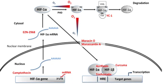
Several anti-‐cancer agents acting at different levels of HIF-‐1 signaling have been tested in clinical trials, or are being investigated in pre-‐clinical studies. These molecules inhibit HIF-‐1 activity by interfering with HIF-‐1α mRNA expression, translation, dimerisation with HIF-‐1β and DNA binding or by promoting HIF-‐1α degradation (Xia et al., 2012). Below is presented a selection of small molecular HIF-‐1 inhibitors along with some experimental data in favor of their potential use in the prevention or treatment of IH-‐ and OSA-‐induced cardiovascular alterations.
Camptothecin, a natural inhibitor of topoisomerase I, prevents HIF-‐1α transcription and has proven beneficial in phase I and II clinical studies (Rapisarda et al., 2004). Interestingly, camptothecin also has the ability to decrease HIF-‐1 expression and activity by up-‐regulating miR-‐155 expression (Bertozzi et al., 2014).
Antisense oligodeoxynucleotide HIF-‐1α mRNA inhibitors, such as EZN-‐2968, have been shown to reduce the expression of its target genes in cell and animal models (Greenberger et al., 2008) as well as in cancer patients (Jeong et al., 2014).
YC-‐1, 3-‐(5-‐hydroxymethyl-‐2-‐furyl)-‐1-‐benzyl indazole, first described as a platelet guanylate cyclase activator (Ko et al., 1994), is an approved cancer drug that prevents HIF-‐1α from interacting with the transcriptional co-‐activators p300/CBP by activating FIH (S. H. Li et al., 2008). In cardiomyocytes, HIF-‐1 inhibition by YC-‐1 effectively represses VEGF induction (Hui et al., 2012).
Acriflavine, a cutaneous antibacterial and antiviral agent, prevents HIF-‐1 dimerisation by binding to the PAS subdomain of HIF-‐1α, thereby inhibiting HIF-‐1 DNA-‐binding and transcriptional activity (Lee et al., 2009). Moreover, acriflavine prevents HIF-‐1 activity in C2C12 mouse myocytes exposed to IH (Cai et al., 2013).
Echinomycin is an antibiotic that can bind DNA in a sequence-‐specific fashion and that interferes with HIF-‐1-‐induced gene transcription by binding to its hypoxia response elements (D. Kong et al., 2005).
Finally, the antifungal antibiotic trichostatin, a class I and II histone deacetylase inhibitor, promotes the proteasomal degradation of HIF-‐1α (Geng et al., 2012; X. Kong et al., 2006). It was recently shown to repress HIF-‐1-‐VEGF signaling and to inhibit choroidal angiogenesis in
vivo (N. Chan et al., 2015).
4.3.3. Natural inhibitors
Various natural substances possess HIF-‐1 inhibiting activities. Moracin O, from Morus
Alba also known as white mulberry, inhibits HIF-‐1 translocation and activity due to its
particular structure containing a 2-‐arylbenzofuran ring (Xia et al., 2011). Curcumin, a yellow spice from Curcuma longa, is known to repress HIF-‐1 activity via its action on HIF-‐1β (Choi et al., 2006). And finally, Manassantin A, from the water plant Saururuschinensis, prevents HIF-‐1α nuclear translocation (Hossain et al., 2005).
5. Conclusion
Although continuous positive airway pressure treatment, the first line therapy for OSA, clearly improves quality of life and sleepiness, positive response to CPAP in terms of cardiovascular and metabolic outcome is hampered by poor patient adherence, suboptimal use or limited efficacy. Novel therapeutic strategies are thus required to help counteract the
key mechanisms responsible for OSA-‐related cardiovascular consequences. These therapeutic agents could be used in OSA patients unable or unwilling to use CPAP or in combination with CPAP in order to increase its efficacy.
This review supports a role for the ROS-‐HIF-‐1-‐endothelin axis in the deleterious cardiovascular consequences of the chronic intermittent hypoxia associated with moderate to severe OSA. It also stresses the importance to extend fundamental and clinical research around the pivotal role of the HIF-‐1 transcription factor in this context.
Conflicts of interest
None.
6. References
Allahdadi, K. J., Cherng, T. W., Pai, H., Silva, A. Q., Walker, B. R., Nelin, L. D., et al. (2008). Endothelin type A receptor antagonist normalizes blood pressure in rats exposed to eucapnic intermittent hypoxia. Am J Physiol Heart Circ Physiol 295, H434-‐440.
Allahdadi, K. J., Walker, B. R., & Kanagy, N. L. (2005). Augmented endothelin vasoconstriction in intermittent hypoxia-‐induced hypertension. Hypertension 45, 705-‐709.
Andreadou, I., Iliodromitis, E. K., Rassaf, T., Schulz, R., Papapetropoulos, A., & Ferdinandy, P. (2015). The role of gasotransmitters NO, H2S and CO in myocardial ischaemia/reperfusion injury and cardioprotection by preconditioning, postconditioning and remote conditioning. Br J Pharmacol 172, 1587-‐1606.
Anunciato, I. F., Lobo, R. R., Coelho, E. B., Verri, W. A., Jr., Eckeli, A. L., Evora, P. R., et al. (2013). Big endothelin-‐1 and nitric oxide in hypertensive elderly patients with and without obstructive sleep apnea-‐hypopnea syndrome. Arq Bras Cardiol 101, 344-‐351. Arnaud, C., Beguin, P. C., Lantuejoul, S., Pepin, J. L., Guillermet, C., Pelli, G., et al. (2011a).
The inflammatory preatherosclerotic remodeling induced by intermittent hypoxia is attenuated by RANTES/CCL5 inhibition. Am J Respir Crit Care Med 184, 724-‐731. Arnaud, C., Poulain, L., Levy, P., & Dematteis, M. (2011b). Inflammation contributes to the
atherogenic role of intermittent hypoxia in apolipoprotein-‐E knock out mice.
Atherosclerosis 219, 425-‐431.
Arzt, M., Hetzenecker, A., Steiner, S., & Buchner, S. (2015). Sleep-‐Disordered Breathing and Coronary Artery Disease. Can J Cardiol 31, 909-‐917.
Arzt, M., Young, T., Finn, L., Skatrud, J. B., & Bradley, T. D. (2005). Association of sleep-‐ disordered breathing and the occurrence of stroke. Am J Respir Crit Care Med 172, 1447-‐1451.