Antibacterial Activity and Anti-Biofilm Formation of Henna (Lawsonia inermis)
Extracts against Pseudomonas aeruginosa
Article · May 2017 CITATIONS 0 READS 212 3 authors, including:
Some of the authors of this publication are also working on these related projects: Lutte contre la formation du biofilm à Pseudomonas aeruginosaView project Aicha Lattab
Université Abdelhamid Ibn Badis Mostaganem
13 PUBLICATIONS 0 CITATIONS
SEE PROFILE
Abed Arabi
Université Abdelhamid Ibn Badis Mostaganem
16 PUBLICATIONS 2 CITATIONS
and Biological Sciences www.textroad.com
Antibacterial Activity and Anti-Biofilm Formation of Henna (Lawsonia
inermis) Extracts against Pseudomonas aeruginosa
LATTAB Aicha*, DJIBAOUI Rachid and ARABI Abed
Laboratory of Microbiology and Plant Biology, Faculty of Natural Sciences and Life, University of Mostaganem, B.P. 227, 27000 Mostaganem, Algeria.
Received: February 9, 2017 Accepted: March 15, 2017 ABSTRACT
Pseudomonas aeruginosa is an opportunistic bacterium causing severs nosocomial infections. It is
characterized by its ability to form attached biofilm to protect itself from various external pressures. These adherent cells of P. aeruginosa make treatment of their infections complicated and demand new therapies. The present work was focused on the study of L. inermis extracts effects on planktonic cells and on biofilm formation by seven strains of P. aeruginosa. The henna plant was harvested from two different regions in Algeria: Adrar and Biskra. Two solvents were used to extract L. inermis flavonoids compounds. The ethyl acetate and butanolic fractions were prepared from a hydro-methanolic extract (70%). The results show that all studied extracts present an antibacterial properties against all the isolates tested. It was also indicated that L.
inermis of Adrar had the highest anti- P. aeruginosa activity compared with henna of Biskra. However the
butanolic fractions of both L. inermis samples have the highest effect with a MIC of 3.12mg/ml and 6.25mg/ml for samples of Adrar and Biskra respectively, compared with the fractions of ethyl acetate which showed a MIC of 6.25mg/ml and 12.5mg/ml for the same samples. Also, the biofilm formation was decreased when incubated with the ½ MIC of butanolic fraction for both L. inermis tested. Whereas the ½ MIC of ethyl acetate extracts did not affect biofilm formation in all isolates of P. aeruginosa. The quantitative estimation showed that ethyl acetate extracts for both varieties of L. inermis are rich in polyphenols (224,83-136,95 mg GAE/g) and flavonoids (20,15-24,92 mg QE/g) compared to butanolic extracts. Finally we can conclude that extraction by butanol makes it possible to better concentrate the antibacterial active ingredients present in the hydro-methanolic extract of L. inermis leaves.
KEYWORDS: Pseudomonas aeruginosa, Lawsonia inermis, Antibacterial activity, MIC, MBC, Biofilm. INTRODUCTION
Pseudomonas aeruginosa is a ubiquitous Gram-negative bacteria found in many environments. It is one
of the major opportunistic pathogens implicated in serious nosocomial infections. It is a versatile microorganism able to tolerate unfavorable conditions. These proprieties allow it to attach itself and survive on many biotic and abiotic surfaces, which favors the beginning of infections especially in immunocompromised patients [1]. Treatment of the infections due to P. aeruginosa are difficult because of their elevated intrinsic resistance against most antibiotics and their capacity to acquire new mechanisms of resistance following the exposure to antibiotic during treatments [2] and are associated with high mortality rates, ranging from 18% to 61% [3].
In addition to its resistance to antibiotics, P. aeruginosa is characterized by its ability to form biofilms. Bacterial biofilms are surface-attached communities of microorganisms embedded in a matrix of complex polysaccharides [4]. Bacteria grown in a biofilm have increased resistance to antibiotics and to the host immune system compared to the non-surface attached (planktonic) bacteria.
Bacterial behavior within biofilms is regulated by the quorum sensing (QS) system [5]. It’s a key regulator of biofilm development [6] resistance to antibiotics [7] and expression of virulence factors [8]. The main components of quorum sensing system are the quorum sensing signal synthesis, the signal molecule and the signal receptor [9].
For many years, it is well known that plants have been used in traditional herbal medicine to prevent or cure infectious diseases. However, in the recent years, many research studies have reported the quorum
sensing-blocking properties of plant extracts by their similarity in chemical structure to quorum sensing signals and also their ability to degrade signal receptors [10].
Lawsonia inermis is commonly called as henna. It is the sole species of the genus Lawsonia, belonging to
the family Lythraceae [11]. It is known by various names in different languages; in Arabic (alhenna), in French (henné) and in English (henna) [12, 13]. It is grown in various tropical regions in Northern Africa, Asia and Australia. L. inermis has long been used for many medicinal purposes including as an astringent, an antifungal, an antihemoragic agent and for its cardio-inhibitory, hypotensive, and anti-bacterial herb for the skin and hair [14]. Phytochemical investigations of L. inermis leaves have shown the presence of flavonoids, coumarins, gallic acid, triterpenoids, naphthalene derivatives, Lawsone, phenolic glycosides and xanthones [15, 16]. Out of these ingredients, the main compound is Lawsone (2-hydroxynapthoquinone). About 0.5-1.5% of leaves of henna made of Lawsone [17].
The purpose of this study was to examine the effect of ethyl acetate and butanolic fractions of L.
inermis on biofilm formation by Pseudomonas aeruginosa ATCC 27853 and six clinical isolates of the same
species. The plant leaves were harvested from Adrar and Biskra regions in Algeria. MATERIALS AND METHODS
Bacterial strains:
P. aeruginosa ATCC 27853 and six clinical isolates of P. aeruginosa obtained from different skin infections
in diabetic patients in a health center of Sidi Lakhdar Mostaganem (Algeria). The bacterial colonies morphology were examined and several tests such as Gram stain, catalase, oxidase, pyocyanin and pyoverdin production, the growth at 42°C and at 4°C were used to identify the bacterial isolates. Each isolate was also identified by an API 20NE system.
Genotypic identification
PCR amplification was carried out in Biorad iCycler thermocycler (Biorad, USA) using the universal primers 1492R (5' GGTTACCTTGTTACGACTT 3') and 27F (5' AGAGTTTGATCCTGGCTCAG 3'). 4μl of a genomic DNA extracted using a rapid method based on thermal shock, are amplified in a 25μl of The
reaction mixture consisted of 1 U Taq polymerase, 0,2mM dNTP, 1X PCR reaction buffer, 1,5mM MgCl2
and 1,25 µl of each of the primers at 10µM. The amplified products were analyzed by agarose gels electrophoresis. 10μl of PCR products were stained with ethidium bromide followed by a migration at a constant voltage. The DNA was visualized and pictures were taken under UV.
Amplified products of three isolates (P2, P3 and P6) obtained were sequenced to validate their identities. Primer 1492R (5' GGTTACCTTGTTACGACTT 3') was used for amplification and sequencing. An additional primer 27F (5’ACTCCTACGGGAGGCAGCA 3’) was used for sequencing.
The sequences obtained were analyzed using GenBank's BLAST (https://blast.ncbi.nlm.nih.gov/Blast.cgi Blast) tool (National Centre for Biotechnology Information).
Antibiotic susceptibility testing
The test was performed on Mueller- Hinton (MH) agar using the standard disc-diffusion method according to the recommendations 2015 of the French Society of Microbiology Antibiogram committee (FSM-AC) (www.sfm.asso.fr). A total of 15 antibiotics were assessed: Penicillin G (P, 10 U), Piperacillin (PRL, 100µg), Ticarcillin (TI, 75 µg), Ticarcillin-clavulanate (TIM, 75/10 µg), Imipenem (IMP, 10mg), Ceftazidime (CAZ, 10 µg), Cefepime (FEP, 30 µg), Gentamycin (CN, 15µg), Amikacin (AK, 30µg), Netilmicin (NET, 10), Tobramycin (TOB, 10µg), Ciprofloxacin (CIP, 5 µg), Levofloxacin (LE, 5 µg), Colistin (COL, 10), Fosfomycin (FF, 50µg). Inhibition zones around the disc were determined after incubation for 24h. Susceptibility results for each strain were reported according to the criteria established by the FSM-AC -2015.
Plant material
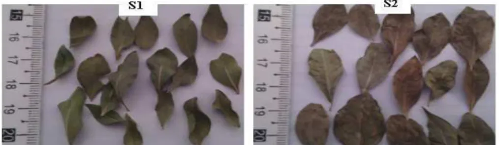
Lawsonia inermis samples used in this study were harvested from two different regions of Algeria: Adrar
and Biskra. The leaves (Figure 1) were dried under shade and powdered with a mechanical grinder and stored away from light at room temperature.
Figure 1. L. inermis samples used in this study, S1: leaves from Adrar region and S2: leaves from Bescra region.
Plant extracts Preparation: Hydro-methanolic extracts
The extraction was done by maceration of 25 g of fine powder in 500 ml of methanol-water (70/30, v/v). The mixture was agitated for 48h at room temperature (27±2 °C) in enclosed flasks [18]. After filtration through whatman N°1 filter paper, the MeOH of resultant hydroalcoholic extracts was evaporated at 40°C under reduced pressure in a rotary evaporator (Modal: Buchi Switzer land, Type: 201) and affording the aqueous extracts.
Ethyl acetate and butanolic fractions
The crude extract was then fractionated by solvent – solvent extraction, the aqueous solution was firstly extracted ethyl acetate, then with n-butanol. 2 fractions of extracts, namely ethyl acetate and butanolic were obtained. The all fractions were concentrated and dried to a constant weight in a vacuum oven at 45°C. The extracts were stored at 4°C [19].
Total phenolic content determination:
The total phenolic content in ethyl acetate and butanolic extracts was determined by colorimetric assay using Folin-Ciocalteu reagent assay. A volume of 0,2 mL of standard solution (Gallic acid with different concentrations) or the extracts was mixed with 1,5 mL of Folin-Ciocalteu reagent diluted 10 times in water. Afterwards, the obtained solution was added to 1,5 mL of 7,5% aqueous sodium carbonate. The absorbance was then measured at 750 nm against a blank after incubation for 90 min in obscurity at room temperature. [20].
Gallic acid was used as a standard for the calibration curve (1-200µg/mL). The Total phenolic content was expressed as milligram Gallic acid equivalents per gram extract (mg GAE/g extract).
Flavonoids content determination:
The total flavonoids content were determined spectrophotometrically using a method described by Pothitirat
et al. [21]. In brief 1,5 mL of diluted extract or standard solution was allowed to 1,5 mL of 2% AlCl3. After incubation for 30 min at ambient temperature, the absorbance of the mixture was measured at 430 nm. Quercetin was used to prepare standard solution (1-400 µg/mL). The flavonoids content were expressed as milligram quercetin equivalents per gram extract (mg QE/g extract).
Antibacterial Activity:
The antibacterial activity of L. inermis extracts was evaluated by using disc diffusion method on Mueller-Hinton agar plates [22, 23]. Overnight grown culture of microorganisms at 37°C for 24h was used for inoculums preparation. A colony was inoculated in 5ml of tryptic soy broth (TSB) at 37°C for 3h. The turbidity of resulting suspension was measured in order to adjust the cellular concentration to 106 CFU/mL. 100µL of the standardized bacterial suspensions of each isolate were inoculated on Mueller-Hinton agar plates. 5min later, filter paper discs (6 mm in diameter) saturated with plant extract (10 μl at 200mg/ml) were placed on the surface of the plates. After pre-incubation for 20min to allow for proper diffusion of the extracts into the media, plates were incubated at 37 °C for 24h. The activity was determined after 24hr of incubation by the measurement of the inhibitory zone diameter.
Determination of MIC and MBC:
The minimal inhibitory concentration (MIC) of the ethyl acetate and butanolic fractions of two samples of L.
inermis were determined by a broth microdilution method by using 96-well microplates. The optical density
The lyophilized ethyl acetate and butanolic extracts stock solution were prepared by dissolution in 10% dimethyl sulfoxide (DMSO). 100μl of the dilution extracts were introduced in each well we to be tested.
Subsequently, 100μL of an inoculum (106 CFU/mL) was added to each well. The plates were incubated at
37°C for 24hr and the MIC was calculated as the lowest concentration of extract that inhibited visible growth of the organism [24]. The MBC was determined by spreading out 0.l ml of the wells contents of concentration ≥ MIC on Mueller-Hinton agar. The MBC is the smallest concentration which permits the survival of 0.01% or more of the bacterial suspension after 24 h incubation.
Effect of plant extracts on P. aeruginosa biofilm formation:
Effect of ethyl acetate and butanolic fractions of the two different samples of L. inermis leaves on biofilm formation was tested by Tissue Culture Plate Method and Tube Method. The first was effectuated on polystyrene 96-well microplates as described by Adonizio et al. [25] and the second was made in glass tubes. Cultures were grown overnight in 5 ml of LB, the optical density at 600 nm of bacterial suspension was measured in order to adjust the cellular concentration to 106 CFU/mL. 100 μl was dispensed into each well of 96 well polystyrene flat-bottomed microtiter plates in the presence of 100 μl of TSB with plants extracts (ranging from final concentration MIC to 1/8MIC). 100 μl of bacterial culture added to the same volume of medium without plant extract were used as control. In the Tube Method, 1ml was inoculated in tubes containing 1ml of TSB (as a control), and TSB with the appropriate concentration of plants extract (ranging from final concentration of MIC to 1/8 MIC). Biofilms were allowed to develop under static conditions at 37°C for 24h. The quantitative analysis of biofilm formation was performed using crystal violet staining (1% w/v) of the attached cells, the supernatant in the tubes and microplates were aspirated and rinsed 3 times with physiological water and fixed by drying at room temperature until they were fully dried out. The total of 200µl and 2 ml of 1% crystal violet was added into each well and tube respectively for 15 min. The excess stain was washed off 3 times with distilled water. The crystal violet that stained the attached cells in glass tubes was distained with 2 ml of 95% ethanol at room temperature for 2 min. The optical density (OD at 580nm) of 2 ml distained solution was examined using a spectrophotometer [26]. The absorbance value is positively correlated to the amount of the bacterial adhesion or biofilm. All tests were performed in three triplicate and the absorbance readings were averaged.
Statistical analysis
The experimental results were expressed as mean ± standard deviation of three replicates. The data were evaluated by analysis of variance using Statbox 6. The comparison of means was performed using the least significant difference were considered statistically significant if P < 0.05.
RESULTS AND DISCUSSION Isolation and identification
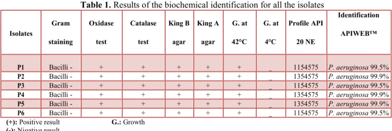
The six clinical isolates obtained from different skin infections were belonged to P. aeruginosa. All of them produce pyocyanin and pyoverdin on King A and King B agar medium respectively. They present a positive oxidase and catalase reactions. They grow at 42°C but not at 4°C. The results of API 20NE system show a percentage of similarity of 99.5% and 99.9% to P. aeruginosa (Table 1).
Table 1. Results of the biochemical identification for all the isolates
Gram Oxidase Catalase King B King A G. at G. at Profile API
Identification
Isolates APIWEB™
staining test test agar agar 42°C 4°C 20 NE
P1 Bacilli - + + + + + _ 1154575 P. aeruginosa 99.5% P2 Bacilli - + + + + + _ 1354575 P. aeruginosa 99.9% P3 Bacilli - + + + + + _ 1154575 P. aeruginosa 99.5% P4 Bacilli - + + + + + _ 1354575 P. aeruginosa 99.9% P5 Bacilli - + + + + + _ 1354575 P. aeruginosa 99.9% P6 Bacilli - + + + + + _ 1154575 P. aeruginosa 99.5%
(+): Positive result G.: Growth
The PCR profile (Figure 2) present similar bands size around 1500 bp showing that all of the isolates belong to the species of Pseudomonas.
Figure 2. Agarose gel electrophoresis of PCR amplified 16S-rRNA gene for the clinical isolates (lanes1-6). Lane M: DNA size marker.
The 16S rRNA sequencing analysis of the isolates P2, P3 and P6 confirmed as P. aeruginosa (P2: 99% P.
aeruginosa CJM, P3: P. aeruginosa NO5 and P6: 99% P. aeruginosa strain F23).
Antibiotic sensitivity
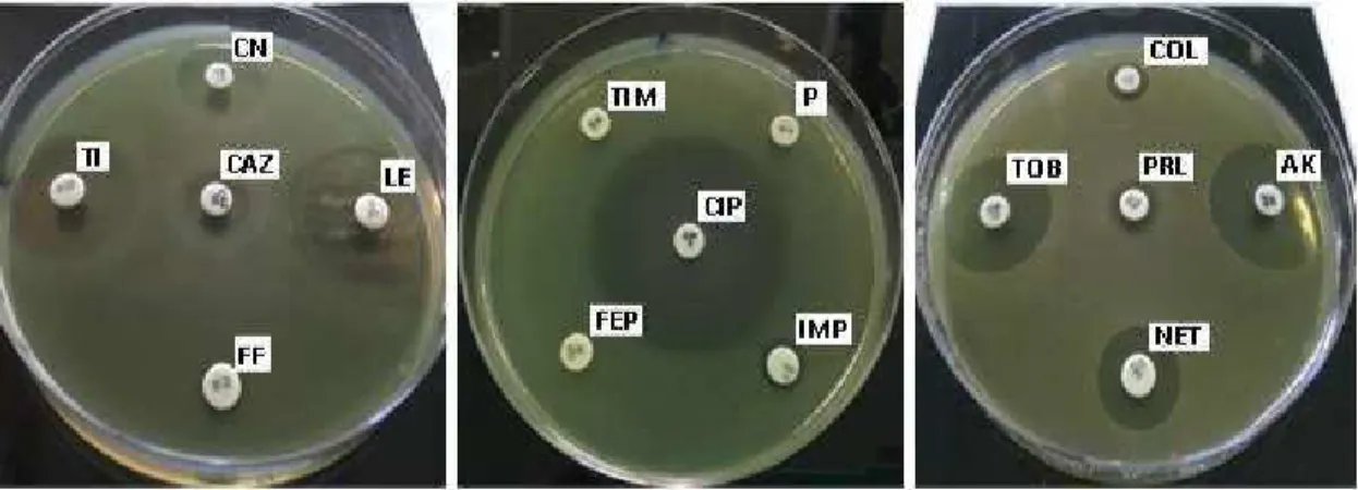
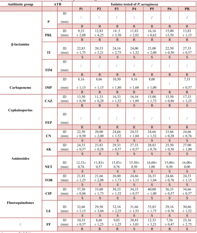
Susceptibility of P. aeruginosa towards studied antibiotics has generated the results shown in Table 2 and Figure 3. All P. aeruginosa isolates showed high resistance towards β-lactams (Penicillin G, Piperacillin, Ticarcillin-clavulanate), Carbapenems (Imipenem), Cephalosporins (Cefepime). We can argue that the decreasing intracellular β -lactams concentration by MBLs producers and increased the expression of efflux pumps as MexAB, MexCD and MexXY [27] are the most prevalent β-lactams resistance mechanisms in our isolates. An increase in P. aeruginosa strains resistant to carbapenems has been observed, which has generated a health problem in several countries (Africa, Europe, America) [28, 29]. The studies presented by Kumar et al. [30] and Sara et al. [31] reported that the high prevalence of P. aeruginosa strains resistant to MBL producers and carbapenems was due to the excessive use of carbapenems.
On the other hand, all the strains showed sensitivity towards Fluoroquinolones (Ciprofloxacin and Levofloxacin) and Aminosides groups tested antibiotic (Gentamycin, Amikacin, Netilmicin, Tobramycin). These results were comparable with results obtained by Tripathi et al. [32], the authors found that the isolates of P. aeruginosa were sensitive to variety of Aminosides antibiotic. The isolates showed variable susceptibility toward the Ceftazidime and Fosfomycin.
Figure 3. Sensitivity of P. aeruginosa towards antibiotics studied: Penicillin G (P), Piperacillin (PRL), Ticarcillin (TI), Ticarcillin-clavulanate (TIM), Imipenem (IMP), Ceftazidime (CAZ), Cefepime (FEP),
Gentamycin (CN), Amikacin (AK), Netilmicin (NET), Tobramycin (TOB,), Ciprofloxacin (CIP), Levofloxacin (LE), Colistin (COL), Fosfomycin (FF).
Table 2. Susceptibility of P. aeruginosa isolates towards antibiotics studied
Antibiotic group ATB Isolates tested of P. aeruginosa
P1 P2 P3 P4 P5 P6 PR ID / / / / / / / P (mm) R R R R R R R ID 9,33 12,83 14 ,5 11,83 16,16 15,00 13,83 PRL (mm) ± 2,08 ± 4,25 ± 3,50 ± 2,02 ± 0,62 ±3,50 ± 1,15 β-lactamins R R R R R R R ID 22,83 20,33 24,16 24,00 23,00 22,50 27,33 TI (mm) ± 1,75 ± 3,21 ± 2,75 ± 1,32 ± 2,00 ± 0,50 ± 0,57 S S S S S S S ID / / / / / / / TIM (mm) R R R R R R R ID 8,16 8,66 10,50 9,16 8,00 / 7,33 Carbapenems IMP (mm) ± 1,15 ± 1,15 ± 1,80 ± 1,60 ± 1,00 ± 0,57 R R R R R R R ID 13,50 11,33 16,33 16,16 15,00 15,50 17,33 CAZ (mm) ± 0,50 ± 0,28 ± 1,52 ± 1,89 ± 1,73 ± 0,86 ± 1,25 Cephalosporins R R S S R R S ID / / / / / / / FEP (mm) R R R R R R R ID 22,50 20,00 24,66 24,33 24,66 23,66 24,66 CN (mm) ± 0,50 ± 2,00 ± 1,52 ± 1,04 ± 1,52 ± 0,28 ± 0,76 S S S S S S S ID 24,33 23,83 29,33 27,33 28,83 25,50 27,00 AK (mm) ± 0,57 ± 0,28 ± 0,57 ± 0,57 ± 0,76 ± 0,50 ± 1,00 Aminosides S S S S S S S ID 12,33± 11,83± 15,83± 15,50± 16,00± 15,00± 16,00± NET (mm) 0,76 0,57 0,76 0,50 1,00 0,50 0,00 S S S S S S ID 21,83 21,66 26,00 24,66 26,33 24,66 24,33 TOB (mm) ± 1,25 ± 2,08 ± 1,73 ± 1,15 ± 1,04 ± 0,76 ± 1,15 S S S S S S S ID 37,50 33,00 38,33 34,33 40,00 36,33 34,66 CIP (mm) ± 0,86 ± 1,73 ± 1,52 ± 0,57 ± 1,00 ± 0,57 ± 0,57 Fluoroquinolones S S S S S S S ID 32,66 29,50 32,16 31,66 35,83 29,16 30,66 LE (mm) ± 1,15 ± 3,04 ± 2,25 ± 1,53 ± 1,75 ± 0,76 ± 1,52 S S S S S S S ID 10,33 8,66 9,83 20,83 12,33 7,50 23,16 / FF (mm) ± 0,57 ± 1,25 ± 1,25 ± 3,01 ± 3,21 ± 0,47 ± 2,75 R R R S R R S /: no zone of inhibition Extraction yields
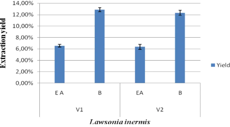
Ethyl acetate and butanolic extracts from leaves of L. inermis obtained from two different regions are shown in Figure 4. According to the results obtained, a low difference was noticed between the tested varieties of L.
inermis. On the other hand, comparison of flavonoids extraction yield of solvents showed that butanol
fraction extract was found: 12,87% and 12,31% for samples of Adrar and Biskra respectively and found less important (6,56% and 6,36%) for ethyl acetate fractions.
Figure 4. Extraction yield of ethyl acetate and butanolic fractions of two different varieties of L. inermis. Total phenols and flavonoids contents:
The table 3 summarizes the results of the phenols and flavonoids contents. It was found that total phenolic and flavonoids compound extracted from henna leaves are affected with solvent system. The quantitative estimation showed that ethyl-acetate extracts for both samples of L. inermis are rich in polyphenols (224,83-136,95 mg GAE/g) and flavonoids (20,15-24,92 mg QE/g) comparatively with butanolic extract.
The solvent is one the factors that can affect the extraction of polyphenols [33]. The extraction of phenolic compounds is also influenced by the extraction method, time and the condition of storage of plant material, chemical structure and sizes of particles forming the phenolic compounds [34].
Table 3. Total phenols and flavonoids contents of ethyl acetate and butanol extracts of L. inermis leaves.
Plant extract Total phenols Flavonoids
(mg GAE/g extract) (mg QE/g extract)
L. inermis (S1) Ethyl-acetate 224,83±20,07 20,15±1,41
Butanolic 147,5±6,60 18,81±2,33
L. inermis (S2) Ethyl-acetate 244,58±25,54 24,92±2,47
Butanolic 136,95±4,76 15,65±1,43
S1: L. inermis from Adrar region
S2: L. inermis from Biskra region
Antibacterial Activity:
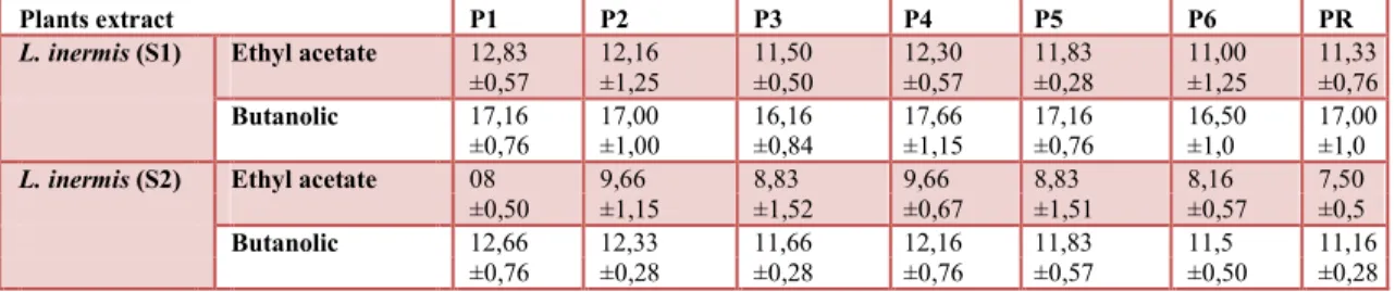
The antimicrobial activity of ethyl acetate and butanolic fractions of the two L. inermis extracts revealed significant antibacterial activity against all tested isolate of P. aeruginosa (Table 4). However, leaves obtained from Adrar region had the highest anti- P. aeruginosa activity compared with L. inermis from Biskra. The comparison of the zones inhibition obtained by the two extracts tested show that the butanolic fractions exhibited a greater antibacterial activity against all isolates tested for both tested varieties comparatively to ethyl acetate fractions.
L. inermis is a natural colorant showing a high compatibility with the environment [35]. It has been widely
used to treat skin infections, such as tinea. Naphthoquinones, including lawsone (2-hydroxynapthoquinone) are the principal constituent responsible for the antimicrobial properties of the plant [36, 37].
Minimal inhibitory concentration:
The results indicate that the butanolic fraction inhibit the tested bacteria more than ethyl acetate, with a CMI of 3,12 mg/ml and 6,25 mg/ml obtained from L. inermis leaves of Adrar and Biskra respectively. However the ethyl acetate fraction exerts an inhibitory activity (MIC) at a concentration of 6.25mg/ml and 12,5mg/ml (Table
5). The minimum bactericidal concentration (MBC) was around 6,25mg/ml and 12,5 mg/ml for the butanolic leaves extract of L. inermis obtained from Adrar region and Biskra region and at concentration of 12,5 mg/ml for ethyl acetate extracts for both varieties (Table 5). The butanolic extract showed the lowest MICs
compared to ethyl acetate extracts and this may be due to the large quantity of bioactive compounds that were retained by the solvent (Butanol) during the extraction process.
Table 4. Antimicrobial activity of two varieties of L. inermis extracts against isolates of P. aeruginosa
Plants extract P1 P2 P3 P4 P5 P6 PR
L. inermis (S1) Ethyl acetate 12,83 12,16 11,50 12,30 11,83 11,00 11,33
±0,57 ±1,25 ±0,50 ±0,57 ±0,28 ±1,25 ±0,76
Butanolic 17,16 17,00 16,16 17,66 17,16 16,50 17,00
±0,76 ±1,00 ±0,84 ±1,15 ±0,76 ±1,0 ±1,0
L. inermis (S2) Ethyl acetate 08 9,66 8,83 9,66 8,83 8,16 7,50
±0,50 ±1,15 ±1,52 ±0,67 ±1,51 ±0,57 ±0,5
Butanolic 12,66 12,33 11,66 12,16 11,83 11,5 11,16
±0,76 ±0,28 ±0,28 ±0,76 ±0,57 ±0,50 ±0,28
S1: L. inermis from Adrar region S2: L. inermis from Biskra region
Table 5. MIC and MBC of ethyl acetate and butanolic extracts of two different varieties of L. inermis
Plants extracts Isolates of P. aeruginosa tested CMB/CMI P1 P2 P3 P4 P5 P6 PR MIC (mg/ml) 6,25 6,25 6,25 6,25 6,25 6,25 6,25 AE MBC 6,25 6,25 6,25 6,25 6,25 6,25 6,25 2 (mg/mL) L. inermis MIC (S1) 3,15 3,15 3,15 3,15 3,15 3,15 3,15 (mg/mL) BE 1-2 MBC 6,25 6,25 3,15 6,25 3,15 6,25 6,25 (mg/mL) MIC 12,5 12,5 12,5 12,5 12,5 12,5 12,5 (mg/mL) AE 1 MBC 12,5 12,5 12,5 12,5 12,5 12,5 12,5 L. inermis (mg/mL) (S2) MIC 6,25 6,25 6,25 6,25 6,25 6,25 6,25 (mg/mL) BE 1 MBC 6,25 6,25 6,25 6,25 6,25 6,25 6,25 (mg/mL)
S1: L. inermis from Adrar region
AE: Ethyl acetate
extract
S2: L. inermis from Biskra
region BE: butanolic extract
Effect of plant extracts on P. aeruginosa biofilm formation:
The non growth inhibition concentration of the flavonoids extracts of L. inermis samples on the P.
aeruginosa cells was selected for the study to allow the growth of planktonic cells to develop biofilms. The
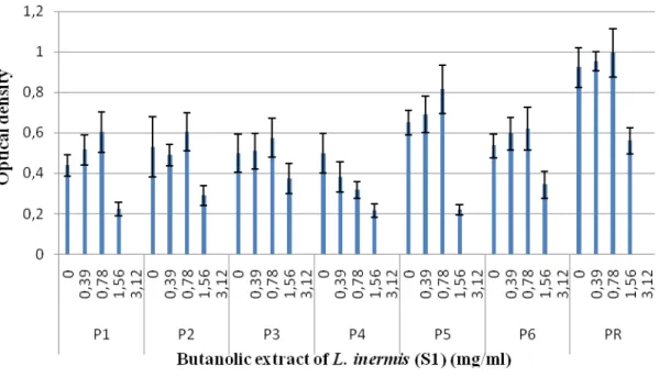
results (Figures 5, 7 and 8) showed that the butanol fractions can reduce biofilm formation for both L.
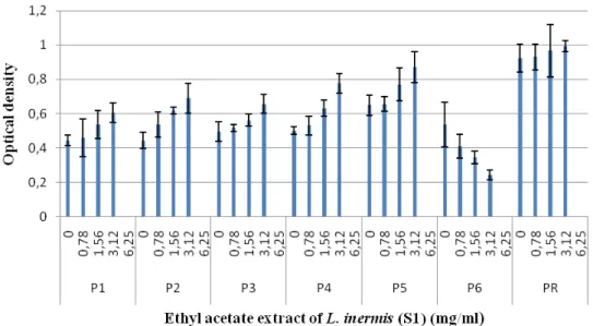
inermis samples tested. However subinhibitory concentrations of ethyl acetate did not affect biofilm
formation in all isolates of P. aeruginosa in both methods (Figures 6, 9 and 10). The significant effect of the butanolic extract was found at the concentration of ½ MIC as indicated from the remarkable decreased in the number of attached cells (biofilm) and significant reduction in the OD580nm value after 24h incubation. Plant extracts and other biologically active compounds isolated from plants are known to exhibit antimicrobial properties. In recent years, plant extracts and phytochemicals have also been highlighted as Quorum Sensing Inhibitors (QSI), providing an opportunity to develop new drugs against these targets to combat biofilm formation. Phenolic substances, including flavonoids are a major class of phytochemicals that have demonstrated significant anti-inflammatory, antioxidant, and anticancer properties. Flavonoids
contents of L. inermis such as quercetin and apigenin have been reported to have quorum sensing inhibitors activities. Vikram et al. [39] demonstrated a potential modulation of bacterial cell to cell communication, decreasing biofilm formation on enterohemorrhagic Escherichia coli (EHEC) and Vibrio harveyi virulence, by the two flavonoids compounds quercetin and apigenin.
A B
Figure 5. Effect of the butanolic extract (A) and ethyl acetate extract (B) of L. inermis (S1) on the biofilm formation by P. aeruginosa on polystyrene microtiter plates.
A B
Figure 6. Effect of the butanolic extract (A) and ethyl acetate extract (B) of L. inermis (S2) on the biofilm formation by P. aeruginosa on polystyrene microtiter plates.
Figure 8. Effect of the ethyl acetate extract of L. inermis (S1) on the biofilm formation by P. aeruginosa.
Figure 9. Effect of the butanolic extract of L. inermis (S2) on the biofilm formation by P. aeruginosa.
CONCLUSION:
According to our results obtained, where all studied extracts present an antibacterial property against all the isolates tested. It was also indicated that L. inermis obtained from Adrar region had the highest anti- P.
aeruginosa activity compared with that obtained from Biskra region. However the butanolic fractions of
both L. inermis samples have the highest effect with a MIC of 3.12mg/ml and 6.25mg/ml for samples of Adrar and Biskra respectively, compared with the fractions of ethyl acetate which showed a MIC of 6.25mg/ml and 12.5mg/ml for the same samples. Also, the biofilm formation was decreased when incubated with the ½ MIC of butanolic fraction for both L. inermis samples tested. Whereas the ½ MIC of ethyl acetate did not affect biofilm formation in all isolates of P. aeruginosa. We conclude that the extraction by butanol is that which makes it possible to better concentrate the antibacterial active ingredients present in the hydro-methanolic (70%) extract of Lawsonia inermis leaves.
REFERENCES
1. Engel, J. and P. Balachandran, 2009. Role of Pseudomonas aeruginosa type III effectors in disease. Curr Opin Microbiol, 12: 61-66.
2. Breidenstein, E.B.M., 2011. De la Fuente-Nuñez C, Hancock REW. Pseudomonas aeruginosa: all roads lead to resistance. Trends Microbiol, 19: 419-426.
3. Poole, K., 2011. Pseudomonas aeruginosa: resistance to the max. Front Microbiol, 2-65.
4. Joo, H.S., M. Otto, 2012. Molecular basis of in vivo biofilm formation by bacterial pathogens. Chem. Biol., 19: 1503–1513.
5. Heurlier, K., V. Denervaud and D. Haas, 2006. Impact of quorum sensing on fitness of Pseudomonas
aeruginosa. Int. J. Med. Microbiol., 296: 93-102.
6. De Kievit T.R., 2008. Quorum sensing in Pseudomonas aeruginosa biofilms. Environ. Microbiol., 11: 279-88.
7. Høiby, N., T. Bjarnsholt, M. Givskov, S. Molin and O. Ciofu, 2010. Antibiotic resistance of bacterial biofilms. Int. J. Antimicrob. Agents, 35: 322-332.
8. Schuster, M., E.P. Greenberg, 2006. A network of networks: quorum-sensing gene regulation in
Pseudomonas aeruginosa. Int. J. Med. Microbiol., 296: 73-81.
9. Williams, P., 2007. Quorum sensing, communication and cross-kingdom signaling in the bacterial world. Microbiology, 153: 3923-3938.
10. Kalia, V. C., 2013. Quorum sensing inhibitors: an overview. Biotechnology Advances, 31(2): 224-245. 11. Singh M., S. K. Jindal, Z. D. Kavia, B. L. Jangid, Khem Chan, 2005. Traditional Methods of
Cultivation and Processing of Henna. Henna, Cultivation, Improvement and Trade: 21-14. Jodhpur, India: Central Arid Zone Research Institute.
12. Borade A. S., B. N. Kale and R. V. Shete, 2011. A phytopharmacological review on Lawsonia inermis (linn.). Int. J. Pharm. Life Sci., 2: 536-41.
13. Yada, S., A. Kumar, J. Dora and A. Kumar, 2013. Essential perspectives of Lawsonia inermis. Int. J. Pharm. Chemical. Sci., 2: 888-96.
14. Shivananda, N.B., G. Isitor, E.M. Davis and G.K. Pillai, 2007. The evidence based wound healing activity of Lawsonia inermis Linn. Phytotherapy Research, 21(9): 827-31.
15. Chaudhary, G., S. Goyal and P. Poonia, 2010. Lawsonia inermis Linnaeus: a phyto-pharmacological review. Int. J. Pharma. Sci. Drug Res, 2: 91-98.
16. Hsouna, A.B., M. Trigui, G. Culioli, Y. Blache and S. Jaoua, 2011. Antioxidant constituents from
Lawsonia inermis leaves: isolation, structure elucidation and antioxidative capacity. Food Chem., 125:
17. Kelmanson, J. E., A. K. Jager and J. V. Staden, 2002. Zulu medicinal plants with antibacterial activity. J. Ethnopharm., 69: 241-246.
18. Fathiazad, F., M. Mazandarani and S. Hamedeyazdan, 2011. Phytochemical analysis and antioxidant activity of Hyssopus officinalis L. from Iran. Advanced Pharmaceutical Bulletin, 1(2): 63 – 67.
19. Bekkara, F., M. Jay, M.R. Viricel, and Rome, S. 1998. Distribution of phenolic compounds within seed and seedlings of two Vicia faba cvs differing in their seed tannin content, and study of their seed and root phenolic exudations. Plant and Soil, 203 (1): 27–36.
20. Othman, A. Ismail, N.A. Ghani and I. Adenam, 2007. Antioxidant capacity and phenolic content of cocoa beans. Food Chem., 100: 1523-1530.
21. Pothitirat, W., M.T. Chomnawang, R. Supabphol and W. Gritsanapan, 2009. Comparison of bioactive compounds content, free radical scavenging and anti-acne inducing bacteria activities of extracts from the mangosteen fruit rind at two stages of maturity. Fitoterapia, 80: 442–447.
22. Sacchetti G, S. Maietti, M. Muzzoli, M. Scaglianti, S. Manfredini, M. Radice and R. Bruni, 2005. Comparative evaluation of 11 essential oils of different origin as functional antioxidants, antiradicals and antimi-crobials in foods. Food Chemistry, 91: 621-632.
23. Celiktas O. Y., E. E. Hames Kocabas, E. Bedir, F. Vardar Sukan, T. Ozek and K. H. C. Baser, 2007. Antimicrobial activities of methanol extracts and essential oils of Rosmarinus officinalis, depending on location and seasonal variations. Food Chemistry, 100: 553-559.
24. Howaida, A. F., N. Skaug and W. Francis George, 2002. In vitro antimicrobial effects of crude miswak extracts on oral pathogens. Saudi Journal of Dentistry, 14: 26–32.
25. Adonizio, A., K.F. Kong and K. Mathee, 2008. Inhibition of quorum sensing-controlled virulence factor production in P. aeruginosa by South Florida plant extracts. Antimicrob Agents Chemother, 52: 198-203.
26. Djordjevic, D., M. Wiedmann and L.A. Mclandsborough, 2002. Microtiter plate assay for assessment of
Listeria monocytogenes biofilm for-mation. Applied and Environmental Microbiology, 68: 2950-2958.
27. Mesaros N., Tulkens P.M., P. Nordmann, P. Plésiat, M. Roussel-Devallez, J. Van Eldere, Y. Glupczynski, Y. Van Laethem, F. Jacobs, P. Lebecque, A. Malfroot and F. Van Bambeke, 2007.
Pseudomonas aeruginosa: Résistance et options thérapeutiques à l’aube du deuxième millénaire.
Clinical Microbiology and Infection, 126 (8): 305-316
28. Romano-Mazzotti L, C. Gayosso, C.M.D. Alcántar, J.I.Santos-Preciado and C.M. Alpuche-Aranda, 2008. Transmisión cruzada: un factor primordial en bacteremias por Pseudomonas aeruginosa resistente a carbapenemes en un hospital pediátrico de tercer nivel. Enferm. Infec. Microbiol., 28(suppl 1): S83. 29. Tsakris, A., A. Poulou, I. Kristo, T. Pittaras, N. Spanakis, S. Pournaras, et al., 2009. Large dissemination
of VIM-2-metallo-b-lactamase-producing Pseudomonas aeruginosa strains causing health care-associated community-onset infections. J. Clin. Microbiol., 47: 3524-3529.
30. Kumar, S.H., A.S. De, S.M. Baveja and M.A. Gore, 2012. Prevalence and risk factors of metallo b-lactamase producing Pseudomonas aeruginosa and Acinetobacter species in burns and surgical wards in a tertiary care hospital. J. Lab. Physicians., 4: 39-42.
31. Sara, A. Ochoa, Fernanda López-Montiel, Gerardo Escalona, Ariadnna Cruz-Córdova, Leticia B. Dávila, Briseida López-Martínez, Yolanda Jiménez-Tapia, Silvia Giono, Carlos Eslava, Rigoberto Hernández-Castro and Juan Xicohtencatl-Cortes, 2013. Pathogenic characteristics of Pseudomonas
aeruginosa strains resistant to carbapenems associated with biofilm formation, Bol. Med. Hosp. Infant
32. Tripathi, P., G. Banerjee, S. Saxena, S.M. Gupta and P.W. Ramteke, 2011. Antibiotic resistance pattern of Pseudomonas aeruginosa isolated from patients of lower respiratory tract infection. African J. Microbiol. Res., 5(19): 2955-2959.
33. Haddad Khodaparast Mohammad Hosein and Dezashibi Zinab, 2007. Phenolic Compounds and Antioxidant Activity of Henna Leaves Extracts (Lawsonia Inermis). World Journal of Dairy & Food Sciences, 2 (1): 38-41.
34. Naczk, M. and F. Shahidi, 2004. Extraction and analysis of phenolics in food. J. Chromatogr. A, 1054: 95–111.
35. Phirke S. S. and M. Saha, 2013. An overview of Lawsonia inermis L: a natural dye plant. Bionano Frontier. 6: 181-4.
36. Arun, P., K. G. Purushotham, J. Jayarani and V. Kumari, 2010. In vitro anti-bacterial activity and flavonoid contents of Lawsonia inermis. Int. J. Pharm. Tech. Res., 2: 1178-81.
37. Mostefa-Kara B., C. Ziani-Cherif, M. Benabdallah, N. M. Rahmoun, D. Villemin, N. Choukchou-Braham, K. Boucherit, 2010. New chemi-cal tools for the assessment of hemolytic anemia induced by naph-toquinones. Der. Pharma. Chemica., 2: 14-21.
38. Atkinson, S. and P. Williams, 2009. Quorum sensing and social networking in the microbial world. Journal of the Royal Society Interface, 6(40): 959-978.
39. Vikram, A., G.K. Jayaprakasha, P.R. Jesudhasan, S.D. Pillai and B.S. Patil, 2010. Suppression of bacterial cell-cell signalling, biofilm formation and type III secretion system by citrus flavonoids. Journal of Applied Microbiology, 109(2): 515-527.