HAL Id: hal-03162040
https://hal.univ-grenoble-alpes.fr/hal-03162040
Submitted on 15 Mar 2021HAL is a multi-disciplinary open access
archive for the deposit and dissemination of sci-entific research documents, whether they are pub-lished or not. The documents may come from teaching and research institutions in France or abroad, or from public or private research centers.
L’archive ouverte pluridisciplinaire HAL, est destinée au dépôt et à la diffusion de documents scientifiques de niveau recherche, publiés ou non, émanant des établissements d’enseignement et de recherche français ou étrangers, des laboratoires publics ou privés.
Maltose-Based Fluorinated Surfactants for
Membrane-Protein Extraction and Stabilization
Moheddine Wehbie, Kenechi Kanayo Onyia, Florian Mahler, Aline Le Roy,
Anaïs Deletraz, Ilham Bouchemal, Carolyn Vargas, Jonathan Oyebamiji
Babalola, Cécile Breyton, Christine Ebel, et al.
To cite this version:
Moheddine Wehbie, Kenechi Kanayo Onyia, Florian Mahler, Aline Le Roy, Anaïs Deletraz, et al.. Maltose-Based Fluorinated Surfactants for Membrane-Protein Extraction and Stabilization. Lang-muir, American Chemical Society, 2021, 37 (6), pp.2111-2122. �10.1021/acs.langmuir.0c03214�. �hal-03162040�
1
Maltose-Based Fluorinated Surfactants for Membrane-Protein
Extraction and Stabilization
Moheddine Wehbie,a Kenechi Kanayo Onyia,b,c Florian Mahler,b Aline Le Roy,d Anais
Deletraz,a Ilham Bouchemal,d Carolyn Vargas,b,e,f,g Jonathan Oyebamiji Babalola,c Cécile
Breyton,d Christine Ebel,d Sandro Keller,b,e,f,g Grégory Duranda*
a
Institut des Biomolécules Max Mousseron (UMR 5247 UM-CNRS-ENSCM) & Avignon University, Equipe Chimie Bioorganique et Systèmes amphiphiles, 301 rue Baruch de Spinoza – 84916 AVIGNON cedex 9, France;
b
Molecular Biophysics, Technische Universität Kaiserslautern (TUK), Erwin-Schrödinger-Str. 13, 67663 Kaiserslautern, Germany;
c
Department of Chemistry, University of Ibadan, 200284 Ibadan, Nigeria;
d
Univ. Grenoble Alpes, CNRS, CEA, CNRS, IBS, F-38000 Grenoble;
e
Biophysics, Institute of Molecular Biosciences – IMB, NAWI Graz, University of Graz, Humboldtstr. 50/III, 8010 Graz, Austria;
f
Field of Excellence BioHealth, University of Graz, Graz, Austria;
g
BioTechMed-Graz, Graz, Austria.
Corresponding Author. Grégory Durand. E-mail: gregory.durand@univ-avignon.fr ; Phone: +33 (0)4 9014 4445.
2
Abstract
Two new surfactants, F5OM and F5DM, were designed as partially fluorinated analogs of
n-dodecyl-β-D-maltoside (DDM). The micellization properties and the morphologies of the
aggregates formed by the two surfactants in water and phosphate buffer were evaluated by
NMR spectroscopy, surface tension measurement (SFT), isothermal titration calorimetry
(ITC), dynamic light scattering (DLS), small-angle X-ray scattering (SAXS), and analytical
ultracentrifugation (AUC). As expected, the critical micellar concentration (CMC) was found
to decrease with chain length of the fluorinated tail from 2.1–2.5 mM for F5OM to 0.3–0.5
mM for F5DM, and micellization was mainly entropy-driven at 25°C. Close to their respective
CMC, the micelle sizes were similar for both surfactants i.e. 7 and 13 nm for F5OM and
F5DM, respectively and both increased with concentration forming 4 nm diameter rods with
maximum dimensions of 50 and 70 nm, respectively, at a surfactant concentration of ~30
mM. The surfactants were found to readily solubilize lipid vesicles and extract membrane
proteins (MPs) directly from Escherichia coli membranes. They were found more efficient
than the commercial fluorinated detergent F6H2OM over a broad range of concentrations (1–
10 mM) and even better than DDM at low concentrations (1–5 mM). When transferred into
the two new surfactants, the thermal stability of the proteins bacteriorhodopsin (bR) and FhuA
were higher than in the presence of their solubilization detergents and similar to that in DDM;
furthermore, bR was stable over several months. The membrane enzymes SpNOX and BmrA
were not as active as in DDM micelles but similarly active as in F6OM. Together, these
findings indicate both extracting and stabilizing properties of the new maltose-based
3
Introduction
Membrane proteins (MPs) are encoded by 20–30% of all genes in most genomes, and they
perform a variety of vital functions like solute transport, signal transduction, intercellular
recognition and cell adhesion, to name but a few. Moreover, MPs are of great importance
since they represent the majority of current drug targets.1 MPs are usually extracted from
native membrane bilayers using a detergent that, ideally, should combine both solubilizing
and stabilizing properties so as to preserve the native structures of MPs in non-native
environments. Sugar-based surfactants are widely considered mild detergents and have been
commonly used as solubilizing and non-denaturing detergents for MP applications including
n-octyl-β-D-glucoside (OG),2-3 n-decyl-β-D-maltoside (DM),2 n-dodecyl-β-D-maltoside
(DDM),4-6 lactobionamides,7 and thioglycoside such as n-octyl-β-D-thioglucopyranoside
(OTG).8-9 Recently, some sugar-based surfactants with branched polar headgroups or
branched hydrophobic tails have been described for the same objective, namely, alkyl
diglucosides (DigluM),10 CALX-173-GK,11 and laurylmaltose neopentylglycol (LMNG).12-14
Fluorinated surfactants (FSs) bear several perfluorinated carbon atoms in their hydrophobic
tail. The presence of such CF2 groups makes them more hydrophobic15 and more
surface-active16-17 than their fully hydrogenated analogues. Moreover, fluorinated chains have a lower
affinity for hydrogenated chains, which makes FSs less denaturing towards MPs as they
hardly compete with protein–protein and protein–lipid/hydrophobic cofactor interactions.18
However, earlier neutral FSs were not able to solubilize membrane lipid bilayers, nor extract
MPs, rendering classical detergents mandatory for solubilisation, while FSs came into play
only at a later stage to stabilize MPs after extraction, when they might already have suffered
from irreversible denaturation. Recently, we have demonstrated that fluorination per se does
not prohibit detergency, with a fluorinated octyl maltoside derivative (F6OM) showing mild
4
solubilizes phospholipid vesicles in a manner reminiscent of conventional detergents without,
however, compromising membrane order at subsolubilizing concentrations, that is, well below
its critical micellar concentration (CMC).
Several series of sugar-based fluorinated surfactants have been synthesized and tested for
handling MPs in aqueous solutions. For example, a series of derivatives with various chain
lengths, and branched diglucose polar headgroups, showed solubilization of preformed
vesicles and efficient extraction of MPs from E. coli membranes.20 Fluorinated analogues of
DDM with a maltose polar headgroup were synthesized and found to stabilize the model MP
bacteriorhodopsin (bR) over extended periods of time.21-22 However, to the best of our
knowledge, in the literature, none of the earlier sugar-based FSs has been studied for both
solubilization and stabilization of MPs.
Currently, however, only a limited number of FSs are available from commercial sources, and
their biochemical properties have not been thoroughly investigated. To render FSs more
widely accessible and useful, our long-term project is intended to synthesize a range of related
compounds in which both head group and tail properties are modified rationally with the aim
of tailoring them to the specific needs of MP solubilization, purification, and handling. In this
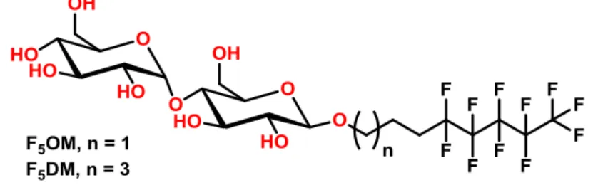
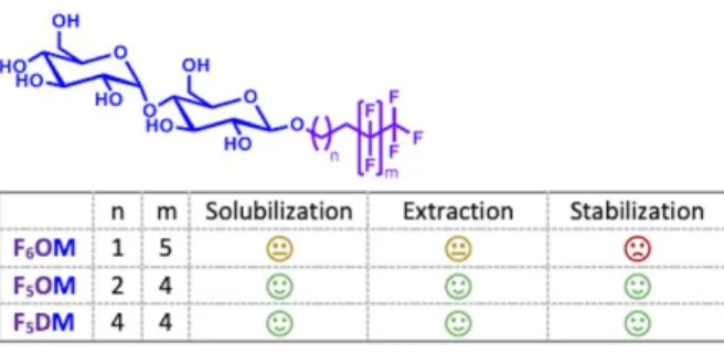
work, we have developed two new maltose-based fluorinated surfactants, called F5OM and
F5DM, which are analogues of the gold standard DDM (Figure 1). The length of the
fluorinated and hydrogenated segments within the hydrophobic chain were chosen in line with
our previous observations that the stabilization of the model MP bR is sensitive to the fluorine
content,22 whereas increasing the fluorine content in the hydrophobic chain tends to favor the
formation of rod-like large and poorly defined micelles19 and/or large protein–surfactant
complexes.21 The micellization properties and morphology of aggregates formed by the two
surfactants in water were evaluated by NMR spectroscopy, surface tension measurement
X-5
ray scattering (SAXS), and analytical ultracentrifugation (AUC). The efficiency of the
synthesized compounds for the extraction and stabilization of MPs were also investigated, on
a variety of different MPs: the E. coli multidrug transporter, BmrA23; a prokaryotic analog of
the eukaryotic NADPH oxidases, SpNOX24; the E. coli outer membrane transporter, FhuA25;
and bR.26 The new detergents showed great potency to solubilize lipid vesicles and to extract
different proteins from E. coli membranes. They also imparted stability to model MPs bR and
FhuA, the former protein being still correctly folded after a year of incubation.
Figure 1. Chemical structures of the new maltoside derivatives.
Experimental Section.
All the fluorinated maltoside derivatives studied in this work are named following the
common nomenclature used for their hydrogenated analogs: Fn#M, where n indicates the
number of the perfluorinated carbons within the chain starting from the last carbon atom, #
indicates the length of the alkyl chain (O for octyl, N for nonyl, D for decyl, UD for undecyl
and DD for dodecyl), and M indicates the maltoside polar head. F5OM indicates the
fluorinated analog of octylmaltoside where the last five carbon atoms of the chain are
perfluorinated whereas F5DM indicates the fluorinated analog of decylmaltoside with the last
five carbon atoms of the chain being perfluorinated.
All starting materials were commercially available and used without further purification. All
solvents were of reagent grade and used as received unless otherwise indicated. CH3OH was
6
under argon. The progress of the reactions was monitored by thin-layer chromatography. The
compounds were detected either by exposure to ultraviolet light (254 nm) or by spraying with
sulfuric acid (5% ethanol), followed by heating at ∼150 °C. 1H, 13C and 19F-NMR analyses were performed at 400, 100 and 376 MHz, respectively. Chemical shifts are given in ppm
relative to the solvent residual peak as a heteronuclear reference for 1H and 13C. Abbreviations
used for signal patterns are: s, singlet; d, doublet; t, triplet; q, quartet; m, multiplet; dd,
doublet of doublet; and dt, doublet of triplet. HRMS (ESI+) was determined on a QStar Elite
mass spectrometer. Milli-Q water (resistivity, 18.2 MΩ cm; surface tension, 71.45 mN/m at 25°C) was employed for all physical−chemical experiments.
Synthesis.
Allyl-2,3,6-tri-O-acetyl-4-O-(α-D-2΄,3΄,4΄,6΄-tetra-O-acetyl-glucopyranosyl)-β-D-glucopyranoside (2a). Under argon, octa-O-acetyl-ß-D-maltose (3.20 g, 4.71 mmol, 1.0
equiv) was dissolved in dry dichloromethane (10 mL) and the resulting solution was cooled
down using an ice bath. Allyl alcohol (0.437 g, 7.54 mmol, 1.6 equiv) was first added
followed by the dropwise addition of boron trifluoride diethyl ether complex (0.87 mL,
7.07 mmol, 1.5 equiv). The mixture was stirred at 0 oC for 2 h and kept at room temperature
overnight. Dichloromethane (20 mL) was added, then the mixture was washed with saturated
NaHCO3 (2 20 mL) and brine (2 20 mL). The organic phase was collected and dried over
anhydrous Na2SO4, filtered, and the solvent was removed under reduced pressure. The crude
compound was purified by column chromatography on silica gel (cyclohexane/ethyl acetate,
4:6, v/v) to get 2a (1.88 g, 59%) as a white powder. Rf (cyclohexane/ethyl acetate, 5:5, v/v) =
0.28. 1H NMR (CDCl3, 400 MHz): δ/ppm 5.89-5.78 (m, 1H), 5.40 (d, J = 3.9 Hz, 1H), 5.35 (t,
J = 9.4 Hz, 1H), 5.28-5.17 (m, 3H), 5.04 (t, J = 9.7, 1H), 4.87-4.82 (m, 2H), 4.57 (d, J = 7.9
Hz, 1H), 4.47 (m, 1H), 4.33-4.19 (m, 3H), 4.11-3.93 (m, 4H), 3.67(m, 1H), 2.14 (s, 3H), 2.09
7
170.5, 170.2, 169.9, 169.6, 169.4, 133.3, 117.7, 99.0, 95.5, 75.4, 72.7, 72.2, 72.1, 70.0, 69.3,
68.5, 68.0, 62.8, 61.5, 20.9, 20.8, 20.7, 20.6, 20.6, 20.6. HRMS (ESI+) m/z: [M +Na]+
calculated for C29H40O18Na:699.2112, found 699.2122.
Pent-4-en-1-yl-2,3,6-tri-O-acetyl-4-O-(α-D-2΄,3΄,4΄,6΄-tetra-O-acetyl-glucopyranosyl)-β-D-glucopyranoside (2b). 2b was synthesized following the same procedure as for 2a, from
octa-O-acetyl-ß-D-maltose (3.0 g, 4.42 mmol, 1.0 equiv), pentyl alcohol (0.571 g, 6.63 mmol,
1.5 equiv), and boron trifluoride diethyl ether complex (0.82 mL, 6.63 mmol, 1.5 equiv).
After purification by column chromatography on silica gel (cyclohexane/ethyl acetate, 3:7,
v/v) compound 2b (1.16 g, 37%) was obtained as a white powder. Rf (cyclohexane/ethyl
acetate, 3:7, v/v) = 0.21. 1H NMR (CDCl3, 400 MHz): δ/ppm 5.76 (m, 1H). 5.40 (m, 1H), 5.35 (t, J = 9.6 Hz, 1H), 5.25 (t, J = 9.0 Hz, 1H), 5.09-4.93 (m, 3H), 4.87-4.79 (m, 2H), 4.50 (d, J = 7.9, 1H), 4.46 (dd, J = 2.6 Hz, J = 12.1 Hz, 1H), 4.25 (m, 2H), 4.05-3.93 (m, 3H), 3.85 (m, 1H), 3.66 (m, 1H), 3.48 (m, 1H), 2.13 (s, 3H), 2.09 (s, 3H), 2.06 (m, 2H), 2.04 (s, 3H), 2.01 (s, 6H), 1.99 (s, 6H), 1.62 (m, 2H). 13C NMR (CDCl3, 100 MHz): δ/ppm 170.5, 170.5, 170.3, 170.0, 169.6, 169.4, 137.8, 115.1, 100.3, 95.5, 75.5, 72.8, 72.2, 72.0, 70.0, 69.3, 69.3, 68.5, 68.0, 62.9, 61.5, 29.8, 28.5, 26.9, 20.9, 20.8, 20.7, 20.6, 20.6, 20.5. HRMS (ESI+) m/z: [M
+H]+ calculated for C31H45O18: 705.2605, found 705.2600. HRMS (ESI+) m/z: [M+Na]+
calculated for C31H44NaO18:727.2420, found 727.2393.
4,4,5,5,6,6,7,7,8,8,8-undecafluoro-2-iodo-octyl-2,3,6-tri-O-acetyl-4-O-(α-D-2΄,3΄,4΄,6΄-tetra-O-acetyl-glucopyranosyl)-β-D-glucopyranoside (3a). To a solution of 2a (1.84 g, 2.72
mmol, 1.0 equiv) in dichloromethane (10 mL), perfluoropentyl iodide (0.71 mL, 3.67 mmol,
1.35 equiv) and triethyl borane 1M in hexane (0.5 mL, 0.5 mmol, 0.2 equiv) were added. The
mixture was flushed with air and stirred at room temperature for 1 h. 50 mL of a diluted
solution of Na2S2O3 was added and the aqueous solution was extracted with CH2Cl2
8
the solvent was removed under reduced pressure. The crude compound was purified by
column chromatography on silica gel (cyclohexane/ethyl acetate, 3:7, v/v) to give compound
3a (2.51 g, 86 %) as a white powder. Rf (cyclohexane/ethyl acetate, 3:7, v/v) = 0.24. 1H NMR
(CDCl3, 400 MHz): δ/ppm 5.41 (m, 1H), 5.36 (td, J = 1.3 Hz, J = 10.2 Hz, 1H), 5.26 (td, J = 0.8 Hz, J = 9.2 Hz, 1H), 5.06 (td, J = 2.2 Hz, J = 9.9 Hz, 1H), 4.88-4.82 (m, 2H), 4.59 (dd, J = 2.5 Hz, J = 7.9 Hz, 1H), 4.48 (m, 1H), 4.40-4.17 (m, 3H), 4.11-3.93 (m, 4H), 3.78 (m, 1H), 3.69 (m, 1H), 2.98 (m, 1H), 2.66 (m, 1H), 2.13 (s, 3H), 2.10 (s, 3H), 2.03 (s, 6H), 2.02 (s, 3H), 2.00 (s, 6H). 19F NMR (CDCl3, 376 MHz): δ/ppm –80.7 (td, J = 2.7 Hz, J = 9.5 Hz, 3F, CF3), –113.8 (m, 2F, CF2), –122.6 (m, 2F, CF2), –123.7 (d, J = 52 Hz, 2F, CF2), –126.3 (t, J = 13.6 Hz, 2F, CF2). 13C NMR (CDCl3, 100 MHz): δ/ppm 170.7, 170.5, 170.3, 170.1, 169.7, 169.7, 169.6, 100.8, 99.8, 95.7, 75.3, 74.9, 73.9, 72.7, 72.5, 71.9, 70.2, 69.5, 68.7, 68.2, 62.7, 61.7, 37.4, 21.0, 20.9, 20.8, 20.8, 20.8, 20.7, 20.6, 13.5. HRMS (ESI+) m/z: [M+H]+
calculated for C34H41F11IO18: 1073.1162, found 1073.1156.
4,4,5,5,6,6,7,7,8,8,8-undecafluoro-4-iodo-decyl-2,3,6-tri-O-acetyl-4-O-(α-D-2΄,3΄,4΄,6΄-tetra-O-acetyl-glucopyranosyl)-β-D-glucopyranoside (3b). Compound 3b was synthesized
following the same procedure as for 3a, from 2b (1.16 g, 1.64 mmol, 1.0 equiv),
perfluoropentyliodide (0.88 g, 2.22 mmol, 1.35 equiv) and triethyl borane 1M in hexane (0.3
mL, 0.3 mmol, 0.2 equiv). After purification by column chromatography on silica gel
(cyclohexane/ethyl acetate, 2:8, v/v), compound 3b (1.70 g, 94 %) was obtained as a white
powder. Rf (cyclohexane/ethyl acetate, 3:7, v/v) = 0.31. 1H NMR (CDCl3, 400 MHz): δ/ppm
5.41 (m, 1H), 5.34 (t, J = 10.2 Hz, 1H), 5.23 (t, J = 9.2 Hz, 1H), 5.06 (t, J = 9.9 Hz, 1H),
4.85-4.76 (m, 2H), 4.50 (d, J = 7.9 Hz, 1H), 4.47 (m, 1H), 4.30 (m, 1H), 4.25-4.17 (m, 2H),
4.05-3.94 (m, 3H), 3.85 (m, 1H), 3.65 (m, 1H), 3.52 (m, 1H), 2.80 (m, 2H), 2.12 (s, 3H), 2.08 (s,
3H), 2.02 (s, 3H), 2.00 (s, 3H), 1.98 (2s, 9H), 1.84 (m, 4H).19F NMR (CDCl3, 376 MHz): δ
9
1F, CF2), –122.6 (s, 2F, CF2), –123.9 (t, J = 12 Hz, 2F, CF2), -126.3 (s, 2F, CF2). 13C-NMR
(CDCl3, 100 MHz): δ/ppm 170.5, 170.4, 170.2, 170.0, 169.6, 169.5, 100.1, 95.5, 75.4, 72.7,
72.2, 72.1, 70.0, 69.3, 68.5, 68.0, 62.8, 61.5, 41.6, 36.8, 29.8, 20.9, 20.8, 20.6, 20.6, 20.5,
20.0, 19.8. HRMS (ESI+) m/z: [M+H]+ calculated for C36H45F11IO18 :1101.1475, found
1101.1476.
4,4,5,5,6,6,7,7,8,8,8-undecafluorooctyl-4-O-(α-D-glucopyranosyl)-β-D-glucopyranoside
(4a). Compound 3a (1.24 g, 1.15 mmol, 1.0 equiv) was dissolved in methanol and 50 mg of
Pd/C and sodium acetate (0.310 g, 3.77 mmol, 3.3 equiv) were added portion-wise. The
resulting solution was stirred under H2(g) (6 bars) overnight. The resulting mixture was
filtered over a pad of celite and the solvent was evaporated under reduced pressure. The crude
product was dissolved in CH2Cl2 (50 mL) and washed with a diluted solution of Na2S2O3 (50
mL). Then the aqueous phase was extracted with CH2Cl2 (2 × 50 mL). The organic fractions
were collected, dried over anhydrous Na2SO4, filtered, and the solvent was removed under
reduced pressure. The resulting compound was dissolved in methanol, then a catalytic amount
of sodium methoxide (27 mg, 0.50 mmol) was added portion-wise. The mixture was stirred
overnight at room temperature. The reaction mixture was neutralized by addition of Dowex
50W×8-100 ion exchange resin (2.0 g). The ion exchange resin was filtered off and the
solvent was removed under reduced pressure. The crude compound was purified by column
chromatography on silica gel (CH2Cl2/CH3OH, 85:15, v/v) to give compound 4a (0.695g,
91%) as a white powder. Rf (CD3OD/ethyl acetate, 2:8, v/v) = 0.27. 1H NMR (CD3OD, 400
MHz): δ/ppm 5.18 (m, 1H), 4.30 (d, J = 7.8 Hz, 1H), 3.98 (m, 1H), 3.94-3.78 (m, 3H), 3.71-3.60 (m, 5H), 3.55 (m, 1H), 3.45 (m, 1H), 3.38 (m, 1H), 3.27 (m, 2H), 2.44-2.25 (m, 2H),
1.92 (m, 2H). 19F NMR (CD3OD, 376 MHz): δ /ppm –82.5 (td, J = 2.7 Hz, J = 10.2 Hz, 3F,
CF3), –115.5 (q, J = 17 Hz, 2F, CF2), –123.8 (s, 2F, CF2), –124.7 (s, 2F, CF2), –127.5 (s, 2F,
10
74.2, 71.5, 69.2, 62.8, 62.2, 28.9 (t, J = 22.5 Hz), 21.9. HRMS (ESI+) m/z: [M +H]+
calculated for C20H28F11O11: 653.1459, found 653.1456.
4,4,5,5,6,6,7,7,8,8,8-undecafluorodecyl-4-O-(α-D-glucopyranosyl)-β-D-glucopyranoside
(4b). Compound 4b was synthesized following the same procedure as for 4a, from 3b (1.69 g,
1.54 mmol, 1.0 equiv), 50 mg of Pd/C and sodium acetate (0.404 g, 4.93 mmol, 3.2 equiv)
under H2(g) (6 bars) overnight, followed by deprotection using sodium methoxide (27 mg,
0.50 mmol) to give compound 4b (0.786 g, 75%) as a white powder. Rf (CD3OD /ethyl
acetate, 2:8, v/v) = 0.32. 1H NMR (CD3OD, 400 MHz): δ/ppm 5.16 (d, J = 3.8 Hz, 1H), 4.27 (d, J = 7.8 Hz, 1H), 3.94-3.78 (m, 4H), 3.71-3.51 (m, 6H), 3.44 (dd, J = 3.8 Hz, J= 9.6 Hz, 1H), 3.36 (m, 1H), 3.27 (t, J = 9.4 Hz, 1H), 3.24 (m, 1H), 2.25-2.0 (m, 2H), 1.65 (m, 4H), 1.53 (m, 2H). 19F NMR (CD3OD, 376 MHz): δ /ppm -82.5 (td, J = 2.3 Hz, J = 10.4 Hz, 3F, CF3), –115.5 (q, J = 17 Hz, 2F, CF2), –123.8 (s, 2F, CF2), –124.8 (s, 2F, CF2), –127.5 (s, 2F, CF2). 13C-NMR (CD3OD, 100 MHz): δ/ppm 104.3, 102.9, 81.4, 77.9, 76.6, 75.1, 74.8, 74.7, 74.2, 71.5, 70.4, 62.8, 62.2, 31.7 (t, J = 22.0 Hz), 30.4, 26.6, 21.1. HRMS (ESI+) m/z:
11
CMC determination by 19F-NMR measurements. Seven samples of each detergent at
different concentrations were prepared from stock solutions (4.0 g/L for F5DM and 6.0 g/L for
F5OM). All samples were dissolved in D2O/H2O (10:90, v/v). CF3COONa was used as an
internal reference (30 μL of a solution at 1 g/L was added). The chemical shifts of the terminal CF3 group of F5DM and F5OM were plotted as a function of the concentration to
derive the CMC linear fitting. Below the CMC, the observed chemical shift (δobs) is the
chemical shift of the monomer (δmon), whereas above the CMC, δobs is the weighted average
of the monomer, and micelle chemical shift, assuming the exchange between the bulk solution
and the micelle, is fast on the NMR time scale. If the monomer concentration is constant
above the CMC, the observed chemical shift can be written as follows:
CMC determination by Surface Tension Measurements. The surface activity of detergents
in solution at the air/water interface was determined using a K100 tensiometer (Kruss,
Hamburg, Germany). Surface tensions were determined by dilution of stock solutions (0.70
g/L for F5DM and 5.8 g/L for F5OM, ∼5 CMC) using the Wilhelmy plate technique. In a
typical experiment, 20−30 concentration steps were used with ca. 5−10 min between each concentration step. All measurements were performed at (25.0 ± 0.5) °C.
CMC determination by ITC. Demicellization experiments were performed at 25°C on a
VP-ITC (Malvern Instruments) by titrating 28 mM F5OM and 5 mM F5DM, respectively, from
the injection syringe into the sample cell containing triple-distilled water or phosphate buffer.
Experimental settings included injection volumes of 5–10 µL, a reference power of 58 μJ/s, a
filter period of 2 s, and time spacings of 5 min to allow the signal to reach the baseline before
the next injection. Automated baseline adjustment and peak integration were done with
least-12
squares fitting was performed in an Excel (Microsoft, Redmond, USA) spreadsheet using the Solver add-in (Frontline Systems, Incline Village, USA), as explained elsewhere.31
Analysis of thermodynamic properties. ITC demicellization experiments directly calculates
the CMC and the molar enthalpy of micelle formation, . From the CMC, the partition coefficient for micellization, can thus be derived as the ratio of the mole fractions of the surfactant in the micellar (m) and the aqueous (aq) phases: The micellar phase consists only of surfactant molecules, 1, whereas , where denotes the water concentration (55.5 M). From this, the standard molar Gibbs free energy change upon micellization was
derived as – ln ln and the entropic contribution to micellization as – – , with denoting the standard molar entropy change upon micellization.
Dynamic light scattering (DLS). DLS measurements were carried out with a Nano Zetasizer
S90 (Malvern, Herrenberg, Germany), utilizing a He–Ne laser at a wavelength of 633 nm as
light source and a detection angle of 90°. Samples were transferred to a 45-μL quartz glass
cuvette (Hellma, Munich, Germany) and equilibrated for 2 min prior to each measurement.
The attenuator was fixed to the maximum position to ensure comparable results for light
scattering intensity measurements while in case of the determination of size distributions,
attenuator settings were automatically set by the software.
Sedimentation velocity experiments. Sedimentation velocity experiments were performed in
a Beckman XL-I analytical ultracentrifuge with a rotor Anti-50 (Beckman Coulter, Palo Alto,
USA) and double-sector cells of optical path length 12 mm equipped of Sapphire windows
(Nanolytics, Potsdam, DE). Samples were centrifuged at 42000 rpm (130 000 g), at 20°C.
13
analyzed in terms of continuous size distribution c(s) of sedimentation coefficients, s,27 by
using SEDFIT. Peak integration and figures were done with the GUSSI software28
(http://biophysics.swmed.edu/MBR/software.html). Standard equations and protocols
described in 29 were used to derive the refractive index increment, the CMC, the
sedimentation coefficient at infinite dilution, s0. We used the Svedberg equation to derive
from s, micelle molar masses, Mmic, from which were derived aggregation numbers, Nagg,
using the information on the calculated surfactant molar masses and partial specific volumes
reported on Table 1, and estimates on the hydrodynamic diameters from DLS.
SAXS experiment. Five samples of each detergent at different concentrations were prepared
from stock solutions in H2O (33.3 mM for F5DM and 29.5 mM for F5OM). SAXS
experiments were conducted on the BM29 beamline at the European Synchrotron Radiation
Facility (Grenoble, France). The data were recorded for 0.004<Q<0.5 Å-1 (Q=(4/)sin is the modulus of the scattering vector, with 2 being the scattering angle, and the wavelength), using a two-dimensional 1M Pilatus detector, at 20 °C, with a monochromatic
X-ray beam with = 0.9919 Å and a sample to detector distance of 2.864 m. Measurements were performed with 50 µL loaded sample, in a quartz capillary, with a continuous flow. 10
acquisitions with 0.5 s irradiation (flows of 5 µL/s), were recorded for the samples and water.
Data reduction was performed using the automated standard beamline software (BSxCuBE),30
and data processing, including the elimination of data suffering from radiation damage,
averaging, buffer subtraction, Guinier plots, and pair distribution functions, using PRIMUS
(V3.1) of the software suite ATSAS.31 Absolute scales were obtained using the scattering of
water. The radii of gyration (Rg) and the intensities scattered in the forward direction (I(0))
were extracted by the Guinier approximation, with RgQ ≤ 1.0. The molar mass of the micelle,
Mmic, was derived from Mmic = (I(0)/cmic)NA/(∂el/∂c)2, with NA Avogadro’s number, cmic the
14
g-1) given in table S#1. the increment of electron scattering length density per g of surfactant.
Aggregation numbers Nagg were then derived from Mmic. The maximum dimensions (Dmax)
were estimated from the pair distribution functions. Shape analysis were done using
shape-dependent models in SASview (V4.2.1) (https://www.sasview.org/). We investigated the
cylinder models. Theoretical SLD values are given in table S1. The form factor included size
polydispersity on radius (fixed at 15 %, this value resulting from preliminary fits) using a
gaussian distribution. The scale factor (i.e. the surfactant concentration in vol/vol unit), and
the SLD of the solvent were constrained (table S1). The core radius and cylinder length were
adjusted. Note that for F5DM scattering curves, we also performed an analysis with core shell
cylinder model. The scale factors, the core and solvent SLDs were fixed (table S1) and core
radius, thickness and SLD shell, and cylinder length were adjusted. The two fits in the
cylinder and core shell cylinder models were equivalent in terms of quality, as evaluated by
the chi2 values. But the later provided inconsistent values for the lowest concentrations; for
the three largest concentrations, the radius and the thickness were constant: 1.79 ± 0.09 and
1.16 ± 0.1 nm, respectively, thus the sum (radius + thickness) was rather large (2.95 nm). The
mean value of the fitted SLD shell (1.07 ± 0.02 10-5 Å-2) corresponded to 75 % water. The
very large dimension of the total radius associated to overestimated hydration in the core-shell
cylinder model can be due to the fact that the SLDs of the anhydrous head and tail are rather
close (1.51 10-5 Å-2 and 1.44 10-5 Å-2) compared to water (9.53 10-6 Å-2), which argues in
favor of the simple cylinder model.
Preparation of lipid vesicles. To prepare LUVs, POPC in powder form was weighed on a
high-precision XP Delta Range microbalance (Mettler Toledo, Greifensee, Switzerland) and
suspended in phosphate buffer (10 mM Na2HPO4/NaH2PO4, 150 mM NaCl, pH 7.4). The
solution was vortexed for 15 min at room temperature and extruded in a LiposoFast extruder
15
polycarbonate membranes with a pore diameter of 100 nm (Avestin). The hydrodynamic
diameter of the LUVs was distributed around 120–130 nm, as shown by DLS.
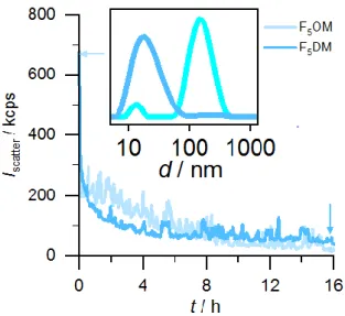
Kinetics of vesicle solubilization. For vesicle solubilization kinetics, measurements were
conducted by adding a high concentration (5 mM) of the respective surfactant above its CMC
to 100 μM POPC LUVs in a 3 mm×3 mm quartz glass cuvette. Measurements were started
immediately after mixing the vesicle suspension to monitor changes in light scattering
intensity, Iscatter.
Solubilization of MPs from native E. coli membranes. E. coli BL21(DE3) cells were
transformed with an empty pET-24 vector and selected by kanamycin resistance. After
incubation in 400 mL lysogeny broth overnight at 37°C under constant agitation (150 rpm),
cells were harvested by centrifugation and washed twice with saline (154 mM NaCl). Cell
pellets were resuspended in ice-cold buffer (100 mM Na2CO3, pH 11.5) and subjected to
ultrasonication in an S-250A sonifier (Branson Ultrasonics, Danbury, USA) twice for 10 min
each. To remove cell debris, the lysate was centrifuged at 4°C for 20 min at 3000 g. The
supernatant was ultracentrifuged at 4°C for 1 h at 100,000 g to separate membrane fragments
from soluble and peripheral proteins. Membrane pellets were washed and suspended in
working buffer, ultracentrifuged again at 4°C for 1 h at 100,000 g to remove any residual
soluble or peripheral proteins. The resulting pellets were resuspended in buffer (50 mM Tris,
200 mM NaCl, pH 7.4) to a final concentration of 100 mg wet-weight pellet per 1 mL of
buffer and mixed in a 1:1 volume ratio with stock solutions of DDM or FSs in buffer.
Surfactant concentrations were chosen on the basis of the CMC values determined in this
study to ensure comparable extraction conditions. All samples were incubated for at least 16 h
at 20°C under constant, gentle agitation (500 rpm) and subsequently ultracentrifuged at 4°C
for 1 h at 100,000 g. The solubilized supernatant containing micelles was analyzed using
16
Sodium dodecyl sulphate polyacrylamide gel electrophoresis (SDS-PAGE). The
solubilization efficiency of the two FSs on biological membranes was assessed by
SDS-PAGE using a NuSDS-PAGE Bis–Tris system (Life Technologies, Carlsbad, USA) with a
polyacrylamide gradient of 4–12%. 14-μL samples were mixed with 5 μL 4x SDS sample
buffer (106 mM Tris HCl, 141 mM Tris base, 2% (w/v) SDS, 10% (w/v) glycerol, 0.51 mM
EDTA, 0.22 mM SERVA Blue G250, and 0.175 mM Phenol Red, pH 8.5) and 1 μL 1 M
dithiothreitol (DTT) and boiled at 95°C for 10 min. 12 μL of each sample was loaded on a
ready-to-use NuPAGE. As reference, a standard-weight marker (Roti-Mark 10–150, Carl
Roth, Karlsruhe, Germany) was used, and the working buffer was used as negative control.
Gel electrophoresis was performed for 45 min in MES buffer (50 mM MES, 50 mM Tris
base, 0.1% (w/v) SDS, 1 mM EDTA) at 200 V and 50 W. Subsequently, gels were fixed for
20 min (10% (w/v) acetic acid, 40% (w/v) ethanol), stained for 30 min (0.025% (w/v)
Coomassie brilliant blue G250, 10% (w/v) acetic acid) and destained overnight in water. For
quantification of solubilization efficiencies, gels were photographed with a C4000Z camera
(Olympus, Tokyo, Japan), and protein bands were analyzed with ImageJ.32
Thermal denaturation assays. Thermal unfolding analysis were performed by differential
scanning fluorimetry coupled to back scattering using a Prometheus NT.48 instrument
(Nanotemper Technologies, Munich, DE), and the provided software PR.thermocontrol
v2.0.4. Up to 48 capillary containing 10 µL of sample are sequentially illuminated at 280 nm,
and fluorescence intensity at 350 (F350) and 330 (F330) nm, and back scattering measured as
a function of temperature. The temperature was increased by 1 °C/min from 15 °C up to 90 or
95°C °C. The derivatives of F350/F330 and of the back scattering were used to estimate the
melting temperature, Tm, and the onset of aggregation, Tagg, respectively. FhuA and bR
17
bR solubilization and detergent exchange by sucrose gradient. Sucrose gradients are a
convenient means to perform both detergent exchange and evaluate the colloidal homogeneity
of the protein-detergent complex. We routinely use this method to evaluate the potentialities
of fluorinated surfactants in the biochemistry of MPs.32 BR retinal molecule, whose visible
absorption spectrum is very sensitive to its local environment, is a convenient reporter of the
state of the protein: the trimeric protein in its native membrane reveals a visible absorption
spectrum with a maximum at λmax = 570 nm; when solubilized in detergent, the protein
monomerizes and displays λmax ~ 550 nm; the protein appears purple/pink. When the protein
denatures, the retinal is released, and λmax shifts to 400–380 nm: the protein solution turns
yellow. We have reported that when the solubilized monomeric protein is transferred into a
fluorinated surfactant, fluorinated surfactant migrates deeper in the gradients, due to the
higher density of the surfactant, and λmax can shift to ~ 610 nm, giving a blue color to the
protein-surfactant complex.32 Diffusion of the absorption curve is a witness of the appearance
of larger particles in the solution, suggesting either aggregation of the protein or the formation
of membrane patches.
Purified purple membrane was solubilized for 40 h at 4°C with 89mM OTG (CMC = 9 mM)
at a membrane concentration of 1.5 g L−1 in 20mM sodium phosphate buffer, pH 6.8. Samples
were diluted to reach a final OTG concentration of 15 mM, supplemented with 2 mM of the
surfactant to be tested, and incubated 15 min prior to being loaded onto a 10–30% (w/w)
sucrose gradient containing 20 mM sodium phosphate buffer pH 6.8 and 6 mM of either
DDM as a control, or the surfactant to be tested. Gradients were centrifuged for 5 h at 55,000
rpm (200,000 g) in the TLS55 rotor of a TL100 ultracentrifuge (Beckman). Bands containing
the colored protein were collected with a syringe, and protein samples were kept at 4°C in the
18
Results and Discussion.
Synthesis. The detergents were synthesized in four steps, as illustrated in Scheme 1. The
synthetic route is inspired by that previously used for the preparation of the poorly fluorinated
analog of undecylmaltoside with two perfluorinated carbons F2UDM (also called F2H9Malt).22
Compounds 2a and 2b were prepared starting from peracetylated maltose by glycosylation
reaction with allyl alcohol and penten-1-yl alcohol, respectively. The double bonds of the
obtained compounds (2a and 2b) were then subjected to free radical reaction with
perfluoropentyl iodide in the presence of 1 M BEt3 in hexane.33 The addition of the
fluoroalkyl chain to the double bonds was confirmed by 1H- and 13C-NMR, which showed the
disappearance of the signals corresponding to the double bond and the formation of new
signals of -CHI. The iodine group of compounds 3a and 3b was reduced under H2 gas and in
the presence of Pd/C as catalyst. The obtained compounds were then deprotected under
Zemplén conditions,34 using a catalytic amount of MeONa in MeOH to obtain the desired
detergents 4a and 4b. The crude detergents were purified by chromatography and freeze–
dried to give the pure detergents in satisfactory global yields of 46% and 26% for F5OM and
F5DM, respectively.
19
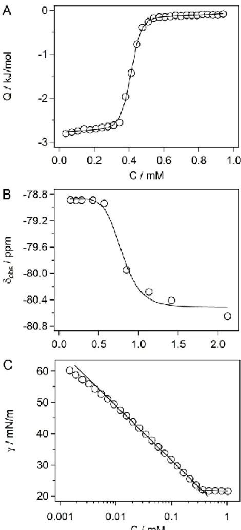
Micellization. Micellization of the two surfactants was characterized by means of ITC, 19
F-NMR, and SFT, from which we derived micellar parameters (Table 1). The critical micelle
concentration (CMC) values were in very good agreement among the three techniques (Figure
2 and Figure S1). While F5OM exhibited a CMC around 2.3 mM, the longer-chain derivative
F5DM had a CMC of ~0.4 mM. Thus, addition of two more methylene groups to the chain led
to a decrease in CMC by a factor of ~5, which is only half the effect predicted by Traube’s
rule35 for adding two methylene groups to an alkyl chain.
The changes in Gibbs free energy , enthalpy , and entropy , accompanying the transfer of surfactant monomers from the aqueous solution into micelles
are also summarized in Table 1. These data showed that micellization was almost exclusively
driven by entropy, with enthalpy making only a minor contribution that decreased with
increasing chain length. The Gibbs free energy of micellization increased in magnitude by –
4.2 kJ/mol upon increasing the chain length by two CH2 groups. In addition, the fluorinated
maltoside-based surfactants displayed a higher tendency to form micelles at lower
concentration than their hydrogenated analogues bearing the same number of carbon atoms in
their hydrogenated chains. The hydrophobic contribution to micelle formation, that is, the
contribution of the alkyl tail of F5DM (CMC = 0.39 mM), which contains 10 carbon atoms,
was greater than that of its decyl hydrogenated analogue DM (CMC = 2.0 mM) and fall
between that of the undecyl derivative UDM (CMC = 0.59 mM) and DDM (CMC = 0.17
mM).36 Similarly, the alkyl tail of F5OM (CMC = 2.21 mM), which contains 8 carbon atoms,
had almost similar hydrophobic contribution as DM. The hydrophobic contribution of CF2 in
highly fluorinated surfactants follows the rule that 1.0 CF2 moiety has about the same effect
as 1.5 CH2 moieties;37 hence, the CMC of F6OM (0.71 mM) is similar to that of UDM (0.59
mM). This is once again confirmed with the two F5DM and F5OM where 1CF2 =
20
observed that the hydrophobic contribution of a CF2 unit depends on the length of the
fluorinated tip at the end of the aliphatic chain.37 For instance F2H9Malt,22 the fluorinated
analog of UDM with two perfluorinated carbons (F2UDM following our nomenclature) has a
CMC of 1.14 mM which is close to that of DM (1.8 mM) and would correspond to 1CF2 =
0.5×CH2.
SFT data were used to construct Gibbs adsorption isotherms (data not shown) to determine
the surface excess concentration at surface saturation, Γmax. The values observed for F5OM
(2.79 ×10⁻¹² mol/mm2) and F5DM (2.87 x10⁻¹² mol/mm2), thus indicate similar packing of the
two detergents at the air/water interface. From these values, the areas occupied per detergent
molecule at the air/water interface, Amin, were determined to be close to 60 Å2 for both
compounds.
Figure 2. (A) ITC data for F5DM. Shown are an experimental isotherm (open symbols) and a
21
versus F5DM concentration. We followed the signal of the terminal CF3 group of the chain.
The solid line represents the nonlinear fit of the experimental points.38 (C) Surface tension versus F5DM concentration. The solid lines represent the linear fit of the experimental points
and the intersection corresponds to the CMC.
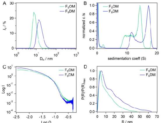
Size and shape of the micelles. We next investigated the self-assembly properties of the FSs
in phosphate buffer using DLS. At 10 mM, volume-weighted particle size distributions for
F5OM and F5DM revealed unimodal distributions of rather small micelles with hydrodynamic
diameters ranging from ∼8 nm for F5OM to ∼15 nm for F5DM (Figure 3A). Upon dilution to
5 mM, no significant difference in the volume-weighted distributions was observed for both compounds yet with a small decrease of the hydrodynamic diameters to ∼7 nm for F5OM and
to ∼13 nm for F5DM (data not shown).
Figure 3. (A) Volume-weighted particle size distributions for F5OM and F5DM at 10 mM in
phosphate buffer. (B) Distributions of sedimentation, c(s), for F5OM at 10.5 mM and F5DM at
7.7 mM. (C) SAXS patterns, and (D) pair distribution functions for F5OM at 23.6 mM and
22
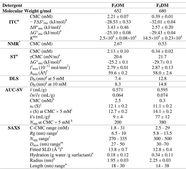
Table 1. Micellar Properties of Maltose Derivatives.
Detergent F5OM F5DM
Molecular Weight g/mol 652 680
ITCa CMC (mM) − TΔS°mic (kJ/mol)b ΔH°mic (kJ/mol)c ΔG°mic (kJ/mol)d Kmic 2.21 ± 0.07 –28.53 ± 0.53 3.43 ± 0.46 –25.10 ± 0.08 2.5×10⁴ ± 0.08×10⁴ 0.39 ± 0.01 –32.01 ± 0.04 2.57 ± 0.20 –29.43 ± 0.04 14.5×10⁴ ± 0.23×10⁴ NMR* CMC (mM) 2.67 0.53 STa CMC (mM) γCMC (mN/m)e ΔG°mic (kJ/mol)d 2.13 ± 0.10 20.6 -25.2 ± 0.1 0.34 ± 0.02 21.7 -29.7± 0.1 Γmax (10–12 mol/mm2) 2.79 ± 0.01 2.87 ± 0.13 Amin (Ų)f 59.6 ± 0.2 58.0 ± 2.6 DLS DH (nm)g at 5 mM 7.4 12.8 DH (nm)g at 10 mM 8.3 14.8 AUC-SV (mL/g) ∂n/∂c (mL/g) CMC (mM)h s0 (S)i s (S) at CMC + 5 mMi k′s (mL/g)j Nagg at CMC + 5 mM k 0.571 0.064 2.5 12.1 ± 0.2 12.7 ± 0.2 9 ± 4 200 0.595 0.074 0.3 11.1 ± 0.2 14.1 ± 0.2 77 ± 12 380 SAXS C-CMC range (mM) Rg (nm) rangel Nagg rangel Dmax (nm) rangem Fitted SLD (Å-2)n
Hydration (g water /g surfactant)n Radius (nm)n Length (nm) rangen 1.8 - 31 6.5 - 10 270 - 335 27 - 50 13.8 ± 0.7 0.18 ± 0.12 1.95 ± 0.03 18 - 30 2.5 - 29 5.8 – 13.5 300 - 500 30 -70 12.8 ± 0.4 0.34 ± 0.11 2.25 ± 0.03 14 - 38
aData are averages of at least two experiments unless noted by * for one experiment only. ± indicates 95% confidence interval boundaries from a nonlinear least-squares fit for ITC. ± indicates standard errors from at least two experiments for SFT, AUC and SAXS. bEntropic contribution to micelle formation. cEnthalpic contribution to micelle formation. dGibbs free energy of micellization. eSurface tension attained at the CMC. fThe surface excess (
max) and the surface area per molecule (Ų) were estimated from the slope of the surface tension curve. gHydrodynamic diameter by volume in phosphate buffer. iSedimentation coefficient at infinite dilution (s0), or linearly interpolated from experimental data at CMC+ 5 mM, in water at 20 °C. jConcentration dependence factor k’s from linear fits. kAggregation number obtained from s, and DH. Error is estimated at 10%. lradius of gyration and aggregation numbers from Guinier analysis. mMaximum distance from P(R) analysis. nFitted scattering length density, derived hydration, radius and length considering a cylinder model.
To further characterize the micellar aggregates, AUC sedimentation velocity experiments
were performed. Figure S2A displays the sedimentation velocity profiles. From the c(s)
23
F5OM and 10-18 S for F5DM (Figures 3B, S2B and S2B’). The s value increased with
concentration, with a more pronounced effect for F5DM than for F5OM (Figure S2D). We
used the dilution series to determine, from the micelle signals versus concentration, the refractive index increment (∂n/∂c) and the CMC, as well as the s value at infinite dilution (s0)
and the concentration dependence factor (k’s) (Figure S2C and Table 1). For both detergents,
AUC provided CMC values relatively similar to those obtained from NMR, SFT, and ITC
(Table 1). The s0 values are similar, but sedimentation coefficients vary with concentration to
different extents for the two surfactants as illustrated by the k’s values. A realistic estimate of
aggregation numbers, Nagg, is obtained by combining the values of s with that of the
hydrodynamic diameters from DLS. Calculated Nagg at the CMC+5mM are lower for F5OM
compared to F5DM (Table 1).
To complete the colloidal characterization of the two compounds, SAXS experiments were
next performed. Figure 3C shows the scattering curves for F5OM and F5DM whose
similarities in the shape suggest similar micelle organization (See Figure S3 for detailed
analysis). Guinier analysis at low angle (Figure S3B) provided mean radius of gyration (Rg)
and Nagg, which increase with concentrations, moderately for F5OM and to a larger extent for
F5DM (Table 1 and Figure S4). Pair distribution functions, P(R), derived from the whole
scattering curves (Figure 3D and S3C) present for F5OM a main maximum at 3.5 nm which
remains invariant while it increases slightly with concentration for F5DM from 3.6 to 4.6 nm.
All curves present at larger R a linear decrease, which indicates a linear rod shape for the
aggregates.39 The largest distance, Dmax, corresponding to P(R) reaching zero, increases with
concentration for both surfactants, and in minor extent for F5OM compared to F5DM (from
30 for both to 30 and 50 nm, respectively). Lastly, we analyzed the scattering curves
considering a cylinder with hard sphere interaction. The fitted and experimental curves are
24
results, and Table 1 reports the main conclusions. The values of the fitted scattering length
density (SLD) do not depend on surfactant concentration, as expected, and are intermediate
between anhydrous surfactant and water SLDs. We derived reasonable hydration of 0.18 ±
0.12 and 0.34 ± 0.11 g of water per g of surfactant. Fitted radius do not vary with surfactant
concentration: 1.95 and 2.25 nm for F5OM and F5DM, respectively. It is comparable to the
sum, determined from SAXS and SANS, of the core radius and shell thickness, for the small dimension ( 2.1 nm) of the slightly elongated DDM micelle,12, 40-41
or for the lateral
dimension (2.2 nm) of the rod-forming detergent LMNG, which also bears maltose heads and
have two dodecyl chains.12 Because the fitted SLD and radius-values are correlated with the
concentration input values, the minor differences in the fitted hydration and radius for the two
surfactants may be irrelevant. The length is 15 nm at the lowest concentrations, and reaches 30 and 38 nm at 30 mM F5OM and F5DM, respectively (Figure S3D). These values
correspond to length/diameter ratio of 8. We note that the fitted length is about half Dmax. A
tentative explanation is that there is a distribution in length. Dmax probes the largest molecules,
while the fit considers the most populated dimensions.
The larger micelle size above 5mM, for F5DM versus F5OM observed from AUC and SAXS
is in line with what is generally observed for hydrogenated42 and fluorinated detergents.20
Comparing with fluorinated compounds with the same OM head-group, while the
hydrogenated DDM forms small slightly elongated micelles of 60 kDa up to at least 10 mM
29
, the poorly fluorinated F2UDM and the nonyl derivative with four perfluorinated carbons
F4NM (also called F4H5Malt)22were described to form small micelles with Nagg < 100 up to
C-CMC 10 mM (s of 4 and 7 S at C-CMC + 5 mM), the later experiencing very slightly attractive interactions evidenced only above 30 mM. The commercial F6OM forms very large
rod micelles with Nagg > 500 at CMC + 10 mM (s of 27 S at CMC + 5 mM). The propensity
25
fluorinated surfactants with a head bearing two glucose groups, which self-assemble into
compact and well-defined globular micelles of 6–8 nm in diameter with aggregation numbers
below 100.20
Solubilization of POPC LUVs by FSs. The detergency reflects the ability of an amphiphilic
compound to both solubilize lipid bilayers and extract MPs. To assess the detergency of
F5OM and F5DM, we tested whether they are able to dissolve large unilamellar vesicles
(LUVs) composed of the singly unsaturated phospholipid
1-palmitoyl-2-oleyl-sn-glycero-3-phosphocholine (POPC). Measurements were conducted at 25°C by adding a rather high
concentration (CMC+5 mM) of the respective FS to 100 μM POPC LUVs, which resulted in a
steady decrease in the light scattering intensity over time. The particle size distributions
shown in Figure 4 support the interpretation that the decreased light scattering intensity was
due to vesicle solubilization, as the vesicular peak at ∼120 nm at the beginning of the measurement completely disappeared after the intensity decreased to the level of pure mixed
micelles. Solubilization was essentially complete after ∼16 h for F5DM but took longer for
F5OM. Most importantly, however, both FSs were able to solubilize synthetic POPC vesicles
at 25°C, which sets them apart from more conventional FSs such as F6OM, F4H2-DigluM,
F6H2-DigluM, and F8H2-DigluM, which require elevated temperatures and prolonged
26
Figure 4. Kinetics of 100 μM POPC LUVs solubilization by 7.2 mM F5OM & 5.4 mM F5DM
at 25°C as monitored in terms of the light scattering intensity recorded at an angle of 90°. The inset shows intensity-weighted size distributions obtained for a mixture of 100 μM POPC and F5DM immediately (green) or after 16h (blue). Buffer: 10 mM phosphate, 150 mM NaCl, pH
7.4.
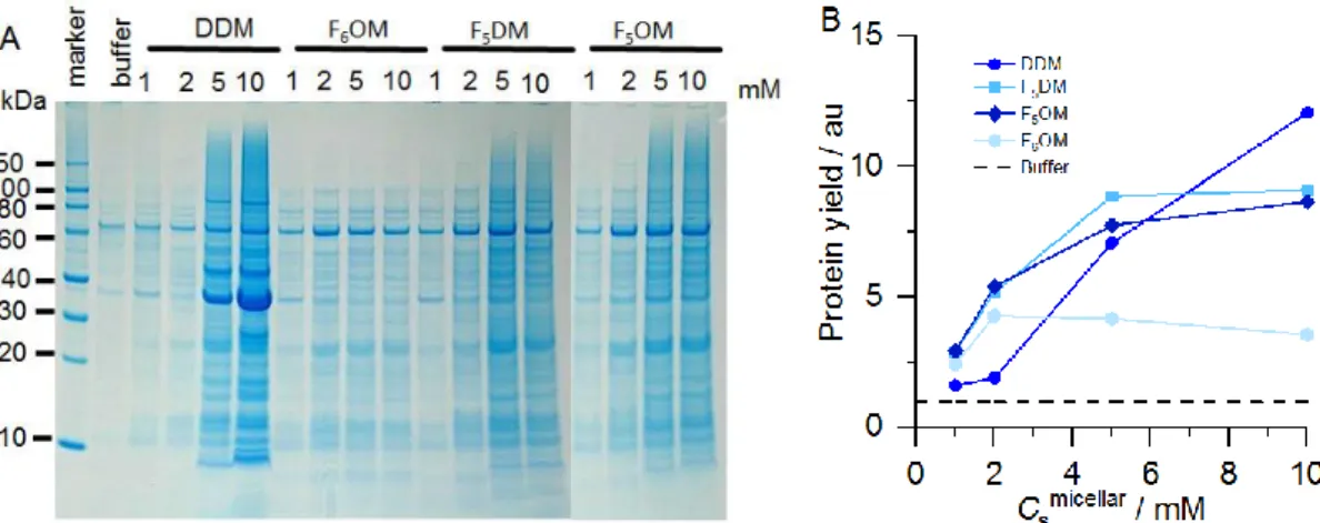
Extraction of MPs from native E. coli membranes. Next, we investigated whether F5OM
and F5DM can also extract MPs from native E. coli membranes. To this end, we quantified the
intensities (i.e., pixel counts) of SDS-PAGE band patterns (Figure 5A) and compared their
efficiencies with those of DDM and F6OM. The overall protein-extraction yields were also
expressed relative to the buffer without any detergent (Figure 5B). Figure 5A indicates that
both F5OM and F5DM extracted similar patterns of MPs spanning a broad size range.
Notably, at low concentrations (i.e., 1–5 mM), both FSs displayed better solubilization
efficiencies than DDM, although DDM was outstanding in extracting a single abundant
protein of ∼35 kDa, namely, outer-membrane protein OmpA, at higher concentrations. By contrast F6OM showed very limited solubilization.
27
Figure 5. (A) SDS-PAGE of E. coli membrane extracts upon exposure to various FSs with micellar concentrations as indicated. (B) Graphical representation of protein-extraction yields (symbols) when using surfactant relative to the yield obtained when no surfactant was added (i.e., only buffer; dashed line). Data are mean values from three experiments.
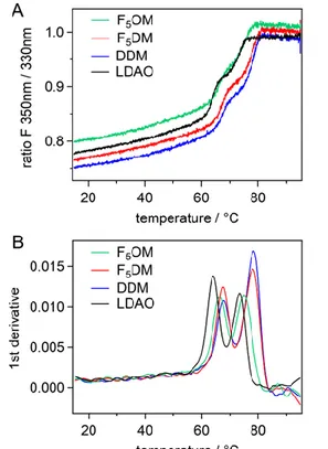
FhuA and bR thermal stability. To assess the stability of MPs in the new FSs, we
investigated the thermal stability of two models proteins. Differential scanning fluorimetry
(DSF) probes conformation changes with temperature. It allows measuring the melting
temperature (Tm) of the protein by measuring the fluorescence emission (F350nm/F330nm ratio)
of the aromatic residues upon increasing temperature. Simultaneous light back-reflection
measurement probes protein aggregation, Tagg being the onset temperature for aggregation.
FhuA is an E. coli outer membrane ferrichrome-iron transporter involved in bacteriophage
infection.43 bR is a light-driven proton pump purified from the archaea Halobacterium. It
binds a covalent cofactor, a retinal molecule that confers a purple color to the protein.44 We
use the two proteins, representatives of the two main structural classes ß-barrels and -helix bundle, of proteins, to investigate their thermal stabilities in the presence of our fluorinated
derivatives. The two proteins were first extracted by lauryldimethylamine oxide (LDAO) for
FhuA and OTG for bR, and then transferred into F5OM and F5DM, as well as in DDM and the
solubilizing detergent, at CMC + 0.2 mM, and CMC + 2 mM. Final residual concentrations of
the initial detergents, LDAO for FhuA and OTG for bR, were 0.05 and 0.4 CMC. FhuA
28
and then of the barrel, at Tm2,45 while bR shows only one transition (Figure S5). For each of
the two proteins, the melting curves general appearance is similar whatever the detergent and
its concentration. Table 2 presents the mean values of Tm and Tagg. For FhuA and bR, in
LDAO or OTG the extracting detergents, Tm are lower than that in F5OM, F5DM or DDM
suggesting a thermostabilizing effect of the three maltoside derivatives.
Figure 6. Thermal denaturation of FhuA by differential scanning fluorimetry. (A) Ratio of the fluorescence emitted at 350 and 330 nm, and (B) derivative (bottom panel) for FhuA at 0.04 mg mL-1, incubated in the presence of F5OM at CMC+2mM (green), F5DM at CMC+2mM
(red), DDM at CMC+0.2mM (blue), LDAO at CMC+2 mM (black).
Table 2. Melting temperatures of FhuA and bR
Protein type
Detergent Concentration (mM) Tm (°C) Tagg (°C)
Detergent Micelle Tm1 Tm2 FhuA LDAO 1.20, 3.00 0.2, 2.0 64 73 72 DDM 0.37, 2.17 0.2, 2.0 68 79 73 F5OM 3.00, 4.80 0.2, 2.0 66 75 70 F5DM 0.60, 2.40 0.2, 2.0 68 78 73 bR OTG 9.2, 11.0 0.2, 2.0 54 43
29
DDM 0.37, 2.17 0.2, 2.0 60 54
F5OM 3.00, 4.80 0.2, 2.0 59 50
F5DM 0.60, 2.40 0.2, 2.0 58 55
Tm1, Tm2, Tm: melting temperatures measured by differential scanning fluorimetry for the first and second transition of FhuA, and the transition for bR, Tagg: onset temperature for aggregation from light back reflexion. The precision on Tm1 is estimated at 1° C, that for Tm2 and Tm, which are above Tagg, at 2° C. The precision on Tagg is 2°C. Micelle concentrations (i.e. above the CMC concentrations) were calculated considering CMC-values of 1, 0.17, 2.8, and 0.4 mM for LDAO, DDM, F5OM, and F5DM, respectively
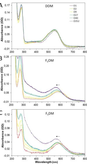
Homogeneity and stability of bR over time. The retinal molecule bound to bR, whose
visible absorption spectrum is very sensitive to its local environment, is a convenient reporter
of the state of the protein.32 Figure 7 reports bR absorption spectra after detergent exchange,
over time, in DDM, F5OM and F5DM. Pink monomeric bR in DDM displays a λmax = 550
nm. In F5OM or F5DM, just after detergent exchange, the λmax ~ 595 nm is compatible with
the observed bR blue color, and reflects a monomeric state, as observed previously in various
fluorinated surfactants.35 After two days incubation, λmax shifts to ~ 575 nm (close to the λmax
of native bR), and remains unchanged for one year; the absence of λmax = 390 nm, reporter of
free retinal, reflects the absence of protein denaturation; over time, the spectra show
scattering, witness of progressive but very minor aggregation. These three observations
suggest clustering of monomeric bR into larger, native-like oligomers. Thus, the protein is
extremely stable as regards its conformation, but its colloidal stability is not as good as that in
DDM. When comparing with the previous fluorinated compounds of the maltose series, some
differences can be noted: 1- in F2UDM and F4NM, bR was blue (max ~ 610 nm) and
remained so unless it denatured; 2- in F2UDM, bR was not stable, denatured and aggregated ;
3- in F6OM, bR was not soluble and aggregated during surfactant exchange.22 Thus, both
F5OM and F5DM appear more solubilizing than F6OM, more stabilizing than F2UDM and
providing a more native environment than F4NM. Thus, an optimized F/H ratio has been
found in those two compounds, providing solubility, stability and close-to-native environment
30
Figure 7. Spectral time course of bR collected from the gradients in (A) DDM, (B) F5OM and
(C) F5OM. Samples were incubated at 4°C in the dark and UV-visible spectra were recorded
at the indicated time (given in days, D). Day 1 curve is displayed in red, day 2 in orange, day 9 in yellow, day 27 in green, day 40 in cyan and day 357 in violet. The arrow indicates the evolution of max with time.
Specific activity of BmrA and SpNOX
We investigated the enzymatic stability of two MPs, SpNOX, a Streptococcus pneumoniae
protein analog to the eukaryotic NADPH oxidase24 and BmrA, a transporter of multiple drugs
with the driving force of ATP hydrolysis.23The activity results following detergent exchange
are displayed on Figure S6. The specific activity of BmrA and SpNOX is partially preserved,
31
Conclusion
We have designed two maltose-based fluorinated surfactants, F5OM and F5DM, whose
hydrophobic tails are made up of linear fluoroalkyl chains. Formation of micelles in water
occurred as governed by the length of the hydrophobic chain. F5OM and F5DM
self-assembled into rod-like micelles, with hydrodynamic diameters and aggregation numbers that
increased with chain length. The potencies of the new FSs to act as detergents was first
demonstrated using synthetic POPC lipid vesicles and were further confirmed through the
extraction of MPs from E. coli membranes. Both F5OM and F5DM showed detergency with
more solubilizing activity than the commercial fluorinated compound F6OM at all
concentrations and even better protein-extraction efficiency than DDM at low concentrations.
F5DM consistently exhibited better solubilizing properties than F5OM, towards both lipid
vesicles and MPs. The detergency of the two derivatives exceeded by far that of the
fluorinated DigluM derivatives F4H2-, F6H2- and F8H2DigluM. This suggests that the linear
maltoside polar head, likely owing to its small size, may promote detergency as compared
with the bulky branched diglucose polar head. BR and FhuA, representatives of -helical and ß-barrel proteins, showed remarkable thermal and, for bR, also functional stability, similar to
DDM, when transferred into both F5OM and F5DM. These surfactants appear, for bR, better
than the commercial F6OM in which the protein aggregatedmore stabilizing than F2UDM and
provided a more native environment than F4NM. This indicates that an optimized F/H ratio
has been identified in those two compounds, providing solubility, stability, and a
close-to-native environment for bR. The enzymatic activities of BmrA and SpNox, were by contrast
rather limited when compared to DDM, and similar for F5OM, F5H5OM and F6OM. Taken
together, these findings support the usefulness of this novel series of fluorinated maltoside
32
ASSOCIATED CONTENT
Supporting Information
The supporting information is available free of charge on the ACS Publication website at
DOI: #
ITC, 19F NMR and surface tension data curves of F5OM; Analysis of sedimentation velocity
experiments; Small Angle X-Ray Scattering complementary data of F5OM and F5DM
including concentration dependence analysis and cylinder shape fitting; Thermal denaturation
of bR by differential scanning fluorimetry; Material and Methods for BmrA and SpNox
production and SpNox and BmrA activity assays; SpNox and BmrA activity data in F6OM,
F5OM and F5DM; 1
H and 13C NMR spectra and mass spectrometry data of compounds 2a and 2b; 1H, 19F and
13
C NMR spectra and mass spectrometry data of compounds 3a, 3b, 4a (F5OM) and 4b
(F5DM).
Acknowledgment.
This work was supported by the Agence Nationale de la Recherche (ANR) through grants no.
ANR-16-CE92-0001, by the Deutsche Forschungsgemeinschaft (DFG) through grant no. KE
1478/7 1, and by the Deutsche Akademische Austauschdienst (DAAD) with a grant to K.K.O.
We acknowledge the financial support of the European Regional Development Fund, the French Government, the “Région Provence Alpes Côte d'Azur”, the “Département de Vaucluse” and the “Communauté d’agglomération Grand Avignon” for access to the NMR
platform (CPER 3A). This work used the platforms of the Grenoble Instruct-ERIC center
(ISBG; UMS 3518 CNRS-CEA-UGA-EMBL) within the Grenoble Partnership for Structural
Biology (PSB), supported by FRISBI (ANR-10-INBS-05-02) and GRAL, financed within the
University Grenoble Alpes graduate school (Ecoles Universitaires de Recherche)
33
Institute of Grenoble (IRIG, CEA) We thank Martha Brennich (ESRF) for help in SAXS data
acquisition and Marine Soulié (IBMM) for carefully checking NMR data. Emmi Mikkola
(UGA) participated to the biochemical evaluation during her master internships at IBS. This
work benefited from the use of the SasView application, originally developed under NSF
Award DMR- 0520547. SasView also contains code developed with funding from the EU
34
References
1. Overington, J. P.; Al-Lazikani, B.; Hopkins, A. L., How many drug targets are there?
Nature Reviews Drug Discovery 2006, 5 (12), 993-996.
2. Arachea, B. T.; Sun, Z.; Potente, N.; Malik, R.; Isailovic, D.; Viola, R. E., Detergent selection for enhanced extraction of membrane proteins. Protein expression and purification 2012, 86 (1), 12-20.
3. Morandat, S.; El Kirat, K., Solubilization of supported lipid membranes by octyl glucoside observed by time-lapse atomic force microscopy. Colloids and Surfaces B:
Biointerfaces 2007, 55 (2), 179-184.
4. Vacklin, H. P.; Tiberg, F.; Thomas, R. K., Formation of supported phospholipid bilayers via co-adsorption with β-d-dodecyl maltoside. Biochimica et Biophysica Acta (BBA) -
Biomembranes 2005, 1668 (1), 17-24.
5. Ai, X.; Caffrey, M., Membrane Protein Crystallization in Lipidic Mesophases: Detergent Effects. Biophysical Journal 2000, 79 (1), 394-405.
6. Rouse, S. L.; Marcoux, J.; Robinson, C. V.; Sansom, M. S. P., Dodecyl maltoside protects membrane proteins in vacuo. Biophysical Journal 2013, 105 (3), 648-656.
7. Lebaupain, F.; Salvay, A. G.; Olivier, B.; Durand, G.; Fabiano, A.-S.; Michel, N.; Popot, J.-L.; Ebel, C.; Breyton, C.; Pucci, B., Lactobionamide Surfactants with Hydrogenated, Perfluorinated or Hemifluorinated Tails: Physical-Chemical and Biochemical Characterization. Langmuir 2006, 22 (21), 8881-8890.
8. Fayolle, D.; Berthet, N.; Doumeche, B.; Renaudet, O.; Strazewski, P.; Fiore, M., Towards the preparation of synthetic outer membrane vesicle models with micromolar affinity to wheat germ agglutinin using a dialkyl thioglycoside. Beilstein Journal of Organic
Chemistry 2019, 15, 937-946.
9. Asada, A.; Sonoyama, M., Solubilization and Structural Stability of Bacteriorhodopsin with a Mild Nonionic Detergent, n-Octyl-β-thioglucoside. Bioscience, Biotechnology, and
Biochemistry 2011, 75 (2), 376-378.
10. Guillet, P.; Mahler, F.; Garnier, K.; Nyame Mendendy Boussambe, G.; Igonet, S.; Vargas, C.; Ebel, C.; Soulié, M.; Keller, S.; Jawhari, A.; Durand, G., Hydrogenated Diglucose Detergents for Membrane-Protein Extraction and Stabilization. Langmuir 2019, 35 (12), 4287-4295.
35
11. Dauvergne, J.; Desuzinges, E. M.; Faugier, C.; Igonet, S.; Soulié, M.; Grousson, E.; Cornut, D.; Bonneté, F.; Durand, G.; Dejean, E.; Jawhari, A., Glycosylated Amphiphilic Calixarene-Based Detergent for Functional Stabilization of Native Membrane Proteins.
ChemistrySelect 2019, 4 (19), 5535-5539.
12. Breyton, C.; Javed, W.; Vermot, A.; Arnaud, C.-A.; Hajjar, C.; Dupuy, J.; Petit-Hartlein, I.; Le Roy, A.; Martel, A.; Thépaut, M.; Orelle, C.; Jault, J.-M.; Fieschi, F.; Porcar, L.; Ebel, C., Assemblies of lauryl maltose neopentyl glycol (LMNG) and LMNG-solubilized membrane proteins. Biochimica et Biophysica Acta (BBA) - Biomembranes 2019, 1861 (5), 939-957.
13. Cho, K. H.; Husri, M.; Amin, A.; Gotfryd, K.; Lee, H. J.; Go, J.; Kim, J. W.; Loland, C. J.; Guan, L.; Byrne, B.; Chae, P. S., Maltose neopentyl glycol-3 (MNG-3) analogues for membrane protein study. Analyst 2015, 140 (9), 3157-3163.
14. Chae, P. S.; Rasmussen, S. G. F.; Rana, R. R.; Gotfryd, K.; Chandra, R.; Goren, M. A.; Kruse, A. C.; Nurva, S.; Loland, C. J.; Pierre, Y.; Drew, D.; Popot, J.-L.; Picot, D.; Fox, B. G.; Guan, L.; Gether, U.; Byrne, B.; Kobilka, B.; Gellman, S. H., Maltose–neopentyl glycol (MNG) amphiphiles for solubilization, stabilization and crystallization of membrane proteins. Nature Methods 2010, 7, 1003.
15. Sadtler, V. M.; Giulieri, F.; Krafft, M. P.; Riess, J. G., Micellization and Adsorption of Fluorinated Amphiphiles: Questioning the 1 CF2 1.5 CH2 Rule. Chemistry – A European
Journal 1998, 4 (10), 1952-1956.
16. Krafft, M. P.; Riess, J. G., Highly fluorinated amphiphiles and colloidal systems, and their applications in the biomedical field. A contribution. Biochimie 1998, 80 (5), 489-514.
17. Riess, J. G.; Krafft, M. P., Fluorinated materials for in vivo oxygen transport (blood substitutes), diagnosis and drug delivery. Biomaterials 1998, 19 (16), 1529-1539.
18. Durand, G.; Abla, M.; Ebel, C.; Breyton, C., New Amphiphiles to Handle Membrane Proteins: “Ménage à Trois” Between Chemistry, Physical Chemistry, and Biochemistry. In
Membrane Proteins Production for Structural Analysis, Mus-Veteau, I., Ed. Springer New
York: 2014; pp 205-251.
19. Frotscher, E.; Danielczak, B.; Vargas, C.; Meister, A.; Durand, G.; Keller, S., A Fluorinated Detergent for Membrane-Protein Applications. Angewandte Chemie International