HAL Id: hal-02396090
https://hal.archives-ouvertes.fr/hal-02396090
Submitted on 30 Nov 2020HAL is a multi-disciplinary open access archive for the deposit and dissemination of sci-entific research documents, whether they are pub-lished or not. The documents may come from teaching and research institutions in France or abroad, or from public or private research centers.
L’archive ouverte pluridisciplinaire HAL, est destinée au dépôt et à la diffusion de documents scientifiques de niveau recherche, publiés ou non, émanant des établissements d’enseignement et de recherche français ou étrangers, des laboratoires publics ou privés.
Dissociation between objective and subjective
perceptual experiences in a population of hemianopic
patients: A new form of blindsight?
Clémentine Garric, Aïda Sebaa, Florent Caetta, Céline Perez, Julien
Savatovsky, Claire Sergent, Sylvie Chokron
To cite this version:
Clémentine Garric, Aïda Sebaa, Florent Caetta, Céline Perez, Julien Savatovsky, et al.. Dissociation between objective and subjective perceptual experiences in a population of hemianopic patients: A new form of blindsight?. Cortex, Elsevier, 2019, 117, pp.299-310. �10.1016/j.cortex.2019.05.006�. �hal-02396090�
p. 1
Title:
Dissociation between objective and subjective perceptual experiences in a population of hemianopic patients: A new form of blindsight? (135)
Short Title:
Sensitivity in the blind visual field (38)
Authors:
Clémentine Garrica,b, Aïda Sebaaa, Florent Caettaa, Céline Pereza,c, Julien Savatovsky d, Claire Sergentb and Sylvie Chokrona,b,c
a Unité Vision et Cognition, Fondation Ophtalmologique de Rothschild, Paris, 75019 France b Laboratoire de Psychologie de la Perception, UMR 8242, CNRS & Université Paris-Descartes,
Paris, 75006 France
c Service de Neurologie, Fondation Ophtalmologique Rothschild, Paris, 75019 France d Service d’Imagerie, Fondation Ophtalmologique Rothschild, Paris, 75019 France
Corresponding author:
Sylvie Chokron
Unité Vision et Cognition, Fondation Ophtalmologique de Rothschild, 29 rue Manin, Paris, 75019 France
+33148036672, sylvie.chokron@gmail.com
Keywords:
blindsight, hemianopia, post-chiasmatic lesion, blind visual field, awareness
Declaration of interest:
p. 2
Abstract
After a post-chiasmatic lesion, some patients may retain unconscious visual function, known as blindsight, in their contralesional visual field. Despite the importance of blindsight in the study of
consciousness, little is known about the nature of patients’ experience in their hemianopic field.
To address this knowledge gap, we measured blindsight, and assessed the perceptual experience in the contralesional visual field, of seventeen homonymous hemianopic (HH) patients. To ensure that the stimuli were shown in a “blind” sector of the visual field, we selected a subgroup of eight complete-HH patients, as determined by automatic perimetry. Firstly, we measured blindsight through a forced-choice task in which the patients had to identify letters displayed on a screen. Secondly, we compared the patients’ binary responses (“Something was presented” vs. “Nothing was presented”) to responses on a new, five-level scale, the Sensation Awareness Scale (SAS), which we designed to include visual as well as non-visual answers (e.g. “I felt something”). Interestingly, only one of the eight complete-HH patients met the criteria for blindsight. More importantly, our SAS enabled us to identify a previously unreported dissociation, which we have named blindsense, in four of the eight complete-HH patients. Specifically, these four patients exhibited better-than-chance sensitivity to the presence of a stimulus on the subjective scale, despite being unable to identify the stimulus during the forced-choice task. Our findings highlight the importance of awareness-assessment methods to investigate perceptual experiences in the contralesional visual field and suggest a low incidence of blindsight in post-stroke HH patients.
Significance Statement
To investigate the nature of the perceptual experience in the blind visual field, we submitted a group of HH patients to visual letter-detection and letter-identification tasks and assessed them by the Sensation Awareness Scale (SAS). Measuring their objective and subjective performance in
their blind visual field enabled us to identify a previously unknown clinical profile, which we have named blindsense and which differs from both types (I and II) of blindsight. Our findings raise important questions on patients’ perceptual experiences in the blind visual field following a post-chiasmatic lesion and underscore the complexity of classifying blindsight and related phenomena.
p. 3
1. Introduction
The most common visual defect induced by a unilateral lesion of the retrochiasmal pathways is homonymous hemianopia (HH) (Cavézian et al., 2010; Zhang, Kedar, Lynn, Newman, & Biousse, 2006), whereby patients are blind in the contralesional visual field of each eye. Hemianopic patients can exhibit implicit residual capacities in the so-called “blind” visual field, despite exhibiting a severe contralesional visual-field defect. Such residual capacities are usually referred to as blindsight, a term coined by Weiskrantz et al. (1974) that has inspired debate (Cowey, 2010; Overgaard & Mogensen, 2015; Sanders, Warrington, Marshall, & Weiskrantz, 1974; Stoerig, 2006; Weiskrantz, 1972). Blindsight was first described in patients who could accurately steer their gaze toward a flashing light presented in their blind field, yet who did not report having perceived the stimulation (Poppel, Held, & Frost, 1973). Another blindsight patient, G.Y., reported awareness of certain stimuli despite not attesting to any concrete visual perception of them (Weiskrantz, Barbur, & Sahraie, 1995). Consequently, blindsight was eventually classified into two types: type I, the ability to discriminate specific attributes of a stimulus in the contralesional visual field without any awareness of it; and type II: exhibiting objective performance and subjective performance in the blind visual field above
chance level, without any conscious detection of the stimuli (Brogaard, 2015; Weiskrantz, 1998).
Diagnosing blindsight implies both accurate measurement of patients’ objective performance, and well-controlled and interpretable measurement of their subjective experience, which can depend on the methodology used to assess their awareness (Fayel et al., 2014). For example, use of a binary scale (i.e. Aware vs. Unaware) may compel a subject to report being “unaware” of a stimulus despite having experienced a minor degree of awareness (Overgaard, 2011; Overgaard, Fehl, Mouridsen, Bergholt, & Cleeremans, 2008). In one study, the patient G.R. exhibited distinct responses in his blind visual field in function of whether a binary scale (Seen vs. Not Seen) or the Perceptual Awareness Scale (PAS) (Ramsøy & Overgaard, 2004) was used, the latter of which comprises four levels: Clear Experience, Almost Clear Experience, Weak Glimpse and Not Seen. Indeed, when asked to process a stimulus in his contralesional visual field and subsequently report a dichotomous answer (Seen vs. Not Seen), G.R. appeared to exhibit a typical type-I-blindsight profile. However, when assessed by the PAS, he strongly reported some degree of awareness in the presence of stimuli in his contralesional visual field, consistent with a type-II profile. Despite this proven drawback, binary questionnaires are still widely used to study blindsight; indeed, very few researchers use graduated measurements of perception (Mazzi, Bagattini, & Savazzi, 2016; Overgaard et al., 2008). Thus, our aim in the present study was to assess blindsight in several HH patients using a subjective report on a refined scale to better understand what these patients perceive and experience in their contralesional visual field.
p. 4
1.1. Questioning the nature of perceptual experience
Among the most controversial issues surrounding blindsight, is that of determining how to understand the nature of patients’ perceptual experience in their blind field. Many authors affirm that blindsight is based on unconscious perceptual abilities (Leh, 2006; Sanders et al., 1974). However, two other, competing hypotheses have been proposed to explain blindsight (for a review, see (Cowey, 2010; Hadid & Lepore, 2017)): that of residual remaining vision; and that of degraded, abnormal vision, which arose from studies on type II blindsight and has been described as a “conscious experience, though of a very different nature to that of normal vision” (Kentridge, 2015; Mazzi et al., 2016; Weiskrantz, 2009). Corroborating the residual normal-vision hypothesis, researchers have shown that blindsight could be triggered by the activation of sparse islands of surviving tissue in the primary cortex (V1) (Campion, Latto, & Smith, 1983; Fendrich, Wessinger, & Gazzaniga, 2001). However, blindsight aptitudes have also been found in patients that lack a functional primary cortex (Ajina, Pestilli, Rokem, Kennard, & Bridge, 2015; Mazzi et al., 2016) and in monkeys with complete ablation of V1 (Stoerig & Cowey, 1997). In those cases, blindsight seems to be mediated by subcortical pathways that bypass V1, such as the superior colliculus pathway and the dorsal lateral geniculate nucleus (dLGN) pathways. Based on a meta-analysis of human and monkey data, researchers have concluded that blindsight can indeed occur without any functional portion of the primary visual cortex (Stoerig, 2006). Moreover, several recent studies have underscored the roles of the ipsilateral hemisphere and the corpus callosum in processing of stimuli in the contralesional visual field of HH patients (Bridge, Thomas, Jbabdi, & Cowey, 2008; Celeghin et al., 2017; Leh, 2006).
Currently, there is a lack of consensus on the nature of perceptual experience in blindsight (i.e. totally unconscious vision, residual conscious vision or degraded abnormal vision), which is probably due to the fact that research in this area mainly comprises case studies, often conducted with highly trained patients (e.g. D.B or G.Y; for a review, see (Cowey, 2010)) that are familiar with the experimental procedures involved. Therefore, even though blindsight is central to the study of consciousness, there remains a lack of basic epidemiologic data on it (e.g. incidence of blindsight among HH patients).
p. 5
1.2. Towards population studies of blindsight
Understanding blindsight at the patient-population level would be beneficial both to clinicians working in rehabilitation and to researchers studying the influence of clinical parameters (e.g. the localization, side or size of the brain lesion; or the type of visual-field defect). Only a few studies have been conducted on (small) patient cohorts, and these provide only indirect information on blindsight incidence. For instance, Perenin and Jeannerod found blindsight in six out of eight (75%) patients (Perenin & Jeannerod, 1975). Ajina et al. studied the anatomical bases of blindsight in seventeen HH patients, twelve (71%) of whom they classified as “blindsight positive” and five (29%) as “blindsight negative”, based on two alternative forced-choice detection tasks related to the contralesional visual field (Ajina et al., 2015). Alternatively, when blindsight was measured in a cohort of twenty patients through a Redundant Signal Effect (RSE) task involving indirect flash presentation to assess visual-information processing (Marzi, Tassinari, Aglioti, & Lutzemberger, 1986), the observed incidence was only 5%. However, in a group study of nineteen patients, in which their pupil responses to visual stimuli presented in their contralesional visual field was used as a proxy for residual visual reflexes, 13 patients (70%) were reported to exhibit blindsight (Sahraie, Trevethan, MacLeod, Urquhart, & Weiskrantz, 2013). The above examples indicate that, despite the many articles on blindsight published over the past four decades, there remains a need for systematic investigation of the dissociation between conscious visual detection and non-conscious visual processing abilities in patient groups.
In the work reported here, our aim was two-fold: firstly, to re-evaluate the incidence of blindsight in a population of HH patients by measuring objective performance in two visual tasks; and secondly, to investigate the nature of the perceptual experience in the blind visual field, which has only been examined in single-case studies so far. According to clinical observations, the PAS, a four-level scale based on healthy subjects’ spontaneous reports, was not entirely appropriate for evaluating the experience of patients with visual-field defects. Thus, we designed the Sensation Awareness Scale (SAS), including a critical level that does not refer to visual experience. Thus, the complete SAS comprises the following assertions: [1] I did not see anything; [2] I don’t think that I saw anything, but I am not sure; [3] I felt something; [4] I saw something; and [5] I clearly saw something and can identify it. Subjective performance was calculated based on the SAS and subsequently compared to the objective performance, which was evaluated through forced-choice detection and identification tasks. Our experimental design enabled us to reclassify our HH patients according to the dissociation between objective and subjective performance and by different concepts of blindsight described in the literature.
p. 6
Thus, we hypothesized that HH (and perhaps other) patients could exhibit the following profiles:
Absence of blindsight: objective performance and subjective performance both at chance
level;
Type I blindsight: objective performance above chance level, without any statistically
significant subjective sensitivity;
Type II blindsight: objective performance and subjective performance both above chance
level, without any conscious detection in the blind visual field;
Blindsense: objective performance at chance level combined with a statistically significant
subjective performance*;
* Note: although this has not yet been defined or labeled as a specific phenomenon, there are literature reports of HH patients that experience some feeling in their blind visual field.
2. Material and Methods
2.1. Participants
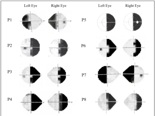
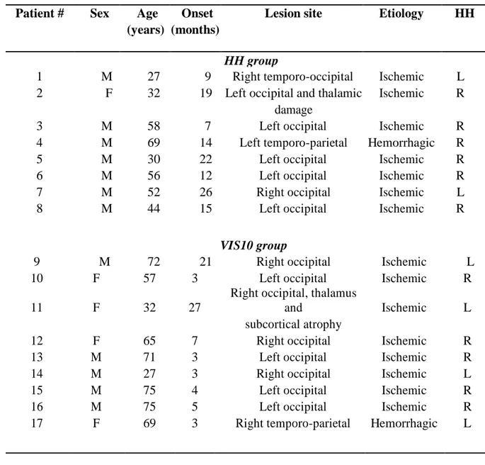
We recruited seventeen patients (five females; mean age = 52.8 years, SD = 17.6 years) suffering from left (n = 7) or right (n = 10) HH, induced by a post-stroke unilateral occipital lesion, and seventeen healthy controls (eight females; mean age = 52.2 years, SD = 14.0 years). All 34 participants were right-handed, and all completed a consent form before participation. After neuropsychological and ophthalmologic examinations, only those patients diagnosed with an isolated hemianopia (using Humphrey or Octopus automated perimeters) with corrected-to-normal visual acuity were included in the study. Clinical measurements of visual deficit were collected and analyzed for each patient. Based on a mean deviation obtained for both eyes, a strict threshold (at -30 dB of the mean deviation) was set for the measurements taken at 10° from the fixation point; above this threshold, the visual deficit was considered to be ambiguous (for technical details of the automated measurements, see (Sayo et al., 2017)). Based on this clinical threshold, nine patients (group VIS10; four females; left HH (n = 4); right HH (n = 5)) were excluded from the blindsight performance analyses; thus, these analyses were restricted to the remaining eight patients (group HH; one female; left HH (n = 2); right HH (n = 6))) with no visual detection capacities at 10° of eccentricity. The clinical details and automated visual perimetry values for the patients are presented in Figure 1, Figure 2 and Table 1.
p. 7
Figure 1: Automated visual perimetry, HH group (n = 8). The axial hash-marks denote
increments of 10º (visual) and the greyscale denotes sensitivity (in decibels).
2-column fitting image
Figure 2: Automated visual perimetry, VIS10 group. (n = 9; This group had a detection score higher than - 30 dB at 10° in the blind field). The axial hash-marks denote increments of 10º
p. 8
2-column fitting image
Patient # Sex Age Onset Lesion site Etiology HH (years) (months)
HH group
1 M 27 9 Right temporo-occipital Ischemic L 2 F 32 19 Left occipital and thalamic Ischemic R
damage
3 M 58 7 Left occipital Ischemic R
4 M 69 14 Left temporo-parietal Hemorrhagic R
5 M 30 22 Left occipital Ischemic R
6 M 56 12 Left occipital Ischemic R
7 M 52 26 Right occipital Ischemic L
8 M 44 15 Left occipital Ischemic R
VIS10 group
9 M 72 21 Right occipital Ischemic L
10 F 57 3 Left occipital Ischemic R
11 F 32 27
Right occipital, thalamus
and Ischemic L
subcortical atrophy
12 F 65 7 Right occipital Ischemic R
13 M 71 3 Left occipital Ischemic R
14 M 27 3 Right occipital Ischemic L
15 M 75 4 Left occipital Ischemic R
16 M 75 5 Left occipital Ischemic R
17 F 69 3 Right temporo-parietal Hemorrhagic L
Table 1: Demographic and clinical data. M, male; F, female; Onset: the time elapsed between lesion
onset and the testing session; L, left; R, right; HH, homonymous hemianopia; “HH group” corresponds to the patients with a critical blind spot at 10° of eccentricity (Humphrey score ≤ - 30 dB for both eyes). “VIS10 group” corresponds to the patients with residual detection at 10° of eccentricity (Humphrey score > - 30 dB for both eyes).
p. 9
2.2. Materials
Participants were seated in a dark room and their heads were held in place using a chin piece and a forehead rest at 57 cm from the computer screen (CRT monitor; refresh rate: 85 Hz; resolution: 1280 x 1024 pixels). To control fixation during the experiment, the participants’ eye movements were tracked by an EyeTribe.
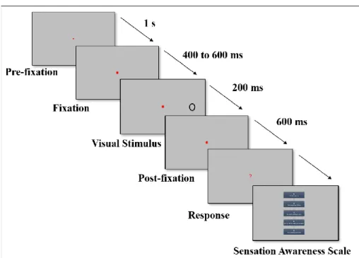
2.3. Stimuli and Procedure
The central fixation point was a red square (1° x 1°). Participants were presented with black letters (X and O; size: 3.5° x 3.5°) at maximal contrast on a gray background, at 10° eccentricity along the horizontal meridian to the left or right of the fixation point. One third of the trials were catch trials with no stimulus presented beside the fixation point. In each session, contralesional and ipsilesional visual fields were stimulated equally. To avoid a habituation bias, the hemifield of stimulus presentation
was semi-randomized: thus, a maximum of three successive trials in the same hemifield could be presented. The experiment comprised two sessions: firstly, a detection task; and secondly, a
forced-choice identification task. The two sessions differed only in the type of objective responses; otherwise, the trials were identical on the screen (see Fig. 3). In each session, two types of performance were recorded per trial: objective sensitivity (keyboard answer) and subjective perceptual experience (reported verbally according to the SAS). Objective performance in the detection task was measured on a binary scale as “Something appeared on the screen” (green button) or “Nothing appeared on the screen” (white button). Objective measurements in the identification task were taken by
forced-choice answers: the subjects were asked to specify manually which letter had appeared (X or O button), even if they did not see anything. Once the objective answer had been recorded, subjective
measurements were taken (using the SAS) at the end of a trial. The awareness scales were similar in the two sessions, although they were adapted to the associated objective task. For the identification task, the last two levels of the SAS were modified relative to those described earlier in the introduction: (4) I saw a letter; and (5) I clearly saw a letter and can identify it. Once the verbal response had been reported by the participant and recorded via keyboard entry by the researcher, the next trial began.
p. 10
Figure 3: Trial structure of the behavioral task. After the pre-fixation period corresponding to a
minimum of 1 second of fixation recorded by the Eyetribe, the fixation square was followed by a gap ranging from 400 ms to 600 ms. A stimulus (letter X or O) was then presented for 200 ms at 10° to the right or left visual field; however, in the catch trials, this time period was substituted with a gap of 200 ms. If the participants did not answer until 600 ms post-stimulus, then a question mark appeared on the screen to remind them that they had to press a key corresponding to their binary answer (detection task: Something appeared vs. Nothing appeared; identification task: X vs. O). Once the subject had given an objective response, they were tested on the awareness scale to obtain the subjective verbal report.
2-column fitting image
2.4. Data analysis
Trials in which the subjects’ gaze deviated from the fixation window were rejected. The objective analyses were performed separately from the subjective analyses. Objective performance was measured using the Signal Detection Theory (SDT; see, for example: (Macmillan & Douglas Creelman, 2005; Stanislaw & Todorov, 1999)). To this aim, key responses were coded as hits in signal trials (i.e. correct detection or identification of the letter) and as false alarms in noise trials (e.g. the button corresponding to “something appeared on the screen” was pressed during a blank trial). Based on these data and the SDT, the objective sensitivity (d’) for detection and for identification was calculated for each participant in the blind and the sighted fields (Green, D.M, & Swets, 1966; Macmillan & Douglas Creelman, 2005). Each subject’s d’ value was compared to a theoretical distribution of chance-level d’ values (obtained by calculating randomized responses to the test 100,000 times) and considered as significantly above chance at an alpha risk of 5%. Subjective sensitivity was calculated in each condition using the Area Under the receiver operating Curve (AUC) extracted from the signed rank test, which also provides a statistical significance of the difference in rating for signal and noise trials. Thus, subjective performance above chance level indicated that the
p. 11
use of the awareness scale was significantly different in the presence of a target than in the absence of one.
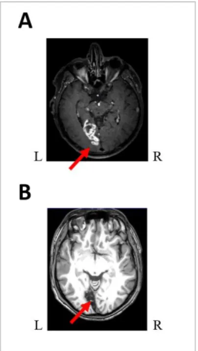
For two patients from the HH group (patients P6 and P8), we were able to perform a lesion analysis. For lesion reconstruction, MRI images were acquired on a Philips 3 Tesla unit, in the 3D FLAIR (Fluid-Attenuated Inversion Recovery) 8000 sequence (Time to repetition: 8000 ms; Echo time: 365 ms; Matrix size: 240 x 243 pixels; Reconstruction size: 240 x 243 pixels; Field of view: 240 x 240 mm; Flip angle: 90°; Voxel size range: from 0.9 mm to 1.25 mm). Anatomic slices were recovered in DICOM format for subsequent 3D visualization using OsiriX Medical Imaging software (OsiriX MD, Pixmeo, Geneva, Switzerland). The lesion contours were manually delineated in each 2D slice, thereby enabling reconstruction of a volume of interest (VOI; in cm3). To determine the anatomic extent of the lesions, each damaged Brodmann area was individually identified with the help of Brain Voyager software (Brain Innovation, Maastricht, The Netherlands) and subsequently checked by a neuroradiologist.
3. Results
3.1. Average group performance
As expected, in the healthy controls (CT) group, the objective and subjective sensitivities were significantly above chance (p < 0.05). A Kruskall-Wallis multiple comparison of objective performance among the HH group, VIS10 group (composed of HH patients with spared vision at 10°) and CT group clearly revealed group effects, both in the detection task (H (2, N = 34) = 21.6; p < 0.01) and in the identification task (H (2, N = 34) = 18.6; p < 0.001) (see Appendices, Figure A.). Thus, whereas the HH patients did not significantly differ from the control group in terms of performance in the sighted hemifield Fig A.1, A.2 (red lines), they did perform significantly lower in the blind field than did the controls (Dunn’s multiple comparison; p < 0.001 in detection task, p < 0.01 in identification task), Fig A.1, A.2 (blue lines). There was also a group effect regarding subjective performance in both the detection task (H (2, N = 34) = 16.57, p < 0.001) and the identification task (H (2, N = 34 = 17.23; p < 0.001). Dunn’s multiple comparison revealed that for both tasks, only the HH patients’ mean subjective sensitivity in the blind field was significantly lower than that of the other groups (Fig A.3, A.4; blue lines). Detailed objective and subjective sensitivities of the HH patients for each task are reported in Figure 4. Use of the SAS is represented in Figure 4, which is ordered according to the subjective sensitivity results.
p. 12
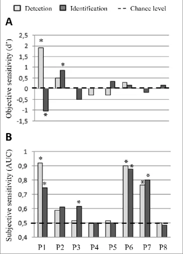
Figure 4: Comparison of objective sensitivities and subjective sensitivities per patient (HH group, n
= 8), per session
(detection session: light grey, identification session: dark grey); A. Objective sensitivities (d’) for each patient in the detection task (keyboard responses to the binary scale) and the forced-choice task (keyboard responses: X or O) are compared to the chance level (*p < 0.05, performance significantly above chance, with a d’ significantly different from a theoretical d’ value generated by random responses). B. Subjective-sensitivity AUC values for each patient were calculated by comparing the distribution of the level reported on the SAS between “letter trials” and catch trials. The signed rank test assessed the significance of the difference of the distribution with a p-value of 0.05. (*p < 0.05,
p. 13
subjective sensitivity significantly different in the presence of stimulus compared to in the catch trials). AUC: Area Under the receiver operating Curve.
1,5-column fitting image
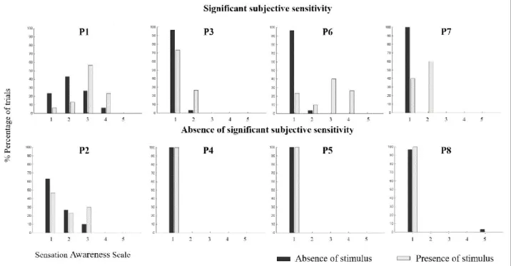
Figure 5: Distribution of responses on the Sensation Awareness Scale (SAS) during stimulation in the blind hemifield and during catch trials, per patient, in the identification task. Patients are
ordered according to the results of subjective sensitivity: P1, P3, P6 and P7 exhibited a significant subjective sensitivity (p < 0.05), compared to P2, P4, P5 and P8. The dark-grey bars denote catch trials and the light-grey bars, target trials in which ‘X’ or ‘O’ was presented in the blind visual field. Horizontal axes: the SAS levels: (1) I did not see anything; (2) I don’t think that I saw anything, but I am not sure; (3) I felt something; (4) I saw something; and (5) I clearly saw something and can identify it. The vertical axis indicates the percentage of trials.
2-column fitting image
Below, we describe the dissociation between the objective performance and the subjective performance of the HH patients.
p. 14
3.2. Type I blindsight: Patient P2
Only one of the eight HH patients (P2) could distinguish between the letters X and O in her contralesional visual field, responding at significantly higher than chance level (p < 0.05; Figure 4A dark grey bar). However, P2’s objective detection was at chance level, suggesting the presence of type I blindsight. This was confirmed by the subjective experience assessment, which revealed her lack of explicit sensitivity to the presence of stimuli in both tasks (Figure 4B). Indeed, P2 responded at different SAS levels but similarly in the presence or absence of letters on the screen (Figure 5).
3.3. Absence of blindsight: Patients P4, P5 and P8
The objective performance and subjective performance of patients P4, P5 and P8 in both tasks did not exceed chance level (Figures 4A & 4B). They reported seeing nothing (SAS level 1) during the catch trials or when a letter had been presented in their blind hemifield (Figure 5).
3.4. Subjective sensitivity without significant objective identification: Patients P1, P3, P6 and P7
The four remaining HH patients (P1, P3, P6 and P7) did not perform the identification task above chance-level (Figure 4A, dark grey bar); however, they demonstrated significant subjective sensitivity (p < 0.05; Figure 4B). To the best of our knowledge, this profile has never previously been described in the literature. We have named this dissociation between significant subjective performance and chance-level stimulus identification, blindsense. Although P3 demonstrated a significant subjective sensitivity only during the identification task (p = 0.01; light grey bar: P3), P1, P6 and P7 exhibited subjective sensitivity in both tasks. Regarding these patients’ SAS responses during the second session, a pattern clearly arose (Figure 5). Their reported awareness when a letter appeared in the blind field is shifted on the scale, mainly at levels 2 (I don’t think that I saw anything, but I am not sure) and 3 (I felt something), whereas when nothing appeared on the screen, the patients reported primarily at level 1 (I did not see anything). Interestingly, and as observed in Figure 5, two of the patients (P3 and P7) answered exclusively at level 2 when presented with the stimulus, whereas the other two patients (P1 and P6) answered mainly at level 3 and marginally at level 2. The results for patient P1 differed notably from this group in two regards. Firstly, he was the only patient to both objectively and subjectively detect the presence of stimuli in his blind field (p < 0.05; P1: light grey bar). Secondly, he demonstrated an abnormal reverse performance in identifying the letters, with an objective sensitivity significantly below chance in the identification task (p < 0.05; P1: light grey bar). This atypical result is discussed later as a case of “reverse blindsight”. The aforementioned blindsight profiles are summarized in Table
p. 15
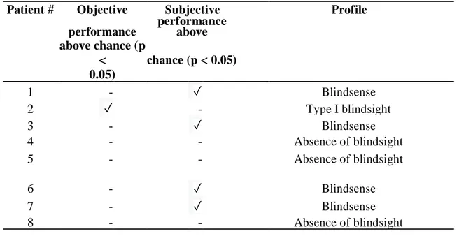
Patient # Objective Subjective Profile performance performance above
above chance (p < chance (p < 0.05) 0.05) 1 - ✓ Blindsense 2 ✓ - Type I blindsight 3 - ✓ Blindsense 4 - - Absence of blindsight 5 - - Absence of blindsight 6 - ✓ Blindsense 7 - ✓ Blindsense 8 - - Absence of blindsight
TABLE 2: Presence and type of blindsight and presence of blindsense, in the HH group, based on
the dissociation between objective performance and subjective performance during the identification task
2-column fitting image
3.5. SAS levels and correctness
We plotted the distributions of correct responses and incorrect responses in the identification task, for each level of the SAS (Appendices, Figure B). Even though P2 successfully identified the letters X and O at a significant level, there was no correlation between a higher reported sensitivity on the scale and the correct answers. In fact, P2 answered mostly at levels 1, 2 and 3, but performed best at level 2. Concerning the remaining seven patients (P1, P3 to P8), their level of sensitivity was not predictive of their objective performance, either.
p. 16
3.6. Lesion analysis
As illustrated in Figure 6, we were able to perform neuroanatomic analysis of the lesions in patients P6 and P8. Interestingly, both patients suffered from a complete right homonymous hemianopia. However, P6 exhibited a blindsense profile (i.e. subjective sensitivity without significant objective identification) whereas P8 did not exhibit any sign of blindsight or blindsense. For P6, neuroanatomic analysis revealed a mean lesion volume of 4.98 cm3 and his lesion involved only Brodmann Areas 17 and 18. In P8, neuroanatomic analysis revealed a mean lesion volume of 14.5 cm3 and his lesion involved Brodmann Areas 17, 18, 19, 29 and 30.
Figure 6: Neuroanatomical lesion analyses: (A) P6, right homonymous hemianopia and blindsense, 3D Flair 8000 sequence acquisition showing the lesion of BA 17 and 18 in the left hemisphere and (B) P8, right homonymous hemianopia with absence of blindsight, 3DTI sequence showing a lesion of BA 17, 18, 19, 29, 30 in the left hemisphere. Please note that conversely to radiological conventions, here, for convenience, the left hemisphere is presented on the left.
1 -column fitting image
4. Discussion
Although the objective tasks that we employed in this study were consistent with the classical format of blindsight experiments, we introduced a novel element in the subjective measure. Indeed, it is not uncommon during clinical examinations that patients with a field defect spontaneously report having a “sensation” in response to a stimulus presented in their blind field—albeit a sensation that they cannot
p. 17
qualify as “visual”. Despite this common observation, this spontaneously-reported level of awareness has never been integrated in systematic tests. In the present experiment, we developed the SAS, which adds the aforementioned level to the previously reported PAS. Using this procedure, we classified the patients’ deficits into different profiles according to the presence or absence of significant sensitivity to the stimulus in the objective measurements and the subjective measurements.
4.1. Scarcity of blindsight
Among the eight tested patients, only one patient, P2, met the criteria for blindsight. Indeed, P2 performed the identification task better than chance while being unable to consciously detect the target (detection performance below chance level). Considering that sensitivity on the subjective scale was not significant, P2’s profile clearly corresponds to type I blindsight. Using a restrictive threshold to eliminate patients that could residually detect a visual stimulus at 10° of eccentricity, we thus found type I blindsight in only one HH patient. Three patients were unable to identify the letters or to report any subjective experience from the stimulus (Table 2: patients P4, P5 and P8). Since we did not observe any patients with type II blindsight, according to our criteria the HH group showed an incidence of blindsight of 12.5% (one of eight patients) and an absence of blindsight of 37.5% (three of eight patients). Finally, the four remaining patients in the HH group (50%) did not meet the criteria previously described in the literature for either incidence or non-incidence of blindsight. These patients performed at chance-level on the objective task, whereas their sensitivity to the stimulus was significantly above chance when measured on the SAS (see patients P1, P3, P6, and P7 in Table 2). We have named their profile as blindsense, as all these patients significantly sensed the presence of the stimuli in their blind field without actually identifying the stimuli. We expound on this concept below.
4.2. Blindsense
Blindsense patients, exhibiting an unusual dissociation between significant subjective performance and objective performance at chance-level, represented half of our group of complete HH patients. Thus, in our study, a markedly higher number of patients exhibited blindsense than type I blindsight, the latter of which has been extensively discussed in the literature. Blindsense patients do not match the type II blindsight description, since their objective detection and identification performance scores were at chance. However, their SAS answers indicate that their perceptual experiences differed significantly when a letter was presented in their blind field compared to when nothing was presented. Interestingly, although such a profile has never been described in the literature, it is consistent with a frequently observed clinical reality. Indeed, neuropsychologists specialized in cortical visual impairment have confirmed that many patients report “feeling something” in their contralesional visual field without being able to precisely distinguish or describe the stimuli that they “felt”. Here we wish
p. 18
to emphasize that, by testing a patient cohort, rather than a single case, and by using an extended awareness scale, we were able to identify the behavioral pattern of blindsense. Assessment of a larger population should enable measurement of the incidence of this phenomenon relative to types I and II blindsight.
One patient, P1, stood out from the blindsense group due to a remarkable result: his identification performance was significantly lower than chance-level, yet he was the only patient to have scored above chance in the binary task, despite being objectively “blind” (based on perimetry). This result suggests that the responses he gave were not completely independent from the stimulus: indeed, P1 pressed the O-key significantly more often when an X appeared on the screen, and vice versa. However, during the same session, he performed far above chance in the trials involving presentation of a stimulus in the intact visual field. Thus, we can exclude the possibility that his performance was due to an erroneous understanding of the response mapping. One interpretation is that P1 presented a sort of “reverse-blindsight”: he processed the visual information, but his behavioral output was somehow reversed. A rather similar phenomenon has been reported in a patient with unilateral neglect (Mijović-Prelec, Shin, Chabris, & Kosslyn, 1994). Again, such behavior raises questions on the nature of patients’ experiences in their blind visual field, as discussed below.
4.3. Awareness measurement
Methods for assessing patients’ awareness levels in blindsight experiments remain controversial (Overgaard, 2011; Weiskrantz, 2009). Undeniably, if one claims to demonstrate unconscious abilities, one must provide strong evidence to this effect. In our study, the juxtaposition between the binary scale and the SAS in the same session (detection task) demonstrated that, after patients had answered “nothing appeared on the screen” in a binary forced-choice objective task, they could nonetheless report “not being sure” or “feeling something”. Thus, our results highlight two main points: firstly, they confirm that a gradual scale enables more sensitive measurement of the subjective report than does a binary scale; secondly, they suggest that the assessment of detection performance through a binary choice in patients strongly depends on the patients’ decision criteria. If researchers choose to use a Yes/No paradigm to assess patient’s detection, then one requirement would be to eliminate the response criteria bias through a highly specific psychophysical experimental design, as previously proposed (Azzopardi & Cowey, 1997). Moreover, we would like to highlight the fact that the patients in our study, instead answering “I saw something” or “I clearly saw something and can identify it”, mainly gave the non-visual answers such as “I am unsure” or “I feel something”. This is highly interesting in terms of phenomenological experience. In fact, researchers have traditionally assumed that blindsight is a visual experience, albeit a degraded one. However, as we discuss below, this may not be the case: patients may be aware of a stimulus but unaware of its specific visual nature.
p. 19
4.4. Nature of the perceptual experience
Is the residual perception experience of patients with visual-field defects triggered by degraded normal vision or is it a “non-visual experience”? (Brogaard, 2015; Kentridge, 2015; Weiskrantz, 2009). The profile observed in type II blindsight patients suggests that they do indeed experience a form of degraded normal vision. Corroborating this interpretation, a positive correlation between patients’ correct responses in a stimulus-identification task and their level of awareness of the stimulus has previously been reported (Overgaard et al., 2008). However, while our blindsense patients displayed above-chance level sensitivity to the stimulus using a subjective scale, they performed at chance level for detection or identification in forced-choice tasks. Thus, it seems that rather than exhibiting normal degraded vision, blindsense patients might instead experience a conscious “non-visual” sensation in response to visual stimulation. These results open a new avenue for future studies on blindsight, suggesting that objective and subjective scales (binary or otherwise) could include non-visual questions to elucidate patients’ perceptual experiences in the contralesional visual field. Another critical point to address in the future will be to ascertain any possible differences between left and right HH patients, as our group has previously demonstrated the effect of lesion lateralization on capacities in the contralesional as well as ipsilesional visual field in HH patients (Cavézian et al., 2015; Chokron, S., Peyrin, C. & Perez, 2018; Perez & Chokron, 2014). Interestingly, in the present study, a left
hemispheric lesion could be associated either with the presence or absence of blindsight, or with blindsense. Accordingly, the association between the lesion lateralization and the subjective experience in the blind visual field is far from being evident and requires further studies among a larger group of HH patients. However, being able to perform a neuroanatomical analysis on patients P6 (blindsense) and P8 (absence of blindsight or blindsense), we found that blindsense in patient P6 was associated to a relatively small lesion that affected only left BA 17 and 18, whereas patient P8, who presented a larger lesion that affected, in addition to left BA 17 and 18, left BA 19, 29 and 30, did not exhibit any sign of blindsense. Our findings suggest that the occurrence of blindsense or blindsight in post-stroke HH patients could require a relatively small lesion that includes, at the very least, loss of the striate cortex, but that spares other occipital areas. Further studies should include analysis of neuroanatomic lesions in larger cohorts of HH patients to elucidate the neuroanatomical correlates of patients’ perceptual experience in their contralesional visual field.
5. Conclusion
Measuring blindsight and perceptual experience in eight patients selected among a group of seventeen post-stroke HH patients, we identified one patient with type I blindsight (12.5%), no cases of type II blindsight (0%), three patients with a total absence of blindsight (37.5%) and most importantly, four patients (50%) exhibiting a never-before described phenomenon, which we have named blindsense.
p. 20
Given that blindsight is frequently used to train HH patients (Chokron et al., 2008; Hadid & Lepore, 2017; Perez & Chokron, 2014), clinicians and researchers must gain a broader understanding of the subtleties of patients’ perception experiences. Further characterization of blindsight types, including the blindsense phenomenon that we have reported here, and understanding of the neuronal correlates of these phenomena, will both require studies of larger cohorts of hemianopic patients.
p. 21
6. Acknowledgments
SC is grateful to the Edmond and Benjamin de Rothschild Foundations for their support.
7. Funding
This research was not funded or financed by any specific grants from public, private or not-for-profit agencies.
p. 22
References
Ajina, S., Pestilli, F., Rokem, A., Kennard, C., & Bridge, H. (2015). Human blindsight is mediated by an intact geniculo-extrastriate pathway. ELife, 4, 1–23. https://doi.org/10.7554/eLife.08935 Azzopardi, P., & Cowey, A. (1997). Is blindsight like normal, near-threshold vision? Proceedings of
the National Academy of Sciences, 94(December), 14190–14194. https://doi.org/10.1073/pnas.94.25.14190
Bridge, H., Thomas, O., Jbabdi, S., & Cowey, A. (2008). Changes in connectivity after visual cortical brain damage underlie altered visual function. Brain, 131(6), 1433–1444.
https://doi.org/10.1093/brain/awn063
Brogaard, B. (2015). Type 2 blindsight and the nature of visual experience. Consciousness and Cognition, 32, 92–103. https://doi.org/10.1016/j.concog.2014.09.017
Campion, J., Latto, R., & Smith, Y. M. (1983). Is blindsight an effect of scattered light, spared cortex, and near-threshold vision? Behavioral and Brain Sciences, 6(03), 423.
https://doi.org/10.1017/S0140525X00016861
Cavézian, C., Gaudry, I., Perez, C., Coubard, O., Doucet, G., Peyrin, C., … Chokron, S. (2010). Specific impairments in visual processing following lesion side in hemianopic patients. Cortex, 46(9), 1123–1131. https://doi.org/10.1016/j.cortex.2009.08.013
Cavézian, C., Perez, C., Peyrin, C., Gaudry, I., Obadia, M., Gout, O., & Chokron, S. (2015).
Hemisphere-dependent ipsilesional deficits in hemianopia: Sightblindness in the ‘intact’ visual field. Cortex, 69, 166–174. https://doi.org/10.1016/j.cortex.2015.05.010
Celeghin, A., Diano, M., de Gelder, B., Weiskrantz, L., Marzi, C. A., & Tamietto, M. (2017). Intact hemisphere and corpus callosum compensate for visuomotor functions after early visual cortex damage. Proceedings of the National Academy of Sciences, 114(48), E10475–E10483.
https://doi.org/10.1073/pnas.1714801114
Chokron, S., Peyrin, C. & Perez, C. (2018). Ipsilesional Deficit of Selective Attention in Left homonymous hemianopia and Left Unilateral spatial Neglect. Neuropsychologia (in press). Neuropsychologia. https://doi.org/10.1016/j.neuropsychologia
Chokron, S., Perez, C., Obadia, M., Gaudry, I., Laloum, L., & Gout, O. (2008). From blindsight to sight: Cognitive rehabilitation of visual field defects. Restorative Neurology and Neuroscience, 26, 305–320.
Cowey, A. (2010). The blindsight saga. Experimental Brain Research, 200(1), 3–24. https://doi.org/10.1007/s00221-009-1914-2
Fayel, A., Chokron, S., Cavezian, C., Vergilino-Perez, D., Lemoine, C., & Dore-Mazars, K. (2014). Characteristics of contralesional and ipsilesional saccades in hemianopic patients. Experimental Brain Research, 232(3), 903–917. https://doi.org/10.1007/s00221-013-3803-y
Fendrich, R., Wessinger, C. M., & Gazzaniga, M. S. (2001). Speculations on the neural basis of islands of blindsight. Progress in Brain Research, 134(June), 353–366.
https://doi.org/10.1016/S0079-6123(01)34023-2
Green, D.M, & Swets, J. A. (1966). Signal detection and psychophysics. New York: Wiley, 702. Hadid, V., & Lepore, F. (2017). From Cortical Blindness to Conscious Visual Perception: Theories
on Neuronal Networks and Visual Training Strategies. Frontiers in Systems Neuroscience. https://doi.org/10.3389/fnsys.2017.00064
Kentridge, R. W. (2015). What is it like to have type-2 blindsight? Drawing inferences from residual function in type-1 blindsight. Consciousness and Cognition, 32, 41–44.
https://doi.org/10.1016/j.concog.2014.08.005
Leh, S. E. (2006). Unconscious vision: new insights into the neuronal correlate of blindsight using diffusion tractography. Brain, 129(7), 1822–1832. https://doi.org/10.1093/brain/awl111 Macmillan, N. A., & Douglas Creelman, C. (2005). Detection Theory, A user’s Guide (TeAM
YYEPG).
Marzi, C. A., Tassinari, G., Aglioti, S., & Lutzemberger, L. (1986). Spatial summation across the vertical meridian in hemianopics: A test of blindsight. Neuropsychologia, 24(6), 749–758. https://doi.org/10.1016/0028-3932(86)90074-6
p. 23
Mazzi, C., Bagattini, C., & Savazzi, S. (2016). Blind-Sight vs. Degraded-Sight: Different Measures Tell a Different Story. Frontiers in Psychology, 7(JUN), 1–11.
https://doi.org/10.3389/fpsyg.2016.00901
Mijović-Prelec, D., Shin, L. M., Chabris, C. F., & Kosslyn, S. M. (1994). When does “no” really mean “yes”? A case study in unilateral visual neglect. Neuropsychologia, 32(2), 151–158. https://doi.org/10.1016/0028-3932(94)90002-7
Overgaard, M. (2011). Visual experience and blindsight: A methodological review. Experimental Brain Research, 209(4), 473–479. https://doi.org/10.1007/s00221-011-2578-2
Overgaard, M., Fehl, K., Mouridsen, K., Bergholt, B., & Cleeremans, A. (2008). Seeing without seeing? Degraded conscious vision in a blindsight patient. PLoS ONE, 3(8), 8–11.
https://doi.org/10.1371/journal.pone.0003028
Overgaard, M., & Mogensen, J. (2015). Reconciling current approaches to blindsight. Consciousness and Cognition, 32, 33–40. https://doi.org/10.1016/j.concog.2014.08.003
Perenin, M. T., & Jeannerod, M. (1975). Residual vision in cortically blind hemiphields. Neuropsychologia, 13(1), 1–7. https://doi.org/10.1016/0028-3932(75)90041-X
Perez, C., & Chokron, S. (2014). Rehabilitation of homonymous hemianopia: insight into blindsight. Frontiers in Integrative Neuroscience, 8(October), 1–12.
https://doi.org/10.3389/fnint.2014.00082
Poppel, E., Held, R., & Frost, D. (1973). Residual Visual Function after Brain Wounds involving the Central Visual Pathways in Man. Nature, 243, 295–296. https://doi.org/10.1038/243295a0 Ramsøy, T. Z., & Overgaard, M. (2004). Introspection and subliminal perception. Phenomenology
and the Cognitive Sciences, 3(1), 1–23. https://doi.org/10.1023/B:PHEN.0000041900.30172.e8 Sahraie, A., Trevethan, C. T., MacLeod, M. J., Urquhart, J., & Weiskrantz, L. (2013). Pupil response
as a predictor of blindsight in hemianopia. Proceedings of the National Academy of Sciences, 110(45), 18333–18338. https://doi.org/10.1073/pnas.1318395110
Sanders, M. D., Warrington, E., Marshall, J., & Weiskrantz, L. (1974). “BLINDSIGHT”: VISION IN A FIELD DEFECT. The Lancet, 303(7860), 707–708.
https://doi.org/10.1016/S0140-6736(74)92907-9
Sayo, A., Ueno, S., Kominami, T., Nishida, K., Inooka, D., Nakanishi, A., … Terasaki, H. (2017). Longitudinal study of visual field changes determined by Humphrey Field Analyzer 10-2 in patients with Retinitis Pigmentosa. Scientific Reports, 7(1), 16383.
https://doi.org/10.1038/s41598-017-16640-7
Stanislaw, H., & Todorov, N. (1999). Calculation of signal detection theory measures. Behavior Research Methods, Instruments, & Computers, 31(1), 137–149.
https://doi.org/10.3758/BF03207704
Stoerig, P. (2006). Blindsight, conscious vision, and the role of primary visual cortex. Progress in Brain Research, 155(1), 217–234. https://doi.org/10.1016/S0079-6123(06)55012-5
Stoerig, P., & Cowey, A. (1997). Blindsight in man and monkey. Brain, 120(3), 535–559. https://doi.org/10.1093/brain/120.3.535
Weiskrantz, L. (1972). Review Lecture: Behavioural analysis of the monkey’s visual nervous system. Royal Society, 455(1069), 427–455. Retrieved from http://www.jstor.org/stable/76163
Weiskrantz, L. (1998). Consciousness and commentaries. In Towards a science of consciousness II— the second Tucson discussions and debates (pp. 371–377). Cambridge.
Weiskrantz, L. (2009). Is blindsight just degraded normal vision? Experimental Brain Research, 192(3), 413–416. https://doi.org/10.1007/s00221-008-1388-7
Weiskrantz, L., Barbur, J. L., & Sahraie, A. (1995). Parameters affecting conscious versus
unconscious visual discrimination with damage to the visual cortex (V1). Proceedings of the National Academy of Sciences of the United States of America, 92(13), 6122–6126.
https://doi.org/10.1073/pnas.92.13.6122
Zhang, X., Kedar, S., Lynn, M. J., Newman, N. J., & Biousse, V. (2006). Homonymous hemianopias: Clinical-anatomic correlations in 904 cases. Neurology, 66(6), 906–910. https://doi.org/10.1212/01.wnl.0000203913.12088.93