HAL Id: tel-01127186
https://tel.archives-ouvertes.fr/tel-01127186
Submitted on 7 Mar 2015HAL is a multi-disciplinary open access
archive for the deposit and dissemination of sci-entific research documents, whether they are pub-lished or not. The documents may come from teaching and research institutions in France or abroad, or from public or private research centers.
L’archive ouverte pluridisciplinaire HAL, est destinée au dépôt et à la diffusion de documents scientifiques de niveau recherche, publiés ou non, émanant des établissements d’enseignement et de recherche français ou étrangers, des laboratoires publics ou privés.
limb muscle activity using surface electromyography :
methodological considerations and practical applications
Karin Lienhard
To cite this version:
Karin Lienhard. Analysis of whole-body vibration exercise effect on lower limb muscle activity us-ing surface electromyography : methodological considerations and practical applications. Education. Université Nice Sophia Antipolis, 2014. English. �NNT : 2014NICE4080�. �tel-01127186�
U
NIVERSITE DE
N
ICE
S
OPHIA
A
NTIPOLIS
Faculté des Sciences du Sport – Unité de Formation et de Recherche en Sciences et Techniques des Activités Physiques et Sportives
A
NALYSIS OF WHOLE
-
BODY VIBRATION EXERCISE EFFECT
ON LOWER LIMB MUSCLE ACTIVITY USING
SURFACE ELECTROMYOGRAPHY
M
ETHODOLOGICAL CONSIDERATIONS
AND PRACTICAL APPLICATIONS
THESE
En vue de l’obtention du grade de Docteur d’Université
En Sciences du Mouvement Humain - Ecole Doctorale 463
Présentée et soutenue publiquement par
Karin Lienhard
Le 7 novembre 2014
Devant le jury composé de :
BERTUCCI WILLIAM (Rapporteur) MCF-HDR Université Reims Champagne Ardenne CABASSON ALINE (Examinateur) MCF Université de Nice Sophia Antipolis COLSON SERGE (Directeur) Professeur Université de Nice Sophia Antipolis MARTIN ALAIN (Examinateur) Professeur Université de Bourgogne
MESTE OLIVIER (Co-directeur) Professeur Université de Nice Sophia Antipolis PORTERO PIERRE (Rapporteur) Professeur Université Paris Est
© Karin Lienhard
Laboratory of Human Motricity Education Sport and Health (LAMHESS, EA 6309) Faculty of Sport Sciences
University of Nice Sophia Antipolis Nice, France
Laboratory of I3S (CNRS, UMR7271) University of Nice Sophia Antipolis Nice, France
A
BSTRACTThe aim of this thesis was to analyze the effect of whole-body vibration (WBV) exercise on lower limb muscle activity and to give methodological implications and practical applications. Two methodological studies were conducted that served to evaluate the optimal method to process the surface electromyography (sEMG) signals during WBV exercise and to analyze the influence of the normalization method on the sEMG activity. A third study aimed to gain insight whether the isolated spikes in the sEMG spectrum contain motion artifacts and/or reflex activity. The subsequent three investigations aimed to explore how the muscle activity is affected by WBV exercise, with a particular focus on the vibration frequency, platform amplitude, additional loading, platform type, knee flexion angle, and the fitness status of the WBV user. The final goal was to evaluate the minimal required vertical acceleration to stimulate the muscle activity of the lower limbs. In summary, the research conducted for this thesis provides implication for future investigations on how to delete the excessive spikes in the sEMG spectrum and how to normalize the sEMG during WBV. The outcomes of this thesis add to the current literature in providing practical applications for exercising on a WBV platform.
R
ÉSUMÉL’objectif de cette thèse a été d’analyser l’effet de l’exercice physique réalisé sur plateforme vibrante (whole-body vibration, WBV) sur l’activité musculaire des membres inférieurs, de développer des outils d’analyse méthodologiques et de proposer des recommandations pratiques d’utilisation. Deux études méthodologiques ont été menées pour identifier la méthode optimale permettant de traiter les signaux d'électromyographie de surface (sEMG) recueillis pendant la vibration et d'analyser l'influence de la méthode de normalisation de l'activité sEMG. Une troisième étude visait à mieux comprendre si les pics sEMG observés dans le spectre de puissance du signal contiennent des artéfacts de mouvement et/ou de l'activité musculaire réflexe. Les trois études suivantes avaient pour but de quantifier l’effet de la WBV sur l’activité musculaire en fonction de différents paramètres tels que, la fréquence de vibration, l'amplitude de la plateforme, une charge supplémentaire, le type de plateforme, l'angle articulaire du genou, et la condition physique du sujet. En outre, l'objectif a été de déterminer l'accélération verticale minimale permettant de stimuler au mieux l'activité musculaire des membres inférieurs. En résumé, les recherches menées au cours de cette thèse fournissent des solutions pour de futures études sur : i) comment supprimer les pics dans le spectre du signal sEMG et, ii) comment normaliser l'activité musculaire pendant un exercice WBV. Enfin, les résultats de cette thèse apportent à la littérature scientifique de nouvelles recommandations pratiques liées à l’utilisation des plateformes vibrantes à des fins d’exercice physique.
I, Karin Lienhard, hereby declare that this thesis and the work reported herein was composed by and originated entirely from me. Information derived from the published and unpublished work of others has been acknowledged in the text and the sources are given in the list of references.
Signed in Nice, France, the 9th of September 2014
T
ABLE OFC
ONTENTSC
HAPTER1
–
G
ETTING TOK
NOWW
HOLE-B
ODYV
IBRATION1.1 What is whole-body vibration? ……….….. 2
1.2 WBV and science ……… 3
1.3 WBV and its effects on muscle performance ……….… 4
1.4 Muscle activity during WBV ……….….. 6
1.5 Problematic of WBV studies ……….…. 7
C
HAPTER2
–
P
OTENTIALM
ECHANISMSD
URINGW
HOLE-B
ODYV
IBRATION 2.1 Overview of the potential mechanisms……….…………. 92.2 Tonic vibration reflex………... 2.2.1 Muscle spindles 2.2.2 Stretch reflex 2.2.3 H-Reflex 9 2.3 Cutaneous mechanoreceptors ………. 14 2.4 Tendon organ ……….. 16 2.5 Damping ……….. 17
C
HAPTER3
–
L
ITERATURER
EVIEW-
D
URINGW
HOLE-B
ODYV
IBRATION 3.1 Tonic vibration reflex……… 203.2 Occurrence of motion artifacts ………..…. 3.2.1 sEMG spikes – motion artifacts or reflex activity? 24 3.3 Methodological issues in WBV studies……….………...…….. 3.3.1 sEMG processing 3.3.2 sEMG normalization 28 3.4 Effect of WBV on muscle activity……….... 30
3.5 Effect of WBV parameters………...
3.5.1 Frequency
3.5.2 Amplitude
3.5.5 Knee flexion angle
3.6 Acc transmission during WBV………...
3.6.1 Amplification of vibrations
3.6.2 Vibration damping
42
3.7 Effect of WBV parameters on acceleration transmission………...……...
3.7.1 Platform
3.7.2 Frequency
3.7.3 Knee angle
3.7.4 Population
48
3.8 Potential side effects ……….………... 51
3.9 Summary ………..……….………... 53 3.10 Hypotheses ……..………..……….………... 54
C
HAPTER4
–
M
ETHODS 4.1 Participants ………..…. 56 4.2 Data acquisition ……….. 57 4.3 Surface electromyography ………..4.3.1 Analysis in the time domain
4.3.2 Analysis in the frequency domain
4.3.3 Relationship between time and frequency domain
4.3.4 sEMG processing
59
4.4 sEMG processing during WBV ………...……..
4.4.1 No filter
4.4.2 Band-stop filter
4.4.3 Band-pass filter
4.4.4 Spectral linear interpolation
66
4.5 Normalization methods ………...
4.5.1 Maximal voluntary contraction
4.5.2 Mmax
4.6 Motion artifact recordings………... 78
4.7 Platform acceleration ………...
4.7.1 Calculation of platform displacement
80
4.8 Platform types ……….………...………... 82 4.9 Acceleration transmission ………..……….……….………... 87 4.10 Body posture ……..……….……….………... 90
C
HAPTER5
–
S
TUDYI:
SEMG
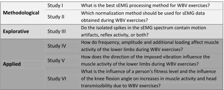
PROCESSING5.1 Preface ……….……….…….. 93
5.2 The influence of sEMG processing on the quantification of
neuromuscular activity during WBV ……….………… 94
C
HAPTER6
–
S
TUDYII:
SEMG
NORMALIZATION6.1 Preface ……….……….. 110
6.2 Does the sEMG normalization method have an influence
on the quantification of muscle activity during WBV exercise? ………..…….. 111
C
HAPTER7
–
S
TUDYIII:
M
OTIONA
RTIFACTS VS.
R
EFLEXA
CTIVITY7.1 Preface ……….……….. 123
7.2 sEMG during WBV contains motion artifacts and reflex activity ………..….……….. 124
C
HAPTER8
–
S
TUDYIV:
T
HEO
PTIMALP
ARAMETERS8.1 Preface ……….….. 139
8.2 Determination of the optimal parameters maximizing muscle activity of the
lower limbs during vertical synchronous WBV ………...………..……….. 140
C
HAPTER9
–
S
TUDYV:
A
CCLERATIONT
HRESHOLD9.1 Preface ……….…….. 160
9.2 Quantification of the vertical acceleration threshold to enhance
10.1 Preface ……….………... 160
10.2 The effect of WBV on muscle activity in active and inactive subjects ………... 161
C
HAPTER11
–
D
ISCUSSION 11.1 Aims ………... 19711.2 Synopsis of Results ...………... 197
11.3 How to filter the sEMG during WBV ...………... 199
11.4 Vibration input ………...………... 200
11.5 Acceleration threshold ………...………….…... 202
11.6 Optimal parameters ………...………….…... 205
11.7 Safety aspect ………...………….…... 207
11.8 Populations ………...………….…... 207
11.9 Methodological considerations and limitations ………... 209
11.10 Practical applications ………... 211
C
HAPTER12
–
O
UTLOOK&
C
ONCLUSIONS 12.1 Outlook ………...…... 21412.2 Conclusions ………... 217
P
REFACEThis thesis was a collaboration between two laboratories of the University of Nice Sophia Antipolis, France; the laboratory LAMHESS (EA 6309) and the laboratory I3S (CNRS, UMR7271). The thesis was co-supervised by Prof Serge S Colson (LAMHESS) and Prof Olivier Meste (I3S).
Studies I-IV of this thesis were carried out at the Faculty of Sport Sciences (STAPS), LAMHESS laboratory, Nice, France, from October 2011 to December 2012. Studies V&VI were completed at the Human Performance Laboratory (HPL), University of Calgary, Alberta, Canada. In agreement with the supervisors, this visit took place from January 2013 to August 2013, under the direction of Prof Benno M Nigg.
This thesis was financed by the University of Nice Sophia Antipolis from October 2011 to September 2014. Additional support for travel expenses was received by the Fondation Partenariale Dream It. This grant allowed participation to several congresses (ECSS 2014, ACAPS 2013, ECSS 2012) and a research visit to the HPL of the University of Calgary, Alberta, Canada. Financial support by the Natural Sciences and Engineering Research Council of Canada (NSERC) permitted participation to the ISEK congress 2014. The vibration platforms were sponsored by Power Plate (France) and Total Image Fitness (Calgary, Canada). The funding institutions were not involved in data collection, data analysis, interpretation of the data, preparation of the manuscript, or decision to publish.
First and foremost, I would like to express my warm thanks to my supervisors, Prof Olivier Meste and Prof Serge S Colson for their aspiring guidance, support, constructive criticism and friendly advice during this thesis. I am also deeply grateful to the University of Nice Sophia Antipolis, in particular the doctoral school of human movement sciences directed by Prof Reinoud Bootsma, and the LAMHESS laboratory directed by Prof Jeanick Brisswalter.
I would also like to thank the University of Nice Sophia Antipolis France, the Fondation Partenariale Dream It France, and the Natural Sciences and Engineering Research Council of Canada for the financial support they have offered me. Many thanks go also to Power Plate and Total Image Fitness for providing the vibration platforms.
I am further appreciative to Prof Benno M Nigg and Sandro Nigg from the University of Calgary, Alberta, Canada, for welcoming me in their group and providing me with an invaluable experience.
My many thanks to Aline Cabasson, Jordyn Vienneau, Florence Verdera, Gilles Roussey, Pierre-David Petit, Fabien Fuchs and Bernd Friesenbichler for their valuable laboratory assistance and tremendous contribution to the successful completion of this thesis. And to my numerous colleagues and office mates that have made this time unique in their own way, thank you.
Last I am eternally grateful to my parents for their unconditional love and support through many years of schooling that has led to the completion of this thesis.
L
IST OFT
HESISR
ELEVANTP
UBLICATIONSARTICLE |Lienhard K, Cabasson A, Meste O, Colson SS (2014). Determination of the optimal
parameters maximizing muscle activity of the lower limbs during vertical synchronous whole-body vibration. Eur J Appl Physiol. 114(7):1493-501.
ARTICLE |Lienhard K, Vienneau J, Nigg S, Meste O, Colson SS, Nigg BM. Quantification of vertical
acceleration threshold to enhance lower limb muscle activity during whole-body vibration exercises. In revision.
ARTICLE |Lienhard K, Cabasson A, Meste O, Colson SS. The influence of sEMG processing on the
quantification of neuromuscular activity during whole-body vibration. Submitted.
ARTICLE | Lienhard K, Cabasson A, Meste O, Colson SS. sEMG during whole-body vibration
contains motion artifacts and reflex activity. Submitted.
ARTICLE |Lienhard K, Vienneau J, Friesenbichler B, Nigg S, Meste O, Nigg BM, Colson SS. The
effect of whole-body vibration on muscle activity in active and inactive subjects. Submitted.
L
IST OFT
HESISR
ELEVANTC
ONFERENCEP
RESENTATIONSORAL PRESENTATION | Lienhard K, Vienneau J, Nigg S, Meste O, Colson SS, Nigg BM (2014). Dual
mode whole-body vibration has a greater effect on muscle activity than the side-alternating mode. 20th Congress of the International Society of Electrophysiology and Kinesiology (ISEK), July 15 – 18, Rome, Italy.
ORAL PRESENTATION | Lienhard K, Cabasson A, Meste O, Colson SS (2013). What is contained in
the excessive spikes in sEMG spectrum during whole-body vibration: motion artifacts or stretch reflexes? 15th International Congress of the Association des Chercheurs en Activités Physiques et Sportives (ACAPS), October 29 - 31, Grenoble, France.
during whole-body vibration exercise. 18th Annual European Congress of Sport Science (ECSS), July 2-5, Amsterdam, Netherlands.
POSTER PRESENTATION | Lienhard K, Vienneau J, Friesenbichler B, Nigg S, Meste O, Nigg BM,
Colson SS (2014). Damping in trained and untrained individuals during whole-body vibration. 10ième Journée de l’Ecole Doctorale, Sciences du Mouvement Humain, June 6, Montpellier, France.
POSTER PRESENTATION | Lienhard K, Cabasson A, Meste O, Colson SS (2012). EMG signal
processing during whole-body vibration: a preliminary study. 17th Annual European Congress of Sport Science (ECSS), July 4-7, Bruges, Belgium.
POSTER PRESENTATION | Lienhard K, Colson SS, Meste O (2012). Analysis and processing of human
electrical and physiological signals during whole-body vibration exercises. Journée des jeunes doctorants et docteurs de l’Université de Nice Sophia Antipolis, January 31, Nice, France.
L
IST OFT
HESISR
ELEVANTA
WARDSTRAVEL GRANT | Received by the Fondation Partenariale Dream It, March 26 2012, Nice, France.
POSTER AWARD | 3rd price for poster presentation at the Journée des jeunes doctorants et
docteurs de l’Université de Nice Sophia Antipolis, January 31 2012, Nice, France.
L
IST OFA
BBREVIATIONSAcc Acceleration
Amp Amplitude
ANOVA Analysis of variance
BF Biceps Femoris CV Coefficient of variation Disp Displacement Freq Frequency GL Gastrocnemius Lateralis GM Gastrocnemius Medialis H-reflex Hoffmann-reflex
ICC Intraclass correlation coefficient
ISO International Organization for Standardization
LOA Limits of agreement
MUAP Motor unit action potential
MVC Maximal voluntary contraction
PSD Power spectral density
RF Rectus Femoris
RMS Root mean square
sEMG Surface electromyography
sEMGRMS Root mean square of the surface electromyography
SD Standard deviation
SOL Soleus
TA Tibialis Anterior
TVR Tonic vibration reflex
VL Vastus Lateralis
VM Vastus Medialis
1.1
W
HAT ISW
HOLE-B
ODYV
IBRATION?
Whole-body vibration (WBV) means transmission of a mechanical repetitive movement to the body. WBV employed as a training device requires a platform that generates sinusoidal oscillations by motors underneath the vibration platform that are transmitted to the person on the machine. The oscillations can be adjusted by manipulating the frequency and/or the displacement of the platform. The resulting acceleration (Acc) is defined by the frequency (Freq) and the amplitude (Amp) of the platform displacement via the equation The person using the machine can adapt various positions on the platform such as sitting, standing, squatting, or hovering.
Whole-body vibration exercises have been and are still currently being used amongst professional athletes such as ice hockey players, skiers, golfers or marathon runners as a warm-up or as a method to increase performance. In recent years, WBV training has increased in popularity amongst recreational athletes. As a result, WBV platforms are available in many gyms and are even affordable for individual purchases. The commercialization of WBV platforms has led to numerous companies manufacturing such platforms, leading to a large variety of products available. As the popularity of WBV platforms has grown faster than the scientific background, manufacturers have claimed benefits that are untested and open to debate. Their claims, based on anecdotal evidence, include weight loss, reduced lower back pain, improved balance, increased bone density, enhanced blood flow and increases in muscle strength and power.
These unwarranted claims of positive effects through WBV have made many researchers rightfully frown upon the use of WBV as a proven performance/health stimulant. While the image of WBV has remained dubious to the scientific community, there has been an increase in research performed, and some research has successfully replaced some of the anecdotal evidence with scientific proof. This increase in WBV’s scientific understanding is directly correlated with the dramatic uptick in research being completed on its effects.
1.2
WBV
ANDS
CIENCEWBV has gained more and more popularity amongst the scientific community mainly over the last two decades. To give a general idea of the increase in publications around WBV, an internet search was conducted using the portal of the US National Library of Medicine
(http://www.ncbi.nlm.nih.gov/pubmed). A first search was launched by using the term
whole-body vibration [All Fields]. The total number of publications until the end of the year 2013 was
1044, with an exponential increase over the past 20 years (Figure 1.1). The highest number of publications per year was reached in 2013 with 130 publications.
Figure 1.1 The amount of publications sorted by year covering whole-body vibration indexed by the US National Library of Medicine. A total amount of 1044 publications was found by the end of the year 2013.
Out of the claims that have been made by the manufacturers, blood flow and weight loss have received the least attention by the scientific community thus far, with only about 70 and 25 publications respectively. Other fields such as back pain, balance, and bone density have been more in the focus with 100 - 200 publications per topic. By far the most prevalent subject has been muscle strength and power, with over 350 publications.
1.3
WBV
AND ITSE
FFECTS ONM
USCLEP
ERFORMANCEThe first scientific studies that have investigated the effect of WBV exercises on muscle strength and power were conducted in the late 90’s. Bosco et al. (1999a; 1999b) have tested the immediate effects of WBV exercises on muscle performance, whereas one limb received the vibration treatment and the other limb served as a control entity that received no vibration treatment. Bosco et al. (1999a) found that 5 x 1 min of vibration applied to a contracted arm increased its average power immediately after the treatment compared to the control arm. Similarly for the lower limbs, Bosco et al. (1999b) found that after exposing the leg to 10 x 60 s of WBV, velocity, force and power was increased in the vibration-treated leg but not in the control leg. Bosco et al. (1998) also examined the short-term effects of WBV exercises by testing jumping performance before and after 10 days of a training period. This consisted of WBV exercises in an intervention group and no exercises in a control group. The measurements after the training period showed that only the intervention group improved their power output and their jump height. While Bosco and his group were amongst the first researchers who showed that WBV exercises can positively affect muscle performance, the draw-back of their study set-up was that the control groset-up/limb did not receive the same treatment as the intervention group/limb but without vibration. Hence, the parameter “vibration” was not isolated in their investigations, meaning that it is not clear if the positive adaptations originated from the vibration per se.
Subsequent studies compared the short-term effects of WBV training to the same training without vibration. De Ruiter et al. (2003) found no effect on muscle strength and power that was solely attributable to WBV, as muscle strength of the knee extensor muscles and counter movement jump performance was unaffected after 11 weeks of training regardless of utilizing WBV or not. On the other hand, Delecluse et al. (2003) found a significant effect on muscle strength solely attributable to WBV, as isometric and dynamic knee extensor strength was increased after 12 weeks of WBV training but not after 12 weeks of the same training without WBV. Further, Delecluse et al. (2003) found that muscle strength was positively affected by the WBV training in equal measures as by resistance training, but that an improvement in counter
movement jump performance was only observed after the WBV intervention. Later studies confirmed beneficial immediate (McBride et al. 2010) as well as beneficial short-term effects (Colson et al. 2010; Petit et al. 2010) of WBV exercises on muscle strength of the lower limbs.
The findings from the early 2000’s studies have led to the conclusion that WBV triggers something in the muscle that improves performance. Therefore, subsequent investigations were focused on the changes in muscle activity while the body is being vibrated. As a result, with the use of surface electromyography (sEMG) recordings, muscle activity during WBV exercises started to be extensively investigated in the years thereafter.
1.4
M
USCLEA
CTIVITY DURINGWBV
In order to illustrate that in the early 2000’s researchers became more and more interested in examining muscle activity during the exposure to WBV, a more refined search was launched to narrow down the publications that have investigated WBV in relation with muscle activity. The used search term was whole-body vibration [All Fields] AND (EMG [All Fields] OR sEMG [All
Fields] OR muscle activity [All Fields]). The total amount of publications until the end of 2013
was 111, whereas the most publications were found in the year 2012 (Figure 1.2).
Figure 1.2 The amount of publications sorted by year covering whole-body vibration together with muscle activity or surface electromyography indexed by the US National Library of Medicine. A total amount of 111 publications was found by the end of the year 2013.
1.5
P
ROBLEMATIC OFWBV
S
TUDIESAlthough it was not distinguished whether muscle activity was assessed before, during, or after the WBV exercise, this search shows that the interest in the effect of vibration on muscle activity as measured by sEMG recordings has been increasing over the last decade. Unfortunately, contradictory findings have been reported in these studies. This makes drawing general conclusions about the effectiveness of WBV exercises challenging. This lack of concise conclusions stems from several issues.
First, although directly applied vibration to the muscle belly or the tendon elicits stretch reflex responses, it is unclear if such responses occur during WBV exercises. Second, it is unknown whether the myoelectrical signal during WBV is contaminated by motion artifacts or not. This disagreement has led to unstandardized methods to process and analyze the sEMG signals during WBV. Third, WBV studies have been using different platform types, vibration frequencies and amplitudes, as well as different body positions. Hence, due to the large variety of the used study parameters, it is challenging to explain the conflicting outcomes. Subsequently, it is crucial to develop a sEMG processing method for a standardized use in WBV studies, and to describe the effectiveness of WBV and its optimal parameters leading to the highest positive changes in sEMG activity during the exposure to vibration.
Therefore, CHAPTER 2 ofthis thesis aims to describe the potential mechanisms explaining the
increased sEMG activity during WBV exercises. CHAPTER 3 expands on the findings and outcomes of the above mentioned scientific literature, and the problematic of WBV studies are further discussed. CHAPTER 4 describes the methodology that was used aiming to (1) find the optimal
method to process and analyze the sEMG during WBV, (2) to clarify whether the sEMG signal during WBV contains reflex activity and/or motion artifacts, and (3) to scrutinize the effect of WBV and its parameters on muscle activity levels. The detailed studies and their outcomes are each presented in a chapter (CHAPTER 5-10). Last, the outcomes of this thesis are discussed in CHAPTER 11,and conclusions are drawn in CHAPTER 12.
C
HAPTER
2
P
OTENTIAL
M
ECHANISMS
D
URING
2.1
O
VERVIEW OF THEP
OTENTIALM
ECHANISMSThe potential mechanisms leading to increased muscle activity during the exposure to WBV are various, and some of them have only been evidenced with directly applied vibration on the muscle belly or the tendon of interest. The most frequently cited mechanism in relation with WBV exercises is the tonic vibration reflex (TVR), mediated by the primary and secondary endings of the muscle spindles (Cardinale and Bosco 2003; Rittweger 2010). Besides the muscle spindles, other somatosensory receptors provide feedback during direct exposure to vibration, including mechanoreceptors in the skin, tendon organs, joint receptors, nociceptors, and thermal sensors (Brooke and Zehr 2006). While these mechanisms are well documented during directly applied vibration, evidence is lacking for their occurrence during WBV. A more recently identified mechanism with demonstrated occurrence during WBV is vibration damping (Wakeling and Nigg 2001). However, some of these mechanisms have only been evidenced during directly applied vibration to the muscle or the tendon. Nevertheless, there is the potential that all these mechanisms occur during WBV. The most prevalent mechanisms are described in the following.
2.2
T
ONICV
IBRATIONR
EFLEXThe occurrence of muscle contractions evoked by vibrations has been called the TVR, and has been experimentally established a few decades ago (Hagbarth and Eklund 1965; Matthews 1966). The TVR is based on stretch reflex responses, which are mediated by the muscle spindles. It needs to be emphasized that in these studies and in the ones mentioned in the following, vibration was directly applied to the muscle belly or the tendon of interest.
2.2.1MUSCLE SPINDLES
Muscle spindles have a fusiform shape and lie in parallel with the skeletal muscle fibers. The muscle spindle fibers are referred to as intrafusal fibers, whereas the skeletal muscle fibers outside the spindle capsule are called extrafusal fibers (Figure 2.1). The muscle spindle has an afferent supply, i.e. Ia-afferent and II afferent, to transmit action potentials to the central nervous system. In addition to that, intrafusal fibers receive efferent input, namely from the
alpha (α) and gamma (γ) motor neurons. Alpha motor neurons innervate extrafusal fibers, whereas gamma motor neurons connect to intrafusal fibers. Normal mode of operation involves concurrent activation of the alpha and gamma motor neurons, which is known as alpha-gamma co-activation. The alpha motor neurons activate the extrafusal fibers to produce the force required for the task, and the gamma motor neurons activate the intrafusal fibers to set the desired level of feedback from the muscle spindle. A stretch of the muscle is detected by the primary (Brown et al. 1967; Roll and Vedel 1982) and secondary (Brown et al. 1967) endings of the muscle spindles which results in innervations of the Ia-afferents (De Gail et al. 1966).
Figure 2.1 Schematic illustration of a muscle spindle.
(Retrieved from http://www.studyblue.com/notes/note/n/movement-science-motor-control/deck/1421471)
2.2.2STRETCH REFLEX
The TVR is based on stretch reflex responses; rapid muscle contractions as a response to a brief, unexpected increase in length (stretch) of the muscle. The stretch reflex consists of a short-latency response (M1) after 30 ms via homonymous Ia excitation. The medium-short-latency response (M2) occurs after 50 - 60 ms and is mediated by the transcortical pathway. Occasionally, a long-latency response (M3) is observed occurring after 90 - 100 ms also mediated by the transcortical
pathway (Petersen et al. 1998). The short-latency response is mediated by monosynaptic and polysynaptic pathways. The monosynaptic pathway evokes a homonymous Ia excitation, which means that efferent motor neurons innervate the same muscle from which the afferent signal originated. The function of this pathway is to reverse the stretch by a rapid muscle contraction. The polysynaptic pathway evokes a reciprocal Ia inhibition, which means that Ia inhibitory interneurons evoke inhibitory postsynaptic potentials in the motor neurons that innervate the antagonist muscles (Figure 2.2). The function of the polysynaptic pathway is to link the inhibition of an antagonist muscle to the activation of the agonist muscle in order to complete the task (e.g. flexion, extension). Reciprocal inhibition declines during tasks that involve co-activation of agonist and antagonist muscles, and increases during postural activity.
Figure 2.2 Schematic representation of the monosynaptic (1, 2) and polysynaptic (3) stretch reflex pathway.
(Retrieved from
The TVR evoked during directly applied vibration has been observed in a frequency range of 20 - 200 Hz (Eklund and Hagbarth 1966; Burke et al. 1976a; Burke et al. 1976b; Martin and Park 1997), with increasing motor unit synchronization the higher the frequency up to 100 Hz (Martin and Park 1997). The TVR is also positively correlated to the amplitude of the vibratory input stimulus (Eklund and Hagbarth 1966). The TVR occurs whether the innervated muscle is relaxed or contracted (Bongiovanni and Hagbarth 1990), and the magnitude of the TVR is dependent on the amount of the background muscle activity (Eklund and Hagbarth 1966; Burke et al. 1976a; Gottlieb and Agarwal 1979; Bedingham and Tatton 1984; Ogiso et al. 2002). Background muscle activity increases the magnitude of the TVR response due to its relation to the excitability of the motoneuron pool. On the contrary, the amplitude of the TVR is reduced when the antagonist muscle is contracted due to reciprocal Ia inhibition (Gottlieb and Agarwal 1979). Additionally, the amplitude of the TVR is greater with increasing muscle length or stretch because of an increased sensitivity of the muscle spindles during the stretch (Burke et al. 1976b; Nordin and Hagbarth 1996). The characteristics of the TVR have further been studied by evoking Hoffmann (H)-reflexes. The H-reflex has been studied before and after a vibratory stimulus as its magnitude, like the magnitude of the TVR, represents the excitability of the motoneuron pool. The difference between the H-reflex and the TVR is that the H-reflex bypasses the muscle spindles as the nerve is directly innervated. Before discussing the behavior of the H-reflex during direct vibration, the pathway of the H-reflex is described in the following section.
2.2.3H-REFLEX
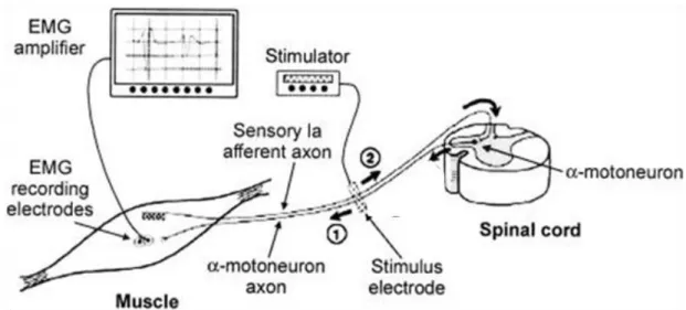
The H-reflex can be evoked by application of a single electrical stimulus to a peripheral nerve, which evokes a twitch response in the muscle, approximately 28 – 35 ms after the stimulus. This response can be measured as the sEMG and/or the force resulting from the twitch. The H-reflex is most commonly studied in the Soleus (SOL), but it can also be evoked in the Quadriceps Femoris, the Hamstrings, and Tibialis Anterior (TA) (Enoka 2008). The pathway underlying the H-reflex involves activation of Ia afferents and subsequent generation of action potentials in the same muscle. Increasing the intensity of the electrical stimulus evokes a short-latency (5 ms) response called the M-wave. While the H-reflex tests the excitability of the motor neuron pool,
the M-wave tests neuromuscular propagation. When the nerve is stimulated, the action potential is propagated both toward the neuromuscular junction and back toward the motor neuron. The action potential (Figure 2.3) propagating toward the neuromuscular junction (1) produces an M-wave, whereas the action potential propagating toward the motor neuron (2) produces an H-reflex. As the pathway of the H-reflex involves a spinal loop, the H-reflex is observable shortly after the M-wave response.
Figure 2.3 Pathway for the Hoffmann (H)-reflex and the M-wave. The electrical stimulus is applied over the peripheral nerve. The action potential that propagates toward the neuromuscular junction (1) produces an M-wave, whereas the action potential that propagates toward the motor neuron (2) produces an H-reflex.
(Retrieved from http://bmsi.ru/doc/61f60d25-6a9e-426f-a16e-5e1101da0811)
Studies have shown that the magnitude of the H-reflex increases with increasing baseline muscle activity (Verrier 1985) and with increasing torque levels (Pensini and Martin 2004), and that the H-reflex is linearly correlated to torque levels ranging from 0 to 50% of the maximal voluntary contraction torque (Butler et al. 1993). These observations are in line with the ones for the TVR, and support the following conclusions.
The magnitude of the TVR during direct vibration
is positively related to the vibration frequency and amplitude of the input stimulus
is positively related to the background muscle activity
is positively related to the amount of the stretch of the muscle
2.3
C
UTANEOUSM
ECHANORECEPTORSCutaneous mechanoreceptors provide information about the acceleration of the skin and the deeper tissues such as movement of the hair, and displacement and stretch of the skin. This feedback is provided by the Meissner corpuscles, Pacinian corpuscles, Merkel disks, Ruffini endings, and free nerve endings (Figure 2.4).
Figure 2.4 Schematic illustration of the cutaneous mechanoreceptors consisting of Meissner corpuscles, Pacinian corpuscles, Merkel disks, Ruffini endings, and free nerve endings.
The cutaneous mechanoreceptors can be divided into fast and slow adapting receptors. The fast adapting receptors are the Meissner corpuscles and the Pacinian corpuscles; the Meissner corpuscles are most responsive to a vibratory stimulus around 40 Hz and the Pacinian corpuscles around 100 Hz and above (Rittweger 2010). More specifically, the Meissner corpuscles are sensitive to local, maintained pressure, and their response fades rapidly. The Pacinian corpuscles, which are the largest receptors in the skin, detect rapid changes in pressure and respond mainly to the acceleration component of a stimulus. The slow adapting receptors, the Merkel disks and the Ruffini endings, are sensitive to sustained pressure. Merkel disks are sensitive to local vertical pressure and do not respond to lateral stretch of the skin. The Ruffini endings respond to stretch of the skin and are sensitive to the direction of the stretch. Last, the free nerve endings provide feedback on the movement of the skin and hair.
As the cutaneous mechanoreceptors provide the central nervous system with information about the magnitude, speed and direction of a mechanical stimulus, they are partly responsible for the sensorimotor alterations during a vibratory stimulus. While it has been believed that only the fast adapting mechanoreceptors are responsible for these alterations during vibration, a study by Ribot-Ciscar et al. (Ribot-Ciscar et al. 1989) suggests involvement of both, the slow and the fast cutaneous mechanoreceptor. It needs to be considered that the fast receptors become quiescent after a certain vibration frequency (Ribot-Ciscar et al. 1989), whereas the slow adapting receptors are able to respond one-to-one to vibration frequencies up to 200 Hz (Vedel and Roll 1982). The study by Ribot-Ciscar et al. (Ribot-Ciscar et al. 1989) also showed that the processing of pressure and touch is masked during vibration, and one of their earlier study found such a masking even after the vibration set-off (Ribot-Ciscar et al. 1996).
2.4
T
ENDONO
RGANThe tendon organ includes one afferent (i.e. Ib afferent) and no efferent connections (Figure
2.5). The Ib afferents are innervated when a muscle and its connective tissues are stretched,
either through pulling of the muscle or through activation of the skeletal muscle fibers. Hence, the tendon organ provides information about the muscle force. In addition to the spindle afferents, Ib-afferents from the Golgi tendon organs are responsive to vibration (Burke et al. 1976a; Hayward et al. 1986). During the reflex contraction, discharge from the primary and secondary spindle endings decline, whereas discharge from the Golgi tendon organs increase (Burke et al. 1976a).
Figure 2.5 Schematic illustration of a tendon organ.
2.5
D
AMPINGA more recently identified mechanism leading to increased sEMG activity during WBV exercises is damping. During vibration, the muscles and tendons act as a spring-mass system that experiences compression during the upstroke of the vibration platform, and expansion during the down stroke of the platform. The stiffness and the mass of the system determine the
natural frequency and are related with . As the mass of the system cannot be altered, adjustment of the natural frequency can only be changed through alteration of the stiffness . When the frequency of the actuator (i.e., the frequency of the vibration platform) coincides with the frequency of the resonator , resonance can occur (Cardinale and Wakeling 2005; Boyer and Nigg 2006). This can lead to accumulation of energy where the vibration amplitude of the resonator is greater than the amplitude of the actuator. This amplitude amplification can lead to destruction – the so-called resonance catastrophe (Figure
2.6).
Figure 2.6 Model of the human body acting as a resonator. A mass is linked to a spring system with stiffness and a dashpot with friction . Resonance can occur in the system when the frequency of the actuator is close to the natural frequency of the system. Transmissibility is defined as and is amplified when > 1. (Adapted from Rittweger et al. 2010)
In order to prevent resonance-related tissue damage, it is speculated that the body aims to minimize vibrations by damping (Nigg 1997). The effects of resonance can be reduced by changing either the stiffness and thus the natural frequency , or the damping characteristics of the tissue, both of which are possible through increased muscle activation (Wakeling and Nigg 2001; Wakeling et al. 2002).
This mechanism has been confirmed during WBV exercises in a study by Wakling et al. (2002). In this study, the subjects were exposed to continuous vibrations and pulsed bursts across a frequency range of 10 - 65 Hz. It was found that muscle activity was elevated together with increased damping of the vibration power when the input frequency was close to the natural frequency of the soft tissue. As the frequency range of commercially available WBV platforms are typically within the range of the natural frequency of the human body, resonance can be expected as well as damping via increased muscle activity.
Besides increased muscle activation, the stiffness of the body can be altered by changing the body position, such as the degree of knee flexion. Effective body stiffness increases with straightening the legs (Lafortune et al. 1996), which increases the natural frequency Therefore, an optimal body position can help to prevent resonance.
In summary, spinal reflexes elicited by e.g. muscle spindles, cutaneous mechanoreceptors, or tendon organs as well as damping via increased muscle activity have been identified as possible mechanisms that could explain the elevated sEMG activity during WBV exercises. However, it is debatable whether spinal reflexes not only occur during directly applied vibration but also during WBV exposure.
C
HAPTER
3
L
ITERATURE
R
EVIEW
–
3.1
T
ONICV
IBRATIONR
EFLEXAlthough the TVR is a well established mechanism during directly applied vibration to the tendon and the muscle, it is not clear if the TVR occurs during WBV. In order to clarify this matter, several studies have compared the behavior of the stretch reflex and the H-reflex before and after WBV. The magnitude of the stretch reflex response was found to be enhanced (Rittweger et al. 2003; Melnyk et al. 2008), unchanged (Hopkins et al. 2009; Cochrane et al. 2010; Fernandes et al. 2013; Yeung et al. 2014), and even depressed (Ritzmann et al. 2011) immediately after the WBV exercise. Similarly, the magnitude of the H-reflex was found to be unchanged (Pollock et al. 2012) and lower (Armstrong et al. 2008; Apple et al. 2010; Ritzmann et al. 2011; Games and Sefton 2013; Hortobágyi et al. 2014) immediately after the WBV exercise. Post-WBV enhancement of reflex responses is rather surprising, as one would expect reflex depression as observed during directly applied vibration for the H-reflex (Arcangel et al. 1971; Roll et al. 1980; Van Boxtel 1986) and the stretch reflex (Roll et al. 1980). Acknowledging that some studies found enhancement of the stretch reflex after directly applied vibration (Van Boxtel 1986; Shinohara et al. 2005), it is difficult to pin point as to why such discrepancies have been observed.
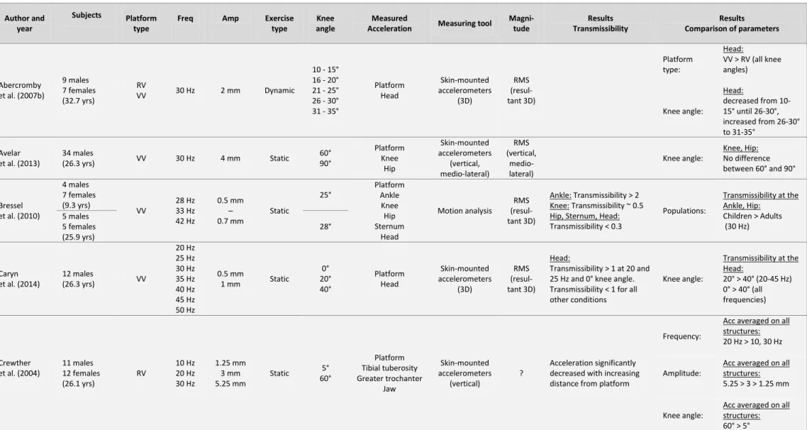
A sparse sample of studies has investigated the occurrence of the TVR/reflex activity during WBV by analyzing responses in muscle activity while the subject was exposed to the vibration (Ritzmann et al. 2010; Pollock et al. 2012; Zaidell et al. 2013). The earliest study (Ritzmann et al. 2010) compared the latencies and spectrograms of sEMG recordings during WBV with recordings of stretch reflex responses evoked by passive dorsiflexion with a custom-made ankle ergometer. The stretch reflex latency during dorsiflexion was defined as the time between the onset of the movement and the onset of the first rise in the sEMG signal. During WBV, the latency was defined as the time from the low point of the vibration platform and the onset of the first spike in the sEMG signal. The outcomes showed that the latencies and the spectrograms of the SOL and Gastrocnemius Medialis (GM) during dorsiflexion were extremely similar to those during WBV (Figure 3.1). In the same study, a pressure cuff was placed on the belly of the SOL muscle as occlusion has been shown to reduce the short-latency response of
the stretch reflex (Leukel et al. 2009). The influence of the pressure cuff on the amplitude of the stretch reflex was then compared during dorsiflexion and WBV, and it was found that the stretch reflex amplitude was reduced by about 56% during dorsiflexion and by 61% during WBV. Hence, the findings of this study showed that the behavior of the sEMG signal during WBV has similarities to the one of stretch reflex responses.
Figure 3.1 (A) Stretch reflex responses of the Soleus evoked by the ankle ergometer at 25 Hz (upper panel) and the mean of the corresponding frequency spectrograms of all participants (lower panel). (B) Surface electromyography signal of the Soleus during whole-body vibration at 25 Hz (upper panel) and the mean of the corresponding frequency spectra of all participants (lower panel).
(Reprinted from Ritzmann et al. 2010.)
In the second study (Pollock et al. 2012), single motor units of the Vastus Lateralis (VL) were recorded during WBV at 30 Hz using intramuscular EMG. Simultaneously, the waveform of the
platform acceleration was measured with the help of a strain gauge (Figure 3.2). The results of this study indicated that motor unit firing is phase locked to the vibration cycle as there was a strong relationship between the timing of the motor unit firing and the phase of the WBV cycle. This observation confirms the presence of reflex muscle activity during WBV, yet it is unclear which somatosensory receptors were involved.
Figure 3.2 Platform acceleration recording (top graph) and intramuscular
electromyography of the Vastus Lateralis (bottom graph) during WBV at 30 Hz. The firing of a single motor unit is phase locked to the phase of the vibration
cycle. (Reprinted from Pollock et al. 2012)
The most recent study (Zaidell et al. 2013) compared sEMG activity of the SOL and TA during WBV and during Achilles tendon vibration. During the procedure, the subjects were seated on a chair with their leg positioned on the platform. WBV and Achilles tendon vibration was induced at 25 and 50 Hz for a time period of 70 s. Further, the effect of background muscle activity on the TVR was tested by passive loading of the leg. The motion artifacts induced by the vibrations of the platform were deleted in the Power Spectral Density (PSD) by subtracting a sine and cosine wave from the sEMG signals. However, the outcomes of this study were that the Root Mean Square (RMS) of the SOL and TA increased over the 70-sec time period during the WBV treatment as well as during the Achilles tendon vibration. This can be explained by increased
muscle spindle sensitivity, which points towards the occurrence of stretch reflex responses during WBV. Also, sEMG activity of the SOL and TA was affected by WBV in equal measures as by Achilles tendon vibration, which further supports the occurrence of the TVR during WBV. According to the findings of these studies, it can be assumed that WBV elicits stretch reflex responses and that the TVR occurs during WBV.
While Ritzmann et al. (2010) highlighted that actual stretch reflex responses display excessive peaks in the frequency spectrum (Figure 3.1A), it is not guaranteed that the excessive peaks that are observed during WBV (Figure 3.1B) are stretch reflex responses. Recently, it has been suggested that the sEMG signals recorded during WBV are contaminated by motion artifacts, caused by the oscillation of the platform. Since the motion artifacts as well as the stretch reflex responses would be phase-locked to the vibration frequency, it is not clear if these excessive peaks contain stretch reflex responses (or reflex activity), motion artifacts, or both.
3.2
O
CCURRENCE OFM
OTIONA
RTIFACTSA typical WBV platform induces vibrations via sinusoidal oscillations, which are transmitted to the person standing on the platform. The vibrations travel through the entire body of the WBV user, which means that the muscles as well as the sEMG electrodes are oscillating. Percussion of the electrodes can result in recordings of motion artifacts. These artifacts are typically visible in the frequency domain as sharp peaks (from now on referred to as spikes) at the vibration frequency and at the multiple harmonics of the vibration frequency ( ).
The first research group who drew attention to this issue was Abercromby et al. (2007a), by illustrating a sEMG signal that was recorded during WBV in the frequency domain. Figure 3.3 is a reprint of the original illustration and shows spikes at the vibration frequency and at a few multiple harmonics . Nevertheless, the excessive spikes per se are not a proof for the occurrence of motion artifacts, as these spikes could originate from stretch reflex responses. In order to verify the existence of motion artifacts during WBV, several studies have measured the artifacts with so-called dummy electrodes. In the study where this set-up has been introduced (Fratini et al. 2009a), electrodes were placed on the patella assuming that any signal recorded during WBV would be due to the shaking of the electrodes. Unfortunately, even though excessive spikes in the sEMG spectrum were located, it cannot be excluded that these peaks were not sEMG recordings from surrounding muscles. To address this limitation, a similar set-up has been repeated (Ritzmann et al. 2010; Sebik et al. 2013) by positioning electrodes on the muscle with several layers of tape between skin and electrodes to ensure exclusive motion artifact related recordings without any cross-talk from surrounding muscles. While Sebik et al. (2013) found motion artifact-contaminated sEMG signals, Ritzmann et al. (2010) observed only a marginal contribution.
Figure 3.3 Surface electromyography signal recorded during whole-body vibration at 30 Hz in the frequency domain. Excessive peaks are observable at the vibration
frequency and its multiple harmonics
. (Reprinted from Abercromby et al. 2007a)
Possible reasons for the observed discrapancy between these studies (Fratini et al. 2009a; Ritzmann et al. 2010; Sebik et al. 2013) could be the type of the used equipment such as the electrodes or the cables (Webster 1984). Using conjoined electrodes vs. single electrodes might have the advantage that the conjoined electrodes are moving in phase to each other, which impedes a difference in potential between the electrodes. Additionally, the degree of cable shielding could have an influence, as motion artifacts are reduced by using shielded cables vs. unshielded cables (Clancy et al. 2002). However, it is unclear as to why Ritzmann et al. (2010) did not observe significant spikes in the dummy electrode signal.
3.2.1 SEMGSPIKES -MOTION ARTIFACTS OR REFLEX ACTIVITY?
While it is very likely that WBV elicits stretch reflex responses, it is unknown if such responses are contained in the isolated spikes. Hence it is not surprising that the scientific community has been debating in the past few years whether the spikes are due to stretch reflex responses, motion artifacts, or both. Although the isolated spikes have never been analyzed, a very recent study (Sebik et al. 2013) gave evidence that motion artifacts and stretch reflex responses may contribute to the sEMG spectrum during WBV.
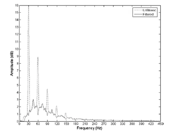
The study by Sebik et al. (2013) used an innovative sEMG filtering regime to demonstrate the occurrence of motor unit synchronization during WBV. First, the sEMG signals were band-pass filtered between 80 - 500 Hz in order to delete the low-frequency sEMG spectrum where motion artifacts might have occurred. Then, the sEMG signals were rectified in the time domain, meaning that all the negative values were multiplied by -1. Last, the sEMG signals were transformed into the frequency domain, where excessive spikes were again observed at the vibration frequency and its multiple harmonics (Figure 3.4B). These new spikes could only originate from motor unit synchronization, as motion artifacts, unlike stretch reflex responses, are strictly sinusoidal. Although the method introduced by Sebik et al. (2013) was useful to prove motor unit synchronization during WBV, its feasibility as a method to delete the motion artifacts is questionable, as a band-pass filter between 80 - 500 Hz rejects a large number of myoelectrical information (see CHAPTER 4).
Figure 3.4 Representative sEMG signals and frequency spectrograms recorded from the Gastrocnemius Medialis during whole-body vibration at 35 Hz. Shown in columns: raw sEMG signal; frequency spectrogram; magnified frequency spectrogram in the 0-100 Hz range. (A) Unprocessed sEMG signal; (B) band-pass (80-500 Hz) filtered sEMG signal; (C) the filtered sEMG signal after rectification, showing dominant peaks at around 35 Hz and 70 Hz due to synchronization.
(Reprinted from Sebik et al. 2013)
While it is strongly assumed that stretch reflex responses as well as motion artifacts are contained in the sEMG signal during WBV, it is important to identify their contribution in the spikes. Even more so it is surprising that to date no scientific study has analyzed the content of the isolated spikes.
3.3
M
ETHODOLOGICALI
SSUES INWBV
S
TUDIES3.3.1 SEMGPROCESSING
As it is unclear if the spikes observed in the sEMG spectrum contain mostly motion artifacts or stretch reflex responses, different strategies have been used to cope with these spikes. Researchers who have assumed that these spikes are mainly stretch reflex responses have been including these spikes in the calculation of the sEMG magnitude (Cardinale and Lim 2003; Cochrane et al. 2009; Di Giminiani et al. 2013; Krol et al. 2011; Perchthaler et al. 2013; Ritzmann et al. 2013; Roelants et al. 2006). Conversely, researchers who have speculated that these spikes are mainly motion artifacts have been withdrawing these spikes and have been excluding them from the calculation of the sEMG magnitude (Abercromby et al. 2007a; Fratini et al. 2009a; Hazell et al. 2007; 2010; Marín et al. 2012a; 2012b; Pollock et al. 2010). Besides the decision whether these spikes should be deleted or not, it seems that researchers have been given the choice to select the appropriate filtering regime. Hence, different methods have been used including a band-stop filter centered at the vibration frequency and its multiple harmonics (Abercromby et al. 2007a; Fratini et al. 2009a; Marín et al. 2012a; 2012b, Pollock et al. 2010) and a band-pass filter rejecting the entire spectrum where the spikes might have occurred (Hazell et al. 2007; 2010). As a consequence, the WBV research studies that have been published used either no filter, a band-stop filter, or a band-pass filter to delete the excessive spikes. Unfortunately, to date it is unknown if the magnitude of the muscle activity is comparable between studies using different filtering regimes. Neither the band-stop nor the band-pass filter has been validated, which means that their respective effect on the quantification of the sEMG magnitude is unknown. Therefore, it is absolutely necessary to analyze how the method of choice influences the magnitude of the sEMG during WBV.
Another draw-back of never having validated the different sEMG processing methods is that none of the methods might be the optimal choice to filter the excessive spikes. For example, using a band-pass filter with a low cut-off frequency of 100 Hz removes the entire sEMG spectrum below that frequency. Hence, although the motion artifacts might be deleted,
important myoelectrical information is lost over a wide range of the frequency spectrum. On the other hand, using a band-stop filter targets the specific frequencies that are contaminated, yet more sEMG activity is deleted than necessary. This can be seen on page 25 in Figure 3.3 originally published by Abercromby et al. (2007a). The dotted line represents the raw sEMG signal in the frequency domain, whereas the solid line represents the band-stop filtered sEMG signal. Evidently, the band-stop filter produces notches in the sEMG spectrum which have no physiological background. These notches are a byproduct of using a band-stop filter and might be a severe draw-back. Therefore it would be of great benefit to develop a new processing method that only deletes the excessive spikes without adding notches to the spectrum.
3.3.2 SEMGNORMALIZATION
After filtering the sEMG signal and calculating the RMS, the next step is typically normalization of the RMS of the sEMG (sEMGRMS). In WBV studies, the sEMGRMS has been expressed as a
percentage of the baseline sEMGRMS during standing (%standing, Marín et al. 2012a; Ritzmann
et al. 2013; Tankisheva et al. 2013), or as a percentage of the sEMGRMS during a maximal
isometric voluntary contraction (%MVC, Hazell et al. 2007; 2010; Perchthaler et al. 2013; Pollock et al. 2010; Roelants et al. 2006). Surprisingly, although normalization of the sEMGRMS is
important for comparison between subjects and muscles (Burden 2010), this step has been skipped in numerous studies (Abercromby et al. 2007a; Avelar et al. 2013; Cardinale and Lim 2003; Cochrane et al. 2009; Di Giminiani et al. 2013; Krol et al. 2011; Marín et al. 2009; 2012b) and the sEMGRMS was reported and compared in Volts (V).
Taken as a whole, measuring the sEMG during WBV is confronted with several methodological issues. First, it is unclear if the excessive spikes in the sEMG spectrum need to be deleted. Second, if they need to be removed, which filter is the most adequate method to do so? Third, it is unknown if the sEMG normalization procedure has an influence of the outcomes of the study. Therefore, before further investigating the instantaneous effects of WBV on muscle activity, these limitations should be addressed and a standardized methodology should be implemented.
3.4
E
FFECT OFWBV
ONM
USCLEA
CTIVITYThe effect of WBV on lower limb muscle activity has been investigated in numerous studies.
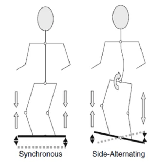
Table 3.1 summarizes the most important findings of these studies and the WBV parameters
that have been used. Comparing the sEMG activity during WBV to the same exercise without vibration, most studies have found significantly higher sEMG during the vibration. This positive effect of WBV on muscle activity has been reported performing static and dynamic exercises (Abercromby et al. 2007a; Hazell et al. 2007) with various knee flexion angles (Ritzmann et al. 2013; Roelants et al. 2006), using a side-alternating or synchronous vertical vibration platform (Abercromby et al. 2007a; Ritzmann et al. 2013), using a frequency range of 6 to 55 Hz and an amplitude range of 0.5 to 5 mm (Abercromby et al. 2007a; Cardinale and Lim 2003; Cochrane et al. 2010; Di Giminiani et al. 2013; Hazell et al. 2007; 2012; Krol et al. 2011; Marín et al. 2009; 2012a; Ritzmann et al. 2013; Roelants et al. 2006; Tankisheva et al. 2013). However, some authors have found that the effectiveness of WBV can vary between muscles (Cochrane et al. 2009; Hazell et al. 2010), the vibration frequency (Di Giminiani et al. 2013; Hazell et al. 2007), the amplitude of the platform (Marín et al. 2009), or the combination of the two latter parameters (Hazell et al. 2007; Krol et al. 2011). For example, vibration frequencies ranging from 25 to 55 Hz have been found to significantly increase muscle activity during WBV (Abercromby et al. 2007a; Cardinale and Lim 2003; Di Giminiani et al. 2013; Krol et al. 2011; Marín et al. 2009; 2012b; Roelants et al. 2006) in combination with amplitudes ranging from 0.5 to 5 mm. On the contrary, no increases in muscle activity have been reported for vibration frequencies below 25 Hz in combination with amplitudes between 0.5 and 2 mm (Di Giminiani et al. 2013; Krol et al. 2011;). The exception to that is the study by Cochrane et al. (Cochrane et al. 2009), where significant increases in sEMG activity were found with 6 Hz and 3.1 mm amplitude WBV in the VL and GM. Certainly, this is only one example where contradictory findings have been reported. It is difficult to seek out the responsible source, as theses studies diverse in a number of parameters. Besides using different frequencies and amplitudes, different vibration platform types, exercise modalities, body postures, and sEMG processing and normalization methods have been used. This makes it difficult to draw general conclusions about the effectiveness of WBV exercises.