HAL Id: tel-01547457
https://tel.archives-ouvertes.fr/tel-01547457
Submitted on 26 Jun 2017
HAL is a multi-disciplinary open access archive for the deposit and dissemination of sci-entific research documents, whether they are pub-lished or not. The documents may come from teaching and research institutions in France or abroad, or from public or private research centers.
L’archive ouverte pluridisciplinaire HAL, est destinée au dépôt et à la diffusion de documents scientifiques de niveau recherche, publiés ou non, émanant des établissements d’enseignement et de recherche français ou étrangers, des laboratoires publics ou privés.
Importance of glucocorticoid receptors in the
physiopathology of Alzheimer’s disease
Scherazad Kootar
To cite this version:
Scherazad Kootar. Importance of glucocorticoid receptors in the physiopathology of Alzheimer’s disease. Agricultural sciences. Université Côte d’Azur, 2017. English. �NNT : 2017AZUR4015�. �tel-01547457�
Université Côte d’Azur, École doctorale 85 Sciences de la Vie et de la Santé Institut de Pharmacologie Moléculaire et Cellulaire, UMR 7275, CNRS/UCA
Thèse de doctorat
Présentée en vue de l’obtention du grade de
Docteur en Sciences
Mention Interactions Moléculaires et Cellulaires
de l’Université Côte d’Azur
Par :
SCHERAZAD KOOTAR
L’importance des récepteurs aux glucocorticoïdes dans la
physiopathologie de la maladie d’Alzheimer
“Importance of glucocorticoid receptors in the physiopathology of Alzheimer’s disease”
Thèse dirigée par Dr Hélène MARIE
Soutenue le 24 Mars 2017 Devant le jury composé de :
Dr Alain Buisson Professeur, Université de Grenoble Rapporteur
Dr Hélène Marie CR1 CNRS/HDR, IPMC Directrice de thèse
Dr Stéphane Martin DR2 INSERM, IPMC Président de Jury
1
Acknowledgments
It gives me great pleasure to express my gratitude to my PhD supervisor Dr. Hélène Marie. Her expertise in electrophysiology, meticulous scientific approach, timely advice, and excellent organizational skills have helped me throughout. I would like to thank her for her patience and ultimate confidence in me. I would like to thank Dr. Jacques Barik for the GRlox/lox mice he introduced to the lab, this taught me much about how to breed mice and systematically record them. His experience in the field of stress, his enthusiasm and forward thinking have been helpful during the thesis. I would also like to convey my thanks to Dr. Ingrid Bethus who has played an important role in my PhD. She taught me various techniques like stereotaxic injections, behavioural paradigms and surgery for cannula implantations. Her enthusiasm for neurobiology has been outstanding as are her teaching methods. A big thank you to Dr. Sebastian Fernandez, who has helped me enormously throughout my project. His experience and vast knowledge in the field of neurobiology has helped me with quick solutions hence saving valuable time. Apart from being a good researcher, his fantastic sense of humour has helped us pass some not so easy days with a smile. I would also like to thank Dr. Paula Pousinha, for all the discussions we have had and the critical analysis of data that helped me to understand things better. I have been captivated by her amazing teaching skills. Her smiles and laughs will be always be remembered. My sincere thanks to Ana Rita
Salgueiro Pereira for all the help over the years. We have had some memorable times working
together and these will always be cherished. A very big thank you to Dr. Marie-Lise Frandemiche. Her participation in my project has been highly appreciated. She has been very helpful and patient in providing input throughout my project. Another big thanks to Xavier Mouska, who has been so kind and patient and helped me in troubleshooting with different techniques and most importantly with his good lab practices. I extend my gratitude to PrécilliaRodrigues. She was the first member
in the lab with whom I worked. I do remember those long nights doing dissections and blood collection! I would also like to thank Loic Broussot and Maria Mensch who have been supportive and kind especially during the last few months of my PhD. I am sure they will both do well in their projects. Overall, the lab members have been a most wonderful team. What was amazing is that we are all from different countries, backgrounds and cultures and yet we made an amazing team. It all shows that “Variety is the Spice of Life”. I cannot forget to thank Dr. Konstanze Beck, who has
been extremely supportive and has helped all of us, the Labex SIGNALIFE students from day one of our stay in France. She has executed all her administrative and bureaucratic work patiently and to perfection. I would also like to thank all the members of the animal facility - Anipro and Animex especially Benjamin, Lucien, Véronique, and Thomas who have taken care of the animal facility throughout the years. The guidance of Dr. Férédic Brau has provided me authorisation to use confocal and other microscopic instruments. Dr. Thomas Lorivel has helped me out in the statistical analysis. A big thank you to both of you. I would also like to appreciate the good moments I spent with my friends apart from the lab, Rohan, Nikita, Magda, Anida, Sochea. Last but not the least I would like to thank my family for their unconditional love and support which has been invaluable.
2
SUMMARY
Alzheimer’s disease (AD) is a neurodegenerative disorder characterized by an irreversible loss of cognitive functions. In the early phase of the disease, there is evidence that synapse dysfunction promotes loss of hippocampus-dependent episodic memories. Strong evidence suggests that the oligomeric forms of the amyloid-ß peptide (oAß) are responsible for these synapse dysfunctions. Concomitantly, AD is associated to dysregulation of the Hypothalamus-Pituitary-Adrenal (HPA) axis as observed in human and mouse models. HPA axis dysregulation in AD produces an increase in glucocorticoids (GCs), which bind to glucocorticoid receptors (GRs). We recently showed that inhibiting these receptors in an AD transgenic mouse model, the Tg2576 (Tg+) mouse, prevented early episodic memory and synaptic plasticity deficits (Lanté et al, 2015 Neuropsychopharmacology).
In this context, we further assessed the extent of the contribution of GRs to AD physiopathology. First, we focussed on the relationship between HPA axis dysregulation and AD early onset in the Tg+ mice. Dysregulated HPA axis was characterized by increased GC levels at 4 and 6 months of age and by loss of GC feedback inhibition in these mice. Secondly, we crossed Tg+ mice with GRfloxed mice to generate GRlox/lox Tg+ double mutant mice, which we characterized phenotypically. Even without removing GRs, these mice already exhibited high GRs levels from weaning period, exacerbated synaptic deficits, low body weight and low survival rate upon surgery. We speculate that, together, presence of the transgene and of the GR floxed allele were too detrimental for adequate development of these double-mutant mice. Hence, we discontinued using this new mouse model. Instead, to identify the functional relationship between GRs and oAß at synapses, we shifted to acute oAß treatment in neurons in vitro and ex-vivo hippocampus slices. In neuron cultures, we observed an increase in GR levels in the post synaptic density upon acute oAß treatment. Using a specific GR antagonist in presence of oAß on ex-vivo hippocampus slices, we observed that the oA-dependent LTP impairment was prevented. This finding was confirmed by reducing GR levels in CA1 neurons using local in vivo injections of Cre-GFP viruses in GRlox/lox mice and then inducing LTP in presence of oAß. Hereby, we demonstrate that reducing GR function prevents the acute oAß effect on LTP.
In conclusion, our results with the Tg+ mice suggest that a neuroendocrine dysregulation occurs during the onset of AD pathology. Additionally, we provide evidence for a functional relationship between oAß and GRs with GRs at the synapse playing an important role in acute Aß-induced synapto-toxicity.
3
RESUME
La maladie d’Alzheimer (MA) est une maladie neurodégénérative caractérisée par une perte irréversible des fonctions cognitives. En début de maladie, il a été mis en évidence que la perte de fonction des synapses de l’hippocampe engendre la perte de mémoires de type épisodique. Les données actuelles suggèrent fortement que les formes oligomériques du peptide ß-amyloïde (oAß), qui s’accumulent dans le cerveau des patients, sont toxiques pour la fonction de ces synapses. La MA est aussi associée a une dérégulation de l’axe du stress, l’axe hypothalamo-pituito-surrénal (HPA), comme observée chez les patients et les modèles animaux de la MA. Cette dérégulation engendre une augmentation de la production des glucocorticoïdes (GCs) qui activent les récepteurs associés (GRs). Nous avons récemment mis en évidence que l’inhibition de ces GRs dans un modèle murin de la maladie, les souris Tg2576 (Tg+), prévient les déficits de mémoire épisodique et de plasticité synaptique (Lanté et al. Neuropsychopharmaco. 2015).
Dans ce contexte, nous avons étudié l’étendue de la contribution des GRs dans la physiopathologie de la MA. D’abord, nous avons étudié la relation entre la dérégulation de l’axe HPA et le début de la pathologie dans les souris Tg+. Nous montrons que cette dérégulation était caractérisée par des niveaux élevés de GCs à 4 et 6 mois d’âge ainsi que part la perte de la boucle de rétroaction négative. Ensuite, nous avons croisé les souris Tg+ avec des souris GR floxées pour générer des double mutants GRlox/lox Tg+, dont nous avons faire la caractérisation phénotypique. Alors même que les GRs étaient encore présents, ces double mutants exhibaient des niveaux élevés de GCs dès le sevrage, une exacerbation des déficits synaptiques, un poids faible et un taux de survie faible lors de chirurgies. Nous concluons, qu’ensemble, la présence du transgène et de l’allèle GR floxé sont trop nuisibles pour permettre un développement adéquat des souris double mutantes. Nous avons donc décidé de mettre fin à cette lignée de souris. A la place, pour identifier la relation fonctionnelle entre les GRs et oAß à la synapse, nous avons utilisé des cultures de neurones et des tranches d’hippocampes soumises à un traitement aigu d’oAß. Dans les cultures, ce traitement a favorisé une augmentation des niveaux de GRs à la synapse. Dans les tranches, ce traitement a provoqué une diminution de la potentialisation à long-terme (LTP), un effet totalement bloqué en présence d’un inhibiteur spécifique des GRs. Confirmant ce résultat, nous n’avons pas vu d’effet d’oAß sur la LTP sur des tranches de souris GRlox/lox
où l’expression génique des GRs dans les neurones CA1 de l’hippocampe avait été supprimée par transduction virale Cre-GFP in vivo. La réduction de la fonction des GRs prévient donc l’action aigue d’oAß sur la LTP.
En conclusion, nos résultats avec les souris Tg+ suggèrent qu’une dérégulation neuroendocrine est présente en début de maladie. Aussi, nous mettons en évidence une relation fonctionnelle entre oAß et les GRs à la synapse, les GRs jouant en rôle clé dans la synapto-toxicité induite par oAß.
4
Table of Contents
SUMMARY ... 2 1 ABBREVIATIONS ... 13 2 INTRODUCTION ... 16 2.1 Alzheimer’s Disease ... 162.1.1 Aging, Dementia and AD ... 16
2.1.2 AD and its discovery ... 17
2.1.3 AD neuropathology: ... 17
2.1.3.1 Senile plaques ... 18
2.1.3.2 Tau neurofibrillary tangles ... 19
2.1.4 Two types of AD: ... 19
2.1.4.1 Familial AD ... 19
2.1.4.2 Sporadic AD ... 20
2.1.5 Environmental risk factors: ... 20
2.1.6 APP and its processing: ... 21
2.1.6.1 Function of APP: ... 21
2.1.6.2 APP processing: ... 22
2.1.6.3 Amyloid cascade hypothesis ... 24
2.1.6.4 Criticism and modifications to the amyloid hypothesis ... 25
2.1.7 Evolution of AD in the brain ... 26
2.1.8 Treatments ... 28
2.2 Memory formation, synaptic plasticity and modulation by A ... 30
2.2.1 Different types of memory ... 30
2.2.1.1 Declarative long term memory ... 31
2.2.2 Memory formation and its stages: ... 31
2.2.2.1 Encoding, Consolidation, Storage and Retrieval ... 31
2.2.3 Episodic memory is affected in AD ... 34
2.2.3.1 Episodic-like object recognition memory in rodents ... 34
2.2.4 Hippocampus organization and pathways for memory formation ... 37
2.2.5 Basal synaptic transmission and synaptic plasticity ... 38
2.2.5.1 Glutamergic synapse ... 40
2.2.5.2 Post synaptic density ... 40
2.2.5.3 AMPAR and NMDAR ... 40
2.2.5.4 Different forms of synaptic plasticity... 41
2.2.5.4.1 LTP ... 41
5
2.2.6 Modulatory action of A on synapse function and plasticity ... 43
2.2.6.1 Physiological conditions ... 44
2.2.6.2 Pathological conditions ... 45
2.2.7 Modulatory action of A on memory ... 47
2.2.8 Putative mechanisms of A actions at synapses ... 48
2.2.9 Mouse models of AD ... 49
2.2.9.1 Different mouse models ... 49
2.2.9.2 Tg2576 ... 50 2.2.9.2.1 A accumulation ... 50 2.2.9.2.2 Memory deficits ... 51 2.2.9.3 A local injections ... 51 2.3 Stress axis ... 52 2.3.1 Stress ... 52 2.3.1.1 Definition ... 52
2.3.1.2 Acute and Chronic stress ... 52
2.3.2 Hypothalamus-Pituitary-Adrenal (HPA) axis ... 53
2.3.2.1 CORT release ... 54
2.3.2.2 Effect of CORT on thymus ... 55
2.3.2.3 ACTH release and its effect on adrenal glands ... 55
2.3.2.4 Negative feedback control of CORT ... 56
2.3.3 GRs and MRs ... 57
2.3.3.1 Gene sequence and structure of the receptors: ... 58
2.3.3.2 Functional role of the receptors: ... 59
2.3.3.2.1 Membrane CORT receptors and their functions ... 59
2.3.3.2.2 Genomic action of receptors ... 60
2.3.4 Techniques to study receptor function ... 61
2.3.4.1 Different GR agonist and antagonists and their drawbacks ... 61
2.3.4.2 Genetic manipulation studies ... 62
2.3.5 Role of the hippocampus in stress/HPA axis ... 63
2.3.5.1 Effect of stress on hippocampus: ... 63
2.3.5.2 Effect of stress/CORT on synaptic plasticity ... 64
2.3.5.2.1 At intermediate CORT level/stress:... 64
2.3.5.2.2 High CORT level/stress ... 65
2.3.5.3 Effect of CORT/ stress on memory ... 65
2.3.6 GR modulators and their use as therapeutic in AD ... 66
6
2.4.1 Stress is a major environmental risk factor for AD ... 66
2.4.2 HPA axis adaptive changes in human AD patients and AD mouse models ... 67
2.4.2.1 CORT levels ... 67
2.4.2.2 ACTH levels ... 68
2.4.2.3 Stress related disorders in AD patients ... 68
2.4.3 Tau and HPA axis ... 69
2.4.4 Role of stress/CORT administration on A pathology ... 69
2.4.5 Role of GRs in AD ... 70
2.4.6 Relationship between A oligomers and GRs ... 70
2.4.7 Common link between A and CORT on the glutamatergic system ... 71
3 OBJECTIVES ... 73
4 MATERIALS AND METHODS: ... 75
4.1 Animal Breeding ... 75
4.2 Genotyping... 76
4.3 Dissection of hippocampus, thymus and adrenal glands ... 78
4.4 Biochemical Techniques ... 79
4.4.1 Estimation by ELISA... 79
4.4.1.1 Plasma corticosterone ... 79
4.4.1.2 ACTH estimation ... 79
4.4.2 Extraction of total proteins from hippocampus ... 79
4.4.3 Immunoblotting ... 80
4.4.4 Aβ oligomer (oAβ) preparation ... 80
4.5 Local in vivo ablation of GR in GRlox/lox mice ... 82
4.5.1 Stereotaxic injections of AAV ... 82
4.5.2 Immunofluorescence staining of GR ... 83
4.5.3 Microscopy and estimation of GR intensity ... 84
4.6 Electrophysiology ... 84
4.6.1 Slice preparation ... 84
4.6.2 Field recordings by electrophysiology ... 85
4.6.2.1 To measure basal synaptic plasticity ... 85
4.6.2.2 For pharmacological studies ... 86
7
4.7 Behaviour ... 88
4.7.1 Episodic-like object recognition memory ... 88
4.7.2 Novel Object Recognition (NOR) after local in vivo injections ... 90
4.8 Statistical analysis ... 92
5 RESULTS ... 93
5.1 Chapter 1 ... 93
5.1.1 Aim: Study of HPA axis dysregulation in Tg2576 (Tg+) AD mouse model ... 93
5.1.1.1 Comparison of CORT levels in WT and Tg+ male mice at 3 and 6-month of age ... 95
5.1.1.2 Comparison of plasma ACTH levels in WT and Tg+ male mice at 4 and 6 months ... 96
5.1.1.3 Comparison of body, thymus and adrenal gland weights between WT and Tg+ male mice at 4 and 6 month of age ... 97
5.1.1.4 Quantification of GR by immunoblotting from hippocampal total protein extract in 4 months male Tg+ and WT mice. ... 99
5.1.1.5 Rescue of episodic memory deficits in 4 month Tg+ male mice with GR antagonist RU486 treatment. 100 5.2 Chapter 2 ... 102
5.2.1 Aim 2: To check the specific role of GRs in AD like phenotypes in GRlox/lox Tg+ mice. ... 102
5.2.1.1 Generation of the GRlox/lox Tg+ mice ... 103
5.2.1.2 Basic characterization of the GRlox/lox Tg + mice ... 105
5.2.1.3 Verification of AD like phenotypes in GRlox/lox Tg+ as seen in Tg+ mice. ... 106
5.2.1.4 Increased CORT levels ... 107
5.2.1.5 Exacerbated LTD phenotype... 108
5.2.1.6 Stereotaxic injections with Cre-GFP in GRlox/lox Tg+ mice to ablate GR gene in CA1 neurons in vivo 109 5.3 Chapter 3 ... 111
5.3.1 Aim 3: To investigate if Aß oligomers act via GRs to promote their acute synaptic effects at hippocampal synapses. ... 111
5.3.1.1 Effect of oAß on levels of GR in PSD ... 112
5.3.1.2 Effect of GR Antagonist compound 13 (C13) on synaptic transmission and LTP ... 113
5.3.1.3 Effect of C13 on LTP impairment caused by oAß ... 114
5.3.1.4 Quantification of GR reduction in the CA1 of the GRlox/lox Tg- mice upon in vivo Cre-GFP transduction ... 116
5.3.1.5 Effect of GR reduction in CA1 on LTP ... 117
8
5.3.1.7 NOR test after oAß local injections ... 121
5.4 Chapter 4 ... 124
5.4.1 Discovery of -secretase APP processing pathway ... 124
5.4.2 Aim 4: Effect of CHO derived Aη- and Aη-ß peptides on LTP ... 124
6 DISCUSSION AND PERSPECTIVES ... 127
7 CONCLUSION ... 139
8 PERSONAL ACCOMPLISHMENTS ... 141
9 ANNEXE ... 142
9
Index of Figures
Figure 1: Photomicrograph of a section of the amygdala from an Alzheimer’s patient showing
the classical neuropathological lesions. ... 18
Figure 2: Scheme of amyloid precursor protein (APP) processing pathways and cleavage products. ... 23
Figure 3: Schematic of A assembly process. ... 25
Figure 4: Gross brain of late stage AD patient. ... 27
Figure 5: Biomarkers of Alzheimer’s pathological pathway. ... 28
Figure 6: Classification of the different memory systems into short and long term storage systems. ... 31
Figure 7: Functional overview of extended hippocampal-diencephalic memory system. ... 32
Figure 8: Standard consolidation model. ... 33
Figure 9: Object recognition tasks. ... 36
Figure 10: Basic anatomy of the hippocampus. ... 38
Figure 11: Simplified representative diagram of excitatory glutamatergic chemical synapse. ... 39
Figure 12: A simplified scheme showing the two forms of synaptic plasticity, LTP and LTD. ... 42
Figure 13: Bell shaped representation of the relationship between A level and synaptic activity... 43
Figure 14: Differential dose-dependent effects of A on LTP in hippocampus. ... 45
Figure 15: Concentration dependent effects of A on synaptic function. ... 46
Figure 16: The HPA axis is under the excitatory control of the amygdala and the inhibitory control of the hippocampus. ... 53
Figure 17: Typical superimposed ultradia (fine line) and circadian (thick line) circulating corticosterone plasma level in mice. ... 54
Figure 18: The major physiological functions of cortisol in the human body. ... 55
Figure 19: Time domains of CORT feedback... 56
Figure 20: Structure of human GR gene and protein. ... 59
10
Figure 22: Specific sites of hippocampus indicating areas responsible for synaptic plasticity
and regulation of HPA axis with respect to the site of GRs. ... 63
Figure 23: Link between A and GRs. ... 71
Figure 24: Gel picture of PCR bands after amplification. ... 78
Figure 25: Profile of the synthetic oAß preparation. ... 81
Figure 26: Summary of the protocol for the Cre-GFP injections in the GRlox/lox mice. ... 82
Figure 27: Cross section of the hippocampus showing the synapses formed between the different sub-structures of the hippocampus ... 85
Figure 28: Timeline used to check the preventive action of the GR Antagonist C13 on oAβ – mediated effect on LTP. ... 87
Figure 29: Representative fEPSP response in clamp fit software... 87
Figure 30: Scheme for What-When-Where object recognition protocol. ... 88
Figure 31: Scheme showing cannula implantation in the CA1 of the hippocampus for local injections of drug/peptide followed by Novel Object Recognition behaviour task. ... 90
Figure 32: Scheme for Novel Object Recognition protocol. ... 91
Figure 33: Timeline of Tg2576 mouse neuropathology showing gradual increase of Aβ oligomers starting at the early stage (3-4 months) and developing plaques only at a later age. ... 95
Figure 34: Comparison of plasma CORT levels in WT and Tg+ mice at 3, 4 and 6 months. . 96
Figure 35: Comparison of plasma ACTH levels in WT and Tg+ mice at 4 and 6 months of age. ... 97
Figure 36: Comparison of body weights, thymus and adrenal gland weights of WT and Tg+ male mice at 4 and 6 month of age. ... 98
Figure 37: Quantification of GR by immunoblotting from hippocampal total proteins in 4 months male WT and Tg+ mice. ... 100
Figure 38: Four-month old Tg+ mice display episodic memory deficits which were rescued by blocking glucocorticoid receptors. ... 101
Figure 39: Summary of the GRlox/lox Tg+ mice breeding. ... 103
Figure 40: Basic characterization of GRlox/lox Tg+ mice ... 105
Figure 41: Comparison of CORT levels in Tg+ and GRlox/lox Tg+ mice. ... 107
Figure 42: Comparison of LFS-LTD in Tg+ and GRlox/lox Tg+... 108
11
Figure 44: Effect of oAß on the GR levels in the post synaptic density. ... 112
Figure 45: Effect of C13 on basal synaptic transmission and LTP... 113
Figure 46: Effect of acute oAß and C13 on LTP. ... 115
Figure 47: Expression of Cre-GFP and reduction of nuclear GR expression in CA1 of GRlox/loxmice ... 116
Figure 48: Effect of GR reduction on LTP. ... 118
Figure 49: Effect of acute oAß on eGFP- and Cre-GFP-transduced adult hippocampal sections. ... 120
Figure 50: Discrimination ratio for oA versus vehicle mice. ... 121
Figure 51: Effect of Aη- (n=4) or Aη-ß (n=4) on baseline activity at the CA3-CA1 synapse. ... 125
Figure 52: Aη-impairs LTP. ... 126
Figure 53: Possible pathways contributing to the dissociation of ACTH and GC levels. ... 129
12
Index of Tables
Table 1: Characteristic symptoms, neuropathology and proportion of the different types of dementia cases. ... 17 Table 2: Comparison between effects of high levels of A and stress/ high levels of CORT/GR activation on the glutmatergic transmission, synaptic plasticity, dendrites and spines, and hippocampus-dependent memory processes. ... 72 Table 3: Primer list for APP transgene and GRloxp PCR ... 77 Table 4: Details of primary and secondary antibodies used for immunoblotting with their respective conditions. ... 80 Table 5: Properties of Compound 13 (C13) as described in (Hunt et al., 2015) ... 86 Table 6: Statistics table for unpublished data from Chapter 3 ... 123
13
1 Abbreviations
ACTH: Adrenocorticotropic hormone AD: Alzheimer’s disease
ADAM: A disintegrin and metalloproteases AICD: APP intracellular domain
AMPA: Alpha-amino-3-hydroxy-5-methyl-4-isoxazole-propionate Amyloid- /A: Amyloid peptide
A-: Amyloid eta- peptide A-myloid eta- peptide AP-1: Activated protein -1
APH1: Anterior pharynx defective homolog 1 APP /APP: Amyloid precursor protein
APLP1 and 2: Amyloid- precursor like protein 1 and 2 APOE: Apolipoprotein E
AR: Androgen receptor AVP: Vasopressin
BACE1: -site APP-cleaving enzyme BDNF: Brain derived neurotrophic factor BST: Bed nucleus of stria terminallis CA: Cornu ammonis
cAMP: Cyclic adenosine monophosphate
CAMKII: Calcium/calmodulin-dependent protein kinase II CBG: Cortisol binding globulin
CD4+/CD8+: Cluster of differentiation CORT: Corticosteroids
CREB: Cyclic AMP response element binding protein CSF: Cerebrospinal fluid
CTF: C terminal fragment DBD: DNA-binding domain DG: Dentate gyrus
DNA: Deoxyribonucleic acid EM: Episodic memory EphB2: Ephrin-type B2
EPSC: Excitatory post synaptic current EPSP: Excitatory post synaptic potential ER: Estrogen receptor
FAD: Familial Alzheimer’s disease GABA: Gamma-Aminobutyric acid GCs: Glucocorticoids
14 GluA1-4: AMPAR subunits
GluN / NR1-3: NMDAR subunits GR: Glucocorticoid receptor
GRE: Glucocorticoid response element Gsk3: Glycogen synthase kinase-3 HFS: High frequency stimulation HPA: Hypothalamus pituitary adrenal HSP: Heat-shock proteins
KO: Knock-out
LBD: Ligand-binding domain LTD: Long term depression LTM: Long term memory LTP: Long term potentiation
MAPK-ERK: Mitogen-activated protein kinases / extracellular signal regulated kinase mGluR: Metabotropic glutamate receptors
MMT: Multiple memory trace mPFC: Medial prefrontal cortex MR: Mineralocorticoid receptor MRI: Magnetic resonance imaging MT: Microtubules
MT5-MMP: Membrane bound metalloproteinase MTL: Medial temporal lobe
MWM: Morris water maze
nAChR: Nicotinic acetylcholine receptors NF-B: Nuclear factor-kappa B
NFTs: Neurofibrillary tangles NMDA: N-methyl-D-aspartate NOR: Novel object recognition
NR3C1: Nuclear receptor subfamily 3 group C membrane 1 NT: Neurotransmitter
NTD: N-terminal domain oA: Oligomers of amyloid-
p38MAPK: p38 mitogen activated protein kinase PEN2: Presenilin enhancer 2
PET: Positron emission tomography PHF: Paired helically wound filaments PMCI: Pre-mild cognitive impairment PP2: Protein phosphatase 2
PR: Progesterone receptor PrPc: Prion protein
PRS: Perceptual representation system PSD: Post synaptic density
15 PSEN-1/ PS1: Presenilin 1
PSEN-2 / PS2: Presenilin 2 PVN: Paraventricular nucleus PVT: Paraventricular thalamus
RAGE: Receptor for advanced glycation end products RNA: Ribonucleic acid
sAPP: Soluble APP alpha SM: Standard model SP: Senile Plaques STM: Short tern memory Tau: Tubulin associated unit Tg: Transgene/transgenic
16
2 Introduction
2.1 Alzheimer’s Disease
2.1.1 Aging, Dementia and AD
Aging is a natural process of cognitive impairment in elderly people causing loss of executive functions as compared to younger people. A huge number of people in our current population are in this group. Eurostats (European statistical Institute) data show 18.9% of the European population in 2015 was 65 years and over. With aging, the incidence of neurodegenerative diseases like dementia increases.
Dementia is a chronic syndrome, characterized by a progressive deterioration in intellect, including memory, learning, comprehension and judgment (World Alzheimer’s report, 2009). 46.8 million people worldwide were estimated to have dementia in 2015 and this is set to rise to 131.5 million people by 2050 (World Alzheimer report 2015). There are two important factual points to note here. First, much of the increase will take place in low and middle income countries. Secondly, the total estimated worldwide cost of dementia in 2015 is US $ 818 billion (World Alzheimer report 2015). This high cost of treatment in lower income countries increases the burden of health care management, hence calling for extensive research in this field.
Dementia syndrome is linked to a large number of underlying brain pathologies. Alzheimer’s disease (AD), vascular dementia, dementia with Lewy bodies and frontotemporal dementia are among the most common (Table 1). AD constitutes the leading cause of dementia in persons over 60 years, reaching 6.7% in subjects 75-79 years old and 31.15% in those over 85 years old (Tromp et al., 2015).
17
Table 1: Characteristic symptoms, neuropathology and proportion of the different types of dementia cases. Alzheimer’s disease is the most common form of dementia consisting of nearly 50-75% of dementia cases (World Alzheimer Report, 2009).
2.1.2 AD and its discovery
As mentioned above, AD is one of the most common forms of dementia contributing to almost 50-75% of cases (see Table 1). According to the Alzheimer’s foundation of America, AD is defined as a progressive, degenerative disorder that attacks the brain's nerve cells, or neurons, resulting in loss of memory, thinking and language skills, and behavioural changes. It was in 1906 that a German physician Alois Alzheimer first described the presence of lesions such as senile plaques and neurofibrillary tangles (NFTs) in the brain (Figure 1) of a patient Auguste Deter, who was suffering from dementia. Her symptoms included cognitive and psychosocial impairments, hallucinations and disorientation (Maurer et al., 1997).
2.1.3 AD neuropathology:
More than 100 years have passed since Alois Alzheimer’s discovery of neuropathological hallmarks of AD. Currently, the pathological features are known to include 1) Senile plaques
18
composed largely of amyloid-ß (A) peptides, 2) intracellular NFTs composed of hyper-phosphorylated microtubule associated protein tau, 3) dysmorphic synapses and 4) neuronal loss (Hardy and Allsop, 1991, Goedert et al., 1991, Palop and Mucke, 2010).
Figure 1: Photomicrograph of a section of the amygdala from an Alzheimer’s patient showing the classical neuropathological lesions. Modified Bielchowsky silver stain demonstrates two senile plaques consisting of extracellular deposits of amyloid surrounded by halo of dystrophic neurites. Some of the pyramidal neurons contain neurofibrillary tangles. (Selkoe et al, 1999)
2.1.3.1 Senile plaques
Senile plaques (also called as neuritic plaques), are spherical lesions, microscopic foci of extracellular deposits of A protein that include abundant amyloid fibrils (7-10 nm) intermixed with non-fibrillar forms of this peptide (Figure 1). They contain degenerating axons and dendrites and intimately surrounded with amyloid deposits. They characteristically contain activated microglia within and near the fibrillary amyloid core, as well as reactive astrocytes surrounding the core (Perlmutter et al., 1992). Plaques are generally found in the limbic and associated cortices.
Along with the neuritic plaques came the discovery of diffused plaques which were amorphous appearing non-fibrillar plaques. These were considered to be diffused “pre-amyloid” deposits.
19
2.1.3.2 Tau neurofibrillary tangles
These are intraneuronal cytoplasmic lesions consisting of non-membrane bound bundles of paired, helically wound approximately 10nm filaments (PHF), sometimes interspersed with straight filaments (Figure 1). Biochemical analysis of neurofibrillary tangles showed that they consist of microtubule associated protein tau in its hyperphosphorylated form. These tangles generally occur in large numbers in AD brain particularly in entorhinal cortex, hippocampus, amygdala and associated cortices of frontal, temporal and parietal lobes. Tau is a highly soluble protein with a predominant expression in the neurons (Trojanowski et al., 1989). A large proportion of this protein present in the axon, interacts with microtubules (MTs) through its C terminal microtubule-binding domain to promote MT polymerization and stabilization (Götz et al., 2013).
Neurofibrillary tangles can occur in multiple uncommon neurodegenerative diseases (like frontotemporal dementia) and in absence of A deposits and neuritic plaques. Therefore, the two classical lesions of AD can occur independently of each other.
2.1.4 Two types of AD:
2.1.4.1 Familial AD
Familial AD (FAD) causes early onset of AD and account for 1-5% of all cases. FAD onset is due to autosomal dominant mutations in the following three genes: Amyloid precursor protein (APP), Presenilin 1, Presenilin 2 (PSEN-1 and 2; catalytic subunit of enzyme responsible for cleavage of APP to produce A). These mutations shift the APP processing towards the amyloidogenic pathway resulting in long, toxic A peptide production (see section 1.1.6 below for more details on APP processing). However, there is an exception in one of the mutations of APP variant in the population of Iceland that does not increase the A peptide load. Instead, this mutation confers protection from AD (Jonsson et al., 2012). Several mutations have been identified supporting studies on FAD, with more than 20 autosomal-dominant APP mutations, ~ 80 mutations in presenilin-1 and 10 mutations in presenillin-2 have been linked to AD (Ashe and Zahs, 2010).
20
2.1.4.2 Sporadic AD
Sporadic AD, which is the more common form of AD, also involves genetic risk factors. This form of AD is seen in patients above 60-65 years and hence generally called as late onset AD (LOAD). The most common mutation in this context is the 4 allelic variant of the APOE gene, which encodes for apolipoprotein E (APOE) (Bertram et al., 2010). This allele is one amongst the three alleles (2, 3, 4), which exist for this protein. Approximately 20-25% of the population carries at least one copy of APOE 4 allele, which increases the risk of AD by ~4 fold (as compared to those with the more common APOE 3/3 genotype). On the other hand, 2% of the population carries two 4 alleles, imparting a ~12 fold increase risk. Studies in mice suggest that APOE regulates A levels in an APOE-isoform dependent manner, such that the 4 isoform promotes A build-up whereas the 2 isoform seems to enhance its clearance (Genin et al., 2011). In addition, a most recent study suggests that APOE isoforms differentially regulate APP transcription (Huang et al., 2017).
Hence both APP mutations and APOE isoforms can either increase A accumulation, which increases the susceptibility towards AD, or reduce its accumulation, which reduces susceptibility to AD. Thus, we can conclude that genetic data strongly support an important etiological role for A accumulation in AD.
Along with the widely studied APOE, recent large genome wide association studies (GWASs) have identified nine other genes/loci (CR1, BIN1, CLU, PICALM, MS4A4/ MS4A6E, CD2AP, CD33, EPHA1 and ABCA7) for LOAD (Kamboh et al., 2012). BIN1 stands as the second most important risk locus in LOAD after APOE. Interestingly it is known to modulate tau pathology, amongst its other functions including endocytosis, calcium homeostasis and apoptosis (Tan et al., 2013).
Together, these data support the notion that LOAD, although neuropathologically and neuroclinically similar to FAD, harbours a complex aetiology.
2.1.5 Environmental risk factors:
LOAD is also subject to environmental risk factors. Besides age, which is the most evident risk factor for LOAD, stress is one of the better known environmental risk factor (Sotiropoulos et al., 2011, Wilson et al., 2003) as there is evidence that it accelerates AD (see Introduction chapter 1.4 below). Also, a rare haplotype in the gene producing cortisol has
21
been associated with a six fold increased risk for sporadic AD (de Quervain et al., 2004), suggesting that genes implicated in stress biology could represent additional genetic risk factors. Other non-genetic risk factors, include hypertension (Kehoe, 2003), metabolic disorders such as diabetes (Sleegers et al., 2004) and diet (Rothman and Mattson, 2010).
2.1.6 APP and its processing:
The etiological role for A accumulation in AD is currently questioned due to contradictory data obtained from AD patients and mouse models (Musiek and Holtzman, 2015, Herrup, 2015, Nelson et al., 2009). Nevertheless, sufficient evidence proves that A and its aggregates cause synaptic dysfunction and memory impairments, strengthening its role as a trigger to initiate AD. To understand better the role of the 4kDa A peptide and its production, it is important to first address the functional role of its precursor APP.
2.1.6.1 Function of APP:
A is a proteolytic product derived from APP (also called APP), which is a type-1 transmembrane glycoprotein expressed in several cells (e.g. neurons, glia, endothelial cells, fibroblasts). There are two other APP-like proteins, APLP1 and APLP2, which seem at least partly homologous in function to APP. However, they do not conserve the A region and till date no AD related mutation have been identified in these genes. To identify the physiological function of APP, different knock-out, knock-in and transgenic studies were created (Wolfe and Guénette, 2007). Single APP KO mice are viable, however, show several phenotypes, which include long term potentiation (LTP) and memory impairment (Dawson et al., 1999, Phinney et al., 1999). These mice do not exhibit any fundamental health problem, like early mortality as this may result due to compensatory action of ALPLs. By contrast, the double knockout APP-APLP2, APLP1-APLP2 and triple KO APP-APLP1-APLP2 result in mortality. Double knock out APP-APLP1 is the only exception that is viable due to the presence of essential APLP2 gene (Heber et al., 2000). These studies indicate that the APP gene family is vital in developmental stages. Overall, APP has been implicated in cell adhesion, cell signalling, protease inhibition and development (Wolfe and Guénette, 2007).
22
In addition, there is heterogeneity of APP which produces three different isoforms APP695,
APP751 and APP770. This is caused by alternative splicing as well as a variety of
posttranslational modifications, including the addition of sugars and phosphate groups (Hung and Selkoe, 1994, Oltersdorf et al., 1990). The importance of these alternative splicing is unknown.
2.1.6.2 APP processing:
A peptides of 39-43 amino acids are generated from the sequential cleavage of APP. APP can be cleaved to produce A at the plasma membrane, the trans-golgi, the endoplasmic reticulum and within endosomal-lysosomal systems (Xu et al., 1997, Greenfield et al., 1999). The structure of APP includes the A domain with several cleavage sites for secretases enzymes. Two well-known pathways compete for the APP substrate, which either leads to amyloidogenic (A production) or non-amyloidogenic (no A production) processing of the protein.
In the non-amyloidogenic pathway (Figure 2), the APP is cleaved in the A domain by -secretases, belonging to the family of membrane glycoproteins called ADAM (a disintegrin and metalloproteases) or ADAM 10 and 17, releasing the soluble ectodomain sAPP and the C-terminal fragment CTF (Buxbaum et al., 1998, Lammich et al., 1999). It was shown that soluble APP harbours numerous neuroprotective functions. These functions include, but are not limited to, proliferation, neuroprotection, synaptic plasticity, memory formation, neurogenesis and neuritogenesis in cell culture and animal models (Mattson et al., 1993, Turner et al., 2003). The subsequent cleavage of CTF by the -secretase produces soluble extracellular p3 peptides and the APP intracellular domain (AICD) (De Strooper et al., 1999).
-secretase is a member of the aspartyl protease family able to regulate intramembrane proteolysis for several type 1 integral membrane proteins, including APP, APLPs, Notch, E-cadherin and many others. Four main components of -secretase have been identified: Presenilin, nicastrin, anterior pharynx defective homolog 1 (APH1), and presenilin enhancer 2 (PEN2) (Steiner et al., 2002, Li et al., 2003). AICD has been reported to interact with different proteins and may be involved in several intracellular pathways including apoptosis, neuronal growth and has transcriptional activity (Cupers et al., 2001).
23
Figure 2: Scheme of amyloid precursor protein (APP) processing pathways and cleavage products. The nonamyloidogenic pathway (upper) involves first cleavage by -secretase to generate sAPP- and C-terminal fragment CTF C83 (not shown). The subsequent cleavage of C83 by -secretase generates APP intracellular domain (AICD) and a short fragment P3. The amyloidogenic APP processing pathway (middle) involves first cleavage by -secretase to generate sAPP and C-terminal fragment C99 (not shown). Subsequent cleavage of C99 by -secretase generates A peptides and AICD. In addition, a recent cleavage site of APP by -secretase to generate sAPP and a membrane bound CTF. This is further cleaved by - and -secretase to release a long A- and a short A- peptide, respectively (Adapted from (Habib et al., 2016)).
On the other hand, the amyloidogenic pathway (Figure 2) involves APP cleavage by -secretases (also known as BACE1, -site APP-cleaving enzyme), (Hussain et al., 1999, Sinha et al., 1999), which releases the soluble ectodomain sAPP and CTF. Cleavage of CTF by
-secretases yields A peptides of varying lengths as well as the AICD fragment. BACE1 (an aspartic protease), beside cleaving APP and its homologs APLP1 and APLP2, acts on
24
other important substrates involved in brain function and development, such as neuregulin and voltage-gated sodium channel (Prox et al., 2012).
There are two C-terminal variants of A (A1-40 and A1-42) which arise from heterogenous
proteolysis at the C terminal of A by -secretase. Physiologically, 90% of A produced is the shorter A1-40, but A1-42 is more prone to aggregate as fibrils and is the main component
of amyloid deposits (Selkoe, 2001, Esler and Wolfe, 2001, Cai et al., 1993, Jarrett et al., 1993). Also, because A1-42 seems to be prone to aggregation and more toxic than the shorter
A1-40, the ratio of A1-42/A1-40 might predict the severity of AD (Hansson et al., 2007).
There are also new pathways for APP processing that are emerging. We contributed to the characterization of the new (eta)-secretase pathway (see Results chapter 4), which yields novel APP fragments: A- and A- (Willem et al., 2015) (Figure 2). In brief, a higher molecular mass carboxy-terminal fragment CTF- is generated by a membrane-bound metalloproteinases such as MT5-MMP, which is referred to as -secretase activity. -secretase cleavage occurs at amino acids 504-505 of APP695, releasing a truncated
ectodomain. CTF- is further processed by ADAM-10 and BACE1 to generate long and short A peptides (termed as A- and A-). These CTF fragments were shown to be enriched in dystrophic neurites in AD patients and mouse models.
2.1.6.3 Amyloid cascade hypothesis
Genetic studies from FAD indicate that different mutations in the APP, PS1, PS2 direct towards increasing the A production. In addition, evidence on problems associated with clearance of A load led to an imbalance in A levels. These two view points led to the hypothesis that A is the causative agent in AD pathology and that neurofibrillary tangles, cell loss, vascular damage and dementia follow as a direct result of this deposition. The original edition of the amyloid cascade hypothesis was proposed in 1992 by Hardy and Higgins (Hardy and Higgins, 1992).
In brief, it is widely believed that chronic elevation of A1-42 in the brain extracellular fluid
and inside the neurons slowly leads to oligomerization of the peptide, eventually fibril formation and its deposition as diffuse and then mature senile plaques (Figure 3). This is followed by a local microglial activation and other various inflammatory processes, which
25
cause oxidative injury to proteins and macromolecules in neurons hence affecting the surrounding neuronal tissue. This eventually gives rise to neuronal dysfunction, tangle formation and cell death. The hypothesis hence attributed dementia to nerve cell death caused by the toxicity of large insoluble amyloid fibrils.
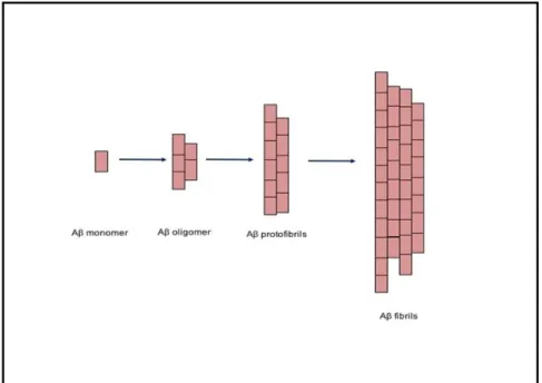
Figure 3: Schematic of A assembly process. A assembly starts from the monomer state to oligomers to protofibrils and fibrils (adapted from Yamin et al, 2009).
2.1.6.4 Criticism and modifications to the amyloid hypothesis
There is accumulating evidence indicating that this hypothesis is not sufficient to explain the multifaceted features of the disease as explained by (Herrup, 2015). The main criticism, which is very evident, is that a direct correlation between amyloid deposits and dementia severity has not been demonstrated, since some patients without amyloid deposition show severe memory deficits, while other patients with cortical A deposits have no dementia symptoms (Terry et al., 1991, Arriagada et al., 1992, Herrup, 2015).
Another point, which arose with time, was the role of the soluble A oligomers. As explained above, during pathological conditions, A1-42 peptide has a tendency to aggregate forming
monomers, oligomers (which could include dimers, trimers, tetramers as aggregates) and later forming protofibrils and fibrils (simplified illustration in Figure 3). Amongst these aggregates, the small soluble A oligomers (between 10 and 100 kDa) were discovered to be toxic and caused synaptic dysfunction and memory impairment (Lambert et al., 1998, Lue et al., 1999, Walsh et al., 2002). Hence these findings modified the hypothesis to the ‘oligomer
26
hypothesis’. It was suggested that A oligomers may have complex effects on surrounding neurons, microglia and astrocytes, the cumulative effect being to subtly alter synaptic function, and thus information storage and retrieval. Memory loss beginning earlier in the disease, was attributed to the oligomer-induced disruption of synaptic plasticity (Selkoe, 2002). The later stages of dementia were attributed to oligomer-induced cellular degeneration and death. The effect of amyloid- peptide/oligomers on synaptic plasticity and memory will be discussed in detail in Chapter 2.
One cannot conclude if it is the large, insoluble deposits or small soluble oligomers that represent the most prominent neurotoxic entity. Recent research shows that there is a dynamic balance between the two entities. The concentration of soluble A oligomers determines the size of the amyloid-beta aggregate, while concurrently this aggregate serves as a reservoir for the soluble amyloid oligomers (Koffie et al., 2009).
Also, although amyloid pathology is still generally considered as a key initiation factor in AD, we shall briefly mention the important contribution of tau. Indeed, the high degree of tau phosphorylation at early stages of AD as a key molecular signature of neurofibrillary degeneration in AD cannot be missed out (Buee et al., 2000). The conversion of physiological tau to pathological aggregates is believed to be a multi-faceted process. Detachment of tau from microtubules to unbound tau could be facilitated by either tau phosphorylation, A-mediated toxicity, oxidative stress, etc (Bramblett et al., 1993, Liu et al., 2005, King et al., 2006). In addition, recent research has opened new insights into the role of tau at the synapse (as mentioned in 2.2.6.2) and in the nucleus. Thus, in combination with amyloid pathology, tau pathology is likely to strongly contribute to neuron dysfunction early in AD progression.
2.1.7 Evolution of AD in the brain
Clinically, the different stages of cognitive symptoms in AD can be divided into three phases: Pre-mild cognitive impairment (PMCI), MCI and AD. A long preclinical stage (PMCI) for almost 15-20 years before any sign of clinical symptoms has been suggested, followed by MCI where NFT and A pathology are increasing along with mild cognitive dysfunction in neuroclinical tests. MCI can be amnestic (aMCI) or multi-domain type (amnesic and other
27
cognitive domains), amongst which those with aMCI are at a higher risk to develop AD (Petersen, 2003).
AD neuropathology commences with thinning of cortex during asympotomatic stages (Dickerson and Wolk, 2012), proceeding with sequential atrophy in the entorhinal cortex, the hippocampus, the neocortex (Killiany et al., 2002, Raz et al., 2004) (Figure 4). In addition, quantitative morphological studies of temporal and cortical biopsies show significant loss of synapse density in AD patients (Davies et al., 1987) supported by low synaptic density and fewer dendritic spines. There is also evidence of a 25% decrease in the presynaptic marker synaptophysin in the cortex of MCI or mild AD patients (DeKosky and Scheff, 1990).
Figure 4: Gross brain of late stage AD patient. A). Generalised atrophy, ventricular dilation and medial temporal atrophy. B). Close up of hippocampal formation of the same patient, red arrow indicating hippocampal atrophy. (Castellani R et al., 2010)
The sequence of appearance of the two neuropathological hallmarks is known to be in a reverse order. Amyloid plaques are initiated in the neocortex and later spread to the entorhinal cortex and hippocampus (specifically, the CA1 sub region) and subcortical regions (Thal et al., 2002a). While NFTs develops sequentially from transentorhinal cortex to entorhinal cortex, hippocampus and neocortex (Thal et al., 1998). The hippocampus is one of the earliest brain structures to develop neurodegenerative changes. There is evidence showing that NFTs develop more profusely than the amyloid plaques in early AD (Arriagada et al., 1992). It is still under debate to determine which of the pathological features is best correlated with the disease state. Clinical studies indicate that NFTs correlate better than amyloid plaques with cognition in AD. Since the discovery that soluble A oligomers have
28
the ability to impair synaptic and memory function (Lesne et al., 2006), the idea is now changing to consider them along with amyloid plaques in pathological studies.
Recent advancement in biomarker research has also helped understand the sequence of abnormalities as the disease progresses. The earliest marker is that of an increase in cerebrospinal fluid (CSF) A42 as shown in the graph below (Figure 5). These changes occur
in preclinical phase and, by the time, the cognitive impairment is clinically detected, A markers have reached a plateau. Subsequently, neuronal injury and neurodegeneration predominate. These are shown by CSF tau and cerebral atrophy on MRI. These markers, which become abnormal later in the disease, correlate more closely with clinical symptoms.
Figure 5: Biomarkers of Alzheimer’s pathological pathway. A is indicated by low CSF A42 or positron emission tomography (PET) A imaging. Tau neuronal injury and dysfunction is shown by CSF tau. Cerebral atrophy is measured with magnetic resonance imaging (Harrison J et al., 2016).
2.1.8 Treatments
AD has till date only a few different treatments targeting specific aspects. The two types of pharmacological therapeutics approved by the FDA are acetylcholine esterase inhibitors and NMDAR antagonist (memantine). In post-mortem AD brains, it was evident that there was cholinergic neuron loss (Whitehouse et al., 1981), although the mechanisms leading to this neuron loss remain largely unknown. Hence, to promote cholinergic signalling in early symptomatic AD patients, acetylcholine esterase inhibitors (donepezil, rivastigmine, and galantamine) have been used for decades to inhibit the acetylcholine degradation and thus maintain higher acetylcholine levels in the extracellular space (Zhu et al., 2013). On the other hand, memantine, an NMDA (N-methyl-D-aspartate) receptor antagonist is used for late
29
stage AD treatment. It is also not yet clear mechanistically how blocking NMDA receptors benefits the diseased brain, but use of memantine probably counteracts the effects of hyperactive excitatory circuits, evidenced in AD patients, and prevents high levels of glutamate from weakening synaptic strength. However, these drugs are only mildly effective and do not significantly halt cognitive deterioration or restore memory function (Zemek et al., 2014).
Other molecular treatments to restore cellular health and to repair circuit and network function are under development. Currently, to target high levels of A, several and -secretase inhibitors and modulators are used in phase III clinical trials (Citron, 2010), but due to severe side effects these clinical trials were aborted (Schor, 2011). Also, active immunization against A is under development, but such endeavours have generally not been successful (Gilman et al., 2005). Indeed, 99% of the A-targeted phase 3 clinical trials in AD have not shown statistically significant benefit on its pre-specified clinical endpoints. These negative results have also largely contributed to question the validity of the amyloid hypothesis. Most recently, some hope was raised by a more positive outcome of a Biogen phase I clinical trial infusing aducanumab in mild AD patients with A pathology as measured by positron emission tomography (PET) (Sevigny et al., 2016). The 3 and 10mg/kg/month doses were effective in reducing brain A levels after 6 months and more so at 12 months. Mild positive effects of these doses on clinical outcome were reported, encouraging rapid transition to phase III trials. Because of the difficulties encountered with anti-A strategies, there resulted an increase of interest in drug trials directed towards tau aggregation (Morris et al., 2011) and APOE (Pedersen and Sigurdsson, 2015). Results of clinical trials using these alternative strategies should be disclosed within the next few years. Directly targeting the activity of brain networks might also help to restore memory. Phase I clinical trials that used deep-brain stimulation techniques to directly manipulate network activity in individuals with AD reported positive memory outcomes (Laxton et al., 2010). This suggests that the development of non-invasive brain stimulation strategies could be a scalable and a safe route to restoring cellular health and network function.
Still, there is a strong urgency to develop new therapeutic strategies as none so far have showed strong benefits in preventing AD occurrence or slowing down AD progression. For this to occur, additional efforts are required to better understand the molecular and cellular mechanisms driving forward AD pathology.
30
2.2 Memory formation, synaptic plasticity and modulation by A
AD is a progressive neurodegenerative disease marked by a constellation of cognitive disturbances. The earliest form of memory affected in AD is episodic memory and is one of the most clinically relevant neurological symptom for AD patients. This form of memory helps us to function smoothly in our basic daily activities and to remember critical events. As the disease progresses the patient develops other cognitive abnormalities such as apraxia (inability to carry voluntary movements), aphasia (loss of ability to speak) and agnosia (inability to recognise objects and their use) eventually leading to global cognitive impairment.2.2.1 Different types of memory
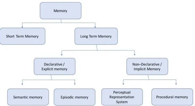
There are mainly two forms of memory depending on the duration of recall: short and long term memory (STM and LTM) (Figure 6).
STM is known to hold a limited amount of information in a very accessible state temporarily (Cowan, 2008). STM mainly relies on cortical neuron activity in the lateral prefrontal cortex (Squire and Wixted, 2011). This structure is specifically known to perform higher executive functions, which include working memory and attention.
The LTM system is divided into two broad classes: explicit (declarative) memory, which requires the conscious recall of data, facts and events, and implicit (non-declarative) memory, which is based on non-conscious memory abilities. Implicit memory includes procedural or skilled-based kinds of learning and perceptual representation system (PRS) (Squire et al., 1987). PRS mainly involves priming effects (where one stimulus influences the response of the other stimulus) and operant or classical conditioning (which involves pairing of two stimuli). Long term memory is stored in the neocortex and distributed in different regions, where the area specific processing occurred at the time of learning.
31
Figure 6: Classification of the different memory systems into short and long term storage systems. Long term memory is further divided into declarative and non declarative memory. Declarative memory represents explicit memory which can be declared and is further classified into semantic and episodic memory. Non declarative memory is the memory used in tasks and skills and can be divided in procedural and perceptual representation system. (Adapted from Tulving, 1995).
2.2.1.1 Declarative long term memory
Declarative memory is subdivided into semantic memory and episodic memory (EM). Semantic memory is related to storage of general facts and knowledge, such as the colour of fruits or the capital of countries. While EM is a system that enables an individual to encode, store, and retrieve information about personal experiences and the temporal and spatial contexts of those experiences (first defined by Tulving in 1972).
2.2.2 Memory formation and its stages:
2.2.2.1 Encoding, Consolidation, Storage and Retrieval
Memory formation and utilization can be divided into the following stages: Encoding, consolidation, storage and retrieval. The critical structures involved in these processes of memory involve hippocampus, the amygdala and the adjacent entorhinal, perirhinal and parahippocampal cortices, which make up the medial temporal lobe (MTL) and the prefrontal cortex (Squire and Wixted, 2011).
Encoding is the first stage of memory, and it is crucial for the storage and retrieval of
32
from external stimuli. The MTL structures are involved and responsible for transforming these sensory inputs to a memory representation (Squire and Wixted, 2011). All these neuronal activities converge to the medial temporal lobe and specifically to the hippocampus, where they bind to form episodic memory (Figure 7) (Kessels and Kopelman, 2012). After binding, they are allocated to areas in the cortex for long-term storage as they are well protected from the influence of new incoming memories (Straube, 2012). The type of information processing that occurs during the encoding stage determines the quality of encoding and recovery of that information.
Figure 7: Functional overview of extended hippocampal-diencephalic memory system. Nonintegrated input from the association areas (such as spatial or object information) is processed in the parahippocampal and perirhinal cortices and then integrated as an “episode” in the entorhinal cortices. Storage then takes place through the hippocampi that are connected via the fornices to the mammillary bodies. Subsequently the thalami projects to the neocortex where the episodic information is permenantely represented (Kessels and Kopelman, 2012).
Consolidation: Encoded memory undergoes consolidation, a process by which short term
memory trace is transferred to stable long term memory before directing it to neocortical areas for long term storage. There are two models of consolidation: the standard model (SM) and the multiple memory trace (MMT) (Nadel et al., 2007). The SM model, proposes that memory storage initially requires the hippocampus to link the different features of memory which are dispersed in several sites in the neocortex. Over time, however, the requirement of the hippocampus decreases and the representation of the memory is solely in the neocortex (Frankland and Bontempi, 2005) (Figure 8). In contrast, the MMT theory poses that all
33
memory traces are combined into a multiple-trace representation. In this model, both the hippocampus and the neocortex continue to interact with each other and that the hippocampus plays a permanent role for the storage and retrieval of the memory. CORT modulate memory consolidation of emotionally aroused experiences (McIntyre et al., 2012) and sleep also plays an active role in memory consolidation (Straube, 2012).
Figure 8: Standard consolidation model. The encoding of perceptual, motor and cognitive information initially occurs in the primary and associative cortical areas. The hippocampus integrates external information coming from specialized cortical areas and fuses them into a coherent memory trace. Successive reactivation of this hippocampal-cortical network leads to progressive strengthening of cortico-cortico connections and over time becomes independent of the hippocampus (Frankland and Bontempi, 2005).
Storage and Retrieval
After the process of consolidation, memories are represented by networks of neurons distributed across the neocortex bound together for rapid storage. For retrieval of memory, MTL structures are necessary along with the cortical regions (Rugg and Vilberg, 2013). During recall of the memory, the representative map in the hippocampus is activated along with the other areas in the cortex, which were involved in forming the memory. While MTL brain structures are necessary for retrieval of recent episode-based memory associations, over time, these associations are expressed and can be recalled independently of the MTL structures (Squire and Wixted, 2011).
During aging, the encoding process seems to be affected, with encoding deficiencies predominating over retrieval deficits (Friedman et al., 2007). Patients with AD show deficits
34
in EM from very early stages of the disease. They show performance deficits in encoding, storing and retrieval (Pena-Casanova et al., 2012).
2.2.3 Episodic memory is affected in AD
Episodic memory refers to the conscious recollection of a unique past experience / event in terms of “what”, “where” and “when” it happened. These three components identify a particular object or person (memory for what happened), the context or environment in which the experience occurred (memory for where it happened) and the time at which the event happened (memory for when it happened) (Nyberg et al., 1996, Clayton and Dickinson, 1998, Tulving and Markowitsch, 1998). It is the first form of memory, which is affected in Alzheimer’s patients (deToledo-Morrell et al., 2007). Recall (free and cued) and
recognition are considered to provide two different ways to measure episodic memory.
Recall depends on declarative memory and recognition on declarative and non-declarative memory. In a free recall experiment, an individual is given a list of items to study and is subsequently asked to recall the items (Arnold and McDermott, 2013), the results of which are noted. While in cued recall, the same protocol is followed with the help of cues. With this test, one can evaluate hippocampus-dependent memory encoding or consolidation. During a recognition experiment, the first phase, called the study phase, a subject is asked to memorize a series of items. Later, in the test phase, randomly ordered items, old and new, are presented and the individual is scored on his ability to distinguish them. In AD patients, both recall and recognition deficits seem to persist, which indicates inability to retrieve information. This could also be related to inability to encode target information (Gold and Budson, 2008).
2.2.3.1 Episodic-like object recognition memory in rodents
It has been challenging to study declarative forms of memory in mice since it is an integrative memory for “what”, “where” and “when” component and also it needs to be expressed non-verbally. Thus, in rodents, the equivalent studies done are hippocampus-dependent spatial and contextual memory tests. The most commonly used tasks to study spatial memory in AD mouse models are the Morris water maze (MWM), the Barnes Maze also called circular platform maze, the continuous and forced-choice spatial alternation task, and the radial arm water maze task (Stewart et al., 2011).
35
To integrate the component of familiarity of items along with recollection of contextual (spatial and/or temporal) information of items, the object recognition test was developed. The novel object recognition task measures spontaneous behaviour in rodents as they readily approach and explore novel objects (Frick and Gresack, 2003). This widely used task consists of a sample trial wherein two identical objects are explored by the mouse, followed by a delayed test trial wherein one familiar object presented during the sample test is replaced by a novel object (see Figure 9A). Animals with adequate memory spend longer time exploring the novel object. The NOR test could be interpreted as the study of the ‘What’ component of episodic memory. This task has been widely used for evaluation of memory in AD mouse models (Balducci et al., 2010). Two key points confirm that novel object recognition can be dependent on hippocampal activity. Firstly, it was noted that hippocampal lesions result in impaired object recognition and secondly a 24-hour inter-trial interval between the two phases (sample and test) confirms retrieval of long term memory consolidated via hippocampal activity (Reed and Squire, 1997). In NOR memory, particularly the dorsal hippocampus plays an important role, especially when spatial or contextual information is a relevant factor (Goulart et al., 2010).