HAL Id: tel-02865212
https://tel.archives-ouvertes.fr/tel-02865212
Submitted on 11 Jun 2020
HAL is a multi-disciplinary open access archive for the deposit and dissemination of sci-entific research documents, whether they are pub-lished or not. The documents may come from teaching and research institutions in France or abroad, or from public or private research centers.
L’archive ouverte pluridisciplinaire HAL, est destinée au dépôt et à la diffusion de documents scientifiques de niveau recherche, publiés ou non, émanant des établissements d’enseignement et de recherche français ou étrangers, des laboratoires publics ou privés.
Modeling cell response heterogeneity to pro-apoptotic
ligands
Luis Carlos Gomes Pereira
To cite this version:
Luis Carlos Gomes Pereira. Modeling cell response heterogeneity to pro-apoptotic ligands. Auto-matic Control Engineering. COMUE Université Côte d’Azur (2015 - 2019), 2019. English. �NNT : 2019AZUR4081�. �tel-02865212�
Modélisation de l’hétérogénéité de la
réponse cellulaire aux ligands
pro-apoptotiques
Luis Carlos GOMES PEREIRA
INRIA Sophia Antipolis / IRCAN CNRS
Présentée en vue de l’obtention du grade de docteur en Informatique d’Université Côte d’Azur
Dirigée par : Madalena CHAVES,
Jérémie ROUX
Soutenue le : 26 Novembre 2019
Devant le jury, composé de :
Elisabeth PÉCOU, Université de Nice Sophia-Antipolis, France
Laurence CALZONE, Institut Curie, France
Laurent TOURNIER, INRA Jouy-en-Josas, France
Rui DILÃO, Instituto Superior Técnico, Portugal
2
Modélisation de l’hétérogénéité de la
réponse cellulaire aux ligands
pro-apoptotiques
/
Modeling cell response heterogeneity to
pro-apoptotic ligands
Jury:
Rapporteurs
Laurence CALZONE, Ingénieur de Recherche Institut Curie, France
Rui DILÃO, Professeur Instituto Superior Técnico, Portugal
Examinateurs
Elisabeth PÉCOU, Professeur Université de Nice Sophia-Antipolis, France
Laurent TOURNIER, Chargé de Recherche INRA, France
Directeur
Madalena CHAVES, Directrice de Recherche INRIA, France
Co-Directeur
4
Abstract
Apoptosis is an essential physiological process through which organisms are able to equilibrate their cell numbers and maintain tissues in healthy and functional conditions. Despite the recent advances in the field, little is known on the molecular mechanisms controlling individual cell decisions to either engage or avoid the activation of this pathway upon cancer treatment, which inevitably impacts on therapeutics development.
To obtain a global view of the intervening proteins and their role on cell response dynamics to anti-cancer drugs, a new and detailed description of the apoptosis pathway at the receptor level was translated into a system of ordinary differential equations. The model was calibrated to single-cell data, from recent experiments on a population of HeLa cells exhibiting a highly heterogeneous response when exposed to the death-inducing ligand TRAIL. The sensitivities of the apoptotic reactions in our model were evaluated using the diversity of experimental behaviors observed in vitro. A series of computational tests and analyses were performed with our model to identify the origins of cell response heterogeneity. New features of the apoptotic pathway emerged from a comparison of different heterogeneity modeling approaches, detecting a set of key reactions to be further expanded.
These analyses yield new biological insights and highlights the importance of refining regulation of death receptor complex activity, possibly through Caspase-10 as suggested from new experimental discoveries. This thesis offers a novel framework that can be used to uncover important biological insights using single-cell data of heterogeneous dynamical pathways.
Keywords: Extrinsic apoptosis; Mass-action differential equations; Cell fate decision;
6
Résumé
L'apoptose est un processus physiologique essentiel permettant aux organismes de maintenir leurs tissus dans des conditions fonctionnelles. Lors du traitement du cancer, les mécanismes moléculaires qui contrôlent la décision cellulaire d’engager ou d’éviter l’activation de cette voie sont mal connus, ce qui a un impact important sur le développement thérapeutique. Pour obtenir une vue globale du rôle des protéines impliquées dans la dynamique de la réponse cellulaire aux anticancéreux, une nouvelle description détaillée de la voie de l'apoptose au niveau du récepteur a été implémentée en un système d'équations différentielles ordinaires. Le modèle a été calibré sur des données expérimentales en cellule unique, de populations de cellules HeLa présentant une réponse hautement hétérogène au ligand, TRAIL. Les sensibilités des réactions apoptotiques dans notre modèle ont été évaluées en utilisant la diversité des comportements expérimentaux observés in vitro. Une série de tests et d'analyses informatiques ont été effectués avec notre modèle pour identifier les origines de l'hétérogénéité de la réponse cellulaire, faisant émerger de nouvelles caractéristiques de la voie apoptotique.
Ces analyses apportent de nouvelles connaissances biologiques et soulignent l’importance de la régulation du complexe des récepteurs de la mort, éventuellement par la Caspase-10, comme le suggèrent de nouvelles découvertes expérimentales. Cette thèse offre une nouvelle approche pour découvrir des informations biologiques importantes en utilisant des données en cellule unique, de voies de signalisation à dynamiques hétérogènes.
Mots-clés : Apoptose extrinsèque ; Equation différentielles d'action de masse ; Décision de la
mort cellulaire ; Hétérogénéité ;Distribution de paramètres ; Régulation de la Caspase-8 ; Modèles prédictifs.
8
Acknowledgments
I would like to start to express my sincerest gratitude to all the jury members for their precious time and attention reading this manuscript. All their observations were extremely valuable and essential for the accomplishment and accurateness of the text. I absolutely appreciate all your efforts.
To my supervisors, Madalena Chaves and Jérémie Roux, a “vrai remerciement” for their kindness and comprehension during these last three years. Despite the difficulties during the different steps of the projectthey were always caring and positive, supporting me through the innumerous challenges I had to face. I hope you keep doing superb research at the level you both deserve and that one day we can work together again. Also, of course, thank you for giving me a position and a scholarship that allowed me to have a PhD on such an extraordinary and motivating field and to have the chance to progress in my personal career.
A big kiss to all my dear friends in Nice for their exclusive comfort and joy. The feedback I received from you helped me to stay motivated and appreciate the wonders of France. I will always remind the dinners and the never-ending talks in our nights-out in Nice. An enormous hug to my fantastic “Italian group”: Serena, Melania, Sofia and Alberto, your kindness and presence is unforgivable. We will be friends to the end.
To a dear close colleague in my office, Mickael Meyer, I am grateful for having meeting you and to have spent three years of my PhD with you in my team. You are an extraordinary person, always with a great smile and ready to hear everyone’s problems. Wish you all the best, you really deserve it.
To Baharia Mograbi, my second and borrowed mother, I am grateful to have shared so many lunches in your company and to have listen to your constants “Courage! Dans la joie et la bonne humeur”. You are a precious and rare human being, I will never forget you.
Last but not least, an enormous gratefulness to my mother and father. Thank you for being the best parents in the world. Your continuous skype calls refilled my motivation and cheered me up in those days where I thought nothing could comfort me and I felt the urge to go back to Portugal. You always gave me reasons to keep going and never give up. I love you both unconditionally.
10
Contents
ABSTRACT ... 4 RESUME ... 6 ACKNOWLEDGMENTS ... 8 LIST OF TABLES ... 16 GLOSSARY... 18 1 INTRODUCTION ... 21 1.1 MOTIVATIONS ... 211.2 APOPTOSIS SIGNALING NETWORK ... 24
1.2.1 TRAIL, a death-inducing molecular agent initiating apoptosis ... 24
1.2.2 DR4 and DR5, decoy death receptors, and clustering modes with TRAIL ... 26
1.2.3 DISC complex, the basic unit structure for activation of Caspase-8, and the role of FLIP as an inhibitor of apoptosis ... 26
1.2.4 Caspase-8, a threshold for cell-fate decision ... 27
1.2.5 Bcl-2 like proteins, pro- and anti-apoptotic roles ... 30
1.2.6 MOMP, an irreversible commitment to cell-death ... 31
1.2.7 Global overview on the extrinsic apoptosis pathway ... 32
1.3 MODELLING IN APOPTOSIS ... 34
1.3.1 Fussenegger’s model, 2000 ... 34
1.3.2 Albeck’s EARM1 model, 2008 ... 37
1.3.3 Schlatter’s & Calzone’s model, two Boolean modeling approaches ... 40
1.3.4 Bertaux’s model, a stochastic protein-turnover approach 2014 ... 42
1.3.5 A summary list of models of apoptosis ... 44
1.4 HETEROGENEITY IN BIOLOGY, INTRINSIC AND EXTRINSIC NOISE ... 46
2 MODELING RECEPTOR-LIGAND INTERACTIONS FOR THE EXTRINSIC APOPTOSIS PATHWAY ... 52
2.1 A NETWORK OF REACTIONS DEFINING A MODEL STRUCTURE FOR THE RECEPTOR LAYER OF THE EXTRINSIC APOPTOSIS NETWORK ... 54
2.2 AVAILABLE EXPERIMENTAL DATA ... 60
2.3 CONVERSION OF FRET-SIGNAL INTO NUMBER OF ICRP-CLEAVED MOLECULES ... 64
11
2.5 ARROM2: A LIGAND-RECEPTOR MODEL WITH AN EXTRA SET OF PROPOSED
REACTIONS ... 68
3 VALIDATION OF ARROM2: A RECEPTOR-LIGAND MODEL IN AGREEMENT WITH EXPERIMENTAL DATA ... 81
3.1 FLIP, A STRONG ANTI-APOPTOTIC PROTEIN WITH IRREVERSIBLE BINDING AT THE DISC STRUCTURE ... 81
3.2 DIMERIC VS.TRIMERIC LIGAND VALENCY, AN UNEQUAL RECEPTOR BINDING RATE ... 84
4 SOURCES OF HETEROGENEITY IN APOPTOTIC CELL-FATE DECISION . 87 4.1 INTRINSIC NOISE:A COMPUTATIONAL APPROACH WITH THE GILLESPIE ALGORITHM . 88 4.2 EXTRINSIC NOISE: A COMPUTATIONAL APPROACH WITH INITIAL CONDITION VARIATION. ... 91
4.3 PARAMETER NOISE: A COMPUTATIONAL APPROACH WITH VARIATION IN REACTION RATES 93 5 FITTING THE ENTIRE CELL POPULATION AND SEARCHING FOR SIGNATURES IN SENSITIVE AND RESISTANT POPULATIONS ... 95
6 DISCUSSION AND PERSPECTIVES ... 103
7 APPENDIX... 108
7.1 APPENDIX 1:ARROM1 MODEL INPUTS ... 108
7.2 APPENDIX 2:ARROM1 FIT TO THE MEDIAN_CELL ... 112
7.3 APPENDIX 3:ARROM2 FIT TO THE MEDIAN_CELL ... 114
7.4 APPENDIX 4 :ARROM1 MODEL EQUATIONS ... 116
7.5 APPENDIX 5:A NEW METHOD TO QUANTIFY PARAMETER VARIABILITY AMONG DIFFERENT PARAMETER POPULATIONS ... 122
13
List of figures
FIGURE 1. A CELL FATE PREDICTION ARISING FROM CONTRIBUTION OF BOTH RATE AND
DURATION OF C8 ACTIVITY ... 29
FIGURE 2. SUMMARY NETWORK OF THE EXTRINSIC APOPTOSIS SIGNALING PATHWAY ... 33
FIGURE 3. FUSSENEGGER’S APOPTOSIS-RELATED MODEL RESULTS ... 35
FIGURE 4. ALBECK’S EARM1 NETWORK OF REACTIONS ... 37
FIGURE 5. BERTAUX’S STOCHASTIC PROTEIN TURNOVER MODEL ... 42
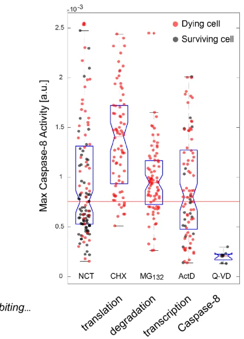
FIGURE 6. VARIABILITY ON MAXIMUM C8 ACTIVITY VALUES FOR A POPULATION OF HELA CELLS UNDER DIFFERENT CO-TREATMENT SCENARIOS. ... 50
FIGURE 7. NETWORK STRUCTURE FOR THE MODELED RECEPTOR-REACTIONS IN THE EXTRINSIC APOPTOSIS PATHWAY ... 59
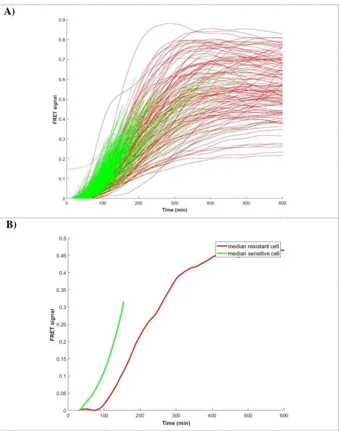
FIGURE 8. GENERAL FEATURES OF THE FRET-SIGNAL OF AN ARBITRARY CELL ... 61
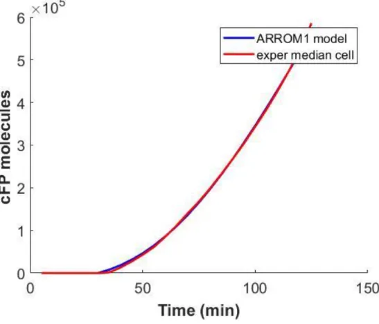
FIGURE 9. SIGNAL IN TIME OF THE CELL POPULATION FOR A TRAIL TREATMENT OF 50NG/ML 62 FIGURE 10. FITTING THE MEDIAN_CELL OF THE HELA-CELL POPULATION TREATED WITH TRAIL AT 50NG/ML ... 69
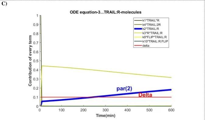
FIGURE 11. MOST CONTRIBUTING TERMS IN THE ARROM1 MODEL FOR THE SET OF ODE EQUATIONS WHERE K2 INTERVENES. THE VECTOR OF PARAMETERS USED TO RUN THE SIMULATION RESULTS FROM THE FIT OF ARROM1 MODEL TO THE MEDIAN_CELL TRAJECTORY. ... 71
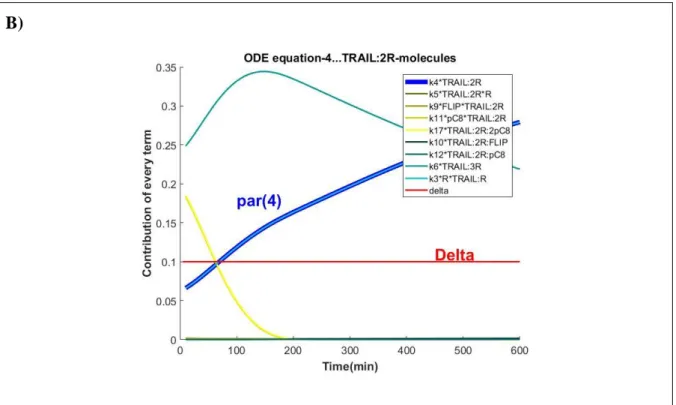
FIGURE 12. MOST CONTRIBUTING TERMS IN THE ARROM1 MODEL FOR THE SET OF ODE EQUATIONS WHERE K4 INTERVENES. THE VECTOR OF PARAMETERS USED TO RUN THE SIMULATION RESULTS FROM THE FIT OF ARROM1 MODEL TO THE MEDIAN_CELL TRAJECTORY. ... 72
FIGURE 13. MOST CONTRIBUTING TERMS IN THE ARROM1 MODEL FOR THE SET OF ODE EQUATIONS WHERE K6 INTERVENES. THE VECTOR OF PARAMETERS USED TO RUN THE SIMULATION RESULTS FROM THE FIT OF ARROM1 MODEL TO THE MEDIAN_CELL TRAJECTORY. ... 73
14
FIGURE 14. MOST CONTRIBUTING TERMS IN THE ARROM1 MODEL FOR THE SET OF ODE EQUATIONS WHERE K16 INTERVENES. THE VECTOR OF PARAMETERS USED TO RUN THE SIMULATION RESULTS FROM THE FIT OF ARROM1 MODEL TO THE MEDIAN_CELL
TRAJECTORY. ... 77
FIGURE 15. FLIP UNBINDING RATES SHOW EVIDENCE OF AN IRREVERSIBLE REACTION ... 83
FIGURE 16. LIGAND VALENCY RESULTS IMPOSE HIGHER BINDING RATES AND/OR LOWER DISSOCIATION RATES OF THE RECEPTOR IN THE TRIMERIC COMPLEX ... 86
FIGURE 17. STOCHASTIC SIMULATION OF THE ARROM2 MODEL USING THE GILLESPIE ALGORITHM ... 90
FIGURE 18. EXTRINSIC NOISE, SIMULATION OF MEDIAN_CELL WITH ONE THOUSAND DIFFERENT
INITIAL CONDITIONS ... 92
FIGURE 19. PARAMETER NOISE SIMULATION ON THE ARROM2 MODEL ... 94
FIGURE 20. FITTING ARROM2 TO SINGLE CELLS REQUIRES PROTEIN NUMBER VARIABILITY AND SMALL PARAMETER VARIATION TO REPRODUCE THE CELL POPULATION TRAJECTORIES
98
FIGURE 21. MOVING-AVERAGE REVEALS TRENDS ON INITIAL CONDITIONS THAT CORRELATE WITH INCREASE IN C8 ACTIVITY. ... 100
FIGURE 22. A POSSIBLE POSITIVE FEEDBACK LOOP BETWEEN X1 AND C8. ... 101
FIGURE 23. INCLUSION OF A POSITIVE FEEDBACK-LOOP IN THE RECEPTOR REACTIONS OF THE EXTRINSIC APOPTOSIS PATHWAY. ... 102
FIGURE A.1) REPRESENTATION OF THE PARAMETER SET OF THE ARROM2 MODEL IN THE NORMALIZED MEAN SPACE AFTER FITTING THE SENSITIVE AND THE RESISTANT CELL POPULATIONS ... 127
FIGURE A.2) REPRESENTATION OF THE PARAMETER SET OF THE ARROM2 MODEL IN THE NORMALIZED STANDARD DEVIATION SPACE AFTER FITTING THE SENSITIVE AND THE RESISTANT CELL POPULATIONS ... 128
16
List of Tables
TABLE 1.SAMPLE OF THE RATE LAWS USED IN FUSSENEGGER’S PUBLICATION ... 34
TABLE 2.VALUES EXTRACTED FROM EARM1’S SUPPLEMENTARY TABLE S5 TO CALCULATE THE
CYTOSOLIC VOLUME FACTOR. ... 64
TABLE 3.COMPARISON OF PARAMETER VALUES BEFORE AND AFTER FITTING THE EXPERIMENTAL
MEDIAN_CELL WHILE ALLOWING THE PARAMETERS [2,4,6,16] TO VARY IN AN UNBOUND INTERVAL. ... 78
TABLE 4.COMPARISON OF PARAMETER VALUES [2, 4, 6,16] AND PROTEIN X2, BEFORE AND AFTER FITTING OF THE EXPERIMENTAL MEDIAN_CELL TO THE ARROM2 MODEL. ... 80
TABLE A1.1.LIST OF AVERAGED NON-ZERO INITIAL CONDITION VALUES FOR ARROM1 AND
ARROM2 MODEL SIMULATIONS. ... 108
TABLE A1.2.LIST OF ARROM1 MODEL REACTIONS AND CORRESPONDING PARAMETER VALUES
………..109
TABLE A2.1:FULL LIST OF RELATIVE DEVIATIONS FOR THE PARAMETER SET IN THE ARROM1 MODEL AFTER FITTING THE MEDIAN_CELL TRAJECTORY ... 112
TABLE A3.1PROTEIN INITIAL CONDITION VALUES BEFORE AND AFTER FITTING THE MEDIAN_CELL
TO THE ARROM2 MODEL. ... 114
TABLE A3.2PARAMETER LIST VECTOR BEFORE AND AFTER FITTING THE MEDIAN_CELL TO THE ARROM2 MODEL. ... 114
18
Glossary
Apaf-1: protease activating factor-1
ARROM: Apoptosis receptor reaction ODE model Bax: Bcl-2-associated X protein
Bak: Bcl-2 homologous antagonist killer Bcl-XL:B-cell lymphoma-extra-large
Bid: Bcl-2 family member Bcl-2 homology domain 3 (BH3) interacting domain C3/C6/C7/C9: caspase 3/6/7/9
C8/C10: Caspase 8/10 CyC: cytochrome c DcR-1/2: Decoy receptor DED: Death effector domain
DISC: Death-inducing signaling complex DR 4/5: Death receptor 4/5
EARM: Extrinsic apoptosis reaction model FADD: FAS-associated death domain
FLIP: FADD-like IL-1-converting enzyme (FLICE)-inhibitory protein IAP: inhibitor of apoptosis protein
IMS: intermembrane mitochondrial space k: Rate of C8 activity
MCL-1: myeloid leukemia cell differentiation proteins MOM: mitochondria outer membrane
MOMP: mitochondria outer membrane permeabilization ODE: Ordinary differential equation
pC8/10: pro-caspase 8/10 R: Receptor
19
tBid: truncated Bid
TNF: Tumor necrosis factor
TRAIL: Tumor necrosis factor-related apoptosis inducing ligand TRAIL-R 1/2/3/4: Trail receptor 1/2/3/4
XIAP: X-linked inhibitor of apoptosis protein
20
“It is not knowledge, but the act of learning, not possession but the act of getting there, which grants the greatest enjoyment. “
21
Chapter 1
1
Introduction
“The unexamined life is not worth living”
- Socrates
1.1 Motivations
Apoptosis: an important pathway in systems biology and medicine
Apoptosis comprises a set of chemical reactions through which cells can be eliminated when exposed to intracellular or extracellular stress signaling events (Renault and Chipuk, 2014; Tait and Green, 2010). When cell damage becomes relevant and endangers the rightful genotypic transmission to the incoming cell generations the cell generally chooses to commit apoptosis in order to protect the tissue from alarming and malignant mutations that can propagate to a tissue-level. The proper control of apoptosis is thus essential for the correct development of biological tissues and maintenance of homeostasis and its deregulation is commonly associated to disease conditions (Jacobson et al., 1997; Kerr et al., 1972). Inhibition of apoptosis often correlates with immune disorders and cancer formation and its excessive activation is an underlying feature of Parkinson, Alzheimer and Huntington’s disease. (Brown and Attardi, 2005; Qi and Shuai, 2016).
22
Apoptosis is defined as either intrinsic or extrinsic depending on the origin of the death-signaling stimulus (Dewson and Kluck, 2009; Rehm et al., 2002). In the intrinsic apoptosis pathway the signal source is due to an intracellular event caused e.g. by a viral infection, excessive exposure to an oxidative stress or high levels of a DNA-damaging agent, such as UV radiation, that lead to the activation of a mitochondrial dependent pathway and the production of effector caspases. The proteins belonging to the effector caspase family are capable for cleaving a subset of intracellular targets, ultimately causing nuclear condensation, DNA fragmentation and consequent cell death (Brenner and Mak, 2009; Brune and Andoniou, 2017). The extrinsic apoptosis pathway is triggered instead by a death-ligand that interacts with the death-receptors on the cell membrane, activating an intra-cellular cascade that results in the production of initiator caspases. These are capable of directly activating effector caspases in the so-called type I cells. However, in some cell-lineages known as type II cells, effector caspases are inhibited by a pool of existing anti-apoptotic proteins and the initiator caspases must follow an indirect pathway and cause first the release of molecular substances from the mitochondria to inhibit the anti-apoptotic proteins present in the cell. In fact, both intrinsic and extrinsic pathways intersect at the level of the mitochondria, differing solely in their signal provenience. In the two pathways the same order is followed: initiator caspases are activated first and only afterwards there’s a rise on the effector caspases concentration inside the cell.
Although massively studied, it is still unclear which step by step conditions or molecular proportions of pro- and anti-apoptotic proteins should be achieved along the several stages of the pathway for the cell to irreversibly commit to death. This has direct implications in clinical applications, for instance in cancer. Cancer cells typically possess innate resistances to both chemotherapy and death-inducing drug agents rendering them almost unaffected to these approaches (Almendro et al., 2013; Fulda and Vucic, 2012; Juin et al., 2013; Lopes et al., 2007). In some cases, resistance arises from specific mutations at the genomic level (Holohan et al., 2013). However, in general, non-genetic factors seem to be the key mechanisms behind resistance to drug and chemotherapy treatments (Cohen et al., 2008; Kreso et al., 2013; Roesch et al., 2010; Roux et al., 2015; Sharma et al., 2010; Spencer et al., 2009). In worse case scenarios these factors can accumulate and eventually persist at the cell population level (Brock et al., 2009; Wakamoto et al., 2013).
The link between the miss-regulation of apoptosis and the resulting phenotypic impairments make it a very challenging topic in both biology and medicine. One of the major drawbacks is
23
to understand why the phenotypic responses of clonal or sister cells are so highly heterogeneous when exposed to identical death signals (Balázsi et al., 2011). This effect, described as “fractional-response”, is a hallmark of apoptosis and is a topic that has been under intense investigation in the past decades.
Multiple modeling efforts have tried to bring to light levels of interactions and mechanisms of control that can make cells to choose between life (apoptosis escape) and death (apoptosis commitment). Along the next lines of the manuscript, the role of the most important proteins of the apoptotic system and a proposed mathematical model will be discussed in parallel as an attempt to tackle this question. In chapter 1, a deep inspection of the literature gathered a theoretical scheme of the extrinsic apoptotic reactions and a general explanation on the most relevant biological events is provided. A quick overview of previous models is also given, with a focus on five models that show how the modeling questions in apoptosis evolved in the past two decades. Chapter 1 ends with a simple description of noise in biology, its different sources and the existing strategies to model each noise source. In Chapter 2, the ARROM1 mathematical model is built from the biological information explained in Chapter 1. The signal of the available data set is briefly explained along with some methods to match the model output with the experimental signal information. Arguments are then presented on the hypothesis that the reactions defined in the literature might be incomplete and an extra set of proteins might exist so that model fittings become more coherent with respect to previous parameter values available in the literature. Chapter 3 provides some examples of how the new model version, with two new proteins, reproduces known biological results, validating the model approach. In Chapter 4, the noise sources introduced in chapter 1 are simulated into the model equations and an inspection is performed to verify which of them can reproduce the heterogeneity observed in the data set. Chapter 5 continues with the fits of the entire cell population and the analysis of which resulting distributions in parameter values and initial conditions contain the largest differences between the population of resistant and sensitive cells. Hypothesis are presented for the dependencies that might exist between the new proposed proteins in Chapter 2 and their connection to FLIP or Caspase-8, the two major apoptotic intervenients at the receptor level. Finally, Chapter 6 summarizes the results of this thesis and proposes new directions in the modeling field of apoptosis.
24
1.2 Apoptosis signaling network
1.2.1 TRAIL, a death-inducing molecular agent initiating apoptosis
Tumor necrosis factor (TNF)-related apoptosis inducing ligand (TRAIL) is a cysteine
polypeptide belonging to the class of death-inducing ligand molecules termed TNF-superfamily, capable of selectively triggering apoptosis in cancer cells in vitro and in vivo (Ashkenazi et al., 1999; Walczak et al., 1999). Its application in the cancer-therapy field has been especially encouraging due to its singular mode of action, not only because of its exclusive impact on cancer cells, but also because of its capability of triggering an intracellular death response independent of the p53 signaling cascade that is commonly mutated in a variety of cancer entities (Kozłowska and Puszynski, 2016; Muller and Vousden, 2013; Olivier et al., 2010).
Like the majority of the TNF-family members, TRAIL’s induction of an apoptotic response depends on its ability to recruit complementary death-receptors to the cell membrane in order to form relatively stable homodimers or heterodimers at the cell surface. In particular, two classes of death-receptors, the class TRAIL-receptor 1 (TRAIL-R1/DR4) and/or the class
TRAIL-receptor 2 (TRAIL-R2/DR5) aggregate with TRAIL to form active structures capable
of propagating the death signal to the interior of the cell (Hymowitz et al., 1999; Jones et al., 1999). Although a natural component of the human bloodstream, TRAIL baseline concentration is insufficient to induce an apoptotic response in natural tissues and its biological role in normal conditions is therefore unknown. (Gibellini et al., 2007; Mariani et al., 1997).
As part of an anti-cancer drug therapy TRAIL shows ulterior advantages as it doesn’t promote the usual toxicity signals derived from cross-activation of pro-survival inflammatory pathways observed in other TNF members (Ashkenazi et al., 1999; Roberts et al., 2011; Walczak et al., 1999). However, it is also long known that besides the desired apoptotic response TRAIL also unleashes the activation of the NF-KB pathway (Chaudhary et al., 1997; Degli-Esposti et al., 1997; Schneider et al., 1997; Trauzold et al., 2001) , MAPK pathway (Tran et al., 2001), and PI3K pathways (Azijli et al., 2012; Xu et al., 2010), all of these pro-survival and undesired secondary signalling pathways. The result is an overall decrease of efficiency of the treatment as at the same time it induces pro-apoptotic signals, TRAIL also unleashes parallel pro-survival signalling responses.
25
Up to these days TRAIL tests in clinical trials have proven rather disappointing as it was shown in a recent metastatic pancreatic cancer study where phase II-trial patients revealed no overall improvement neither in 12-month survival rate nor on overall survival rate in comparison with a gemcitabine monotherapy [gemcitabine is a common chemotherapeutic agent] (Kretz et al., 2018). Other DR agonistic antibodies including mapatumumab, drozitumab, conatumumab , lexatumumab and LBY135 , all triggering extrinsic apoptosis in a manner similar to TRAIL, have all been discontinued after revealing also unfruitful results in clinical applications (Herbst et al., 2010; Merchant et al., 2012; Rocha Lima et al., 2012; Sharma et al., 2014; Younes et al., 2010). The reasons explaining their unsuccess are still to be properly characterized. In future applications, the optimal direction strategy will pass by the development of more potent and stable drugs that can successfully stimulate the extrinsic apoptosis pathway in a selective manner, avoiding the activation of undesired pro-survival responses. For that matter it is necessary to unveil the dynamics of these interlinked pathways and to understand the different thresholds that allows the cell to activate a so called “fractional survival”, where some cells avoid the apoptotic death-fateand resist the therapy (Shlyakhtina et al., 2017).
Recent findings suggest that the interaction between the death-ligand TRAIL and the complementary death receptors is already a decisive factor that distinguishes the downstream activated pathways. Incomplete trimeric interactions with just two binding sites attached in a TRAIL-R1-TRAIL complex are sufficient for activation of a NF-kB signaling response (Morton et al 2015) and a specific oligomerization between trimeric complexes may be required for the proper activation of the apoptotic signal (Wajant, 2019). A detail understanding of these interactions is thus essential.
The time of application has also shown to be important. A pulse-like TRAIL dosage instead of a continuous treatment has demonstrated to be associated with therapy efficacy. In a study from 2016 it was shown that a 1-min pulse of TNF was more efficient in inducing a cell apoptotic response than a 30-min or 60-min pulse and it was as efficient as a continuous treatment with a 10-hours dosage. The conclusion was that the timing of cell exposure impacts cell fate decision and long applications may stimulate the death receptors in such a way that they can be more prone to activate secondary pro-survival responses. Short-pulse scenarios are then related with weaker NF-kB signaling and stronger pro-apoptotic outcomes (Lee et al., 2016).
26
The application of the drug, and specifically the time of successive dosages, has also shown interesting dynamics. As a consequence of the augmented levels of pro-survival proteins during the hours that follow a first TRAIL dosage, the subsequent rounds may end up ineffective if a “drug-holiday” is not considered. In this sense, and similar to other chemotherapy drug scenarios, it can be beneficial to establish neutral periods of non-administration until the pro-survival protein levels return to their basal levels (Becker et al., 2011; Das Thakur et al., 2013).
The adequate treatment may pass by the right usage and combination of multiple drugs while also considering the timing of TRAIL application so that in the end the cell system can be sensitized into a correct cell death direction.
1.2.2 DR4 and DR5, decoy death receptors, and clustering modes with TRAIL
TRAIL can bind to a variety of death-receptors (DR) in the cell membrane. The receptor isoforms propagating the apoptotic signal are the DR4 and DR5 that exist in a stable equilibrium of monomer and dimeric versions, being the monomeric the most common based on molecular studies of CD95 (Liesche et al., 2018). The binding affinity between monomeric receptors is low, justifying the existence of a higher proportion of monomeric receptors (Chan, 2000; Clancy et al., 2005; Neumann et al., 2014). Despite less common, not all the receptors in the cell membrane are functional and another class, termed decoy receptors (DcR), is able to interfere with the death-ligand signal and disrupt the interaction with the adequate receptors DR4/DR5. These are the TRAIL-R3 (DcR1) and the TRAIL-R4 (DcR2) receptors which not only compete for direct contact with the ligand but are also capable of sequestering free DR4/DR5 receptors and bind to them, forming non-functional complexes (LeBlanc and Ashkenazi, 2003).1.2.3 DISC complex, the basic unit structure for activation of Caspase-8, and
the role of FLIP as an inhibitor of apoptosis
TRAIL sequentially adds receptor molecules DR4/DR5 until a trimeric complex is formed. After the trimerization, other molecules join with the death receptors to form a functional
death-inducing signalling complex (DISC), capable of propagating the signal to the remaining
steps of the network. These are the FAS-associated death domain (FADD) molecule and the initiator pro-caspase 8 (pC8), added in the same order and in a sequential manner (Dickens et al., 2012). While the receptor multimerization and association with the ligand is a quick
27
process, the addition of FADD is a limiting and delayed step, essential for the binding of pC8 to the DISC platform (Pennarun et al., 2010). FADD binds to one assembled trimeric receptor complex and acts as an adaptor molecule for the recruitment of several pro-caspases, allowing a first pC8 to be fixed and directly bind to its death effector domain (DED). From there, a second pro-caspase can bind to the free DED of the first pro-caspase and a chain of interlinked molecules is sequentially formed (Schleich et al., 2016). Once a dimerization occurs, by proximity-induced catalytic activation, a functional form of the pro-caspases is released from the receptors, in the form of caspase 8 (C8), sending the apoptotic signal from the receptors to downstream reactions occurring in the cytoplasm of the cell (de Miguel et al., 2016; Dickens et al., 2012) .
A controlling step in this process depends on a homolog molecule with a very similar structure to that of the pro-caspases, the FADD-like IL-1-converting enzyme (FLICE)-inhibitory protein (c-FLIP), that competes for a binding position with FADD suppressing the elongation chain of linked pro-caspases and obstructing their activation (Hughes et al., 2016). Its major role is to hinder the apoptotic signal setting a threshold between quantities of pro-apoptotic caspases vs. the quantity of pro-survival FLIP molecules in the DISC unit.
1.2.4 Caspase-8, a threshold for cell-fate decision
The requirements for the activation of C8 is the presence of a DISC assembled structure with at least two attached pC8 chains, interlinked through their DED domains. In this configuration, the two pC8 proteins inter-activate themselves and form a single structure with catalytic activity that is next released from the DISC unit and navigates into the cytoplasm to participate in the cleavage of downstream molecules. The amount of C8 released from the receptor in this way is essential as a first step towards an apoptotic cell response but yet little is known about the actual mechanisms that control C8 numbers inside the cell.
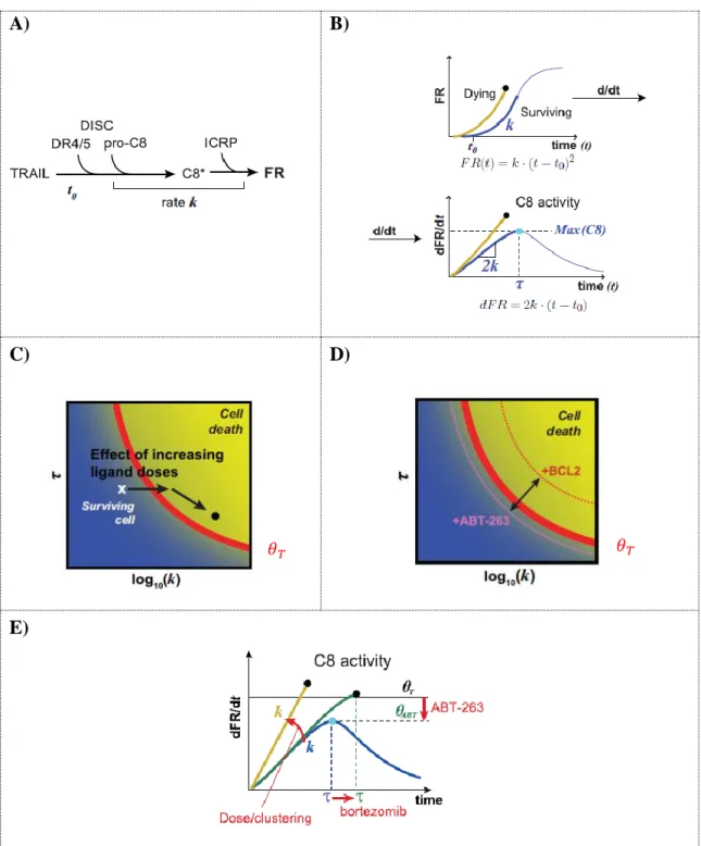
Recently, with the scope of understanding and quantifying the C8 activity level in apoptotic committed cells, Roux and colleagues analyzed hundreds of HeLa-cells treated with TRAIL at multiple concentrations and concluded that the dynamics of the C8 protein plays a central role in cell-fate decision, unveiling a necessary relationship between the rate of C8 activity (k) and the time of maximum C8 activity (𝝉) for cells to properly engage into apoptosis (Roux et al., 2015). Each cell was fitted to the parametric model 𝒌 ∗ (𝒕 − 𝒕𝟎)𝟐 and the optimal maximum derivative value, 𝒎𝒂𝒙(𝟐𝒌 ∗ [𝒕 − 𝒕𝟎]), that best separated the group of sensitive cells from the
28
group of resistant cells was defined as 𝜽. The maximum derivative value assigned to 𝜽 can then be interpreted as a biological threshold, able to distinguish surviving cells from dying cells in the (k, 𝝉) space (red line in figure 1-C).
The work was pioneer in the field by presenting for the first time a metric to predict apoptosis propensity on a given cell. Although simplified by the usage of the lumped parameters k and 𝝉, the approach was accurate enough to correctly forecast the cell fate of more than 80% of the cell population in several cell lines. This number, as a result of a model approximation that considered uniquely DISC-related events, agreed with the result of Gaudet and colleagues that mentions that the non-DISC related proteins contribute to the overall heterogeneity of the apoptotic signal in a range of 20% (Gaudet et al., 2012).
An interesting conclusion of this study was that cells can be sensitized to cross 𝜽 in multiple ways such as by increasing the ligand (TRAIL) concentration (by stabilizing the DISC and consequently augmenting the C8 rate of activation (k)) or by adding drugs that stabilize the C8 molecule (e.g. bortezomib, an inhibitor of protein degradation triggered in a proteasome dependent way) [increasing the time of C8 activation (𝝉)]. In the opposite direction, cells can also turn more resistant to apoptosis by overexpressing anti-apoptotic proteins as the FLIP isoforms (specially the FLIP-s (FLIP short) configuration). All these scenarios are DISC-related events and do not change 𝜽 = 2.61x10⁻³ s−1. However, when adding a drug acting downstream of C8 activation, the value of the threshold changes in a concomitant way. This was verified by the addition of the pro-apoptotic BCL-2 inhibitor, ABT-263, that by decreasing the mitochondrial outer membrane permeabilization (MOMP) “sensitivity”, it was able to lower 𝜽 in those cell populations, making them more prone to die. In the end, the framework was especially relevant as it compiled information from C8 trajectories that varied importantly in signal and had (k, 𝝉) pair values changing tenfold and threefold, respectively, across the cell populations (Roux et al., 2015).
Although simple in the description of the underlying apoptotic pathway, the work still has the potential to be further explored and extended so as to include the role of other molecules also activated under an apoptotic stimulus but known to be C8-independent (Scott et al., 2017). A more complete version would be most useful to understand in better detail how to make a cell irreversibly enter in the death-region space (figure 1-C, D). The framework suggests several ways in which cells can be sensitized to cross the theta-threshold line but cells still remain close to the theta vicinity, calling for more effective co-drugging treatments.
29
A) B)
C) D)
E)
Figure 1. A cell fate prediction arising from contribution of both rate and duration of C8 activity.
A) A simplified scheme for the activation of C8 from the DISC and consequent cleavage of its substrate ICRP (a
reporter fluorescent protein), returning the output signal of the cleaved fluorescence reporter (FR). The lumped parameter k represents the rate of C8 activation and t0 the time required to start receiving the signal from the first cleaved ICRP molecule. B) The shape of an output signal produced by an averaged surviving and dying cell. Dying cells have a more pronounced parabolic trajectory curve, a sign of higher C8 activities. The model 𝑭𝑹(𝒕) = 𝒌(𝒕 − 𝒕𝟎)𝟐 describes the period after ligand stimulation, when C8 activity increases and follows a quadratic
polynomial curve and is valid until the maximum value of dFR/dt, corresponding to the maximum C8 activity value at time t = 𝝉. C) Dosage increase of the ligand causes an overall increase in C8 activity (k) and a small decrease on the time interval required to reach the associated maximum activity (𝝉). The 𝜽-value of the population remained unchanged D), E) Co-treatments cause a deviation on the (k, 𝝉) values of a mean-cell of the population and on the landscape of the 𝜽 curve. Figure adapted from (Roux et al., 2015).
30
1.2.5 Bcl-2 like proteins, pro- and anti-apoptotic roles
Once the DISC complexes are activated and functional C8 is generated, a group of proteins of the BCL-2 family group intervenes into the apoptosis cascade and holds the next decision to block the incoming signal or propagate it to the mitochondria outer membrane (MOM). This decision point, specific of type II cells, starts with the C8 cleavage of the Bcl-2 family member
Bcl-2 homology domain 3 (BH3) interacting domain (Bid) into a truncated form tBid that, in
this form, is translocated from the cytosol to the MOM where it can interact with other elements of the Bcl-2 family. This step is of major importance and should be a result of sustained C8 activation in order to reach an important pool of tBid molecules and surpass an irreversible pro-apoptotic threshold.
The molecular targets of tBid are the Bcl-2-associated X protein (Bax) and Bcl-2
homologous antagonist killer (Bak), both responsible for MOMP and propagation of the
apoptotic signal (Green, 2004; Huang et al., 2016). The agent tBid allows the oligomerization of Bax and Bak to occur, changing them into an active form that is capable of perforating the MOM and transform it into an all-or-none MOMP signal.
A tight level of control is also set by a group of pro-survival proteins, unbalancing the signal from Bax and Bak and allowing the cell to control spurious apoptotic stimulus. Under these conflicting forces a minimum limit is then set for Bax and Bak pro-apoptotic intensity. Among the pro-survival proteins there is the B-cell lymphoma-extra-large (Bcl-XL) and the myeloid
leukemia cell differentiation proteins (Mcl-1) which bind to Bax and Bak and convert them
into their inactive and non-oligomerized form not capable of triggering MOMP (Llambi et al., 2011; Willis, 2005).
As a synthesis, the Bcl-2 family members can be divided into three groups: pore-forming effector proteins, Bax and Bak; BH3-like proteins such as tBid; and the pro-survival proteins such as Bcl-2, Bcl-xL and Mcl-1. The effector proteins are capable of forming mitochondrial pores but only after a conformational change induced by the BH3-like proteins (Chipuk et al., 2010). On the other hand the pro-survival proteins sequester the pro-apoptotic ones, rendering them inactive until a sufficiently high stoichiometric proportion of BH3-like proteins participates and sets the effector proteins free from the inhibitory effect of the pro-survival proteins (Sarosiek et al., 2013). This occurs by direct competition where the BH3-like proteins interact with the pro-survival Bcl-2 members and release them from the effector proteins, making them accumulate at the MOM. Lower intensity apoptotic signals are not able to
31
summon sufficient BH3-like proteins and in these conditions effector proteins are constantly translocated by the pro-survival proteins from the MOM back to the cytosol (Edlich et al., 2011; Schellenberg et al., 2013; Todt et al., 2015).
A positive feedback loop has been proposed for the BCL-2 effector family member Bax, where already activated Bax could reinforce the apoptotic signal by contributing to the activation of still inactive Bax molecules (Cui et al., 2008; Tan et al., 2006). In this way the cell could decrease the necessary levels of tBid required to trigger MOMP or reinforce the response into an all-or-none switch. This complicated network of interactions is not completely understood and may hide other still unknown dynamics.
1.2.6 MOMP, an irreversible commitment to cell-death
Upon a sufficient amount of tBid accumulation and Bax / Bak oligomerization at the MOM, the MOMP event ensues and molecular agents such as cytochrome c (CyC) and the second
mitochondrial activator of caspases (Smac) are released from the mitochondrial intermembrane space (IMS) into the cytoplasm (Tait and Green, 2010). Both of these agents
are implicated in effector caspases activation (Galluzzi et al., 2009). CyC accumulation together with the apoptotic protease activating factor-1 (Apaf-1) create the apoptosome, a stimulation platform for caspase-9 (C9) activation. C9 positively contributes for the activation of the effector caspases -3 (C3), -6 (C6) and -7 (C7) and from this point apoptosis is irreversibly unleashed (Riedl and Salvesen, 2007). Smac contributes to effector caspases onset by causing direct binding and inhibition of X-linked inhibitor of apoptosis protein (XIAP), ensuring a fast activation of C3, C7 and C9 and a stronger pro-apoptotic response (Deng, 2002).
While the onset of MOMP is highly variable and can last up for several hours after an extrinsic or intrinsic apoptotic stimulus exposure, the time frame of a MOMP response after tBid translocation to the membrane is in the order of minutes and suggests a switch type like-response (Albeck et al., 2008; Goldstein et al., 2000; Rehm et al., 2009). The speed of MOMP event depends on the accumulation of Bax/Bak at the MOM. Since only 10% of the Bax cytosolic pool is already sufficient to cause MOM depolarization and consequent MOMP, the speed of these ending processes is fast and tightly regulated among all the molecular intervenients (Hantusch et al., 2018).
32
After MOMP signaling, all the above identified processes are reinforced by a positive feedback look of the form C3 → C6 → C8 that strengthens the apoptotic response inside the cell (Cowling and Downward, 2002; Sohn et al., 2005) and convert it into a “point of no return” towards cell death (Bhola and Letai, 2016).
1.2.7 Global overview on the extrinsic apoptosis pathway
To summarize the contents explained in sections 1.2.1 - 1.2.6 a scheme is presented in
figure 2, with a global overview on the most representative reactions of the extrinsic apoptosis
pathway. On a general outlook, two types of signaling events counteract the correct progression of the apoptotic signal: native anti-apoptotic proteins and active degradation of the existing pro-apoptotic proteins. The former includes the negative effect of decoy receptors on TRAIL, the action of FLIP on the inhibition of the DISC, the impact of the Bcl-2 pro-survival proteins on sequestration of the Bcl-2 effectors and also the binding and inactivation of C3 by XIAP. The second line of defense against apoptosis relies on the ubiquitin-mediated degradation of pro-apoptotic proteins by the proteasome complex. That is achieved for instance by XIAP that induces not only direct contact inhibition over the effector caspase C3 but also degrades it through this mechanism (Suzuki et al., 2001). The presence of multiple layers of control is an opportunity and a challenge for scientists in the field of biology and mathematical modelling in the incoming years, not only for the local dynamics that mediate every step decision but also on the global aspects that inter-regulate the pathway.
33
Figure 2. Summary network of the extrinsic apoptosis signaling pathway.
TRAIL interacts with the death-receptors DR4/DR5 forming a trimeric structure that attaches FADD in the cell membrane. Pro-caspase-8/10 and FLIP compete for FADD-binding, assembling one active caspase-8/10 molecule when two procaspase-8/10 interact without the interference of FLIP. Bid is then cleaved to tBid by the active caspases and migrates into the mitochondrial outer membrane wall where it oligomerizes Bax and Bak. These two effector proteins have to increase in number until surpassing the level of the anti-apoptotic Bcl-2 members Bcl-2 and Bcl-XL. When MOMP is triggered cytochrome C and Smac lead to the activation of C3,C6 and C7 that further reinforce the apoptotic signal by increasing the level of active C8 through a positive feedback loop. Figure extracted from (R Safa, 2013).
34
1.3 Modelling in Apoptosis
1.3.1 Fussenegger’s model, 2000
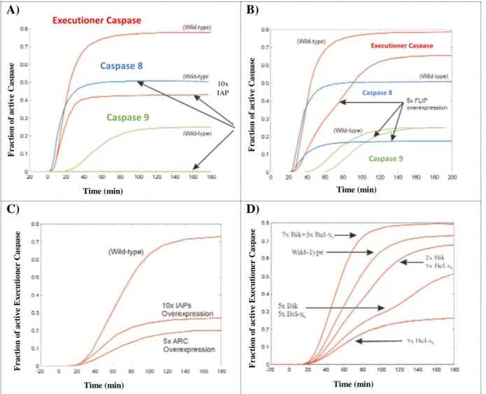
Models of Apoptosis date as far back as the beginning of the century when Fussenegger and colleagues proposed a model for apoptosis with both extrinsic and intrinsic associated pathways (Fussenegger et al., 2000). The model included some of the knowledge of the time such as the receptor role in the activation of functional C8 and the importance of mitochondria in intersecting and transducing both exterior and stress-induced signals. Assembling those facts, the authors were capable of describing known phenomena like the ability to inhibit the apoptotic cascade by overexpressing inhibitors of the apoptotic proteins (IAPs) and to define quantitative proportions of anti-apoptotic BCL-2 molecules necessary to reduce the amount of produced effector caspases in the system. Also, supported by cancer-like scenarios with an overexpression of BCL2-anti-apoptotic proteins, the model permitted the calculation of compensation ratios of pro-apoptotic proteins that could reverse the resistant phenotype and a relevant role was given to the reactions happening at the receptor level in what concerns their impact on the overall response of the system (figure 3-C) ).
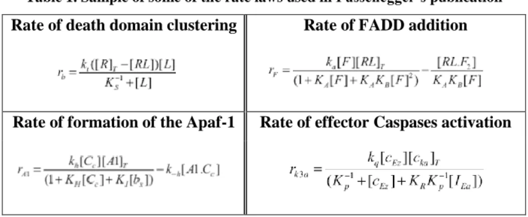
Although not entirely innovative in its conclusions it was a pioneer work that launched the usage of ODE’s in the mathematical field of apoptosis in a manner that agreed with the biological description of the known chemical reactions. In what concerns the underlying mathematical approach, the model did not follow standard mass-action description and the expression for the rates laws are somehow unclear. Same examples are provided in table 1.
Table 1. Sample of some of the rate laws used in Fussenegger’s publication
Rate of death domain clustering Rate of FADD addition
35
A) B)
C) D)
Figure 3. Fussenegger’s apoptosis-related model results.
A), B), C) The authors have shown that different anti-apoptotic proteins cause dissimilar effects on the level of
initiator caspases (C8) and executioner caspases (C3/C6/C7). A) Overexpressing inhibitor of apoptosis protein (IAP) diminishes the final steady state values of C9 and that of the executioner caspases. C8 evolution is unchanged after IAP overexpression (middle blue-line). B) FLIP overexpression impacts the levels of all three represented caspases. C) Increasing five times the amount of decoy-receptor proteins causes a larger decrease in the fraction of executioner caspases than overexpression of IAP ten times the standard levels. The remark is that receptor events have a larger impact on the output of the simulated signaling cascade. D) A cancerous cell, defined by high levels of anti-apoptotic proteins such as Bcl-XL, regains executioner caspases basal levels when submitted to elevating levels of pro-apoptotic proteins like the Bcl-2-interacting killer (Bik). Figure extracted from (Fussenegger et al., 2000).
10x IAP
Time (min) Time (min)
Time (min) Time (min)
Fr a ct io n o f a ct iv e C a sp a se Fr a ct io n o f a ct iv e C a sp a se Fr a ct io n o f a ct iv e E x ec u ti o n er C a sp a se Fr a ct io n o f a ct iv e E x ec u ti o n er C a sp a se Executioner Caspase Caspase 8 Caspase 9 Executioner Caspase Caspase 8 Caspase 9
36
Summary
Global description of the underlying biological processes
Therapeutic strategies point to the apoptotic receptor reactions as an important target that should be up- or down- regulated in the case of cancer and Alzheimer’s disease, respectively
Role of the receptor compartmentas an important layer of the network Mathematical rate laws are not clear
Incomplete and oversimplified representation of the receptor valency Caspase-8 dynamics, as explained in section 1.2.4 of this manuscript,
37
1.3.2 Albeck’s EARM1 model, 2008
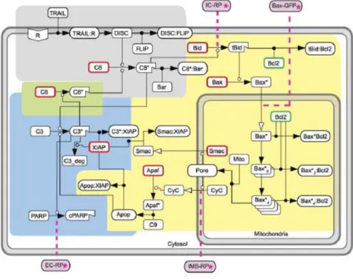
An upgrade on the Fussenegger’s model was given by Albeck and colleagues on a global vision of the extrinsic apoptosis signaling pathway (Albeck et al., 2008). The authors increased the previous description to a four-compartment model named EARM1 (extrinsic apoptosis
reaction model 1) containing a receptor compartment (figure 4, gray box), a mitochondrial compartment (figure 4, yellow box), a C3-associated positive feedback loop compartment
(figure 4, green box) and a posT-MOMP compartment (figure 4, blue box).
Figure 4. Albeck’s EARM1 network of reactions.
A four-compartment model was proposed by Albeck and colleagues for the extrinsic apoptosis pathway. In the receptor compartment (gray area) the formation of the DISC is simplified by the attachment of a single ligand, after which C8 is directly activated into C8*. The production of C8 is simplified with a lumped representation that does not correspond to the dynamics explained in section 1.2.4 of this manuscript. The mitochondrial compartment (yellow area) includes a vast number of reactions from the cleavage of Bid into tBid promoted by C8, down to the formation of the apoptosome after MOMP. A simplified illustration of the positive feedback loop reinforcing C8 activation is included (green area). The feedback loop was shortened and C6 was activated by C3* without any intermediate steps. Finally, in blue, active C3* cleaves PARP into cPARP and irreversible apoptosis is reached. All the boxes highlighted in pink refer to nodes were available experimental data existed in the form of a signal emitted by a reporter protein (RP). Figure extracted from (Albeck et al., 2008).
38
The authors were able to explain two-time separated events: a pre-MOMP variable time delay period followed by a posT-MOMP spontaneous event “snap-action” signal corresponding to effector caspases activation in cells treated with cycloheximide (a potent inhibitor of protein synthesis). The high heterogeneity on the delay period was associated to the level of activity of the initiator caspase C8, which was confirmed experimentally (Albeck et al., 2008). This activity depended on the dose of the ligand but the influence dissipated at the mitochondria level with the MOMP, formation of pores, SMAC release and effector caspases activation being dose independent through an irreversible cell fast switch-type activation signal. In fact, in order to agree with the dynamics of the experimental data, the snap-action signal topology depended critically on the resnap-actions happening in the mitochondrial compartment, setting a local control that is independent from the upstream parts of the network such as the receptor layer. The snap-action signal was also shown to be independent of the downstream feedback loop C3 → C6 → C8, not requiring the reinforcement of a positive feedback interaction to establish the all-or-none behavior of MOMP.
At the time, no quantifications were established on the topic of “fractional killing”. This was a direct consequence of treating cells with cycloheximide, which creates a biological context that inevitably leads to cell death, independently of activation of apoptosis. In this setting, the authors were unable to study different contributions of the network for the underlying cell-fate death or survival decision.
The EARM1 was further on extended up to EARM1.4 version. Spencer and colleagues used EARM1.1 to show that variability in protein initial conditions could reproduced the experimental distributions in time-of-death (Spencer et al., 2009). Variability in time-of-death was found to be caused by specific proteins of the apoptosis pathway, which were defined as sensitive nodes of the network, and were obtained with simulations on the EARM1.3 version (Gaudet et al., 2012). Finally, an EARM1.4 model proposal allowed a study to establish the ratio of XIAP/C3 as a distinguishing factor in cells executing a type I (mitochondrial-independent) or type II (mitochondrial-dependent) apoptosis. Type I occurs when this ratio is small and type II is preferable when the same ratio attains higher values (Aldridge et al., 2011).
39
Summary
First steps in the positioning of C8 dynamics as a central node of the network controlling the variability in time-of-death
Description of the MOMP event as a switch type response that depends on the movement of proteins through different compartments where the scales of the reaction rates change
A small number of pore molecules is enough to release a wave of SMAC and CyC to the cytosol, resulting in a snap action signal independent of upstream or downstream events
Units of [molecules per cytosolic compartment] are not clear and difficult to reproduce
Incomplete representation of the receptor valency
Caspase-8 dynamics, as explained in section 1.2.4 of this manuscript, was not known back at the time of the publication
40
1.3.3 Schlatter’s & Calzone’s model, two Boolean modeling approaches
The transduction and processing of intracellular signals often results from the contribution of multiple chemical agents and signaling pathways whose exact dynamics are frequently unknown. This lack of information greatly compromises the usage of continuous-time modelling approaches, which depend on precise model parameters, and can eventually bias model predictions. In this sense a simpler but useful alternative to study the interactions in a reaction network is to consider a Boolean or logical modeling framework to qualitatively assess the interactions and dependencies between the considered molecular species. The concentration of every element is replaced by a binary variable {0, 1}, in either an “on” or “off” state, and the collection of interactions is embed in an oriented graph that can integrate several types of dependencies, including activation and inhibition effects and positive and negative feedback loops.
In the field of apoptosis two studies based on Boolean formalisms stand out by the complexity and relevance of their conclusions, the work of (Schlatter et al., 2009) and (Calzone et al., 2010). In the former, a large network of reactions describing the intrinsic and extrinsic apoptosis and a myriad of associated pathways was proposed to analyze the effect of an input of Fas ligand, TNF-a, UV-B irradiation, interleukin-1b and insulin into the phenotype outcome of the system. The complexity of the reported interactions made the authors choose for a multi-value node representation where each variable could assume multiple states, instead of the common “1” or “0” and all-or-none definition, to account for “low-active amount” and “high-active amount” and establish higher-valued states where one variable could surpass the inhibitory role of another or instead reinforce the inhibition of a given substrate. The inclusion of more detailed timescale dynamics, with subsets of reactions being active only at certain time points, proved also essential to reproduce threshold dependencies and reaction delays that are known to be apoptotic signatures. The phenotypic outcome of the system was found to depend considerably on the feedback loops and highly connected nodes which included crosstalks with the survival and insulin pathways. A non-reported negative feedback loop from Smac to RIP (a central molecule in the necroptosis pathway inhibiting DISC and C8 activation) was suggested as a mechanism to enhance the stability of the DISC structures and lead to more effective apoptotic responses (Schlatter et al., 2009). Although hypothetical, the possibility of uncovering a new level of regulation in an already complex network is definitely one of the potential benefits of using modeling approaches and overall strengthen the results of this model.
The model of (Calzone et al., 2010) tried to establish a functional relationship between the NF-kB survival pathway and the cellular decision for either apoptosis or RIP1-dependent necrosis, after activation of the death-receptors on the cell membrane. By assembling a large network of reactions with the most representative elements of these three signaling pathways, the C3 activation, a significant drop in ATP levels and emergence of NF-kB were set as representative hallmarks of apoptosis, necrosis and survival decisions, respectively. A Boolean variable was assigned to each node and rules were imposed to define multiple events, such as the activation of a protein. For instance, C8 was considered to effectively change into an active state after direct stimulation by either DISC-TNF or DISC-FAS but only in the absence of FLIP. In this case, the associated logical rule was defined as (DISC-TNF OR DISC-FAS) AND NOT FLIP. By analyzing the multiple steady-states of their system, the authors could
41
propose novel insights about cellular decisions towards different cell-death modalities. Even in the presence of C8-mediated cleavage of RIP1 the system was shown to contain attractors eventually converging into a necrotic phenotype, suggesting that the presence of C8 per se does not imply a cellular apoptotic response. For cells in which important apoptotic elements were mutated (APAF1, BAX, C8, FADD deletions and z-VAD treatment), simulations uncovered the existence of an optimal TNF treatment coincident with maximum proportions of obtained necrotic cells. The authors also found the role of the positive feedback loop from C3 to C8 to be non-essential when TNF or FAS levels were constant in the cell medium. Oppositely, when the same ligands are to be administrated in pulses the feedback loop ensures the persistence of the apoptotic signal (Calzone et al., 2010). This result reassures the importance of positive feedback loops in natural tissues where the cell receives non-sustained death-signals from its surrounding environment. The work still has the potential to uncover, with the availability of more data, the paradigms of cell decision towards necrosis, necroptosis or apoptosis and enlighten how we can force the cell into a specific type of death.
In what concerns the modeling framework, many formulations are available (among Boolean, ODE’s or PDE’s) and the modeler can choose for a more qualitative or quantitative approach according to the data at hand and the desired analysis or conclusions to extract. Some authors chose to build hybrid models combining both Boolean and ODE components and some are available in the field of apoptosis, such as the model for the NF-kB module proposed by (Chaves et al., 2009).
42
1.3.4
Bertaux’s model, a stochastic protein-turnover approach 2014
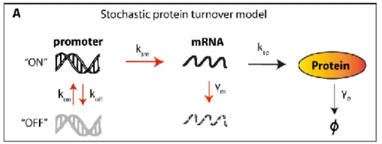
It is long known that identical sister cells often respond in different ways when exposed to equivalent stimulus. In apoptosis these differences have been attributed to inequalities in protein numbers that, when summed up for the plethora of intra-signaling reactions of a network, can influence the phenotype decision. In an effort to study these effects, Bertaux and colleagues used the EARM1 model, previously proposed by Albeck, and added a second contribution in the form of a gene stochastic layer (Bertaux et al., 2014). Each protein of the network interacted with other proteins of the system and also received an extra-input signal resulting from the fluctuations of the corresponding gene expression (figure 5). The goal was to follow the impact of random fluctuations in time in the absolute protein quantities, instead of the traditional sampling of variable initial conditions at the beginning of the simulation that is a common tool used to reproduce the behavior of non-identical cells. This analysis allowed for the simulation of a population of sister cells whose proteome decorrelate in time due to the inherent fluctuations of the underlying gene expression, as observed experimentally (Spencer et al., 2009).
Figure 5. Bertaux’s stochastic protein turnover model.
All the network of proteins of the EARM1 model was equipped with a stochastic layer using a random telegraph approach: mRNA production and degradation rates were modeled as stochastic processes with protein production and degradation rates treated as deterministic processes. Simulations for the stochastic elements were executed with the Gillespie algorithm. Figure extracted from (Bertaux et al., 2014).
The approach allowed the authors to evaluate the contribution of intrinsic noise, produced at the gene layer, and conclude that it was enough to justify the temporal and reversal resistance to TRAIL observed in HeLa cells after an initial TRAIL exposure (Flusberg et al., 2013). Their congruence with the experimental results required the fluctuations of the short-lived protein Mcl1 to be large and rare, imposing low value ranges for the ON-OFF promoter switching rates of this protein. In this case modelling was able to explain the acquired resistance of cells already
43
treated with TRAIL as a complex interplay of three distinct effects: selection acting on the cells with the highest anti-apoptotic protein amounts; transcriptional noise making protein production to switch between on and off states and protein degradation as a force that tends to decay protein numbers back to their original values (making resistance only temporary). The co-participation of these environmental pressures could not be deduced without a modelling approach and reinforced the need of mathematical laws to determine and quantify individual system-level contributions.
By assembling Mcl1 dynamics with rare and large fluctuations, FLIP with promoter fast turnover rates and a default model of protein turnover for the remaining proteins of the system, the authors could also reproduce the MOMP time distributions registered by Spencer and colleagues (Spencer et al., 2009). The work was important as it defined a strategy to introduce fluctuations in a principled manner and study the contribution of gene expression variability in networks of interacting proteins.
Summary
A methodology was proposed to analyze the effect of gene expression fluctuations on a given network of interacting proteins
A justification was presented for the temporal and reversal resistance acquired by HeLa cells after a TRAIL exposure
After cell division, the loss of synchrony of sister cells with time was reproduced, in line with the findings of Spencer and colleagues
Model parameters were fitted from a data set concerning cells treated with TRAIL and cycloheximide. Cycloheximide blocks protein production and changes the universe of interactions at the protein level. This may cause the model predictions to be unrealistic in HeLa cells treated exclusively with TRAIL
Incomplete representation of the receptor valency
Caspase-8 dynamics followed the same representation as in Albeck’s EARM1 model
Under-estimation of the role of FLIP, which is known to be a potent inhibitor of apoptosis
44
1.3.5 A summary list of models of apoptosis
Author / Year
Focus of the model
(Fussenegger et al., 2000) Intrinsic + Extrinsic apoptosis
(Bentele et al., 2004) Extrinsic apoptosis
(Eissing et al., 2004) Positive feedback loop of C3 on C8
(Hua et al., 2005) Extrinsic apoptosis
(Stucki and Simon, 2005) Mitochondria + IAP + C3 (Bagci et al., 2006) C8 + Mitochondrial reactions (Legewie et al., 2006) Intrinsic + Extrinsic Apoptosis
(Rehm et al., 2006) Effector caspases dynamics (Chen et al., 2007b) Bcl-2 apoptotic switch (Chen et al., 2007a) Bax + Bcl-2 interaction
(Albeck et al., 2008) Extrinsic apoptosis
(Cui et al., 2008) Bcl-2 dynamics
(Chaves et al., 2009) Apoptosis + NF-kB dynamics
(Schlatter et al., 2009) Intrinsic + Extrinsic apoptosis
(Zhang et al., 2009) Intrinsic + Extrinsic apoptosis (Calzone et al., 2010) Apoptosis + NF-kB + Necrosis pathways
(Howells et al., 2011) Bcl-2 dynamics
(Aldridge et al., 2011) Extrinsic apoptosis: Type I vs Type II cells (Huber et al., 2011) Bcl-2 family members dynamics
(Gaudet et al., 2012) Extrinsic apoptosis
(Stoma et al., 2013) Extrinsic apoptosis Type I vs Type II cells
(Bertaux et al., 2014) Extrinsic apoptosis
(Kallenberger et al., 2014) C8 dynamics
(Würstle et al., 2014) Intrinsic apoptosis