Chirality dependent corona phase molecular
recognition of DNA-wrapped carbon nanotubes
The MIT Faculty has made this article openly available. Please share how this access benefits you. Your story matters.Citation Salem, Daniel P. et al. “Chirality Dependent Corona Phase
Molecular Recognition of DNA-Wrapped Carbon Nanotubes.” Carbon 97 (February 2016): 147–153 © 2015 Elsevier Ltd
As Published http://dx.doi.org/10.1016/j.carbon.2015.08.075
Publisher Elsevier
Version Author's final manuscript
Citable link http://hdl.handle.net/1721.1/113650
Terms of Use Creative Commons Attribution-NonCommercial-NoDerivs License
1
Chirality dependent corona phase molecular recognition of
DNA-wrapped carbon nanotubes
Daniel P. Salem1, Markita P. Landry1, Gili Bisker1, Jiyoung Ahn1, Sebastian Kruss2, Michael S. Strano1,*
1
Department of Chemical Engineering, Massachusetts Institute of Technology, Cambridge, MA 02139
2
Institute for Physical Chemistry, Göttingen University, Germany *Corresponding author. Email: [email protected], Phone: (617)324-4323
ABSTRACT
Corona phase molecular recognition (CoPhMoRe) is a phenomenon whereby a polymer or surfactant corona phase wrapped around a nanoparticle selectively recognizes a particular molecule. The method can potentially generate non-biological, synthetic molecular recognition sites analogous to antibodies for a broad range of biomedical applications, including new types of sensors, laboratory and clinical assays, as well as inhibitors and targeted therapeutics. In this work, we utilize near infrared fluorescent single-walled carbon nanotubes (SWNTs) wrapped
with specific single stranded DNA sequences to explore the (n,m) chirality dependence of
CoPhMoRe. Specific DNA oligonucleotide sequences are known to recognize and interact uniquely with certain (n,m) SWNTs enabling their enrichment in ion exchange
chromatography. We explore the CoPhMoRe effect using corona phases constructed from a library of 24 sequences, screening against a biomolecule panel that includes common
neurotransmitters, amino acids, saccharides and riboflavin. Example sequences include (ATT)4,
(TAT)4 and (ATTT)3 which recognize (7,5), (6,5) and (8,4) SWNTs respectively. We find that
2 chiralities, and appear to pack more densely as to exclude analyte adsorption on the chirality they recognize. These results have encouraging implications for the controlled design of CoPhMoRe phases for biomedical applications.
1. Introduction
The polymer corona phase of individually-dispersed single-walled carbon nanotubes (SWNTs) forms the basis of many active areas of investigation including SWNT separation technologies [1, 2], targeted biomolecule delivery [3, 4], molecular detection [5], materials development and energy applications [6]. The inherent fluorescence of semiconducting single-walled carbon nanotubes has prompted their widespread use as signal transducers for optical sensing applications [7-9]. Their nearly indefinite fluorescence profile provides a significant advantage over conventional fluorophore-based detection platforms that are susceptible to photobleaching [10], and the overlap of their near infrared (nIR) fluorescence profile with the tissue transparency window makes them especially useful for in vivo applications [11-15]. These properties, along with the capability of single molecule detection [16], have made SWNT
nanosensors valuable tools to study physical, chemical and biological processes at short length and time scales otherwise not obtainable from other detection platforms. As a result,
considerable effort has been devoted to the design and development of SWNT-based optical sensors for a variety of target analytes including neurotransmitters [17], proteins [18-20], carbohydrates [21-23] and assorted small molecules [5, 16, 24].
Corona phase molecular recognition (CoPhMoRe) is a phenomenon whereby a
heteropolymer or surfactant phase encapsulating a nanoparticle (i.e. a corona phase) recognizes a specific set of analytes because of the 3D structure displayed on the particle surface.
3 Individually-dispersed semiconducting SWNTs have advantages for the discovery and utilization of such CoPhMoRe phases due to their nIR fluorescent response to molecular adsorption [7-9]. The study of CoPhMoRe phases is important for their ability to potentially generate non-biological, synthetic antibodies that may benefit a broad range of biomedical applications, including new types of sensors, laboratory and clinical assays, as well as inhibitors and targeted therapeutics [25]. However, to date, direct control over the design of such phases remains elusive, although recent work in our lab has made progress in defining and addressing the CoPhMoRe inverse design problem for the rational design of a helically wrapping
heteropolymer, which renders the underlying SWNT a sensor for an analyte of interest [26]. The effects of ssDNA length and sequence identity on recognition ability of a DNA-SWNT hybrid have not been thoroughly explored [27]. However, the impact of sequence length and composition on the strength and conformation of binding to SWNTs have been the focus of numerous studies [28-34]. In particular, Tu and coworkers identified specific DNA
oligonucleotide sequences known to recognize and interact uniquely with certain (n,m) SWNTs enabling their enrichment in ion exchange chromatography [35]. Follow-up studies investigated the structure and binding strength of select recognition sequences and concluded that the
sequences were able to bind more tightly to their respective chirality partners than other chiralities [31, 33, 34].
In this work, we investigate the chirality dependent fluorescence modulation of DNA-wrapped SWNT sensors to a biomolecule panel that includes common neurotransmitters, amino acids, saccharides and riboflavin, in an effort to address the problem of CoPhMoRe design. We find that several recognition sequences indeed form CoPhMoRe phases that are distinct among SWNT chiralities and appear to pack more densely as to exclude analyte adsorption on the (n,m)
4 chirality they recognize. Our study is the first effort to address chirality dependent CoPhMoRe, which establishes the framework for future applications including multiplexed analyte detection and ratiometric sensing for biomedical sensing and therapeutic applications.
2. Experimental 2.1 Materials
All chemicals were purchased from Sigma-Aldrich (U.S.A.) unless specified otherwise. All ssDNA sequences were purchased from IDT (USA). HiPco-SWNTs were purchased from Unidym (lot R1831) and processed according to the manufacturer’s recommendation to remove residual impurities.
2.2 Suspension and Characterization of ssDNA-wrapped SWNTs
HiPco-SWNTs were suspended in a 100 mM NaCl solution containing twice as much ssDNA by mass. Nanotubes were suspended using a 24-probe tip sonicator (Qsonica Q700) containing 3 mm probe tips and operating at a power of 96 W (4 W per sample) for 10 minutes, while in a cooling block. After sonication, each mixture was centrifuged twice at 16,000g for 90 minutes to remove SWNT aggregates and residual solid impurities. The supernatant was
collected and characterized via ultraviolet (UV)-visible-nIR absorbance spectroscopy (Cary 5000, Agilent Technologies) to determine the concentration of each SWNT suspension [19]. Suspended SWNTs were stored at 4°C to mitigate aggregation.
2.3 Near Infrared Fluorescence Measurements
The fluorescence spectrum of each SWNT suspension was measured before and after the addition of an analyte. Prior to these measurements, SWNT solutions were diluted to a
5 µL were added in triplicate to a 96-well plate (Tissue Culture Plates, Olympus Plastics) and allowed to equilibrate for at least 45 minutes. Samples were excited by a 785 nm laser (450 mW) using a Zeiss AxioVision inverted microscope and a 20x objective. Fluorescence was measured using a PI Acton SP2500 spectrometer and a Princeton Instruments InGaAs OMA V array detector. The 96-well plate was attached to an automated motorized stage above the objective to allow for high-throughput data collection. An exposure time of one second was used for all wrappings and the spectrum of each sample was background-corrected using a spectrum of PBS in an equivalent volume. The fluorescence spectrum of each sample was measured immediately after the addition of 2 µL of 10 mM analyte solution in PBS, resulting in a final analyte
concentration of 100 µM. All analyte solutions were prepared immediately before addition to the sample wells.
2.4 Spectrum Deconvolution
Fluorescence spectra were deconvoluted using a modified Matlab script developed previously by the Strano group (described in detail in the supporting information of ref. [5]). Normalized fluorescence responses were calculated for each chirality upon the addition of each analyte using the peak areas output by the deconvolution code.
3. Results and Discussion
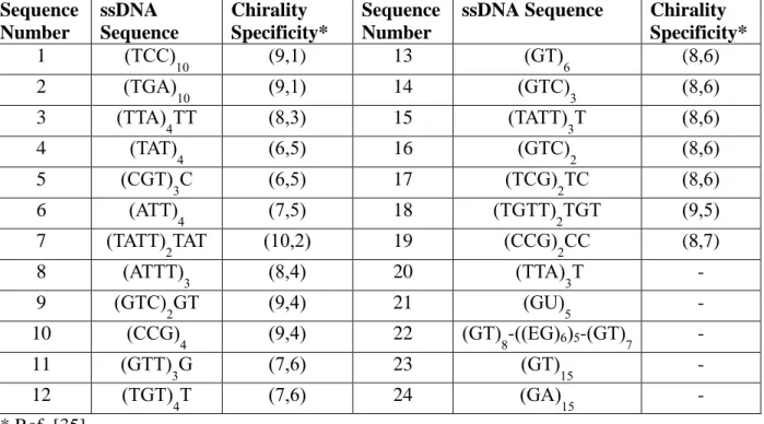
The ssDNA sequences used in this study are presented in Table 1, along with the chirality specificity of each sequence during ion exchange chromatography (if applicable) as determined by Tu and coworkers [35]. Nineteen sequences exhibiting chirality specificity were chosen, encompassing a variety of nanotube chiralities and diameters (1-19, Table 1). Five additional sequences of varying length and structure were also investigated in this study (20-24,
6 Table 1). The absorbance spectra of all 24 sequences are provided in the supplementary data (Figure S1).
Table 1. List of ssDNA wrappings tested in this study. (EG)6 is a hexa-ethyleneglycol spacer.
Sequence Number ssDNA Sequence Chirality Specificity* Sequence Number
ssDNA Sequence Chirality
Specificity* 1 (TCC) 10 (9,1) 13 (GT)6 (8,6) 2 (TGA) 10 (9,1) 14 (GTC)3 (8,6) 3 (TTA) 4TT (8,3) 15 (TATT)3T (8,6) 4 (TAT)4 (6,5) 16 (GTC)2 (8,6) 5 (CGT)3C (6,5) 17 (TCG)2TC (8,6) 6 (ATT) 4 (7,5) 18 (TGTT)2TGT (9,5) 7 (TATT) 2TAT (10,2) 19 (CCG)2CC (8,7) 8 (ATTT) 3 (8,4) 20 (TTA)3T - 9 (GTC)2GT (9,4) 21 (GU)5 - 10 (CCG)4 (9,4) 22 (GT)8-((EG)6)5-(GT)7 - 11 (GTT) 3G (7,6) 23 (GT)15 - 12 (TGT) 4T (7,6) 24 (GA)15 - * Ref. [35]
Each fluorescence emission spectrum was processed by a deconvolution Matlab script to isolate the fluorescence contributions of individual chiralities (Figure 1a). Since the (9,4) and (7,6) peaks are largely overlapping, we tracked the peak area values when calculating the fluorescence response of each chirality and we combined the peak areas of the (9,4) and (7,6) peaks in order to avoid potential artifacts introduced by the fitting algorithm during the
deconvolution. The 24 ssDNA wrappings were screened against nine different analytes (Figure 1b), several of which were previously shown to induce strong sensor responses for
DNA-wrapped nanotubes [5, 17]. Glucose and sucrose were added to the sensors at a concentration of 10 mM, which resembles the glucose concentration in human blood. Moreover, riboflavin was added at a concentration of 1 µM due to its limited solubility in PBS.
7
Figure 1. (a) Deconvolution of a fluorescence spectrum of (GT)15-wrapped SWNT: original spectrum (blue curve),
individual chirality contributions (dashed black curves) and the sum of the individual peaks (dashed red curve). (b) Small molecule analytes used in this study. Dopamine, epinephrine, norepinephrine and ascorbic acid have been reported to elicit turn-on responses for DNA-wrapped SWNTs [17], while riboflavin was previously reported to quench fluorescence for (GT)15-wrapped SWNTs [5].
As expected, all three catecholamines (dopamine, epinephrine, and norepinephrine) as well as ascorbic acid induced similar, strong turn-on responses across nearly all of the sensor wrappings tested. While each SWNT-wrapping exhibited some degree of variability in the response of each chirality, as demonstrated for the (GT)15-SWNT data in Figure 2, most wrappings did not exhibit systematic chirality dependence attributable to their recognition abilities during ion exchange chromatography (supplementary Figure S2). However, as shown in Figure 2, the sequences (TAT)4 and (ATTT)3 did exhibit chirality dependent responses that appear to correlate well with their reported chirality specificities of (6,5) and (8,4) respectively. Although the (8,4) chirality was not included in the deconvolution algorithm, the (9,4) and (7,6) chiralities, which emit at roughly the same wavelength as (8,4) SWNT, are nearly unresponsive to the addition of dopamine and other catecholamines (supplementary Figures S4, S5).
8 Additionally, the smaller diameter SWNTs for the (TAT)4 wrapping, particularly the (6,5)
chirality, were much less responsive than larger diameter chiralities including (12,1) and (11,3).
Figure 2. Fluorescence spectra before (black) and after (red) the addition of 100 µM dopamine for the wrappings
(GT)15, (TAT)4 and (ATTT)3 as well as the normalized fluorescence response of each chirality.
The chirality dependence observed following dopamine addition (Figure 2) was consistent for other analytes in this study. Figure 3 presents the chirality dependent sensor responses to 100 µM epinephrine, 100 µM norepinephrine and 1 µM riboflavin. Consistent with the catecholamine results, the (9,4) and (7,6) chiralities of (ATTT)3-wrapped SWNTs were nearly unresponsive to riboflavin, exhibiting a 5 percent decrease in fluorescence compared to a 40 percent decrease exhibited by (GT)15-wrapped SWNTs (supplementary Figure S6) and a 30 percent decrease exhibited by (TAT)4-wrapped SWNTs. Analogous plots for the remaining analytes contain the same trends as Figure 3 and are provided in the supplementary data. The detection limits for (TAT)4- and (ATTT)3-wrapped SWNTs were determined for epinephrine by measuring the sensor responses to different concentrations of analyte and constructing a
9 calibration curve for each chirality (Figure S8). Each limit of detection was calculated as the concentration on the calibration curve corresponding to the sum of the mean control response (just PBS buffer) and three times the standard deviation. While there was a small degree of variability in the detection limits among different chiralities, (TAT)4- and (ATTT)3-wrapped SWNTs exhibited average detection limits of 20 nM and 11 nM, respectively.
Figure 3. Chirality dependent responses of (a) (TAT)4-wrapped and (b) (ATTT)3-wrapped SWNTs upon the
addition of 100 µM epinephrine (black), 100 µM norepinephrine (blue) and 1 µM riboflavin (red).
To ensure that the chirality dependent sensor responses were triggered by analyte – SWNT interaction rather than by polymer wrapping rearrangement while equilibrating in solution, we monitored the fluorescence emission over a period of 4 hours after dilution to 1 mg/L. Recently, Landry et al. reported that RNA-wrapped SWNTs slowly equilibrated upon dilution in PBS, as manifested by a gradual fluorescence modulation over several hours [36],
10 whereas DNA wrappings showed a much more stable fluorescence signal after dilution.
Although 23 of the 24 the sequences used in this study were DNA, many sequences were much shorter than the 30-mer oligonucleotides used in the previous work and may not bind as tightly. In order to rule out equilibration artifacts, the fluorescence spectra of (GT)15-, (TAT)4- and (ATTT)3-wrapped SWNTs were collected every ten minutes for 4 hours after dilution to 1 mg/L with PBS, and were then deconvoluted and inspected for chirality dependent stability. We found that the oligonucleotide wrappings were remarkably stable after PBS dilution over the course of the experiment, demonstrated by an invariant fluorescent emission signal over time (see
supplementary Figure S9).
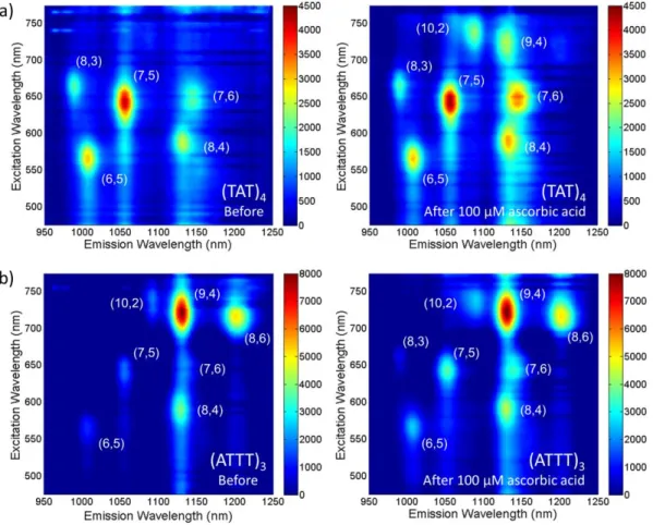
The use of 785 nm laser excitation for the nIR fluorescence experiments did not allow us to isolate the fluorescence contribution of (8,4) SWNTs ((ATTT)3 recognition partner), which fluoresce more strongly at shorter excitation wavelengths. To address this problem and better visualize the fluorescence responses of each chirality, we measured the excitation/emission profiles for (TAT)4- and (ATTT)3-wrapped SWNTs before and after the addition of 100 µM ascorbic acid. These experiments were completed using a custom-built setup containing a xenon arc lamp and an Acton SpectraPro 2150i monochromator. The SWNT samples were excited at wavelengths between 500 nm and 800 nm in intervals of 5 nm and the data were processed to account for the variation in the power profile of the xenon lamp. Figure 4 provides the resulting photoluminescence maps of both ssDNA-SWNT conjugates. As expected, (TAT)4-wrapped (6,5) SWNTs did not exhibit a strong response to 100 µM ascorbic acid, in contrast to larger diameter SWNTs including (8,4), (7,6), (10,2) and (9,4). Moreover, (ATTT)3-wrapped (8,4) and (9,4) SWNTs did not respond strongly to ascorbic acid, while other SWNT chiralities--including (7,6)—did exhibit a response. These data further support our earlier results of
chirality-11 dependent CoPhMoRe, and in particular, our findings that the (TAT)4 and (ATTT)3 ssDNA wrappings induce a reduction in sensor responsivity for their respective chirality partners.
Figure 4. Excitation-emission profiles for (a) (TAT)4-wrapped and (b) (ATTT)3-wrapped SWNTs before and after
the addition of 100 µM ascorbic acid. Spectra were collected in intervals of 5 nm using exposure times of 5 min. for the (TAT)4-SWNTs and 5.5 min. for the (ATTT)3-SWNTs.
In order for a molecule to induce a sensor response, it must be able to access the nanotube surface and displace a portion of the DNA wrapping, as demonstrated by Kruss et al. using SWNTs wrapped with Cy3-labeled DNA [17]. When adsorbed onto the SWNT surface, the Cy3 dye is completely quenched; however, upon the addition of dopamine, Kruss et al. reported a restoration of the Cy3 fluorescence, indicating the desorption of the Cy3 from the nanotube surface. Thus, the chirality dependence observed in this study can be explained by corona structure variability—and hence, variability in analyte accessibility to the SWNT surface—
12 across different chiralities for the same ssDNA wrapping. Molecular dynamics simulations have been extensively used to study the corona architecture of DNA-SWNT hybrids and a
combination of molecular dynamics and surfactant exchange experiments have shown that corona architecture and binding strength vary across different chiralities for several sequences exhibiting separation capability including (TAT)4 [32-34]. Both corona structure and binding strength are dependent on a combination of nanotube diameter and electronic properties, which can be clearly observed in the chirality dependent responses of (TAT)4-wrapped SWNTs. While smaller diameter nanotubes, in general, exhibited attenuated responsivity, the (6,5) chirality consistently responded the least for all analytes tested. Given the chirality dependent response trends observed for both (TAT)4 and (ATTT)3, it is likely that the chiralities exhibiting weaker sensor responses contain more tightly-packed corona phases. The ability of these sequences to exhibit stronger chirality dependence than the other recognition sequences tested could be caused by an increased sensitivity of binding strength on SWNT chirality. This phenomenon was
previously reported for the sequences (TAT)4, (CCA)10 and (TTA)4TT, where surfactant
exchange experiments indicated that the difference in binding strength across various chiralities was much larger for (TAT)4 than for the other sequences [34].
With the growing interest in utilizing fluorescent SWNTs as biomedical sensors capable of label free analyte detection [25], single chirality sensors would be extremely valuable for many applications. Owing to the unique emission wavelength associated with each chirality, multiple targets can be detected simultaneously by hyperspectral imaging techniques. Such capabilities open up new possibilities in the biomedical field by allowing researchers to study complex biological processes by monitoring several important bio-markers at the same time, using selective sensors with different chiralities. SWNT sensors were utilized for monitoring
13 nitric oxide in vivo [13], vitamin dynamics within cells [5], and protein-protein interactions [18]. In addition to multiplexed detection and improved signal to noise ratio, chirality specific sensors would allow for an internal fluorescent standard that would eliminate the need for signal
calibration or allow for ratiometric sensing. This was recently demonstrated for the detection of nitric oxide and hydrogen peroxide within living plants [37]. Moreover, chirality dependent CoPhMoRe may be particularly useful in distinguishing between closely related analyte molecules which tend to induce similar sensor responses. A variation in chirality dependent responses induced by two or more structurally similar molecules will allow for their selective detection by utilizing the responses of several chiralities. Such a strategy may be needed to design SWNT sensors that can effectively discriminate between structurally similar carbohydrate molecules or between various catecholamines and ascorbic acid, both of which remain subjects of ongoing studies [38-41].
4. Conclusions
The recent emergence of optical detection methods for molecular recognition has spurred interest in understanding the parameters which contribute to molecular specificity and selectivity. Based on a previous study reporting ssDNA sequences capable of separating single chirality SWNTs from a mixture during ion exchange chromatography, we examined the effect of such sequences on SWNT fluorescence and molecular recognition. In particular, we studied the chirality dependent photoluminescence responses of polynucleotide-wrapped SWNT to a library of relevant biomolecules including catecholamines, carbohydrates and amino acids. The
experimental results identified two particular ssDNA sequences, (TAT)4 and (ATTT)3, that formed DNA-SWNT hybrids exhibiting significant chirality dependent fluorescence responses to
14 all of the analytes tested. The observed chirality dependence was consistent with previously reported results indicating that the ssDNA sequences capable of single chirality separation are more tightly bound to their respective chirality partners. Our results open up a new path towards single chirality SWNT sensors allowing for multiplexed and ratiometric label-free detection of multiple biomolecules for biomedical research and applications. Moreover, our work motivates the continuous search for antibody analogs for molecular recognition as well as the optimization of currently available optical nanosensors using novel nanoscience and nanotechnology tools.
Acknowledgements
We gratefully acknowledge discussions with Strano group members regarding this manuscript. D.P.S. is grateful for a National Science Foundation Graduate Research Fellowship under Grant No. 2388357. M.P.L. acknowledges an NSF postdoctoral research fellowship under Award No. 1306229, and a Burroughs Wellcome Fund Career Award at the Scientific Interface (CASI). M.S.S acknowledges support by the National Science Foundation under award number 1213622, and the Juvenile Diabetes Research Foundation.
References
1. Arnold, M.S., A.A. Green, J.F. Hulvat, S.I. Stupp, and M.C. Hersam, Sorting carbon
nanotubes by electronic structure using density differentiation. Nature Nanotechnology,
2006. 1(1): p. 60-65.
2. Zheng, M., A. Jagota, E.D. Semke, B.A. Diner, R.S. Mclean, S.R. Lustig, et al.,
DNA-assisted dispersion and separation of carbon nanotubes. Nature Materials, 2003. 2(5): p.
338-342.
3. Karmakar, A., S.M. Bratton, E. Dervishi, A. Ghosh, M. Mahmood, Y. Xu, et al.,
Ethylenediamine functionalized-single-walled nanotube (f-SWNT)-assisted in vitro
delivery of the oncogene suppressor p53 gene to breast cancer MCF-7 cells. International
Journal of Nanomedicine, 2011. 6: p. 1045-1055.
4. Giraldo, J.P., M.P. Landry, S.M. Faltermeier, T.P. McNicholas, N.M. Iverson, A.A. Boghossian, et al., Plant nanobionics approach to augment photosynthesis and
15 5. Zhang, J.Q., M.P. Landry, P.W. Barone, J.H. Kim, S.C. Lin, Z.W. Ulissi, et al.,
Molecular recognition using corona phase complexes made of synthetic polymers adsorbed on carbon nanotubes. Nature Nanotechnology, 2013. 8(12): p. 959-968.
6. Wang, Q.H., D.O. Bellisario, L.W. Drahushuk, R.M. Jain, S. Kruss, M.P. Landry, et al.,
Low Dimensional Carbon Materials for Applications in Mass and Energy Transport.
Chemistry of Materials, 2014. 26(1): p. 172-183.
7. O'Connell, M.J., S.M. Bachilo, C.B. Huffman, V.C. Moore, M.S. Strano, E.H. Haroz, et al., Band gap fluorescence from individual single-walled carbon nanotubes. Science, 2002. 297(5581): p. 593-596.
8. Bachilo, S.M., M.S. Strano, C. Kittrell, R.H. Hauge, R.E. Smalley, and R.B. Weisman,
Structure-assigned optical spectra of single-walled carbon nanotubes. Science, 2002.
298(5602): p. 2361-2366.
9. Barone, P.W., S. Baik, D.A. Heller, and M.S. Strano, Near-infrared optical sensors based
on single-walled carbon nanotubes. Nature Materials, 2005. 4(1): p. 86-U16.
10. Heller, D.A., S. Baik, T.E. Eurell, and M.S. Strano, Single-walled carbon nanotube
spectroscopy in live cells: Towards long-term labels and optical sensors. Advanced
Materials, 2005. 17(23): p. 2793-+.
11. Welsher, K., S.P. Sherlock, and H.J. Dai, Deep-tissue anatomical imaging of mice using
carbon nanotube fluorophores in the second near-infrared window. Proceedings of the
National Academy of Sciences of the United States of America, 2011. 108(22): p. 8943-8948.
12. Boghossian, A.A., J.Q. Zhang, P.W. Barone, N.F. Reuel, J.H. Kim, D.A. Heller, et al.,
Near-Infrared Fluorescent Sensors based on Single-Walled Carbon Nanotubes for Life Sciences Applications. Chemsuschem, 2011. 4(7): p. 848-863.
13. Iverson, N.M., P.W. Barone, M. Shandell, L.J. Trudel, S. Sen, F. Sen, et al., In vivo
biosensing via tissue-localizable near-infrared-fluorescent single-walled carbon nanotubes. Nature Nanotechnology, 2013. 8(11): p. 873-880.
14. Hong, G.S., S. Diao, J.L. Chang, A.L. Antaris, C.X. Chen, B. Zhang, et al., Through-skull
fluorescence imaging of the brain in a new near-infrared window. Nature Photonics,
2014. 8(9): p. 723-730.
15. Bisker, G., N.M. Iverson, J. Ahn, and M.S. Strano, A pharmacokinetic model of a tissue
implantable insulin sensor. Advanced healthcare materials, 2015. 4(1): p. 87-97.
16. Zhang, J.Q., A.A. Boghossian, P.W. Barone, A. Rwei, J.H. Kim, D.H. Lin, et al., Single
Molecule Detection of Nitric Oxide Enabled by d(AT)(15) DNA Adsorbed to Near Infrared Fluorescent Single-Walled Carbon Nanotubes. Journal of the American
16 17. Kruss, S., M.P. Landry, E. Vander Ende, B.M.A. Lima, N.F. Reuel, J.Q. Zhang, et al.,
Neurotransmitter Detection Using Corona Phase Molecular Recognition on Fluorescent Single-Walled Carbon Nanotube Sensors. Journal of the American Chemical Society,
2014. 136(2): p. 713-724.
18. Ahn, J.H., J.H. Kim, N.F. Reuel, P.W. Barone, A.A. Boghossian, J.Q. Zhang, et al.,
Label-Free, Single Protein Detection on a Near-Infrared Fluorescent Single-Walled Carbon Nanotube/Protein Microarray Fabricated by Cell-Free Synthesis. Nano Letters,
2011. 11(7): p. 2743-2752.
19. Zhang, J.Q., S. Kruss, A.J. Hilmer, S. Shimizu, Z. Schmois, F. De La Cruz, et al., A
Rapid, Direct, Quantitative, and Label-Free Detector of Cardiac Biomarker Troponin T Using Near-Infrared Fluorescent Single-Walled Carbon Nanotube Sensors. Advanced
Healthcare Materials, 2014. 3(3): p. 412-423.
20. Nelson, J.T., S. Kim, N.F. Reuel, D.P. Salem, G. Bisker, M.P. Landry, et al., The
Mechanism of Immobilized Protein A Binding to IgG on Nanosensor Array Surfaces.
Analytical Chemistry, 2015. 87(16): p. 8186-8193.
21. Barone, P.W., R.S. Parker, and M.S. Strano, In vivo fluorescence detection of glucose
using a single-walled carbon nanotube optical sensor: Design, fluorophore properties, advantages, and disadvantages. Analytical Chemistry, 2005. 77(23): p. 7556-7562.
22. Mu, B., J. Ahn, T.P. McNicholas, and M.S. Strano, Generating Selective Saccharide
Binding Affinity of Phenyl Boronic Acids by using Single-Walled Carbon Nanotube Corona Phases. Chemistry-a European Journal, 2015. 21(12): p. 4523-4528.
23. Reuel, N.F., J.H. Ahn, J.H. Kim, J.Q. Zhang, A.A. Boghossian, L.K. Mahal, et al.,
Transduction of Glycan-Lectin Binding Using Near-Infrared Fluorescent Single-Walled Carbon Nanotubes for Glycan Profiling. Journal of the American Chemical Society,
2011. 133(44): p. 17923-17933.
24. Jin, H., D.A. Heller, M. Kalbacova, J.H. Kim, J.Q. Zhang, A.A. Boghossian, et al.,
Detection of single-molecule H2O2 signalling from epidermal growth factor receptor using fluorescent single-walled carbon nanotubes. Nature Nanotechnology, 2010. 5(4): p.
302-U81.
25. Kruss, S., A.J. Hilmer, J. Zhang, N.F. Reuel, B. Mu, and M.S. Strano, Carbon nanotubes
as optical biomedical sensors. Advanced Drug Delivery Reviews, 2013. 65(15): p.
1933-1950.
26. Bisker, G., J. Ahn, S. Kruss, Z.W. Ulissi, D.P. Salem, and M.S. Strano, A Mathematical
Formulation and Solution of the CoPhMoRe Inverse Problem for Helically Wrapping Polymer Corona Phases on Cylindrical Substrates. The Journal of Physical Chemistry C,
17 27. Landry, M.P., S. Kruss, J.T. Nelson, G. Bisker, N.M. Iverson, N.F. Reuel, et al.,
Experimental Tools to Study Molecular Recognition within the Nanoparticle Corona.
Sensors, 2014. 14(9): p. 16196-16211.
28. Albertorio, F., M.E. Hughes, J.A. Golovchenko, and D. Branton, Base dependent
DNA-carbon nanotube interactions: activation enthalpies and assembly-disassembly control.
Nanotechnology, 2009. 20(39).
29. Johnson, R.R., A.T.C. Johnson, and M.L. Klein, Probing the structure of DNA-carbon
nanotube hybrids with molecular dynamics. Nano Letters, 2008. 8(1): p. 69-75.
30. Manohar, S., T. Tang, and A. Jagota, Structure of homopolymer DNA-CNT hybrids. Journal of Physical Chemistry C, 2007. 111(48): p. 17835-17845.
31. Roxbury, D., A. Jagota, and J. Mittal, Sequence-Specific Self-Stitching Motif of Short
Single-Stranded DNA on a Single-Walled Carbon Nanotube. Journal of the American
Chemical Society, 2011. 133(34): p. 13545-13550.
32. Roxbury, D., A. Jagota, and J. Mittal, Structural Characteristics of Oligomeric DNA
Strands Adsorbed onto Single-Walled Carbon Nanotubes. Journal of Physical Chemistry
B, 2013. 117(1): p. 132-140.
33. Roxbury, D., X.M. Tu, M. Zheng, and A. Jagota, Recognition Ability of DNA for Carbon
Nanotubes Correlates with Their Binding Affinity. Langmuir, 2011. 27(13): p.
8282-8293.
34. Shankar, A., J. Mittal, and A. Jagota, Binding between DNA and Carbon Nanotubes
Strongly Depends upon Sequence and Chirality. Langmuir, 2014. 30(11): p. 3176-3183.
35. Tu, X.M., S. Manohar, A. Jagota, and M. Zheng, DNA sequence motifs for
structure-specific recognition and separation of carbon nanotubes. Nature, 2009. 460(7252): p.
250-253.
36. Landry, M.P., L. Vukovic, S. Kruss, G. Bisker, A.M. Landry, S. Islam, et al.,
Comparative Dynamics and Sequence Dependence of DNA and RNA Binding to Single Walled Carbon Nanotubes. Journal of Physical Chemistry C, 2015. 119(18): p.
10048-10058.
37. Giraldo, J.P., M.P. Landry, S.Y. Kwak, R.M. Jain, M.H. Wong, N.M. Iverson, et al., A
Ratiometric Sensor Using Single Chirality Near-Infrared Fluorescent Carbon Nanotubes: Application to In Vivo Monitoring. Small, 2015.
38. Ju, H.X., X.J. Zhang, and J. Wang, Carbohydrate Detection Using Nanostructured
Biosensing. Nanobiosensing: Principles, Development and Application, 2011: p.
18 39. Mitra, I., Z.X. Zhuang, Y.N. Zhang, C.Y. Yu, Z.T. Hammoud, H.X. Tang, et al.,
N-Glycan Profiling by Microchip Electrophoresis to Differentiate Disease States Related to Esophageal Adenocarcinoma. Analytical Chemistry, 2012. 84(8): p. 3621-3627.
40. Colleran, J.J. and C.B. Breslin, Simultaneous electrochemical detection of the
catecholamines and ascorbic acid at PEDOT/S-beta-CD modified gold electrodes.
Journal of Electroanalytical Chemistry, 2012. 667: p. 30-37.
41. Chaicham, A., S. Sahasithiwat, T. Tuntulani, and B. Tomapatanaget, Highly effective
discrimination of catecholamine derivatives via FRET-on/off processes induced by the intermolecular assembly with two fluorescence sensors. Chemical Communications,