HAL Id: hal-02933537
https://hal.sorbonne-universite.fr/hal-02933537
Submitted on 8 Sep 2020
HAL is a multi-disciplinary open access
archive for the deposit and dissemination of
sci-entific research documents, whether they are
pub-lished or not. The documents may come from
teaching and research institutions in France or
abroad, or from public or private research centers.
L’archive ouverte pluridisciplinaire HAL, est
destinée au dépôt et à la diffusion de documents
scientifiques de niveau recherche, publiés ou non,
émanant des établissements d’enseignement et de
recherche français ou étrangers, des laboratoires
publics ou privés.
To cite this version:
Anne-Sophie Pavaux, Elisa Berdalet, Rodolphe Lemée. Chemical Ecology of the Benthic Dinoflagellate
Genus Ostreopsis: Review of Progress and Future Directions. Frontiers in Marine Science, Frontiers
Media, 2020, 7, �10.3389/fmars.2020.00498�. �hal-02933537�
doi: 10.3389/fmars.2020.00498
Edited by: Dongyan Liu, East China Normal University, China Reviewed by: Gustaaf Marinus Hallegraeff, University of Tasmania, Australia Haifeng Gu, Third Institute of Oceanography, Ministry of Natural Resources, China *Correspondence: Anne-Sophie Pavaux annesophie.pavaux@gmail.com Specialty section: This article was submitted to Marine Ecosystem Ecology, a section of the journal Frontiers in Marine Science Received: 07 April 2020 Accepted: 03 June 2020 Published: 10 July 2020 Citation: Pavaux A-S, Berdalet E and Lemée R (2020) Chemical Ecology of the Benthic Dinoflagellate Genus Ostreopsis: Review of Progress and Future Directions. Front. Mar. Sci. 7:498. doi: 10.3389/fmars.2020.00498
Chemical Ecology of the Benthic
Dinoflagellate Genus Ostreopsis:
Review of Progress and Future
Directions
Anne-Sophie Pavaux1* , Elisa Berdalet2and Rodolphe Lemée1
1Sorbonne Université, CNRS, Laboratoire d’Océanographie de Villefranche, Villefranche-sur-Mer, France,2Institut
de Ciències del Mar (CSIC), Barcelona, Spain
The genus Ostreopsis includes some species that produce high biomass blooms and/or synthesize toxic compounds that can be transferred through the marine food webs or aerosolized causing ecological, human health and socio-economic impacts. Ostreopsis species are increasing their biogeographic distribution from tropical to more temperate waters and causing recurrent blooms in certain coastal areas, thus constituting an emerging concern worldwide. The proliferation capacity of Ostreopsis is due to a complex and poorly understood combination of multiple factors, and may be a paradigm of chemical ecology reviewed here. A first section summarizes the basic knowledge on the different Ostreopsis species, the toxins they produce and the described foodborne and airborne effects of Ostreopsis toxins on humans. Secondly, direct and indirect interactions between Ostreopsis species and their environment are reviewed. Mucopolysaccharide substances produced by the cells to attach to different substrates appear to be a key element on the chemical ecology and requires further study. However, this research is challenged by technical limitations to conduct ecologically realistic and harmonized studies where organisms can be in direct contact with Ostreopsis cells, their mucus and/or the released extracellular toxic compounds. Understanding the transfer mechanisms of these substances within the food web, potentially affecting humans is critical and requires further study with new analytical tools. Still, the progress in knowledge achieved in the last years, combined with experimental and field studies using cutting edge methods will facilitate to address the open questions on the chemical ecology of Ostreopsis and understand its bloom dynamics now, and under future climate and anthropogenic change scenarios.
Keywords: marine chemical ecology, Ostreopsis spp., HABs, ovatoxins, dinoflagellates
INTRODUCTION
Marine Chemical Ecology (MCE)
Marine Chemical Ecology (MCE) is a cross-disciplinary and emergent research field which gives insights into chemical compounds that shape interactions among organisms and their environment and thus influence the structure, functioning and ecology of marine communities (Hay, 1996;
McClintock and Baker, 2001;Pohnert et al., 2007). Chemical signals and cues are omnipresent in marine systems and play critical roles at different scales, by e.g., affecting cells communication, mating choices and behavior, feeding and habitats selection, food web dynamics, etc.,
with subsequent effects on other ecosystem-level processes (Bakus et al., 1986; Pawlik, 1992; Hay, 1996, 2009; Sieg et al., 2011; Schwartz et al., 2016). In general, chemical cues and signals occur at low concentrations in water and in air and are rapidly degraded likely to prevent miscommunication. This feature makes their sampling, isolation and structural elucidation very difficult and has limited their research. Fortunately, recent improvements in chemical technologies with the rise of high-throughput approaches have led to major advances in the understanding of the role of these compounds, notably in benthic systems (Prince and Pohnert, 2010; Kuhlisch and Pohnert, 2015). The terms “chemical signals” and “chemical cues” refer to compounds released intentionally and not intentionally, respectively. In the following, “chemical signals” comprise both concepts for simplicity.
MCE of Harmful Algal Blooms (HABs)
Chemical communication is particularly important in the life history and evolutionary strategies of eukaryotic microalgae, which live at low Reynolds numbers scale, while larger marine macro-organisms are functionally dependent on optical, acoustical and tactile sensing (Cembella, 2003;Ianora et al., 2011;
Sieg et al., 2011). Chemical signals synthesized at the small cell level are dispersed by molecular diffusion assisted by small-scale turbulence and thus can operate at relatively larger small-scales influencing trophic interactions and life history. By fostering intra- and interspecific cooperation, or as defense mechanisms against predators, parasites or competitors (Prince et al., 2008;
Poulson et al., 2009; Schwartz et al., 2016) chemical signals can play a role in the blooms of certain species. However, the information of chemical signals in the domain of HABs is very limited mainly due (as noticed above) to technical limitations. It has been suggested that allelopathy, i.e., the chemical inhibition of one organism by another due to the release into the environment of substances acting as growth inhibitors (Rizvi and Rizvi, 1992), could be an effective mechanism reinforcing high cell density blooms (Granéli and Hansen, 2006) although not the main or direct cause of HABs (Jonsson et al., 2009). Benthic marine ecosystems could be a source of great diversity of chemical signals since the competition for space and substrate is higher than in pelagic systems (Hay, 2009). Moreover, compared to the three-dimensional space experienced by planktonic cells, benthic systems are bi-dimensional, which imposes unique constraints with distinct advantages for the effectiveness of chemical ecological interactions. The importance of chemical signals is been progressively recognized as a key factor to understand the dynamics of the benthic HAB dynamics and requires complex and sophisticated experimentation and field sampling.
Objectives of the Review
Overall, 30% of the studies onOstreopsis species focus on species identification and biogeography, 25% on toxins, 20% on the life cycle and physiology, and only 14% on ecological aspects including food web impacts, allelopathy, competition and links with the substrate. In other words, a small part of the research focuses directly on the chemical ecology ofOstreopsis species.
This work aims to review the present knowledge on the MCE concerning the biology and dynamics of blooms of the
benthic dinoflagellate genusOstreopsis. This genus has attracted scientific and social interest due to their negative impacts on certain benthic marine fauna, the production of toxins chemically close to the potent palytoxin (PLTX) associated to seldom but dramatic seafood borne poisonings in tropical areas, and the link of its blooms to mild respiratory and cutaneous irritations in beach users in temperate latitudes. The diversity, distribution and toxins produced by theOstreopsis species are presented first, followed by a summary of the knowledge on the health impacts associated to aerosolized toxins. Next, a review of the potential chemical interactions betweenOstreopsis species and their environment, from virus and bacteria to large herbivorous and carnivorous macroorganisms is presented. Overall, these aspects reveal the diverse and relevant aspects of the chemical ecology ofOstreopsis, which can constitute a model organism in this research field. The diverse produced toxins are considered until now, the main substance with a chemical ecology significance. However, very recent studies suggest that other compounds could be also at play. In the last section, some key scientific aspects and future steps to improve knowledge on MCE ofOstreopsis are suggested.
THE GENUS Ostreopsis
Species and Biogeographic Distribution
The genusOstreopsis and the type species Ostreopsis siamensis were first described a century ago from the Gulf of Siam (Thailand) by Schmidt (1901). Since then, 10 other species of Ostreopsis and 7 ribotypes have been identified (Tester et al., 2020): O. ovata (Fukuyo, 1981)O. lenticularis (Fukuyo, 1981) O. heptagona (Norris et al., 1985) O. labens (Faust and Morton, 1995) O. mascarenensis (Quod, 1994) O. belizeanus, O. caribbeanus and O. marinus (Faust, 1999) and more recently,O. fattorussoi (Accoroni et al., 2016a) andO. rhodesae (Verma et al., 2016). Traditionally, the description ofOstreopsis species was based on morphological approaches combining size, shape and thecal plates pattern (as illustrated for instance in Hoppenrath et al., 2014). Nevertheless, a big part of the taxonomy of the genus remains unclear and controversial due to the high morphological and cell size variability within the different species and the fact that original descriptions lacked genetic analysis. Application of molecular approaches based on the nuclear rDNA internal transcribed spacer region (ITS1 and ITS 2), partial nuclear LSU (D1/D2, D8-D10 domains) and ITS-5.8S rDNA gene (Penna et al., 2005, 2010; Tawong et al., 2014; Chomérat et al., 2019, 2020) are nowadays key in the identification and determination of phylogeny links. These methods allowed, for example, the description ofOstreopsis sp. byTartaglione et al. (2016)as the new speciesO. fattorussoi, and clarified uncertainties about former descriptions ofO. lenticularis andO. mascarenensis (Chomérat et al., 2019, 2020). In particular, there is still debate regarding the taxonomy of the most widespread species,O. ovata and O. siamensis since their first description was based on morphological features only. For this reason, these two taxa are often referred to asO. cf. ovata and O. cf.siamensis. In the following, including all the tables, the species are cited as written in the corresponding published document.
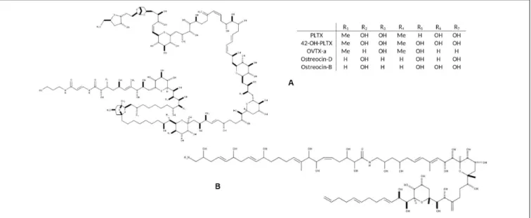
FIGURE 1 | Chemical structure of the main toxic compounds found in Ostreopsis species: (A) Palytoxin and its main analogs, the most common; and (B) Ostreol-A, present in some Korean strains.
FIGURE 2 | Summary of interactions between Ostreopsis species and virus, bacteria and parasites. Mucus could provide a surface for bacteria; bacterial metabolic activity could both provide N and P to Ostreopsis species and use excreted organic C, N and P from the microalga.
With the most recent progresses in chemical analysis, toxin profile could also become a taxonomic criterion.
Ostreopsis was initially considered a tropical genus, but it has been increasing its biogeographic distribution to temperate latitudes (Rhodes, 2011; Selina and Levchenko, 2011; Parsons et al., 2012; Tester et al., 2020 see Figure 5 in that paper-). In recent years, some reports have noticed the expansion of Ostreopsis cf. ovata in coastal waters of Australia, Brazil, China, Cook Islands, Caribbean Sea, French Polynesia, Hawaii, Indian Ocean, Japan, Malaysia, New Caledonia, New Zealand, Russia, Venezuela, Vietnam and specially in the Mediterranean Sea (Table 1). OtherOstreopsis species that appeared to have more confined distributions are also found in new areas. For instance, Ostreopsis fattorussoi isolated from the Eastern Mediterranean
could be also growing in the Atlantic coast of the Iberia Peninsula (Portugal) although it has not been demonstrated yet (Accoroni et al., 2016a) and it was found in the Canary Islands ( Fernández-Zabala et al., 2019). The wide expansion of the genusOstreopsis is considered a potential problem for human health and wellbeing (economy, tourism, beach quality) issues due to the production of PLTX analogs which mode of action in humans is still not clear.
Toxins Synthesized by Ostreopsis
Species: The Main Chemical Substances
Involved in the MCE of the Genus
Several species in theOstreopsis genus are considered potentially harmful to humans and other marine organisms due to the
FIGURE 3 | Summary of known interactions between Ostreopsis cf. ovata and flora.
FIGURE 4 | Summary of direct interactions of Ostreopsis ovata and fauna.
production of palytoxin (PLTX) analogs (Table 1). PLTX is the most potent non-bacterial toxin of biological origin, initially isolated in 1971 from the tropical soft coral genusPalythoa, and now recognized as 42-OH-PLTX (see revision byCiminiello et al., 2011and references inPoli et al., 2018).
Progresses in chemical analysis technology, in particular on HR LC-MS/MS and Nuclear Magnetic Resonance (NMR) have revealed the high diversity of PLTX analogs (Table 1 and
Figure 1) and eight Ostreopsis species have been described as toxic. Ostreocin D (Figure 2) was the first analog isolated from O. siamensis by Usami et al. (1995) and other PLTX analogs, namely, ovatoxins, ostreocins or mascarenotoxin have been characterized forO. fattorussoi, O. mascarensis, O. cf. ovata and O. siamensis (Mercado et al., 1994; Deeds and Schwartz, 2010;
Parsons et al., 2012). However, the toxicity of O. lenticularis remains uncertain.Mercado et al. (1994)reported the production
of Ostreotoxins 1 and 3 by cultures of O. lenticularis isolated from the Caribbean. This study was suggested to be taken with caution in the review byParsons et al. (2012)since subsequent research indicated that bacteria could be involved in the toxicity of O. lenticularis strains from the Caribbean (Mercado et al., 1994, 1995; Meunier et al., 1997; Pérez-Guzmán et al., 2008). Recently, Chomérat et al. (2019) have reported no toxicity of O. lenticularis strains using CBA-N2a and LC-UV-MS/MS analysis.Toxicity ofO. heptagona, O. labens and O. rhodesae has only been described using biological assays and toxins involved in these effects have not been identified yet (Table 1). In the case of O. cf. ovata, up to 12 ovatoxins (OVTX), from a to l (including j1 and j2) and the isobaric PLTX (isoPLTX, García-Altares et al., 2015corresponding to the putative PLTX in other studies) have been identified among different isolated strains, and the toxinology profile is contributing to the taxonomic
FIGURE 5 | Summary of biotic relationships between Ostreopsis spp. and its environment. Arrows in full lines represent the direct interactions, those in dashes represent the indirect interactions. Gray dotted arrows represent unknown interactions which could provide useful guidelines for future research studies. The yellow triangular symbol represents possible human poisoning pathways by toxins produced by Ostreopsis spp. Icons on this figure are copyrighted by Flaticon.
characterization (Accoroni et al., 2016a). In most species and strains, OVTX-a and OVTX-b are the most common toxin forms, and isoPLTX is often absent or only accounting for no more than 1% of the total toxins (Tartaglione et al., 2017;Gémin et al., 2019). A main limitation to ascertain the health impacts risks associated toOstreopsis blooms (and establishing legal toxin thresholds in seafood) is the lack of standards for the variety of toxins, all them complex and with high molecular weight (molecular formulas varying between C129H223N3O55 and C131H227N3O52, Table 1).
As for species identification, information in Table 1 may change in the near future to rapid progresses in toxin analytical methods. Phycotoxins in general and the PLTX group as well, are considered as secondary metabolites and their physiological role in the cells is unknown. Environmental factors such as temperature, salinity, light or nutrients, depth or macroalgal substrate, but also strain characteristics (isolation site, time kept in culture, growth phase of the culture) may influence toxin production (Granéli et al., 2011; Ciminiello et al., 2012;
Pezzolesi et al., 2012, 2014; Scalco et al., 2012; Vanucci et al., 2012b; Gémin et al., 2019). However, it has been suggested that other microorganisms co-occurring along the Ostreopsis blooms could be involved in the toxin production as well. Tosteson et al. (1989) andAshton et al. (2003) found that bacteria associated with the surfaces or the extracellular matrices of O. lenticularis were correlated with the high
dinoflagellate toxicity during the stationary phase of the cultures. Biological assays and analytical chemistry analysis are both used for toxin presence detection and/or quantification. Mouse Bioassay (prohibited in Europe since 2005 for ethical reasons), Haemolysis Neutralization Assay (HNA) or Artemia bioassay have been long recognized for toxicity detection (Delaney, 1984; Bignami, 1993; Botana, 2014). Advantages provided by analytical chemistry, i.e., LC-MS/MS, are the absence of restrictions in terms of reproducibility, specificity, precision and consistency. Unfortunately, this methodology relies on very expensive technical equipment, it requires high-qualified personnel skills and it produces significant chemical waste. The variety of toxins produced by the differentOstreopsis species are considered the main substances involved in the chemical ecology of these dinoflagellates, and with this perspective are considered in this revision. However, recent studies indicate that other non-identified compounds could also be involved in the chemical ecology ofOstreopsis (sections 3.3.1. and 5.2).
Ostreopsis Marine Chemical Ecology
Affecting Human Health
Health impacts in humans due to exposure to PLTX analogs synthesized by Ostreopsis constitute an indication of its related chemical ecology. Health impacts can occur: i) after
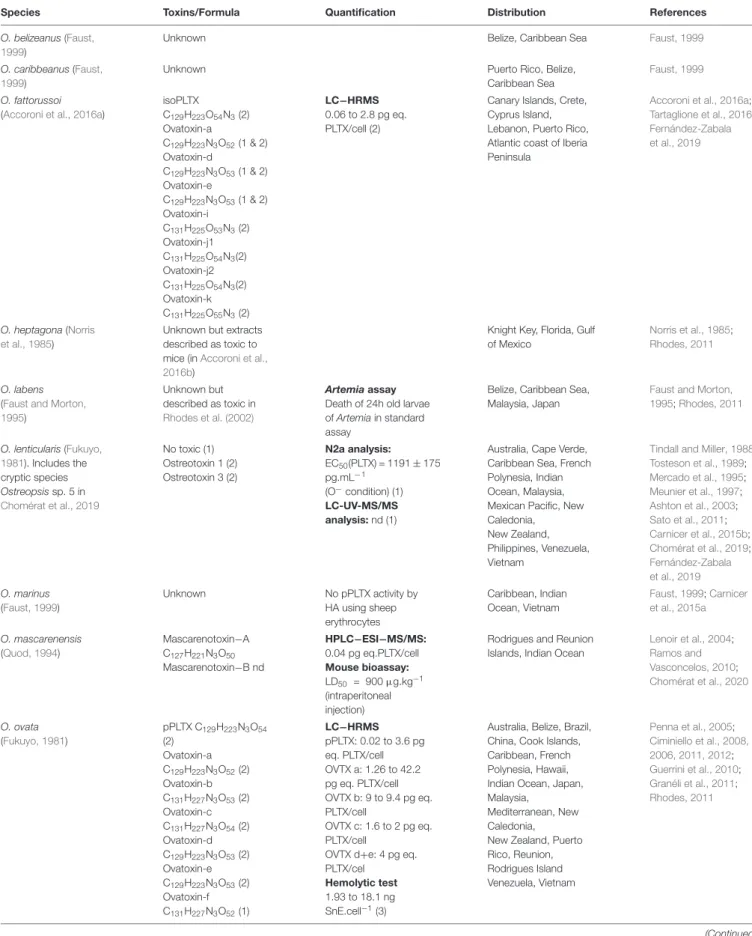
TABLE 1 | Representative information about the range of toxin content on Ostreopsis species from different locations reported in the literature.
Species Toxins/Formula Quantification Distribution References O. belizeanus (Faust,
1999)
Unknown Belize, Caribbean Sea Faust, 1999
O. caribbeanus (Faust, 1999)
Unknown Puerto Rico, Belize, Caribbean Sea
Faust, 1999
O. fattorussoi (Accoroni et al., 2016a)
isoPLTX C129H223O54N3(2) Ovatoxin-a C129H223N3O52(1 & 2) Ovatoxin-d C129H223N3O53(1 & 2) Ovatoxin-e C129H223N3O53(1 & 2) Ovatoxin-i C131H225O53N3(2) Ovatoxin-j1 C131H225O54N3(2) Ovatoxin-j2 C131H225O54N3(2) Ovatoxin-k C131H225O55N3(2) LC−HRMS 0.06 to 2.8 pg eq. PLTX/cell (2)
Canary Islands, Crete, Cyprus Island, Lebanon, Puerto Rico, Atlantic coast of Iberia Peninsula
Accoroni et al., 2016a;
Tartaglione et al., 2016;
Fernández-Zabala et al., 2019
O. heptagona (Norris et al., 1985)
Unknown but extracts described as toxic to mice (inAccoroni et al., 2016b)
Knight Key, Florida, Gulf of Mexico
Norris et al., 1985;
Rhodes, 2011
O. labens (Faust and Morton, 1995)
Unknown but described as toxic in
Rhodes et al. (2002)
Artemia assay Death of 24h old larvae of Artemia in standard assay
Belize, Caribbean Sea, Malaysia, Japan
Faust and Morton, 1995;Rhodes, 2011 O. lenticularis (Fukuyo, 1981). Includes the cryptic species Ostreopsis sp. 5 in Chomérat et al., 2019 No toxic (1) Ostreotoxin 1 (2) Ostreotoxin 3 (2) N2a analysis: EC50(PLTX) = 1191 ± 175 pg.mL−1 (O−condition) (1) LC-UV-MS/MS analysis: nd (1)
Australia, Cape Verde, Caribbean Sea, French Polynesia, Indian Ocean, Malaysia, Mexican Pacific, New Caledonia, New Zealand, Philippines, Venezuela, Vietnam
Tindall and Miller, 1988;
Tosteson et al., 1989; Mercado et al., 1995; Meunier et al., 1997; Ashton et al., 2003; Sato et al., 2011; Carnicer et al., 2015b; Chomérat et al., 2019; Fernández-Zabala et al., 2019 O. marinus (Faust, 1999) Unknown No pPLTX activity by HA using sheep erythrocytes Caribbean, Indian Ocean, Vietnam Faust, 1999;Carnicer et al., 2015a O. mascarenensis (Quod, 1994) Mascarenotoxin−A C127H221N3O50 Mascarenotoxin−B nd HPLC−ESI−MS/MS: 0.04 pg eq.PLTX/cell Mouse bioassay: LD50 = 900µg.kg−1 (intraperitoneal injection)
Rodrigues and Reunion Islands, Indian Ocean
Lenoir et al., 2004; Ramos and Vasconcelos, 2010; Chomérat et al., 2020 O. ovata (Fukuyo, 1981) pPLTX C129H223N3O54 (2) Ovatoxin-a C129H223N3O52(2) Ovatoxin-b C131H227N3O53(2) Ovatoxin-c C131H227N3O54(2) Ovatoxin-d C129H223N3O53(2) Ovatoxin-e C129H223N3O53(2) Ovatoxin-f C131H227N3O52(1) LC−HRMS pPLTX: 0.02 to 3.6 pg eq. PLTX/cell OVTX a: 1.26 to 42.2 pg eq. PLTX/cell OVTX b: 9 to 9.4 pg eq. PLTX/cell OVTX c: 1.6 to 2 pg eq. PLTX/cell
OVTX d+e: 4 pg eq. PLTX/cel
Hemolytic test 1.93 to 18.1 ng SnE.cell−1(3)
Australia, Belize, Brazil, China, Cook Islands, Caribbean, French Polynesia, Hawaii, Indian Ocean, Japan, Malaysia,
Mediterranean, New Caledonia, New Zealand, Puerto Rico, Reunion, Rodrigues Island Venezuela, Vietnam Penna et al., 2005; Ciminiello et al., 2008, 2006, 2011, 2012; Guerrini et al., 2010; Granéli et al., 2011; Rhodes, 2011 (Continued)
TABLE 1 | Continued
Species Toxins/Formula Quantification Distribution References O. cf. ovata pPLTX C129H223N3O54 Ovatoxin-a C129H223N3O52 Ovatoxin-b C131H227N3O53 Ovatoxin-c C131H227N3O54 Ovatoxin-d C129H223N3O53 Ovatoxin-e C129H223N3O53 Ovatoxin-f C131H227N3O52(2) Ovatoxin-g C129H223N3O51(3) Ovatoxin-h C129H225N3O51(1) Ostreol A C67H112N2O23(4) Mascarenotoxin-a C127H221N3O50(5) Mascarenotoxin-c C129H221N3O51(5) LC−HRMS pPLTX: 0.1 to 24.8 pg eq. PLTX/cell OVTX a: 0.065 to 171 pg eq. PLTX/cell OVTX b: 2 to 205 pg eq. PLTX/cell OVTX c: 0.7 to 37 pg eq. PLTX/cell OVTX d+e: 2 to 80 pg eq. PLTX/cell OVTX f: nd - 17 pg eq. PLTX/cell OVTX g: 0.35 to 24.8 pg eq. PLTX/cell
Australia, Brazil, China, Canary Islands, Caribbean, Egypt, Hawaii, Indonesia, Japan, Korea, Malaysia, New Caledonia, New Zealand, Madeira, Mediterranean Sea, Thailand, South West Indian Ocean, Vietnam
Rossi et al., 2010; David et al., 2013; Honsell et al., 2013; Nascimento et al., 2012a,b;Pezzolesi et al., 2012;Sechet et al., 2012;Hwang et al., 2013;Brissard et al., 2014, 2015; Dell’Aversano et al., 2014;García-Altares et al., 2015;Giussani et al., 2015;Ben et al., 2017 O. rhodesae (Verma et al., 2016) Unknown but described as toxic in Verma et al. (2016) LC-MS/MS: No PLTX analogs
Fish gill cell line assay for toxicity: ≥ 98.5% decrease of gill cell line RTgill-W1 viability
Heron Reef Lagoon, southern Great Barrier Reef, Coral Sea, Australia Verma et al., 2016 O. siamensis (Schmidt, 1901) Ostreocin-A C127H219N3O54 Ostreocin−D C127H219N3O53 Ostreocin-E1 C127H217N3O52 Ostreocin−B C127H219N3O53 Haemolysis Neutralization Assay 0.3 pg PLTX eq/cells (Rhodes, 2011)
Belize, Gulf of Siam, Iriomote Island, Madagascar, Mayotte, Mexico, Puerto Rico, Reunion, Rodrigues Island Ukena et al., 2001; Rhodes et al., 2000, 2002; Fernández-Zabala et al., 2019;Terajima et al., 2019 O. cf. siamensis Ostreocin−D C127H219N3O53 Ostreocin−B C127H219N3O53 LC-MS/MS: 0.17 pg. eq PLTX. cell−1(1) Mouse bioassay: LD50 = 25.1 mg.kg−1 (intraperitoneal injection) (2) Atlantic, Australia, Belize, Canary Islands, Caribbean, Eastern Atlantic coast (Portugal, Cape Verde), Japan, Mediterranean Sea, Mexico, New Caledonia, New Zealand, Southwest Indian Ocean, Tasmania, Thailand, Vietnam Cagide et al., 2009; Ciminiello et al., 2013; David et al., 2013; Verma et al., 2016
The review is not exhaustive and progresses in molecular and toxin analyses may result in rapid changes concerning both identity of species and their toxicity (Chomérat et al., 2019, 2020). More information on toxicity tests as well as on other Ostreopsis strains (that can become new species) is found inTester et al. (2020). *Indicates that the isolate(s) were genetically identified, confirming described or cryptic (ribotype) species designations. eq.: Equivalents; HA: Hemolytic Assay - ability of cellular extract to induce erythrocyte cell lysis vs. a saponin control; HPLC−ESI−MS/MS: High-Performance Liquid Chromatography-ElectroSpray Ionization tandem Mass Spectrometry; OVTX: Ovatoxin; LC-HRMS: Liquid Chromatography – High Resolution Mass Spectrometry; MBA: Mouse BioAssay - toxicity determined by intraperitoneal injection of Ostreopsis cellular extracts; MU: Mouse Units, the 24 h LD50dose estimated using ∼20 g mice; nd: Not Detectable, below detection limits; N2a: neuroblastoma cell line
cytotoxicity assay; pPLTX: putative palytoxin; isoPLTX: isobaric palytoxin (García-Altares et al., 2015).
consumption of seafood potentially contaminated with PLTX analogs, or ii) after direct exposure to seawater and/or aerosol during Ostreopsis blooms. The characterization of human poisonings potentially due to exposure to PLTX
analogs was reviewed by Tubaro et al. (2011) and it is briefly presented here. The effects of Ostreopsis species in different marine organisms have also been reported and are presented in section 3.
TABLE 2 | Reports of mild respiratory, cutaneous and/or general malaise symptoms in humans after exposure to aerosols in Ostreopsis spp. blooms affected beaches. Year Species Location Affected people (n) References
1998, 2004 Ostreopsis sp. NW Med (Spain) 74 (estimated∼200) Vila et al., 2008
1998, 2000, 2001 Ostreopsis ovata Tirrenian (Italy) ∼100 Sansoni et al., 2003
2001, 2003, 2004 Ostreopsis sp. S Adriatic (Italy) 28 Gallitelli et al., 2005
2005, 2006 Ostreopsis ovata Ligurian (Italy) 228, 19 Brescianini et al., 2006
2005 Ostreopsis ovata Genoa (Italy) 209 Durando et al., 2007
2006 NW Med (Spain) 37 Vila et al., 2016
2006-2009 Ostreopsis ovata & Ostreopsis siamensis France 47 Tichadou et al., 2010
2006 Ostreopsis sp. SW Med (Spain) 57 García et al., 2008
2009 Ostreopsis sp. SW Med (Algeria) 150-200 Illoul et al., 2012
2010 Ostreopsis cf. ovata Adriatic (Croatia) 7 Pfannkuchen et al., 2012
2013 Ostreopsis cf. ovata NW Med (Spain) 13 Vila et al., 2016
2014, 2015, 2016, 2017, 2019 Ostreopsis sp. NW Med (Spain) N/A Abós-Herràndiz et al., unpublished
Seafood Borne PLTX Poisonings
Severe illness and even lethal fatalities after consumption of seafood potentially contaminated with PLTX analogs have been rarely reported in tropical and subtropical latitudes since the 1970s (see Tables 1–3 in Tubaro et al., 2011). Among them, only a few cases were confirmed through direct PLTX analogs detection in seafood leftovers. In many other cases, screening tests on seafood samples collected or purchased after or before the poisoning episode combined or not with clinical observations and symptoms, concluded that consumption of PLTX contaminated seafood was the causative reason of the health disorders. PLTX poisoning symptoms include nausea, tiredness, diarrhea, vomiting, dizziness, numbness of the extremities, muscle cramps, paresthesia, restless-ness, respiratory distress and bradycardia. In dramatic cases, patients died after 15h to 4 days following seafood poisonings, even after hospitalization. Unusual metallic taste of the seafood is one of the common signs noted by potential PLTX intoxication.
In the Mediterranean coasts, the proliferation ofO. cf. ovata and O. cf. siamensis since the end of the 21th century raised concern about the potential risk of seafood intoxication. Amzil et al. (2012),Biré et al. (2013),Biré et al. (2015), and Brissard et al. (2014)reported the presence of toxin content in macrofauna (mainly sea urchin, rock shell, crab and flathead mullet) sampled during O. cf. ovata blooms. OVTX-a was the most abundant toxin detected, and in some cases PLTX equivalents accounted on average for 223 to 392 µg·kg−1
of whole flesh, exceeding the threshold of 30 µg·kg−1
in shellfish recommended by the European Food Safety Authority (EFSA). The key questions are whetherOstreopsis was the ultimate organism synthesizing the PLTX-like compounds that contaminated the seafood and, how toxins can be transferred through the food webs. Fortunately, until now, seafood poisonings due to PLTX analogs have not been reported in the Mediterranean zone.
Mild Respiratory and Cutaneous Symptoms After Direct Exposure to Seawater and/or Aerosol During Ostreopsis Blooms
In the last 15 years, in several Mediterranean and Brazilian beaches, Ostreopsis blooms have been associated to mild skin
and respiratory symptoms and general malaise in beach users, workers and inhabitants of the first coastline, after direct contact with water and/or after inhalation of marine aerosols (Table 2). Symptoms include rhinorrhea, pharyngeal pain, dry or mildly productive cough, nose irritation, general malaise, headache, fever (≤38◦
C), eye irritation and/or dermatitis (Durando et al., 2007;Tichadou et al., 2010). Most of the symptoms disappeared within a few hours without specific medication when people moved away from the affected area in the vicinity of the Ostreopsis bloom; only a few severe cases required hospitalization (Durando et al., 2007). A metallic taste has also been reported by people when snorkeling and windsurfing, or by researchers (including the authors of this review) when samplingOstreopsis blooms (Pfannkuchen et al., 2012). It has been hypothesized that PLTX analogs could be the most plausible agent causing the observed symptoms, in part because the observed signs were similar, although much milder, to those experienced by people exposed to aerosols from home aquaria containing zoanthids (Deeds and Schwartz, 2010). Indeed, culturingPalythoa corals have become popular among aquarists. Unfortunately, potent irritative compounds released after pouring boiling water or bleach in aquaria with the aim to eliminate a colony of zoanthids caused within 20 min after exposure, rhinorrhoea, coughing, difficult breathing and light headedness which progressed to severe fits of coughing and chest pain 4h later. At the local hospital the patient was administered an anti-inflammatory corticosteroid and pain medication, an inhaled steroid treatment and cough suppressant for one month. A follow-up examination by a pulmonary specialist two weeks post exposure diagnosed the patient with asthma-like symptoms of bronchial inflammation and bronchoconstriction. Similar cases have been reported in the last years (see Tables 4 and 5 inTubaro et al., 2011) one last case affecting a whole family, with three little kids in the UK in August 2019. Metallic taste was also reported by people when snorkeling and windsurfing in coral reefs containingPalithoa-like corals.
However, during Ostreopsis blooms OVTX-a and isoPLTX (putative) have been only rarely found in the aerosol and in very low concentrations (2.4 pg ovatoxins per liter of air by
Ciminiello et al., 2014) and not coinciding with respiratory symptoms in humans. Several studies (Brescianini et al., 2006;
TABLE 3 | Noxious effects of direct interaction of Ostreopsis species on different fauna.
Species/Strain Organisms Stage Effects References Interactions with epibenthic cells of Ostreopsis species
Ostreopsis ovata isolated by A. Penna
Tigriopus fulvus (Fisher, 1860)
I-II stage Nauplii Mortality LC50−48h−20◦C= 1486.74
cells.mL−1
LC50−48h−25◦C= 250.12
cells.mL−1
Faimali et al., 2012
Ostreopsis cf. ovata Tigriopus fulvus (Fisher, 1860)
Mortality LC50−24h−20◦C> 4000 cells.mL−1 Giussani et al., 2016
Ostreopsis ovata isolated by A. Penna Amphibalanus amphitrite (Darwin, 1854) II-III stage Nauplii Mortality LC50−48h−20◦C= 1416 cells.mL−1 LC50−48h−25◦C= 192 cells.mL−1 Faimali et al., 2012
Ostreopsis cf. ovata Amphibalanus amphitrite (Darwin, 1854)
Mortality LC50−24h−20◦C> 4000 cells.mL−1 Giussani et al., 2016
Ostreopsis cf. ovata Natural samples, Ligurian Sea (44◦3’27”N; 9◦55’43”E) Dinophylus gyrociliatus (Schmidt, 1857)
Juveniles Mortality LT50−24◦C(3500 cells.mL−1) = 2h
LT50−24◦C(1500 cells.mL−1) = 3.4h LT50−24◦C(200 cells.mL−1) = 12.25h ± 3.18 Simonini et al., 2011 Ostreopsis ovata isolated by A. Penna Paracentrotus lividus (Lamarck, 1816)
Juveniles Mortality LC50−48h−20◦C= 168 cells.mL−1 Privitera et al., 2012
Ostreopsis cf. ovata Natural samples, Gulf
Tigriopus fulvus (Fisher, 1860)
Nauplii Mortality LC50−96h−25◦C= 10.11 cell.mL−1 Prato et al., 2011
of Taranto, Ionian Sea Corophium insidiosum (Crawford, 1937) 2-4 mm body length Mortality LC50−96h−25◦C= 11.81 cell.mL−1 Sphaeroma serratum (Fabricius, 1787) 2-4 mm body length Mortality LC50−96h−25◦C= 214.81 cell.mL−1 Ostreopsis siamensis CAWD73/74/75/96 Haliotis virginea (Gmelin, 1791)
Larvae Mortality Not lethal within 24h Rhodes et al., 2000
Ostreopsis cf. ovata MCCV 054
Sarsamphiascus cf. propinquus (Sars G.O., 1906)
Adults Mortality LC50−24h−24◦C> 20 000 cell.mL−1 Pavaux et al., 2019
Ostreopsis cf. ovata Natural samples from French and Italian NW Med. Coasts
Phytal meiofauna Community composition
72 % decrease of nauplii Guidi-Guilvard et al., 2012 Ostreopsis cf. ovata MCCV 054 Sarsamphiascus cf. propinquus (Sars G.O., 1906)
Females Reproduction Decrease of fertility and fecundity rations Pavaux et al., 2019 Ostreopsis cf. ovata Natural samples (French Mediterranean coasts) Paracentrotus lividus (Lamarck, 1816) Toxin quantification LC-MS/MS: 77 to 361µg eq PLTX.kg−1 Amzil et al., 2012 Ostreopsis cf. ovata Field sample -Paracentrotus lividus (Lamarck, 1816) Toxin quantification LC-MS/MS: 103 to 423µg.kg−1 Hemolytic test: 179 to 270µg.kg−1 Brissard et al., 2014 Villefranche-sur-Mer, Mediterranean Sea Sarpa salpa (Linnaeus, 1758) Toxin quantification LC-MS/MS: 33 to 152µg.kg−1 Ostreopsis ovata Field samples (Tyrrhenian Sea)
Sea urchins Toxin quantification HNA: 5 to 99 pPLTXµg.kg−1
LC-MS/MS:< LOQ pPLTX µg.kg−1 87 to 164 OVTXaµg.kg−1 Milandri et al., 2010 Ostreopsis cf. ovata Field samples Nice harbour and
Paracentrotus lividus (Lamarck, 1816)
Toxin quantification Haemolytic assay: 256.6µg/kg (WF) LC-MS/MS: 192.5µg/kg (WF) Biré et al., 2013 Villefranche-sur-Mer, Mediterranean Sea Sarpa salpa (Linnaeus, 1758)
Toxin quantification Haemolytic assay: 230.0µg/kg (DT) LC-MS/MS: 191.2µg/kg (WF) Ostreopsis cf. ovata Field samples -Paracentrotus lividus (Lamarck, 1816)
Toxin quantification Haemolytic assay: 159.2µg/kg (DT) Biré et al., 2015 Rochambeau site, Villefranche-sur-Mer, Mediterranean Sea Sarpa salpa (Linnaeus, 1758)
Toxin quantification Haemolytic assay: 361.0µg/kg (DT)
TABLE 3 | Continued
Species/Strain Organisms Stage Effects References Interactions with planktonic cells of Ostreopsis species
Ostreopsis ovata isolated by A. Penna Paracentrotus lividus (Lamarck, 1816) Competent larvae
Mortality LC50−48h−20◦C= 168 cells.mL−1 Privitera et al., 2012
Ostreopsis cf. ovata CBA29-2012 (Genoa)
Aurelia sp. Polyp Reproduction/early stages
Unaffected (100 cells.mL−1) Giussani et al., 2015
Ostreopsis cf. ovata Marche coast (Italy NW Adriatic Sea)
Mytilus galloprovincialis (Lamarck, 1819)
Adults Histological impairments
Immune system: - Decrease of granulocytes
Phagocytosis activity decrease Reduced lysosomal membrane stability
Histology: - Decrease of the digestive gland wall thickness Dilatation of tubules Haemocytes infiltration into digestive gland
Decrease of neutral lipids quantity Oxidative stress: limited role
Gorbi et al., 2013
Ostreopsis cf. ovata Field sample from Adriatic Sea (Portonevo & Passetto sites)
Mytilus galloprovincialis (Lamarck, 1819) Adults Histological impairments Inhibition of Na+ /K+ ATPase and acetylcholine esterase activity Immune system: Reduced lysosomal membrane stability Oxidative stress: limited role
Gorbi et al., 2012
Ostreopsis spp. Cultures + field samples (North Aegean coasts)
Mytilus galloprovincialis (Lamarck, 1819) Venus verrucosa Odiolus barbatus
Toxin quantification MBA: Qualitative analysis HNA: 33.3 to 97 pPLTXµg.kg−1 Aligizaki et al., 2008 Ostreopsis cf. ovata Natural samples (French Mediterranean coasts) Mytilus galloprovincialis (Lamarck, 1819) Toxin quantification LC-MS/MS: 28 to 217µg eq PLTX.kg−1 Amzil et al., 2012 Ostreopsis cf. ovata Filed samples (Adriatic Sea)
Mytilus galloprovincialis (Lamarck, 1819)
Toxin quantification MBA + HNA: Qualitative analysis Gorbi et al., 2012
Ostreopsis ovata Field samples (Tyrrhenian Sea)
Mussels Toxin quantification HNA: 103 pPLTXµg.kg−1
LC-MS/MS:< LOQ pPLTX µg.kg−1- 131 to 228 OVTXa
µg.kg−1
Milandri et al., 2010
Ostreopsis siamensis CAWD75 (New Zeland)
GreenshellTM mussels Pacific oysters Scallops
Toxin quantification MBA: qualitative analysis HNA: 0.3 pg eq. PLTX.cell−1
Rhodes et al., 2000
Ostreopsis siamensis CAWD75 (New Zeland)
Perna canaliculus (Gmelin, 1791) Pecten novaezalandiae (Reeve, 1852) Crassostrea gigas (Thunberg, 1793)
Toxin quantification MBA (qualitative analysis): none to death HNA: trace to 1 ng.g−1 Rhodes et al., 2002 Ostreopsis cf. ovata Field samples -Rochambeau site Villefranche-sur-Mer Arca noae (Linneaus, 1758)
Toxin quantification Haemolytic assay:<10 µg/kg (WF) Biré et al., 2015
Ostreopsis cf. ovata CBA29-2012 (Genoa)
Aurelia sp. Ephyrae Reproduction/early stages
% Alteration of Pulsation Frequency EC50−24h= 5.32 cells.mL−1 Immobility EC50−24h= 10.50 cells.mL−1 Giussani et al., 2015 Ostreopsis cf. ovata ITAC501 Paracentrotus lividus (Lamarck, 1816) Eggs Reproduction/early stages
Gametes: % fertilization decrease; less toxic for eggs than sperms Embryos: no segmentation
Caroppo and Pagliara, 2011
TABLE 3 | Continued
Species/Strain Organisms Stage Effects References Ostreopsis cf. ovata
Natural samples (Gaiola MPA, Italy)
Paracentrotus lividus (Lamarck, 1816)
Embryos Reproduction/early stages
Fertilization and developmental impairments
Alteration of several marker genes
Migliaccio et al., 2016
Ostreopsis cf. ovata Strain UNR-05 Armaçao dos Bùzios (Rio de Janeiro)
Lytechinus variegatus (Lamarck, 1816)
Embryos Reproduction/early stages
Stronger effect on larvae than on fertilization success: - Reduced fertilization
- Arrest of embryonic development (gastrula stages at 400 cells.mL−1)
- Increase of dead and abnormal larvae
Neves et al., 2018
Ostreopsis cf. ovata (whole cells + lysate) Natrural samples (Ionian Sea) Paracentrotus lividus (Lamarck, 1816) Embryos Reproduction/early stages
Whole cells: Developmental delay or anomalous cell division Cell lysate: Growth inhibition at 1.87 mg.mL−1
Pagliara and Caroppo, 2012
Interactions with surface cells of Ostreopsis species Ostreopsis cf. ovata
Field samples Nice harbour and Villefranche-sur-Mer
Patella spp. Toxin quantification Haemolytic assay: 12.7µg/kg (WF) Biré et al., 2013
Mugil cephalus (Linneaus, 1758)
Toxin quantification Haemolytic assay: 392.2µg/kg (DT) LC-MS/MS: 359.1µg/kg (WF) Ostreopsis cf. ovata Field samples -Rochambeau site Villefranche-sur-Mer
Patella spp. Toxin quantification Haemolytic assay:<10 µg/kg (WF) Biré et al., 2015
Ostreopsis cf. ovata Field samples Genoa, Nice, Villefranche-sur-Mer Paracentrotus lividus (Lamarck, 1816)
Density Any decrease of P. lividus densities related to high abundances of O. cf. ovata
Blanfuné et al., 2012
Patella spp. No significant decrease of densities in Villefranche-sur –mer and Nice; Significant decrease in Genoa potentially linked to O. cf. ovata blooms
EC50: Half maximal Effective Ostreopsis Concentration; DT: Digestive Tube; HNA: Hemolysis Neutralization Assay; LC50: Median Lethal Concentration; LC-MS: Liquid
Chromatography – Mass Spectrometry; LT50: median Lethal Time; MBA: Mouse BioAssay; RT: Remaining Tissue; WF: Whole Flesh.
Mangialajo et al., 2011) suggested that Ostreopsis fragments, mucus, and/or associated biota (including bacteria, cyanobacteria or other pico- or nanoeukaryotes) could be present in the aerosol and cause the health disorders. It should be noted here that symptoms do not occur concurrently with high cell Ostreopsis densities neither during the bloom, but only in certain periods.Vila et al. (2016)conducting a parallel epidemiology and ecology survey found that symptoms occurred at the end of the exponential phase and initial stationary phase of the bloom, at least in 2013 in a Mediterranean hot spot. This trend has been approximately repeated in the following years in that hot spot (Abós-Herràndiz et al., unpublished). Still an open question is how toxins and/or fragment ofOstreopsis can be aerosolized and whether these toxins are the causative agent of the disorders.
Toxicity on Mammalian Cells and Mode of Action
The main biological target of PLTX seems to be the Na+
/K+
-ATPase, a plasma membrane pump involved in the maintenance of trans-membrane ionic gradients of animal cells, essential for cellular functions and life (Habermann, 1989;Wu, 2009;Rossini and Bigiani, 2011). Impairment of the membrane pump by PLTX results in its conversion into a non-selective pore for
monovalent cations, thereby destroying the transmembrane ionic gradient and triggering several adverse biological effects, some life threatening (Habermann, 1989;Artigas and Gadsby, 2003).
However, these studies have been conducted in vitro on mammalian cell tissues and the actual in vivo toxicity of the molecule is still poorly understood. In the 1970’s, semi-purified PLTX was tested in a broad range of animal species through several routes of administration. Thus, a reduced or even increased toxicity of the pure molecule are possible. More recently, high purity PLTX administration in mice showed that toxicity is dependent upon the route of administration (Munday, 2011). Poli et al. (2018) characterized the toxicity and basic histopathological effects of PLTX (derived from Japanese Palythoa tuberculosa), the analog 42-OH-PLTX (from Hawaiian P. toxica) and ovatoxin-a (isolated from a Japanese strain of Ostreopsis ovata) after intraperitoneal and aerosol administration to rats. All toxin preparations showed similar potency towards mouse erythrocytes in the erythrocyte hemolysis assay and interactions with the anti-PLTX mAb (antibody). Intraperitoneal LD50 values between 0.92 and 3.26 µg·kg−1
consistent with published values, and aerosol LD50values of 0.031
TABLE 4 | Effects on organisms exposed indirectly to Ostreopsis species.
Species/Strain Organisms Authority Effects References Ostreopsis ovata
Field samples (Tyrrhenian Sea)
Octopuses Toxin quantification HNA: 446 pPLTXµg.kg−1
LC-MS/MS: 115 pPLTXµg.kg−1
971 OVTXaµg.kg−1
Milandri et al., 2010
Ostreopsis cf. ovata Diplodus annularis Linnaeus, 1758 Toxin quantification Haemolytic assay: 10.6µg/kg (DT) Biré et al., 2013
Field samples Diplodus sargus Linnaeus, 1758 Toxin quantification Haemolytic assay: 5.1µg/kg (DT) Nice harbour and
Villefranche-sur-Mer
Eriphia verrucosa Forskål, 1775 Toxin quantification Haemolytic assay: 38.4µg/kg (WF) LC-MS/MS: 58.6µg/kg (WF) (Mediterranean Sea) Maja squinado Herbst, 1788 Toxin quantification Haemolytic assay: 8.3µg/kg (WF)
Mullus surmuletus Linnaeus, 1758 Toxin quantification Haemolytic assay:< LOQ Muraena helena Linnaeus, 1758 Toxin quantification Haemolytic assay:< LOQ Octopus vulgaris Cuvier, 1797 Toxin quantification Haemolytic assay: 18.5µg/kg (RT) Scorpaena porcus Linnaeus, 1758 Toxin quantification Haemolytic assay: 3.7µg/kg (DT) Serranus scriba Linnaeus, 1758 Toxin quantification Haemolytic assay:< LOQ Stramonita
haemastoma
Linnaeus, 1767 Toxin quantification Haemolytic assay: 34.1µg/kg (WF) LC-MS/MS: 26,4µg/kg (WF) Symphodus tinca Linnaeus, 1758 Toxin quantification Haemolytic assay: 1.8µg/kg (DT)
Ostreopsis cf. ovata Coris julis Linnaeus, 1758 Toxin quantification Haemolytic assay:<10 µg/kg (WF) Biré et al., 2015
Field samples Rochambeau site,
Diplodus vulgaris Geoffroy Saint-Hilaire, 1817
Toxin quantification
Villefranche-sur-Mer Eriphia verrucosa Forskål, 1775 Toxin quantification Haemolytic assay: 13.0µg/kg (WF) Hexaplex trunculus Linnaeus, 1758 Toxin quantification Haemolytic assay: 40.4µg/kg (WF) Gobiidae Toxin quantification Haemolytic assay:<10 µg/kg (WF) Maja squinado Herbst, 1788 Toxin quantification Haemolytic assay: 51.3µg/kg (WF) Mullus surmuletus Linnaeus, 1758 Toxin quantification Haemolytic assay:<10 µg/kg (WF) Muraena helena Linnaeus, 1758 Toxin quantification Haemolytic assay:<10 µg/kg (WF) Octopus vulgaris Cuvier, 1797 Toxin quantification Haemolytic assay: 19.9µg/kg (RT) Saurida
undosquamis
Richardson, 1848 Toxin quantification Haemolytic assay:<10 µg/kg (WF) Scorpena porcus Linnaeus, 1758 Toxin quantification Haemolytic assay:<10 µg/kg (WF) Serranus scriba Linnaeus, 1758 Toxin quantification Haemolytic assay:<10 µg/kg (WF) Symphodus
melops
Linnaeus, 1758 Toxin quantification Haemolytic assay:< 10 µg/kg (WF) Symphodus
ocellatus
Linnaeus, 1758 Toxin quantification Haemolytic assay:<10 µg/kg (WF) Symphodus roissali Risso, 1810 Toxin quantification Haemolytic assay:<10 µg/kg (WF) Symphodus
rostratus
Bloch, 1791 Toxin quantification Haemolytic assay:<10 µg/kg (WF) Symphodus tinca Linnaeus, 1758 Toxin quantification Haemolytic assay:<10 µg/kg (WF) Thalassoma pavo Linnaeus, 1758 Toxin quantification Haemolytic assay:<10 µg/kg (WF) Xantho poressa Olivi, 1792 Toxin quantification Haemolytic assay:<10 µg/kg (WF) Ostreopsis ovata
isolated by A. Penna
Dicentrarchus labrax
Linnaeus, 1758 Mortality LC50−48h−20◦C= 540 cells.mL−1 Faimali et al., 2012
Ostreopsis cf. ovate Dicentrarchus labrax
Linnaeus, 1758 Mortality LC50−48h−20◦C= 679 cells.mL−1 Giussani et al., 2016
Ostreopsis cf. ovata Strain OOAN0601
Dicentrarchus labrax
Linnaeus, 1758 Mortality 100% mortality after 45 to 72 h exposed to O. cf. ovata (between 425 to 2367 cells.mL−1)
Pezzolesi et al., 2012
EC50: Half maximal Effective Ostreopsis Concentration; LC50: Median lethal concentration; LT50: Median lethal time. the literature. Most commonly affected tissues were the lungs,
liver, heart, salivary glands, and adrenal glands.
Mechanisms of Toxins Release in Sea Water and Aerosolization
It is well recognized that microorganisms including some microalgae can be dispersed by air and survive in the aerosols
(e.g., Genitsaris et al., 2011; revision by Tesson et al., 2016). Microorganisms become airborne mainly due to drop formation at the water surface microlayer, which is enriched with biological material in all aquatic habitats (Schlichting, 1974;Wilson et al., 2015). Foam drops of a diameter larger than 40µm are formed by wind friction and breaking wave crests at wind speeds exceeding 7 to 11 m·s−1. Bubble bursting due to waves, rainfall, boat traffic, or
supersaturation of gases in the water produces film drops (1µm to 10µm diameter), projected in various directions, and vertically emitted jet drops (6 µm to 100 µm diameter). Recreational activities (i.e., waterskying, using personal watercraft, swimming, or wading) can also generate aerosols. Once emitted, airborne turbulent kinetic energy drags the microorganisms further up into the atmosphere.
The few available studies indicate that microalgae are found at very low concentrations (10−4 to 104 cells per m−3,Mayol
et al., 2014) and with heterogeneous distributions, which requires high-volume sampling technology. High-volume sampling on filters or agar plates can be achieved by different instruments, but high-volume sampling into liquids is more complicated, as high airflows may lead to evaporation and reduce collection efficiency. Once collected, a combination of microscopy and molecular tools contribute to the identification of the airborne microalga. The main microalgal groups found in bioaerosols are chlorophytes (“green algae”), bacillariophytes (diatoms, mainly small Nitzschia) and cyanobacteria. Airborne microalgae are recognized as allergens and antigens. They are the cause of severe medical issues, including respiratory allergies (e.g., hay fevers), asthmatic attacks, dermatitis and skin lesions, rhinitis, and disturbances in lymphatic systems or vital organs (e.g., protothecosis, see review inAshok and Gupta, 1998). In addition, certain aerosolized algal toxins, namely, microcystins produced by cyanobacteria (Backer et al., 2010) brevetoxins synthesized by Karenia brevis (Pierce and Henry, 2008;Fleming et al., 2011) and OVTX and PLTX analogs generated byOstreopsis species, are the causes of further human illnesses.
So far, the detection of Ostreopsis cf. ovata and its related toxins in the aerosol was conducted byCasabianca et al. (2013)
and Ciminiello et al. (2014). Casabianca et al. (2013) used air pump samplers (CAV-A/mb, Barcelona, Spain) equipped with 15 cm diameter QM-A quartz filters (Whatman, Maidstone, United Kingdom). Two air samplers located at 0.5 m and 3 m above sea level, respectively, pumped air continuously for 3 days during anO. cf. ovata bloom at a flow rate of 30 m3·h−1. Filters
were replaced every 6 or 7 h and were divided in two portions to analyze, respectively, the concentration of Ostreopsis cell concentrations by qPCR and toxins by the hemolytic assay and by liquid chromatography with fluorescence detection (LC-FLD). Ostreopsis cells were detected in the aerosol by qPCR, exhibiting a marked variability during the bloom although not always directly related to the concentrations of the epiphytic Ostreopsis in the water; the authors noted that wind and other environmental factors could have affected these findings. In the seawater, epiphytic Ostreopsis contained ca. 1.2 pg total PLTX analogs per cell but toxins were not detected in the aerosol. Different technical aspects (e.g., toxin binding to glass tubes, not efficient retention by the filters) were suggested as potential technical limitation. Ciminiello et al. (2014) also employed AirCube COM2 (Analitica Strumenti srl., Italy) equipped with 47 mm diameter glass/quartz microfiber filters (Whatman, Maidstone, United Kingdom) and SAS PCR VWR International (PBI, Italy). In this case, air and a collecting fluid (50 mL distilled water in 2009, and seawater in 2010) flow together through a nozzle were poured into a vessel with a coil; the collecting fluid was
circulated continuously to prolong the contact time with aerosols and to enrich itself in toxins and/or cells. Both air samplers were located 10 m from the seashore and worked continuously for 1.5-6 h. Air flow speed was 10 L·min−1in 2009 and 30 L·min−1in
2010. The presence of OVTX in the aerosol at levels of 2.4 pg of OVTX per liter of air was confirmed for the first time but PLTX by inhalation exposure toxicological data were not available. Overall, the presence of both cells and toxins in the aerosols provided by these studies opened new possibilities to increase the understanding of theOstreopsis-related respiratory syndrome.
MARINE CHEMICAL ECOLOGY OF
Ostreopsis IN THE AQUATIC
ENVIRONMENT
This section reviews the interactions of Ostreopsis cells with different marine organisms and environments as part of the MCE ofOstreopsis.
Interactions With Bacteria, Viruses and
Parasites
Interactions between microalgae species and bacteria remain poorly investigated although the available studies reveal connections playing important roles in the biology and the ecology of these microorganisms. Bacteria may interact with algae within the phycosphere (Bell and Mitchell, 1972) as free-living bacteria, attached to the algal cell surface or occurring as intracellular symbionts. Algae-bacteria interactions can be synergistic and mutualistic, as commensalism or symbiotic, e.g., by producing cytokinins – phytohormones (Ramanan et al., 2016). Some vitamins and ion chelators produced by bacteria are involved in nutrient recycling facilitating nutrient incorporation by algae, while, in return, bacteria use microalgal exudates (Sarmento and Gasol, 2012). Furthermore, competition, parasitism or antagonist interactions also occur. Bacteria could interfere microalgal life cycle and growth of some HAB species. Bacterial exopolymers can increase the tendency of cells to flocculate and enhance sinking rates and degradation of decaying (harmful and innocuous) blooms (Decho, 1990). In turn, algae could also control bacterial growth by the production of antibiotic or hydroxyl radicals and other selected anti-bacterial growth substances (Bell et al., 1974). Information on these different interactions focused on HAB species can be found, e.g., inFukami et al., 1992;Oda et al., 1992;Sawayama et al., 1993;
Doucette, 1995;Hare et al., 2005; Jasti et al., 2005;Maruyama et al., 2008;Kim et al., 2009;Trick et al., 2011. These interactions exhibit spatio-temporal variability scales (Doucette, 1995) which, in the case of HABs can also involve phylogenetically and metabolically diverse bacterial groups along the bloom dynamics (Mayali et al., 2011). While some bacteria could coexist with microalgal cells during the initiation and exponential phases of the blooms, antagonist or parasitic bacteria would proliferate before the declining period and saprophytic or scavenger bacteria will occur after bloom termination when the availability of phytodetritus is high.
Bacterial communities associated to Ostreopsis lenticularis appear to be species and even clone specific (Tosteson et al., 1989) they vary with the bloom phase, and likely, with the habitat. Pérez-Guzmán et al. (2008) identified single species belonging to the Cytophaga-Flavobacter-Bacteroides complex. Vanucci et al. (2016) and Guidi et al. (2018) found Alphaproteobacteria (83 % relative abundance - genera Ruegeria, Jannaschia and Erythrobacter-), Gammaproteobacteria (13%) and Flavobacteria (7%) during the early O. cf. ovata bloom period, whileAlphaproteobacteria (66%) and Flavobacteria (19%) were predominant in peak bloom phases. In the case of dinoflagellates, bacteria-algae interactions can be very strong given that some microalgae are unable to grow in axenic conditions in culture confirming the predominant role of bacteria in algal cycle. This has also been observed inOstreopsis cultures although not always (Vanucci et al., 2012a).
Some studies indicate that bacteria could directly or indirectly modulate production and degradation of microalgal toxins (Albinsson et al., 2014) although the results appear study- and species- dependent and contradictory. While associated bacteria were linked to the production of paralytic shellfish toxin by Alexandrium spp. (Gallacher et al., 1997) higher toxin levels were found in bacteria-free than in non-axenic cultures of Alexandrium tamarense (Hold et al., 2001) andA. lusitanicum (Wang et al., 2004).Ashton et al. (2003)reported a toxicity loss of Ostreopsis lenticularis when exposed to elevated temperature and explained it by the reduction of Ostreopsis associated Pseudomonas and Alteromonas bacteria. Axenic cultures of Ostreopsis cf. ovata (Vanucci et al., 2012a) were also characterized by lower toxin content in late stationary phase compared to non-axenic cultures. Ostreopsis cf. ovata cell concentration along a bloom was correlated with the abundance of planktonic, free-livingVibrio (Bellés-Garulera et al., 2016) and a high proportion of Vibrio were found attached to particles, suggesting that Ostreopsis bloom could provide biological colonizable surfaces (fragments of thecae or mucilage) for bacteria. The study suggested that bacteria could be involved in wound infections reported by beach users during Ostreopsis blooms. In fact, Ostreopsis blooms were reported in heavily antropogenically impacted environments (Cen et al., 2019). However, the role of bacteria, potentially colonizing the mucus produced byOstreopsis (a mucilaginous matrix, described as a network of trichocysts embedded with acidic polysaccharides filaments byHonsell et al., 2013) in the respiratory irritations associated to the microalgal bloom has not been clarified yet.
Parasite infection may play critical roles in the regulation of HABs. It has been already described for the planktonic toxic dinoflagellate Alexandrium fundyense which presented an up-regulation of genes associated to defense against the parasite Amoebophrya and thus controlling host mortality (Garcés et al., 2013a; Lu et al., 2016, 2014). Ostreopsis spp. (among other phytoplankton species) was infected by the generalistic parasitoid Parvilucifera sinerae (Garcés et al., 2013b).
To our knowledge, there has been no research on the interactions betweenOstreopsis species and viruses while viruses have been proposed as natural agents to control HABs. This is the case of the virus HcRNAV, specific of the dinoflagellate
Heterocapsa circularisquama (Nagasaki and Yamaguchi, 1997;
Nagasaki et al., 2004;Nakayama and Hamaguchi, 2016).
Interactions With Heterotrophic Protists
and Ciliates
Heterotrophic dinoflagellates (HTDs) and ciliates have been described as effective grazers on HAB species (Matsuyama et al., 1999;Tillmann, 2004;Jeong et al., 2015). However, so far, only one study has investigated feeding on Ostreopsis cf. ovata by a ciliate and various HTDs, all them potentially able to ingest it based on cell size criteria (Yoo et al., 2015). O. cf. ovata was ingested byGyrodinium dominans, G. moestrupii, Oxyrrhis marina, and P. kofoidii by engulfment and by Pfiesteria piscicida using a peduncle. The study suggested that these HTDs can feed on O. cf. ovata by enzymes involved in the detection, capture, ingestion and digestion of toxic dinoflagellates. In contrast, lack of these enzymes in the naked ciliate Strombilidium sp. and the HTDs Gyrodinium spirale, Protoperidinium bipes, and Stoeckeria algicida would impede feeding on O. cf. ovata. Overall, information on microzooplankton grazing onOstreopsis species in the field are lacking, and there is a major uncertainty on whether the success of this benthic microalga is caused by lack of effective predation. It has also been suggested that the mucus could be deterrent agent.
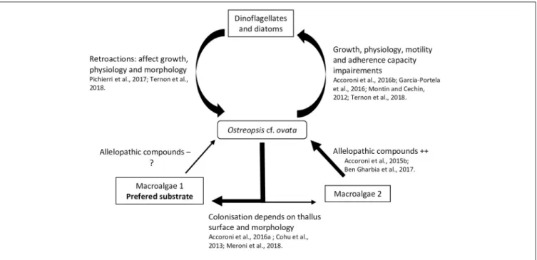
Interactions With Flora
A relatively small but recent number of studies focus on interactions between Ostreopsis species and flora, microphytobenthos and macrophytes, suggesting different allelopathic interactions during the dinoflagellate bloom dynamics. In these studies, the degree of interaction has been measured using different physiology and morphology parameters of the tested organisms, namely, growth rate (García-Portela et al., 2016; Ben et al., 2017) fluorescence (Ben et al., 2017;
Ternon et al., 2018) swimming speed (Yoo et al., 2015) adherence capability (García-Portela et al., 2016) cysts formation (Accoroni et al., 2016b) cellular integrity (variation of shape, size, nucleus position using lipid and nuclear staining;Pichierri et al., 2017) and toxin production (Ben et al., 2017). The interactions with micro- and macroalgae are presented next and summarized in the Figure 3.
Microalgae
Some studies have shown that particular secondary metabolites i.e., low molecular weight compounds (as opposed to peptids or proteins) not directly involved in basic life processes, could considerably affect HABs dynamic and growth (Uchida et al., 1999; Fistarol et al., 2003; Prince et al., 2008; Yamasaki et al., 2010). These sorts of interactions have also been addressed concerning the impact ofOstreopsis cf. ovata on diatom and other dinoflagellates, as well as the retroactions of these microalgae on O. cf. ovata.
A strong inhibitory effect ofO. cf. ovata on microalgae was observed in the in situ study of Accoroni et al. (2016b). In particular, it was manifested as a significant decrease in the diversity index and an increase of motile diatoms abundance during theO. cf. ovata bloom. Motile life diatom phase would
be favored by the Ostreopsis mucous matrix, illustrating the importance of the mucus in the interactions between O. cf. ovata and its environment. Recently,Ternon et al. (2018)using co-cultures assays (with diatoms) suggested a contact-mediated toxicity ofOstreopsis cells, probably by the mucus, which could retain the toxins and other compounds and avoid their dilution in the surrounding environment.
Monti and Cecchin (2012) observed weak effects on the growth of the dinoflagellatesCoolia monotis and Prorocentrum minimum when exposed to filtrates of O. cf. ovata cultures obtained during its exponential phase. This highlights the importance of growth stages considered in the experiments. Indeed, variation of the toxin content among the growth stage has been described in different studies (Vanucci et al., 2012a;Brissard et al., 2014;Pezzolesi et al., 2014) onOstreopsis cf. ovata cultures, with a trend of higher concentration in the stationary phase.
García-Portela et al. (2016)using mixed cultures of different Ostreopsis strains and toxic and non-toxic dinoflagellates, demonstrated differences of toxicity among Ostreopsis strains. Indeed,O. cf. ovata was more toxic than O. sp. «Lanzarote type», highlighting the necessity to study the intraspecific variability of the interactions of Ostreopsis species with other organisms. The chemical compounds involved in the negative interaction ofOstreopsis and other microalgae (specially diatoms) are poorly known and secondary metabolites other than the OVTXs group could play a role, as suggested byTernon et al. (2018).
In turn, it has been observed that planktonic and benthic diatoms can negatively affectO. cf. ovata physiology, morphology and growth. An inhibition of its growth rate by 57 % and 78 % was reported in cultures exposed to, respectively, filtrates of the planktonic diatoms Skeletonema marinoi and Thalassiosira sp., and similar growth inhibition, deleterious effects and genotoxic damages were also observed when exposed to filtrates of other benthic diatoms (e.g., Tabularia affinis, Proschkinia complanatoides and Navicula sp.) (Pichierri et al., 2017). These studies suggested the production of allelopathic compounds by all tested diatoms, especially released after Ostreopsis cell destruction (Pohnert et al., 2007). These diatoms are well known to produce a large family of polyunsaturated aldehydes (PUAs
-Wichard et al., 2005).Pichierri et al. (2017)found that exposure to a range of PUAs (2E,4E-decadienal, 2E,4E-octadienal and 2E,4E-heptadienal) concentrations (from 3 to 36 µmol·L−1)
causedO. cf. ovata growth inhibition, decrease in photosynthesis efficiency, increase of abnormal cells (motionless, decrease in dimensions, contraction of cytoplasm and formation of abnormal vesicle-like structure) and a decrease in cell integrity (chromatin dispersion, lack of autofluorescence of the chlorophyll and larger lipid bodies).
Macroalgae
Biotic substrate preferences of Ostreopsis species has been demonstrated in several studies (Totti et al., 2010). Substrate choice could be explained in part, by the macroalgal thallus surface and morphology: complex, three dimensional flexible and branched thalli favored Ostreopsis cell attachment (Cohu et al., 2013; Accoroni et al., 2016a; Meroni et al., 2018). These thalli would also be more suitable for O. cf. ovata (Vila
et al., 2001; Accoroni et al., 2012) resistance to wave action. Higher abundances of O. cf. ovata were also found on the turf dominated community i.e., in a complex intricate matrix of small Corallinales, Ceramiales (both Florideophyceae) and other filamentous algae (Meroni et al., 2018) as reported inVila et al. (2001)forEllisolandia elongata and inBlanfuné et al. (2015)for Jania rubens. Lee and Park (2018) also showed that Ostreopsis sp. ingested rhodophytes because of their soft cell membrane containing microfibrillar and sulfated galactans (see section 4.2). The production of allelopathic compounds by macroalgae could also explain Ostreopsis species substrate preferences. Macroalgae are known to produce a large number of secondary metabolites sometimes used in biotechnology as antibiofouling coatings (Hellio et al., 2002) or as a way to control HABs (Hu and Hong, 2008; Tang et al., 2015). Thus, it is reasonable to hypothesize that these metabolites could selectively determine substrate preference ofOstreopsis species. Rhodymenia pseudopalmata (Florideophyceae) had little effect onOstreopsis growth and only in powder form (Accoroni et al., 2015). The seagrasses Zostera noltei and Cymodocea nodosa has also a low impact onO. cf. ovata growth although Z. noltei have been described as a producer of phenolic acids inhibiting the growth of Alexandrium catenella (Laabir et al., 2013).Ulva rigida (Ulvophyceae) and Dyctyota dichotoma (Phaeophyceae) had a strong effect on O. cf. ovata by decreasing its growth, physiology, morphology, toxin production and behavior with a decrease of cell adherence (Accoroni et al., 2015;Ben et al., 2017). The potential allelochemical compounds produced by this macroalgae are still poorly known but could mainly include phenols and polyunsaturated fatty acids (PUFAs
-Ben et al., 2017).
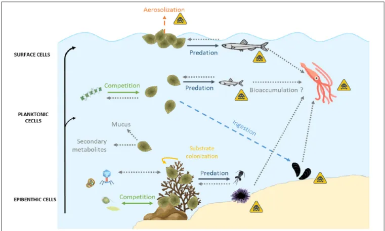
Interactions With Fauna
Toxins produced byOstreopsis have been documented in various marine organisms suggesting the potential bioaccumulation among the trophic web reaching levels above the alert threshold recommended by EFSA. The toxin transfer, which could be analogous to that of Gambierdiscus species synthesized ciguatoxins and maitotoxins (Ledreux et al., 2014), requires more research. Meanwhile, the few existing studies have reported strong differences of sensitivity (e.g., reprotoxicity potentially leading to changes in benthic communities) between organisms exposed toOstreopsis toxins, suggesting unknown mechanisms of acclimation and detoxification.
Impacts of Ostreopsis Blooms on Fauna in Tropical and Temperate Areas
As indicated above, human intoxications have been reported in tropical areas mainly due to the consumption of fish or crustaceans contaminated with PLTX or its analogs without any indication of the effects of these toxins on the vector organisms. This suggests a direct ingestion, at least by herbivorous ingesting macroalgae supporting Ostreopsis cells. Even if no human intoxication due to the consumption of fish or crustaceans was reported in temperate areas, massive mortality of invertebrates occurred during Ostreopsis sp. bloom. Mass mortalities of sea urchins were reported in Italy (Vale and Ares, 2007) Algeria
(Illoul et al., 2012) Brazil (Granéli et al., 2002) New Zealand (Shears and Ross, 2009) Spain (Vila et al., 2008) and France (Blanfuné et al., 2012). Impacts on other sessile and mobile benthic organisms such as cirripeds, mussels, limpets and cephalopods were also observed (Ciminiello et al., 2006; Totti et al., 2010). Even if the role of oxygen depletion and high seawater temperature could be involved in those mass mortalities (Shears and Ross, 2009) more and more authors rather suggest that these mortalities were a consequence of some ecotoxic effects associated toOstreopsis sp. blooms (Sansoni et al., 2003;Simoni et al., 2003;Vila et al., 2008).
Direct Interactions Between Marine Fauna and Ostreopsis Species
Ostreopsis is an epibenthic genus, growing attached to macroalgae or rocks (Totti et al., 2010; Cohu et al., 2013, 2011; Penna et al., 2014; Carnicer et al., 2015a) thanks to the production of a mucilaginous matrix (Honsell et al., 2013; Escalera et al., 2014). Cells are also released in the water column, even if the link between planktonic and benthic abundances exhibits a high variability (Mangialajo et al., 2011) and can also aggregate on the water surface, forming what was called “sea-flowers”.Ostreopsis cells could thus have different impacts on marine ecosystems depending on whether cells are benthic, planktonic or aggregated at surface. These interactions are summarized in Figure 4.
Interactions of fauna with benthic Ostreopsis cells
When Ostreopsis cells attach on benthic substrates, they can interact with its surrounding community and notably with meiobenthic organisms. Indeed, these benthic organisms live in close contact with Ostreopsis cells and could potentially feed on this toxic dinoflagellate. To study the impact of O. cf. ovata on benthic organisms, bioassays are commonly used (Table 3). For instance, the benthic copepod Tigriopus fulvus and the amphipod Corophium insidiosum are both sensitive organisms since their Ostreopsis median lethal concentration (LC50) values after 96 hours of exposition are, respectively, 10.11
et 11.81 cell·mL−1
(Prato et al., 2011). Other organisms, such as the harpacticoid copepodSarsamphiascus cf. propinquus, co-occuring withO. cf. ovata in summer, has been demonstrated as very resistant to toxins produced by O. cf. ovata (Pavaux et al., 2019) with LC50(48h) values higher than 20,000 cells·mL−1
so. These numbers are at least 250 times higher than those reported for another benthic copepod,Tigriopus fulvus (Faimali et al., 2012) suggesting potential acclimation phenomenon to the presence of Ostreopsis. However, these studies are difficult to compare since they used different life stages of the targeted organism, different parameters to determine the sensitivity, and different determination of LC50 values, e.g., at 24, 48, or 96 h,
the experimental conditions (e.g., temperature) during LC50
determination. As an example,Faimali et al. (2012)showed that toxicity increased with the temperature of culture: LC50values of
the barnacleAmphibalanus amphitrite were 1416.95 and 192.26 at 20 and 25◦
C, respectively. This overall situation suggests the need of standardizing the methods to test the toxicity of Ostreopsis species on benthic organisms.
Additionally, a reprotoxic (negative effect on reproduction) effect of O. cf. ovata has been suggested by the significant decrease of the number of copepod nauplii described in the in situ study of Guidi-Guilvard et al. (2012)and confirmed by laboratory experiments byPavaux et al. (2019). The two studies showed a decrease of fecundity and fertility ratios of the benthic copepodSarsamphiascus cf. propinquus. These reprotoxic effects lead to changes in benthic communities by decreasing the number of nauplii.
By forming aggregates that cover macroalgae, Ostreopsis cells are also ingested by macroherbivores such as sea urchins or herbivorous fish. Using biological assays (HNA) or toxin analysis by HPLC-MS, several studies have already quantified PLTX and analogs of these two groups in these herviborous (Milandri et al., 2010; Amzil et al., 2012; Biré et al., 2013, 2015; Brissard et al., 2014). Paracentrotus lividus (an edible and herbivorous sea urchin) and Sarpa salpa (an herbivorous fish) accumulated high concentrations of PLTX-group toxins (respectively, 423 and 361µg·kg−1), making these
organisms good sentinel species for the presence of PLTXs in marine organisms (Biré et al., 2015). Bioaccumulation of toxins seems to be organ-dependent: no toxins were found in sea urchin gonads although it reached up to 423 µg eq PLTX·kg−1 in the digestive tract (Brissard et al., 2014).
However, Bauder et al. (2001) described different affinity of diarrheic shellfish toxins (DST) produced by Prorocentrum lima for tissues leading to a quicker detoxification in the gonads. This low affinity of toxins for tissues except for viscera, could explain the absence of toxins in sea urchin gonads documented in the work of Biré et al. (2015, 2013).
Neves et al. (2018) also reported that the ingestion of O. cf. ovata cells by adult sea urchins had an impact on their offspring (dead and abnormal larvae, arrest of embryonic development – Table 3). These development anomalies persist at least 8 months after the exposition to O. cf. ovata and seems to be transmitted from the female gonads with the involvement of the nitric oxide pathway since the transcription of several genes directly or indirectly modulated by nitric oxide showed transcription variations (Neves et al., 2018;
Tibiriçá et al., 2019).
Interactions of fauna with planktonic Ostreopsis cells
During its planktonic phase, Ostreopsis cf. ovata could reach cell concentrations in the range of 105–106 cell·L−1
(Mangialajo et al., 2011) and thus affect planktonic organisms but also bivalves and other benthic filter-feeders. Some monitoring of toxin concentration in bivalves is conducted to prevent sanitary impacts during these toxic events (Efsa Panel on Contaminants in the Food Chain, 2009; Accoroni et al., 2011; Amzil et al., 2012;
Lemée et al., 2012). Concentrations of toxins quantified in mussels (Mytilus galloprovincialis) varied from 28 to 228 µg·kg−1 (Table 3). Quantities of toxins found in
other filter-feeders such as the mussel Perna canaliculus or the bivalve Arca noae were well below and frequently found only in trace amounts (Rhodes et al., 2000, 2002) probably due to the use of biological assays to quantify