A new BATLS manual has now been prepared under the Authority of the Professor of Military Surgery. In order to disseminate this information widely within the Corps, this Manual will appear in sections in the Corps Journal. This version of the Manual supercedes all previous versions and is now in use on current and future courses.
History of Advanced Trauma
Life Support
0101. There are marked differences in the epidemiology of trauma in different countries. In 1983, only eight people died from gunshot wounds in the United Kingdom. In the same year, the figure in West Germany was 53, approximately one per million of population. Canada had the same ratio with 20 deaths, but in the United States of America with a population of 226 million the figure was 10,838. By 1994, this figure had risen to a shattering 37,500 deaths due to firearms related injury from all causes. In the same year, the United Kingdom with a population of 58.5 million had 261 deaths from firearms. It is not surprising the Americans have a concept of trauma that we in Britain do not have. In America there is also a geographical problem: the nearest major trauma centre can be 150 miles away.
0102. In February 1976, a surgeon piloting his own aeroplane crashed in a rural cornfield in Nebraska. He was badly injured, his wife was killed, three of his children had critical injuries and a fourth child minor injuries. He considered the treatment that he and his children received to be woefully inadequate and said “When I can provide better care in the field with limited resources than what my children and I received at the primary care facility, there is something wrong with the system and the system has to be changed.” (Sic). 0103. As a consequence of this incident the need for training was identified.With the help of the Lincoln Medical Education Foundation and the Southeast Nebraska Emergency Medical Services, a prototype Advanced Trauma Life Support (ATLS©) course was devised.
0104. The project developed and was adopted in 1979 by the American College of Surgeons Committee on Trauma. As a result and with further revisions, a national trauma
CHAPTER 1 INTRODUCTION
programme was established. The original targets were those doctors who do not normally deal with trauma as part of their daily lives, but now all doctors are expected to be capable of managing the trauma patient during the immediate post-trauma phase.
0105. The Advanced Trauma Life Support courses are now run in many centres throughout the United Kingdom. It has also been modified in various parts of the world. In Australia, for example, it has become the Early Management for Severe Trauma (EMST) course.
0106. In the United Kingdom, road traffic accidents are the most common cause of trauma deaths in peacetime; this is still true in most states in America but the proporation of penetrating trauma (gunshot and stab wounds) is much higher.
In America and Australia, trauma services are regionalized with all major cases bypassing the smaller hospitals in favour of a regional trauma centre. In general, this does not apply in the United Kingdom.
History of Battlefield Advanced
Trauma Life Support
0107. Following the attendance on one of the American courses by the late Brigadier Ian Haywood, a former Professor of Military Surgery, the need was identified for a similar course modified for military requirements. The Department of Military Surgery at the Royal Army Medical College [now the Royal Defence Medical College (RDMC)] and the Army Medical Services Training Group [now the Defence Medical Services Training Centre (DMSTC)] were tasked with devising a course for the British Army.
0108. Although the Battlefield Advanced Trauma Life Support (BATLS) Course is about training doctors for war, there is nothing new in this. Medical officers in former times had to deal with the injuries of the day - contusions, lacerations, penetrating wounds and broken bones - and under the primitive conditions prevailing at the time.
The Modern Era
0109. Today’s medical services still have to deal with similar wounds, but they also have to contend with injuries produced by modern weapons - including not only gunshot wounds, but more importantly, multiple injuries produced by fragments with relatively high velocities and capable of
J R Army Med Corps 2000; 146: 110-114
BATLS
Battlefield Advanced Trauma Life Support
(BATLS)
producing high energy-transfer wounds. They also have the problems of the effects of blast and the horrors of extensive burns.
Fragments and bullets
0110. Bombs, shells, grenades and other explosive devices, cause death and injury due to victims being hit by primary and secondary fragments and due to the effects of blast. In older weapons, primary fragments were derived from the weapon casing and, as such, had wide variation in size, shape and weight. These weapons produced random fragmentation.
0111. Modern fragmentation munitions are designed to deliver many hundreds of preformed fragments of different types. These fragments are much more uniform in size, shape and weight. Examples include, the pre-notched wire in a hand grenade, flechets in bomblets and etched plates in shells and mortar bombs. These weapons are referred to as improved (pre-formed) fragmentation devices.
0112. Improved fragmentation devices are designed not to increase lethality but, to increase the likelihood of a hit. In fact, the lethality has fallen (See Table 1-1). The concept of the use of these weapons is a simple one: increase the likelihood of a hit, generate more enemy casualties and choke his logistic evacuation chain. The same concept also applies when these weapons are used by the enemy against friendly forces!
Table 1-1 Lethality of penetrating missiles
Type Lethality
Random fragmentation 1 in 5 (Shell) devices 1 in 10 (Grenade) Improved (Pre-formed) 1 in 7 (Shell) Fragmentation devices 1 in 20 (Grenade) Military bullet 1 in 3
0113. Early rifle bullets depended on their mass and shape in order to produce injury, velocity was less important. For modern rifle and machine gun bullets, mass has fallen considerably but velocity risen dramatically. Given that the energy of a missile is derived from the formula 1/2JMV2 [M = mass, V = velocity], this means the available energy in a modern military bullet has risen several fold. The potential lethality of these bullets is shown in Table 1-1 and well illustrated by the figures in Table 1-2.
Table 1-2 Site of injury and outcome for 67 soldiers injured by Armalite rifle bullets (5.56mm), Northern Ireland 1969-1979 (1)
Site of Injury Dead on Died in Survived arrival hospital Head (brain) 7 1 Head (face) 4 Neck 2 Chest 7 2 9 Thoracoabdominal 1 1 Abdominal 4 5 Upper limb 11 Lower limb 13 Total 14 8 45
Injuries caused by fragments
and bullets
0114. The severity of injuries caused by fragments and bullets is not just a question of what hits you and what is struck, it is multifactorial. Amongst the many factors that determine the severity of injury caused by missiles are:
• Mass • Velocity
• Shape and stability: both are determinants of the amount of energy deposited in the tissue struck.
• Density of tissue: the denser the tissue the greater the retardation of fragment or bullet allowing for higher absorption of energy with more tissue damage.
• Length of wound track: the longer the track the greater the chance of energy exchange.
• Cavitation/shock wave: high-energy transfer wounds caused by military rifle bullets cause both. The role of cavitation in causing tissue damage remains controversial, the role of the shock wave in damaging structures such as bone, arteries and nerves is well established.
0115. The ballistic characteristics of a fragment or bullet should not be confused with the pathophysiological effects of injury. A bullet may enter a thigh, hit the femur and deposit all its energy. In so doing, it can shatter the femur causing considerable tissue damage.The same bullet can enter the thigh, miss the femur and exit without hitting any vital structures causing considerably less tissue damage. The ballistic characteristics are the same, the pathophysiological results are fundamentally different.
0116. Despite advances in weapon technology, the hospital mortality following battlefield injury has been reduced significantly during this century; from 8.1% in World War One, to half that in World War Two (due mainly to advances in resuscitation and surgery) and reduced still further in Korea and Vietnam.
0117. In the Falklands Campaign of 1982 there were over 250 deaths; the majority occurring before arrival at a surgical facility. Those casualties who reached a field hospital had a survival rate of 99.5%. In the Gulf War of 1991, casualties were fewer than anticipated and the survival rate was high. In one British Army field hospital, 63 casualties with penetrating missile injuries underwent surgery; most had multiple fragment wounds involving two or more body systems, ranging from 1 to 47 hits with an average of 9 hits. The average lag-time before surgery was over 10 hours for allied casualties and 24 hours for enemy casualties.
0118. To continue to improve survival rates we must look at the situation before casualties reach hospital. They must be kept alive from point of wounding to arrival at a
surgical centre, otherwise the expert facilities there will be to no avail.
The aim of BATLS is to give the surgeon a live casualty.
Scope of the BATLS/BARTS
Course
0119. Much research has been devoted to finding out why and when casualites die. A United States Army Medical Corps officer, Colonel Ronald F Bellamy, has analysed a large number of deaths in battle. His data are supported by analysis of various British campaigns including that of Urban Terrorist activity in Northern Ireland. These data provide a model for a military trauma population and for the role of resuscitation in the field.
Killed in action (KIA)
0120. This group are those who die on the battlefield before reaching a fixed medical facility; 90% of US soldiers fatally wounded in Vietnam were in this category and 70% of these died within five minutes of wounding.
0121. The common causes of KIAs are: • Exsanguinating haemorrhage (46%) (2): • 80% bleeding from major vessels and
structures within the torso (non compressible haemorrhage).
• 20% bleeding from major vessel and soft tissue injury in one or more limbs (compressible haemorrhage).
• Penetrating brain injury (21%)(2). These are devastating injuries and offer little potential for improved outcome even with early surgical intervention.
• Respiratory injury (4.5%)(2): • Airway obstructions.
• Open penumothorax. • Tension penumothorax.
• Combinsations of the above (9%)(2). A combination of brain injury and exsanguination is the most common. • Mutilating blast injury (10%)(2).
Died of wounds (DoW)
0122. These are casualties who expire after reaching a fixed treatment facility and constitute 10% of this military trauma population. Available data indicate half of them die within 24 hours of wounding. There are three main causes of death: • Brain injury (4%).
• Hypovolaemic shock (2%). This was usually due to continued bleeding from the liver and/or pelvis with or without coagulopathy.
• Sepsis and multi-organ failure (4%). 0123. Analysis of mainly civilian trauma data shows a trimodal death distribution: • Instantaneous. These occur within seconds
to minutes of injury and include injuries
to the brain, spinal cord, heart and major vessels.
• Early. These extend from the first few minutes to a few hours. Examples include airway and respiratory compromise, continuing haemorrhage and subdural and extra-dural haematomas. It is in this phase, often referred to as The Golden Hour of trauma management, that properly trained individuals can save many lives.
• Late. These occur from a few hours to days or even weeks after injury. The majority are due to sepsis with associated multi-organ failure.
The role of BATLS and BARTS
0124. The concept of managing the severely injured within the golden hour may be applicable during operations other than war. In war fighting, this concept will be severely constrained by the tactical situation on the battlefield, particularly the problems of time and distance. So where does BATLS fit into the concept of medical care on the battlefield in war fighting? Rather than consider a trimodal death distribution, it is probably better to think of three casualty population groups:• KIAs (17-20%). This group have fatal injuries and will die irrespective of the level of sophistication of available medical care.
• Moderate to minor injuries (65-70%). This group require the well documented methods of battlefield care for example, analgesia, antibiotics, limb splintage and soft tissue wound excision. The majority of this group are limb injuries.
• Severe but potentially survivable injury (10-15%). A favourable outcome for this group is effected by timely application of sophisticated trauma care. This group is recognised by appropriate triage and includes; airway and respiratory
compromise, management of
compressible haemorrhage, recognising those with non compressible haemorrhage needing urgent or early surgery and appropriate use of intravenous fluid resuscitation. BATLS and BARTS training is aimed at giving you the skills to save lives in this third group.You can also significantly influence the late outcome (death due to multi-organ failure) by vigorous and correct initial management, for example: restoration of tissue perfusion - oxygenation, minimizing wound sepsis and, vitally, by recognising the need for early surgical intervention, followed by intensive medical and nursing care.
0125. The battlefield casualty’s chance of survival improves significantly after arrival at a field hospital. Military constraints limit how far forward hospital surgical facilities can be deployed. This is why it is vital to
provide trauma life support in the pre-hospital phase of casualty management. Despite changing concepts of the deployment of surgical teams on the battlefield of the future, it is vital that:
Every medical attendant possesses the basic skills to keep casualties alive at
Role One and Role Two or until they reach a surgical facility.
0126. The BATLS course concentrates on teaching these skills. You must learn to assess casualties using the five senses, tempered by the sixth sense -commonsense! These are likely to be the only diagnostic aids available to you until the casualty is moved to a field medical facility. The correct application of BATLS/BARTS principles, particularly in an austere and potentially hostile environment with limited equipment and diagnostic aids, will enable you to save lives. 0127. The responsibility for BATLS training rests with the Professor of Military Surgery. Under his guidance, a group of experienced lecturers from all three Services - both Regular and Reserve, form the BATLS Training Team.
0128. Satisfactory performers receive a BATLS or BARTS certificate. The certificate is valid for six years for BATLS and four years for BARTS, after which a revalidation course is necessary.
References
1. Derived from Hostile Action Casualty System Survey of British Service personnel injured - NI. 2. Percentage figures derived from the collective date
referred to in paragraph 0119.
0201. On successfully completing this topic, you will be able to:
• Identify the correct sequence to be followed in assessing and managing battlefield casualties.
• Understand the concept of primary and secondary survey.
• Carry out an initial assessment and management survey on a casualty.
Trauma Management
0202. Managing trauma is stressful even in a good working environment. On the battlefield, conditions are far from ideal. It may be dark and uncomfortable, noisy, wet and cold; it will certainly be dangerous and you may be tired, hungry and frightened.
0203. Training allows you to respond automatically regardless of fear and environment. In military terms you acquire a drill. In the heat of battle you can perform, with a minimum of mental effort, a drill ( a
CHAPTER 2 INITIAL
ASSESSMENT & MANAGEMENT
practical skill) that you have learnt in peacetime.
0204. When dealing with casualites you must consider their management in four phases:
• Primary survey Identify life-threatening problems • Resuscitation Deal with these
problems • Secondary survey Top-to-toe
examination • Definitive care Specific
management
Primary Survey
0205. The primary survey is the most important phase; it is easily remembered as
A B C D E
Airway and cervical spine control. Breathing and ventilation.
Circulation and haemorrhage control. Disability (Displaced brain ) or
neurological status.
Exposure depending on environment. 0206. Do the primary survey as follows: • Airway and cervical spine control. Do not be
distracted by other injuries; the airway must take priority. BATLS does not attribute the same emphasis to potential cervical spine injury as the equivalent civilian course (ATLS©). Nevertheless, the integrity of the cervical spine must be considered. It is always safer to assume a cervical spine fracture in casualties with multiple injuries, especially if there is blunt injury above the level of the clavicle or in an unconscious casualty. Then consider:
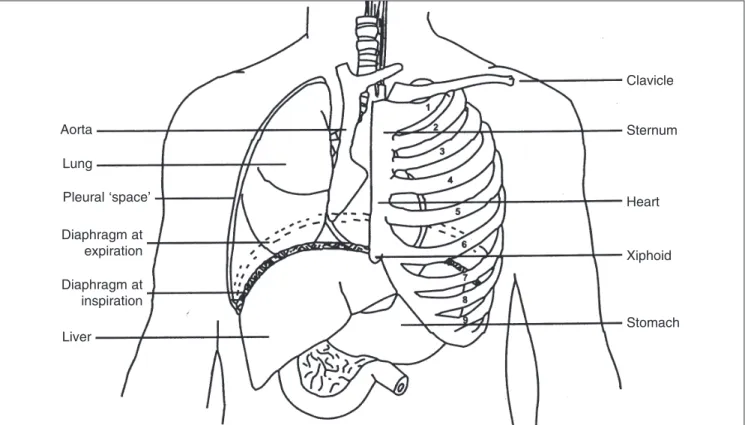
• Breathing and ventilation. Look at the neck to see if the trachea is deviated or the neck veins engorged. Look at the casualty’s chest to see if it is expanding equally and for obvious open chest wounds. If there is compromised ventilation:
What is the reason? Do something about it! Remember that of all those who die from chest injuries, 25% die unnecessarily and 85% of these could be saved by primary care! Then consider:
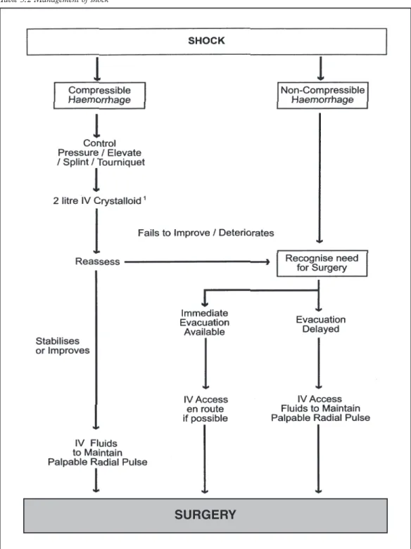
• Circulation and haemorrahge control. Haemorrhage must be arrested if possible and the circulating volume restored to an acceptable level. This applies to casualties with compressible haemorrhage. Uncontrollable (non compressible) haemorrhage requires urgent surgical intervention and a different approach to fluid volume restoration (see paragraph 0527 and Table 5.2). Only now should you consider:
• Disability or neurological status. This is a simple AVPU assessment of the casualty’s
level of consciousness and pupil state.You want to know if the casualty is:
Alert. Voice responsive. Pain responsive. Unresponsive.
You also want to know if the pupils are equal, the pupillary size and if they react to light (indicating Displaced brain). There is not much you can do about the neurological state at this stage other than ensuring cerebral oxygenation and perfusion, but you need to record the findings so that any change can be appreciated later. Remember that the level of consciousness not only refelcts the neurological status, but can be influenced by hypovolaemia and hypoperfusion.
• Exposure. In hospital, this must be total but at Role One and Role Two there may be constraints.
0207. Remember also to speak to the casualty at the very beginning; an alert response can tell you a lot about his respiration and cerebral perfusion. To be able to speak, the casualty must:
• Have a patent airway.
• Have a reasonable tidal volume to phonate.
• Have reasonable cerebral perfusion to comprehend and answer.
Resuscitation
0208. The resuscitation phase is carried out simultaneously with the primary survey, with life-threatening conditions not only identified but managed as they are found.
0209. If available, administer supple-mentary oxygen to all serious casualties with maximum flow rate through a tight-fitting mask and reservoir. Establish and maintain a minimum of two large-calibre intravenous lines; 16 gauge is the smallest adequate size. Assess resuscitation efforts and monitor the casualty by measuring physiological parameters. These include: • Alertness, is it improving or deteriorating?
• Respiratory rate • Pulse rate and rhythm • Pulse pressure
• Capillary refill time
• Blood pressure (presence of radial, femoral, or carotid pulse (See paragraph 0517)
• Urinary output
• Arterial blood gases (if facilities are available)
0210. Consider the insertion of urinary and nasogastric catheters during this phase. Once you have successfully completed the primary survey and resuscitation phases, you can then proceed to the secondary survey.
Secondary Survey
0211. You carry out the secondary survey when the casualty is stable. Remember that casualties have backs and sides as well as fronts; bottoms as well as tops; and lots of holes, both natural and as a result of injury. You must be systematic, going through a top-to-toe process as follows:
• Scalp and vault of skull • Face and base of skull • Neck and cervical spine • Chest
• Abdomen • Pelvis
• Remainder of spine and limbs • Neurological examination
0212. Do not forget the holes. Every orifice merits a finger, a light or a tube.
Definitive Care
0213. In the forward areas, you will rarely be concerned with definitive care. This is more likely to take place in the rear areas. Nevertheless, it is important to realize that definitive care forms the fourth and final phase in BATLS management. It is equally important to remember that if you do not get the primary survey and resuscitation phases correct, definitive care may be in the hands of the War Graves Commission!
Summary
• No matter where you are, remember - as you approach every casualty the following questions should be going through your mind;
• Is the airway patent? • Is the casualty breathing?
• Is there life-threatening external or internal blood loss?
A consistent, systematic approach to the primary survey is vital to the
casualty’s survival.
The BATLS manual is prepared by the BATLS Training Team under the authority of the Professor of Military Surgery who remains responsible for its technical content.
Aim
0301. On successfully completing this topic you will have a sound understanding of how to prioritise casualties for treatment and evacuation, so that the survival of the maximum number is ensured.
Introduction
0302. The management of a single seriously injured casualty in peacetime military or civilian practice is frequently problematic. On the battlefield, problems are compounded by: environment, difficult terrain and tactical constraints. The situation is even more difficult when faced with large numbers of casualties.
0303. If a system for prioritisation of care of the injured is not in place, many salvageable casualties may die unnecessarily. Triage (from the French verb trier, to sieve or to sort), has evolved through military conflicts dating from the Napoleonic Wars to recent civilian disasters.
Definition
0304. The process of triage is complex. The preferred definition is:
Sorting casualties and the assignment of treatment and evacuation priorities to wounded at each role of medical care.
Triage Priorities
0305. There are four triage priorities: • Priority One (P1). Those needing
immediate life-saving resuscitation and/or surgery.
• Priority Two (P2). Those needing early resuscitation and/or surgery, but some delay is acceptable.
• Priority Three (P3). Those who require treatment but where a longer delay is acceptable.
• Dead.
0306. This is the P (Priority) System, of triage. Triage must be repeated at every link of the evacuation chain and the priority adjusted to reflect deterioration or improvement in the casualty’s clinical condition.
Mass Casualties
0307. A mass casualty situation overwhelms the available medical and
CHAPTER 3 TRIAGE logistic capabilities (JSP 110). In thesecircumstances the aim of the medical
services must be to give care to the greatest benefit of the largest number - that is ‘to do
most for the most’.
0308. The term mass casualties is reserved for a situation when medical resources are overwhelmed. When resources are adequate, the incident is said to be
‘compensated’. In a military setting, an ‘uncompensated’ situation may exist temporarily or over a prolonged period. It may be appropriate for the local commander to introduce mass casualty triage without a formal declaration having been made by a higher authority.
0309. The triage system in an uncompensated situation thus becomes; • P1 - Immediate Treatment. Those needing
emergency life-saving treatment. Procedures should not be time consuming and concern only those with a high chance of good quality survival. Examples are remedial airway obstruction, accessible haemorrhage and emergency amputations.
• P2 - Delayed Treatment. Those needing major surgery (after initial sustaining treatment such as intravenous fluids, antibiotics and splinting), or medical treatment, but where conditions permit delay without endangering life. Examples are open fractures of long bones, large joint dislocations and burns covering 15-30% BSA.
• P3 - Minimal Treatment. Those with relatively minor injuries who can effectively take care of themselves or be helped by untrained personnel. Examples are minor lacerations and uncomplicated fractures.
• P1 Hold - Expectant Treatment. Those with serious multiple injuries needing extensive treatment or with a poor chance of survival. These casualties receive appropriate supportive treatment compatible with resources, for example, analgesia. Examples are severe head and spinal injuries, extensive burns and large doses of radiation.
0310. The T (Treatment) System of triage, is an alternative to the P System and is routinely used by the RN, the RAF, J R Army Med Corps 2000; 146: 215-227
BATLS
Battlefield Advanced Trauma Life Support
(BATLS)
BATLS Chapters 3 & 4 216
NATO allies, the International Committee of the Red Cross, civilian ambulance services and in civilian disaster programmes.
0311. The relationship between the two systems is as follows;
• P1 is equivalent to T1 • P2 is equivalent to T2 • P3 is equivalent to T3 • P1 Hold is equivalent to T4 • Dead is still Dead.
Triage for Treatment
0312 A simple, safe, rapid and reproducible system is required that can be applied by any Serviceman with appropriate medical training. Physiological systems that look at the consequences of injury (a change in the vital signs: Respiratory Rate, Pulse Rate and Capillary Refill Time [CRT] are more reliable than anatomical systems (which require extensive clinical knowledge and a need to undress the casualty).
0313. A widely accepted physiological method of triage for treatment is the Triage
Sieve. This involves an assessment of the
casualty’s mobility, then an assessment of
the airway, breathing and circulation (see Table 3.1).
0314. Triage is only a ‘snapshot’ of how the casualty is at the time of assessment. In order to identify changes in the casualty’s condition, the triage sieve must be repeated at each link of the evacuation chain. It is important initially not to try to predict how a casualty may deteriorate, this will lead to over-triage (a higher than necessary triage category) and can overwhelm the system with P1 and P2 casualties.
Triage for Evacuation
0315. Limited time and personnel resources may prohibit a more detailed triage assessment other than that given by the triage sieve. When possible, the Triage
Sort can be used to refine triage sieve decisions. Triage sort uses the respiratory rate, systolic blood pressure and Glasgow Coma Scale, to numerically score the casualty from 0 to 12 and give an indication of priority for evacuation and/or the need for further intervention. This score has a proven direct relationship to outcome from severe injury.
Table 3.2 Triage sort coded values
Physiological variable Measured value Score
Respiratory rate 10-30 4
>30 3
6-9 2
1-5 1
0 0
Systolic blood pressure >90 4
89-76 3
75-50 2
<49 1
0 0
Glasgow Coma Scale 15-13 4
12-9 3
8-6 2
5-4 1
3 0
0316. Priorities are assigned as follows:
• P1 (T1) 1-103
• P2 (T2) 11
• P3 (T3) 12
• P1 Hold (T4) 1-33
• Dead 0
0317. The coded values for the Triage Sort are given in Table 3.2. After coding each of the three parameters, add them together to give a score ranging from 0 (dead) to 12 (physiologically normal).
0318. Evacuation will be delayed when the number of casualties outstrips available transport. In this situation, the greater time spent with the casualty will allow additional anatomical assessment of injuries. Where the priority determined by physiology does not match the anatomical severity of injuries, the priority can be upgraded.
Example: A soldier loses his left leg in a
landmine incident. Immediate first aid is effective in stopping haemorrhage. He is
Now TREAT the injured
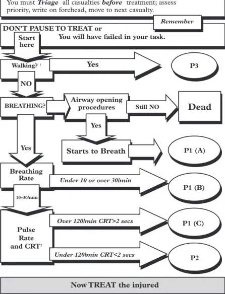
Table 3.1 The triage sieve
1 It must also be realised that some in this group, despite being ambulatory, may have injuries of sufficient magnitude to cause a clinical deterioration requiring a change in priority. 2 Is unreliable in the cold or dark.
3. The overlap in scores allows for the seriously injured to be placed in either category, depending on number of casualties and resources available for evacuation.
Dead
You must Triage all casualties before treatment; assesspriority, write on forehead, move to next casualty.
DON’T PAUSE TO TREAT or
You will have failed in your task. Start here Remember Walking? 1 BREATHING? NO Yes Yes Yes Breathing Rate Pulse Rate and CRT2 10-30/min P3 Airway opening procedures Starts to Breath
Under 10 or over 30/min
Over 120/min CRT>2 secs
Under 120/min CRT<2 secs
P1 (A)
P1 (B)
P1 (C)
P2
transported to the RAP. He cannot walk, his respiratory rate is 22 and his pulse is 110/minute. He is triaged P2 for treatment (Triage Sieve). He then receives intravenous fluids and analgesia. His systolic BP is 115 mmHg, his respiratory rate is 20, he is fully alert, with a GCS of 15. He scores 12 on his Triage Sort, which is P3 for evacuation. Clearly, he requires early surgical treatment and the RMO upgrades his priority to P2 for evacuation to the field hospital.
0319. To help understand priority allocations for evacuation, it is appropriate to consider the standard casualty evacuation chain which is usually through the logistical Lines of Support. These lines of support relate to the Combat Services Support (CSS) provided at levels of operational deployment, that is, First Line at unit level, Second Line at brigade or divisional level, Third Line between the divisional rear boundary and point of entry, and Fourth Line at the base4. These should not be
confused with Roles of Medical Support which is the term used throughout NATO to define the levels of medical capability5.
• Role 1 Treatment to restore and stabilise vital functions. (Regimental Aid Post/Medical Section(s)). • Role 2 Resuscitation and stabilising
treatment - may include stabilising (Damage Control) surgery (Dressing Station).
• Role 3 Hospitalisation and life-saving surgery, definitive surgery (Field Hospital).
• Role 4 Time consuming specialist and long term treatment (NHS Hospital).
0320. It should be assumed that early surgery takes place at the field hospital, with subsequent care taking place back in the United Kingdom, within the National Health Service. There will be occasions when surgery, through a field surgical team being attached, is available forward of field hospitals. This will be the norm in airborne and airmobile operations and when FIRST 6teams are attached to Close
Support Squadrons of Medical Regiments. Sepcialist teams, such as burns teams and head and neck teams, can be allocated to selected field hospitals: this will influence the disposal and transfer of candidate casualties.
0321. United Kingdom military operations are increasingly Joint Service in nature, for example, surgical support at Role Three may be found from the RN in the form of a Primary Casualty Receiving Ship (PCRS). An intermediate hospital may be set up, at a convenient location on either the tactical or strategic LOCs.
Application at Role One and
Role Two
0322. For the regimental medical officer (RMO) and the dressing station medical officer, casualties are likely to be graded for treatment and/or evacuation as follows: • P1
Airway:
Obstruction including that due to maxillofacial injury. Airway burns with the potential for obstruction.Breathing:
Tension pneumothorax - openchest wound - flail chest/pulmonary contusion. Respiratory rate <10>30. Triage Sort Score 1-10.
Circulation: Major haemorrhage - external
or internal - compressible or non compressible. CRT >2S. Pulse rate >120/mm. Massive muscle injuries. Multiple fractures and/or multiple wounds. Burns between 15 and 30% Body Surface Area (BSA).
• P2 Lesser visceral injuries. Vascular injuries not having features of P1. Cerebral injuries that are deteriorating (See table 8.2). Burns of less than 15% BSA involving face, eyelids, hands, perineum and across joints (see paragraph
1222). Large joint
dislocations.
• P3 Lesser fractures and
dislocations. Lesser soft tissue injuries ultimately requiring surgery. Maxillofacial injuries with no airway problems. Eye injuries. Other burns less than 15% BSA.
0323. Expectant (P1 Hold [T4]) cases would include, for example, burns of greater than 30% BSA, gunshot wounds to the brain and other injuries with a poor prognosis.
0324. Medical cases are categorised in exactly the same way in relation to their need for resuscitation and timely intervention by a physician. Psychiatric cases invariably tend to fit into the P3 bracket, with the particular caveat they should be treated as far forward as possible; this approach will result in the maximum number being rendered fit and returned to duty. The further rearward a psychiatric case is evacuated, the less likely this is to happen.
Application at Role Three
0325. The critical decision at the field hospital is whether the casualty needs resuscitation and surgery now, or whether
217 BATLS Chapters 3 & 4
4. See Army Doctrinal Publication Vol 3 Logistics (Army Code 71566). 5. For example, there will be Role 1 and Role 2 medical units at Third Line. 6. Forward Immediate Resuscitation and Surgery Teams.
BATLS Chapters 3 & 4 218
he can withstand further delay. The identification of casualties requiring expert treatment by specialist teams must also be considered.
0326. At this level, in the presence of mass casualties, the P1 Hold (T4) category will represent:
• Cases whose survival is uncertain. • Cases who require prolonged surgery,
who must wait until time and facilities are available.
• Major burns cases who are kept for 48 hours prior to transfer to a specialist burns team.
0327. When there are many casualties, the expedient of the greatest good of the greatest number must prevail.
Summary
• Triage is the sorting of casualties into orders of priority for treatment and avacuation. The triage process is dynamic and needs reassessment throughout the casualty evacuation chain. It will be coloured by doctrinal and organisational factors which affect time between and location of, medical echelons in the chain. Even when faced with large numbers of casualties, the A B C routine must be followed in order to identify life-threatening problems and indicate priorities.
• The principles of management of the injured remain as primary survey, resuscitation, secondary survey and definitive care, albeit that the last two may be carried out at a more rearward echelon. The philosophy of treatment for large numbers of casualties is:
• To evacuate rearwards all those who can withstand the journey.
• To address the medical resources towards those who have the best chance of survival.
This is achieved through effective and efficient triage.
Aim
0401. On successfully completing this topic you will be able to:
• Recognize those conditions causing airway and breathing difficulty in the battlefield casualty.
• Discuss the principles of airway and ventilatory management.
• Demonstrate basic and advanced methods of airway management.
Anatomy and Physiology of the
Airway
0402.The upper airway consists of the nose, mouth and pharynx. The lower airway
CHAPTER 4 AIRWAY MANAGEMENT AND VENTILATION
consists of the larynx, trachea and lungs. An open airway allows air (containing oxygen) to enter the lungs, oxygenate the blood, which is then carried to the tissues. A reduction in oxygen levels in the blood or tissues, is termed hypoxia. Tissue hypoxia causes cell damage and, if prolonged, can lead to organ failure and death. Some organs are more sensitive to hypoxia than others. For example, cerebral hypoxia even for a short period of time will cause agitation, then a decreased level of consciousness and eventually, irreversible or fatal brain damage. 0403. Carbon dioxide is produced by cellular metabolism and carried in the blood to the lungs; it is then exhaled. If there is airway obstruction, there is a build up of carbon dioxide in the blood (hypercarbia). This causes drowsiness.
Supplementary oxygen must be administered to all seriously injured battle casualties as early as possible in
the evacuation chain.
0404. Obstruction to either the upper or lower airway can compromise ventilation of the lungs and quickly result in cerebral hypoxia. Management of the upper and lower
airway is the first concern in any battlefield
casualty.
The airway must be opened, maintained and protected, with ventilatory support provided if necessary.
Airway and Breathing
0405. Early preventable deaths from airway problems after injury are frequently due to:
• Failure to recognize the urgent need for intervention in casualties with a compromised airway.
• Limited experience in airway clearing skills.
• Faulty judgement in selecting the correct airway manoeuvre.
• Failure to secure the airway prior to evacuation.
• Becoming distracted by less urgent problems.
Awareness
0406. Particular problems that will endanger the airway:
• Head injury with decreased level of consciousness (see paragraph 0822). • Other causes of decreased level of
consciousness (poisoning, alcohol, low oxygen, carbon monoxide).
• Maxillofacial injuries:
• Mid face fractures can move backwards and block the airway. • Mandible fractures can allow the
tongue to fall backwards.
• Bleeding and secretions caused by these injuries can block the airway. • Injuries to the neck:
• Direct trauma to the larynx and supporting structures.
• Bleeding inside the neck
compressing the hypopharynx or trachea.
• Burns to the face and neck:
• Swelling of the upper and lower airway due to direct burns or inhaling hot smoke, gases or steam, will cause airway obstruction. 0407. Airway problems may be:
• Immediate (block the airway quickly) or, • Delayed (come on after a time delay
-minutes or hours) or,
• Deteriorate with time; this is often insidious because of its slow progression and is easily overlooked.
0408. An airway that has been cleared may obstruct again if the casualty’s level of consciousness decreases, there is further bleeding into the airway or there is increasing swelling in or around the airway.
Recognition
0409. Talk to the casualty! Failure to
respond implies an altered level of consciousness with the potential for airway compromise. A positive appropriate reply in a normal voice indicates that the airway is patent, breathing normal and brain perfusion adequate. Any inappropriate or incomprehensible response may suggest airway or breathing compromise, or both.
0410. Look to see if the casualty is agitated, drowsy or cyanosed. The absence of cyanosis does not mean the casualty is adequately oxygenated.
Remember that a casualty who refuses to lie down quietly may be trying to sit up in an attempt to keep his airway open and/or his breathing adequate.
0411. Listen for abnormal sounds. Snoring, gurgling and gargling sounds are associated with partial obstruction of the pharynx. Hoarseness implies laryngeal injury. The abusive casualty may be hypoxic and should not be presumed to be merely insubordinate or intoxicated. Total obstruction equals total silence!
0412. Feel for air movement on expiration and check if the trachea is in the midline.
Management
0413. Management comprises: • Clearing the obstructed airway. • Maintaining the intact airway. • Protecting the airway at risk.
0414. Techniques for clearing, maintaining and protecting the airway need to be modified in the trauma casualty when cervical spine injury is suspected or present. Cervical spine injury is suspected:
• In falls from a height. • In vehicle collisions.
• When pedestrians have been hit by a vehicle.
• Where casualties have been thrown by explosions.
• In the unconscious casualty (especially where there is blunt injury above the clavicle).
• In the conscious casualty complaining of neck pain or loss of sensation or motor function in one or both arms. (see paragraph 1002-1006).
0415. Moving a casualty with bony spinal injury risks damaging the spinal cord. Ideally, these casualties should only be moved when the appropriate spinal immobilization devices are in place. (see paragraph 1007).
In situations where there is danger to the casualty or rescuer, rapid extraction of the casualty using improvised immobilisation or none at
all, will be needed.
Priority is CLEAR THE AIRWAY but stay safe.
0416. Penetrating missile neck wounds that directly involve the bony cervical spine or spinal cord carry a 95% mortality. They can be ignored in terms of cervical spine protection; get on and manage the airway! A combination of blunt and penetrating neck injuries should be managed as for blunt injury.
Clearing the airway
0417. In the casualty with suspected cervical spine injury, manual inline immobilization of the cervical spine and airway clearance are carried out together. In a casualty with an altered level of consciousness, the tongue falls backwards and obstructs the hypopharynx. This obstruction can be readily corrected by the
jaw-thrust or chin-lift manoeuvres. Blood and
debris can be cleared by suction and finger sweeps.
219 BATLS Chapters 3 & 4
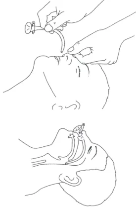
0418. Jaw-thrust. Grasp the angles of the mandible, one hand on each side and move the mandible forward. The jaw-thrust is used for the injured casualty because it does not destabilise a possible cervical spine fracture and risk converting a fracture without spinal cord injury to one with spinal cord injury. This maneouvre will open 95% Fig 4.1 Jaw-thrust Fig 4.2 Chin-lift
BATLS Chapters 3 & 4 220
of obstructed upper airways. The jaw-thrust is illustrated at Fig 4.1.
0419. Chin-lift. Place the fingers of one hand under the chin and gently lift it upwards to bring the chin anteriorly. To open the mouth, use the thumb of the same hand to depress the lower lip slightly. The thumb may also be placed behind the lower incisors and, simultaneously, the chin gently lifted.This will open the upper airway in 70-80% of casualties. The chin-lift is illustrated at Fig 4.2.
Make sure you do not hyperextend the neck.
0420. Suction. Remove blood and secretions from the oropharynx with a rigid suction device (for example, a Yankauer sucker). If there is bleeding at the external nares clear this with suction. A casualty with facial injuries may also have a cribriform plate fracture; this means that suction catheters should not be inserted through the nose as they could enter the skull and injure the brain.
0421. Finger sweeps. Finger sweeps into the back of the mouth and pharynx may be useful for dislodging foreign bodies, particularly if no suction device is available.
Make sure that your fingers do not inadvertently push foreign bodies
further into the airway.
0422. Displacement of fractured maxilla or
mandible. Where airway obstruction results
from a fractured maxilla, insert the index or middle finger or both, through the mouth behind the hard palate and pull it forward to disimpact the displaced bone. When an anterior mandibular fracture allows the tongue to fall back into the hypopharynx you may have to pull the mandible forward manually.
Maintaining the airway
0423. How to maintain the casualty’s airway will depend on:
• The casualty’s injuries.
• The casualty’s level of consciousness. • The equipment available.
0424. Clearing the casualty’s airway may result in his level of consciousness improving and being able to maintain his own airway.
0425. If the casualty cannot maintain his own airway you (or an assistant) need to
continue with the jaw-thrust or chin-lift or try using an oropharyngeal airway or nasopharyngeal airway.
0426. Oropharyngeal airway. The oropharyngeal airway (Guedel type) is inserted into the casualty’s mouth over the tongue. It stops the tongue falling back and provides a clear passage for air flow. The preferred method is to insert the airway concavity upwards until the tip reaches the soft palate and then rotate it 180°, slipping it into place over the tongue (see Fig 4.3).
Make sure that the airway does not push the tongue backwards as this will block rather than open the airway.
0427. A casualty with a gag reflex may reject the oral airway. If so, move on to the nasopharyngeal airway.
0428. Nasopharyngeal airway. A
nasopharyngeal airway can be used when
Fig 4.3 Nasopharyngeal airway
there is oral injury, a fractured mandible or massetter spasm. It is better tolerated than the Guedel by the more responsive casualty and is less likely to be dislodged during evacuation (see Fig 4.4). A suspected fractured base of skull is not a contraindication for use of this airway if an oropharyngeal airway cannot be inserted.
0429. Lubricate the airway and insert it through either nostril, straight backwards -not upwards - so that its tip enters the hypopharynx. A safety pin should be applied across the proximal end before insertion to prevent the tube disappearing into the casualty’s airway. If this happens it could compromise rather than maintain the airway. A complication of inserting the Fig 4.4 Nasopharyngeal airway
the lower airway. • To protect the airway from:
• Obstruction due to swelling. • Aspiration of gastric contents or
other fluid. (Note: A cuffed endotracheal tube or cuffed surgical airway maintains a clear passage and the cuff provides a seal). • To allow accurate control of oxygenation
and ventilation.
• As part of head injury management by helping to control oxygen and carbon dioxide levels in cerebral blood flow (see Chapter 8):
• In the management of some chest injuries.
• In surgery and anasthesia.
DEFINITIVE AIRWAY This is a cuffed tube placed in the trachea by the surgical route or by
endotracheal intubation.
SURGICAL AIRWAY
0434. A surgical airway is used when: • A casualty needing a definitive airway forresuscitation or evacuation is too awake to tolerate endotracheal intubation without the use of anaesthetic drugs.
• Trauma to the face and neck make endotracheal intubation impossible. • A casualty with face and neck burns
requires airway protection to pre-empt delayed obstruction but expert anaesthetic help is unavailable to carry out endotracheal intubation.
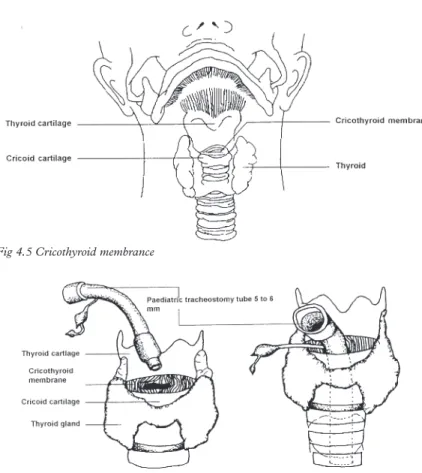
Surgical cricothyroidotomy
9435. Surgical cricothyroidotomy places a tube into the trachea via the cricothyroid membrane (See fig 4.5). A small tracheostomy tube (5-7 mm) is suitable. This will be practised in skill station 2 and is illustrated in Fig 4.6. During the procedure, appropriate cervical spine protection must be maintained when indicated. There are also commercially available cricothyroidotomy sets in use with some NATO armies. A cricothyroidotomy can be replaced by a formal tracheostomy (if needed) at a later time.
Emergency tracheostomy
0436. A formal surgical tracheostomy takes longer and is more difficult, than a surgical cricothyroidotomy. Commercial sets are available for rapid tracheostomy using a Seldinger (guide wire) technique.
Endotracheal intubation
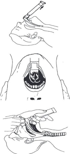
0437. This technique uses a laryngoscope to visualise the vocal cords. A cuffed endotracheal tube is placed through the vocal cords into the trachea. This skill is illustrated at Fig 4.7 [shown in Skill Station 2] and will be practised in the Skill Station.
221 BATLS Chapters 3 & 4
nasopharyngeal airway is bleeding from the nose. Gentle insertion, good lubrication and using an airway that passes easily into the nose will decrease the incidence of bleeding.
The oropharyngeal and nasopharyngeal devices MAINTAIN the airway but do not protect it from
aspiration.
0430. A casualty whose airway has been cleared and maintained by the techniques described above may need help with their breathing if this is depressed due to injuries or toxic agents.
0431. Ventilation is covered at paragraphs 0446-0451 and is practised in Skill Station 1.
Protecting the airway at risk - advanced airway techniques
0432. Advanced airway techniques include: • Surgical cricothyroidotomy.
• Surgical tracheostomy. • Endotracheal intubation.
0433. Indications for performing advanced airway techniques are:-
• Inability to clear or maintain an airway using simple manoeuvres and airways in, for example:
• Injury around the face.
• Face and airway burns (causing swelling of the airway, see paragraphs 0406 and 1213). • An obstruction in the upper airway
that cannot be removed and needs to be by-passed.
• Neck injury or swelling blocking
Fig 4.5 Cricothyroid membrance
BATLS Chapters 3 & 4 222
0438. At Role 1 and Role 2, in the absence of anaesthetic skills and drugs, endotracheal intubation will only be achieved in deeply unconscious casualties (GCS 4 or less). Attempting intubation in the semiconscious casualty will result in gagging on the laryngoscope and coughing on the endotracheal tube. In casualties with raised intracranial pressure this will increase the pressure even further and worsen the situation. If the casualty gags or coughs, stop and try another method of airway management.
Note: This presents a dilemma with head
injuries in coma (GCS 8 or less). Ideally these casualties should be intubated and ventilated to prevent cerebral hypoxia and hypercabia worsening. In many of them, attempting to do this without the aid of anaesthetic drugs worsens the situation by raising ICP. A surgical airway with ventilation and oxygenation my be required. 0439. In casualties who are not deeply unconscious, maintain the airway with simple techniques. If protection is necessary, insert a surgical cricothyroidotomy under local anaesthesia.
0440. Where anaesthetic skills and drugs are available or can be brought to the casualty by an Incident Response Team (IRT), endotracheal intubation can be achieved using:
• Rapid sequence induction of anaesthesia with:
• Application of cricoid pressure (Sellick’s manoeuvre) and,
• Maintenance of cervical spine immobilisation when indicated.
0441. Intermittent oxygenation during
difficult intubation. Intubation of the hypoventilating or apnoeic casualty may require several attempts and even then, may not be successful. If oxygen is available, you must avoid prolonged efforts to intubate without intermittently oxygenating and ventilating the casualty. You should practise taking a deep breath when starting an attempt at intubation; if you have to take further breath before successfully intubating the casualty, abort the attempt.
0442. Correct placement of the endotracheal tube is checked in Skill Station 2. The main points are:
• See if the endotracheal tube has passed between the vocal cords. Remember to maintain immobilisation of the cervical spine when indicated.
• Listen on both sides in the mid-axillary line for equal breath sounds.
• Listen over the stomach for gurgling sounds during assisted ventilation for evidence of oesophageal intubation. • Feel and listen for air movement at the
proximal end of the tube if the casualty is breathing spontaneously.
• Monitor end-tidal carbon dioxide levels if equipment is available.
• If in doubt about the position of the endotracheal tube, take it out and oxygenate the casualty by another method.
It is failure to oxygenate the casualty that kills, not inability to intubate.
If you need to take over care of an intubated casualty make sure the endotracheal tube is in the correct place
and has not moved during evacuation or transfer of the casualty.
Note: Other methods for maintaining the
airways. Both the laryngeal mask airway and combitubes have been used in civilian practice to manage the trauma casualty’s airway. Their role in military trauma has still to be evaluated.
Oxygenation and Assisted
Ventilation
Oxygenation
0443. The primary goal in providing supplementary oxygen is to maximise the delivery of oxygen to the cells. This is done by providing the highest possible oxygen concentration to the lungs using high flow oxygen at 10-15 litres per minute, as soon as oxygen is available. A disposable face mask without a reservoir bag can deliver 35-60% oxygen, depending on type of mask and oxygen flow. A face mask with an oxygen reservoir can be used to deliver up to 85% oxygen. A correctly fitting bag-valve-mask system with a reservoir, can be used to deliver up to 100% oxygen to the lungs.
Ventilation
0444. Spontaneous ventilation (self ventilation) means the same as breathing. Assisted (artificial) ventilation means the casualty is receiving help with breathing. The aim is to improve gaseous exchange in the lungs and to breathe for the casualty if spontaneous ventilation has stopped or is inadequate. Indications for assisted ventilation include:
• Head injury. • Chest injury.
• Respiratory depression due to drugs (such as nerve agents and opiates). 0445. Assisted ventilation can be achieved by the following techniques:
• Mouth to mouth (or nose). • Mouth to mask.
• Bag-valve-mask.
• Bag-valve-endotracheal tube or surgical airway.
• Automatic ventilation (used by specialist resuscitation teams in field hospitals for prolonged casualty ventilation).
0446. Assisted ventilation is described in Skill Station 1 and will be practiced there.
0447. Mouth to mouth (one man)
valve and oxygen inlet is not available, you can give assisted ventilation by removing the mask from the bag-valve-mask device and blowing into the connecting port.
Note: Do not use the mouth to mask
technique if there is a chemical agent vapour
hazard either in the environment or on the
casualty.
0448. Bag-valve-mask. This technique can be performed one or two handed and is best performed with an oral or nasal airway in place. 0449. Bag-valve-endotracheal tube/surgical
airway. The technique you will use will
depend on the circumstances and equipment available. If a manual bag-valve-endotracheal tube/surgical airway technique is used, the bag is squeezed to achieve obvious chest movement at a rate of approximately 12-15 breaths per minute, that is, one second squeeze - three to four seconds release. An oxygen source and a reservoir should be attached to the bag as soon as they are available.
Evacuation of Airway
Compromised Casualties
0450. Casualties who have lost their normal protective airway reflexes are in danger of aspirating gastric contents, blood and debris and developing airway obstruction; they become hypoxic.
Caring for and monitoring a casualty in the back of military vehicles or helicopters can be difficult, especially in low light conditions. A balance has to be made between:
• The need to move the casualty.
• The safety of the casualty during evacuation.
• The resources available to move the casualty.
• Distance for evacuation. • The tactical situation.
If available, seek specialist advice from hospital teams, aeromed teams
or incident response teams.
The casualty with a definitive airway in place 0451. Best practice is that they are evacuated by trained personnel with appropriate anaesthetic and intensive care skills and electronic monitors. If there is concern about the ability to care for an intubated casualty (for example, replacing a displaced endotracheal tube) during evacuation, consider providing a definitive surgical airway. In casualties being evacuated by air, inflate the cuff with saline; air expands at altitude.
The casualty at risk from aspiration but a definitive airway cannot be provided
0452. Maintain the airway with a Guedel or nasopharyngeal airway and transfer the casualty in the lateral position with an escort who has oxygen, suction and can care for the casualty en route. Continue cervical
spine immobilization as can best be managed but airway management takes precedence.
Unconscious casualties without a definitive airway must not be transferred lying on their backs.
The casualty at risk from airway blocking during transfer.
0453. Where the casualty’s airway is at risk from blocking during transfer, for example, airway burns:
• Get specialist advice.
• Consider providing a surgical airway, or endotracheal intubation if anaesthetic expertise is to hand.
Note: On balance, a surgical airway is the
method of choice. It may be difficult in the extreme incising through burnt skin. Aim not to go through burnt skin but, your prime concern is to protect the airway during casualty transfer!
Summary
• Airway obstruction must be recognised and relieved quickly.
• Beware of cervical spine injury during airway management.
• Start with simple techniques such as jaw-thrust/chin-lift/oropharyngeal suction. • Try Guedel or nasopharyngeal airways. • Give high flow oxygen from a mask with a
reservoir.
• Definitive airways protect against aspiration of gastric contents and blood. • Definitive airways include:
• Surgical cricothyroidotomy. • Endotracheal intubation.
Note: Choice of definitive airway will
depend on skills and equipment available: • Casualties with inadequate respiration
will need assisted ventilation.
• This can be done by mouth, by bag and mask or by automatic ventilators.
Aim
The aim of these skills stations is to demonstrate and practise basic and advanced airway management. You will also discuss the indications for oral endotracheal intubation and associated complications.
On successfully completing these stations, you will be proficient in:
• Basic airway clearing techniques. • Oropharyngeal airway insertion. • Nasopharyngeal airway insertion. • Ventilation without intubation. • Orotracheal intubation.
SKILLS STATIONS:
1. Basic Airway Management and Ventilation 2. Advanced Airway Management: Adult
Orotracheal Intubation
BATLS Chapters 3 & 4 224
Equipment
Airway management in the casualty with a fractured maxilla.• Using the Mr Hurt manikin with maxillary fractures, practice pulling the fracture forward to clear the airway.
Airway management in the casualty whose head is not in the neutral position.
• Gently move the casualty’s head into the neutral position if the airway cannot be cleared and maintained in the non-neutral position.
• Stop moving the head if:
• There is resistance to movement. • The casualty complains of pain. Airway management in the combative casualty who will not accept sand bags and tape
• Compromise by using a semi-rigid cervical collar only.
Oropharyngeal Airway Insertion
• Select the correct sized airway. Place the airway against the casualty’s face. The correct sized airway will extend from the centre of the casualty’s mouth to the angle of the jaw.• Open the casualty’s mouth with the chin-lift manoeuvre.
• Insert the tip of the airway along the roof of the mouth to the soft palate (see fig 4.3).
• Rotate the airway 180°, directing the concavity of the airway towards the feet and slip the airway over the tongue. • If necessary, ventilate the casualty with
mouth-to-mask or bag-valve-mask technique.
Nasopharyngeal Airway
Insertion
• Assess the nasal passages for any apparent obstruction (fractures, haemorrhage, polyps). Choose a nostril that is patent. • Select the correct size airway. Size 7 for
the adult female and size 8 for an adult male.
• Insert the safety pin across the nostril end of the airway.
• Lubricate the nasopharyngeal airway with a water-soluble lubricant or water. • Insert the tip of the airway into the nostril
and direct it posteriorly and towards the ear lobe.
• Gently slide the nasopharyngeal airway through the nostril into the hypopharynx with a slight rotating motion until the flange rests against the nostril.
• If an obstruction is encountered try the other nostril or try a smaller nasopharyngeal airway. Trying to force the
nasopharyngeal airway past an obstruction may cause severe bleeding.
• If necessary, ventilate the casualty with mouth-to-mouth or bag-valve-mask technique.
Aim
This skill station allows you to practice basic airway techniques and develop a system for airway management.
The order for performing tasks will depend on the situation, the personnel available and their skills. Some tasks will be done simultaneously if more than one person is available.
This skill station assumes there is an assistant to help you.
Sequence of actions:
• Approach and reassure casualty. React to the casualty’s response.
• Provide manual inline immobilisation of the cervical spine when indicated. • Hand over manual inline immobilisation
to your assistant (get them to place their hands over yours then gently remove your hands as they apply immobilisation). If assistance is not available your sole aim is to clear the airway.
• Clear the airway using:
• Finger sweeps to remove solid debris (if safe to do so). • Magill forceps to remove solid
debris.
• Yankauer sucker to remove blood and fluid from the oropharynx. • Jaw-thrust.
• Chin-lift.
• Provide high flow oxygen from a mask with a reservoir bag.
• Decide if the casualty needs an oropharyngeal or nasopharyngeal airway.
SKILLS STATIONS 1
BASIC AIRWAY MANAGEMENT AND VENTILATION
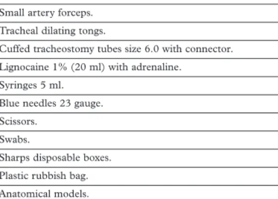
Adult intubation manikins.
Adult endotracheal tubes size 7.0, 8.0 and 9.0. Laryngoscope handles.
Laryngoscope blades, adult, curved. Extra batteries for laryngoscope handles. Extra laryngoscope bulbs.
Stethoscopes.
Lubricant (for example, silicone spray that accompanies intubation manikin). Semi-rigid cervical collar applied to one adult intubation manikin, or sandbags. Magill’s forceps.
Malleable endotracheal introducers. Oropharyngeal airways size 2, 3 and 4. Nasopharyngeal airways size 6.0 and 7.0. Bag-valve-mask devices.
Pocket face masks (with a one-way valve to prevent back-flow of air and secretions). Rigid suction devices (Yankauer sucker).
Safety pins. Scissors.
Ventilation Without Intubation
Mouth-to-mask ventilation, adult (one man technique)• Attach oxygen tubing to the Laerdal pocket mask.
• Adjust the oxygen flow rate to 10 litres per minute.
• If there is no oxygen inlet, place the oxygen tubing under the side of the mask. • Insert an oropharyngeal airway.
• Apply the face mask to the casualty using both hands.
• Open the airway using the jaw-thrust manoeuvre, three fingers on the underside of the jaw.
• Take a deep breath and place your mouth over the mouth port and blow.
• Assess the ventilatory efforts by observing the casualty’s chest movement. • Ventilate the casualty in this manner
every five seconds.
Bag-valve-mask ventilation, adult (two man technique)
• Select the correct sized mask to fit the casualty’s face.
• Connect the oxygen tubing to the bag-valve device and adjust the flow of oxygen to 10 litres per minute.
• Ensure the patency of the casualty’s airway.
• The first person applies the mask to the casualty’s face, making a tight seal with both hands.
• The second person ventilates the casualty by squeezing the bag with both hands. • Assess the adequacy of ventilation by
observing the casualty’s chest movement. • Ventilate the casualty in this manner
every five seconds.
Bag-valve-mask ventilation, adult (one man technique)
• Follow the first three steps for the two man technique.
• Use one hand to apply the mask to the casualty’s face. Use the index finger and thumb to hold the mask in place and use the other fingers to perform a jaw-thrust.• Squeeze the bag with the other hand and ventilate as before.
Aim
This skill station allows you to practice adult orotracheal intubation (see Fig 4.7). It also helps you to develop systems to substitute manual immobilization by using a cervical collar, sandbags and tape, and check that the endotracheal tube is correctly placed.
Sequence of actions:
SKILLS STATIONS 2
ADVANCED AIRWAY MANAGEMENT ADULT OROTRACHEAL INTUBATION
• Ensure that adequate ventilation and oxygenation are in progress.
• Connect the laryngoscope blade and handle; check the bulb for brightness, also check both small and larger endotracheal tubes are to hand, their cuffs are working and the suction is working.
• If indicated, have an assistant manually immobilize the head and neck.
• If indicated, have an assistant apply cricoid pressure.
• Hold the laryngoscope in the left hand. • Insert the laryngoscope into the right side
of the casualty’s mouth, displacing the tongue to the left.
• Look for the epiglottis and place the tip of the blade in the vallecula. Lift the epiglottis forward by pulling the handle of the laryngoscope forward and visualize the vocal cords.
• Gently insert the endotracheal tube into the trachea without applying pressure to the teeth or oral tissues.
• Check the placement of the endotracheal tube by bag-valve tube ventilation. • Inflate the cuff with enough air to provide
a gas-tight seal.
225 BATLS Chapters 3 & 4