HAL Id: tel-03194990
https://tel.archives-ouvertes.fr/tel-03194990
Submitted on 10 Apr 2021
HAL is a multi-disciplinary open access archive for the deposit and dissemination of sci-entific research documents, whether they are pub-lished or not. The documents may come from teaching and research institutions in France or abroad, or from public or private research centers.
L’archive ouverte pluridisciplinaire HAL, est destinée au dépôt et à la diffusion de documents scientifiques de niveau recherche, publiés ou non, émanant des établissements d’enseignement et de recherche français ou étrangers, des laboratoires publics ou privés.
Combining a real-time closed-loop system with
neuromodulation : an integrative approach to prevent
pathological repetitive behaviors
Sirenia Lizbeth Mondragon Gonzalez
To cite this version:
Sirenia Lizbeth Mondragon Gonzalez. Combining a real-time closed-loop system with neuromodula-tion : an integrative approach to prevent pathological repetitive behaviors. Neurons and Cognineuromodula-tion [q-bio.NC]. Sorbonne Université, 2019. English. �NNT : 2019SORUS259�. �tel-03194990�
Sorbonne Université
Ecole doctorale 391 - Sciences mécaniques, acoustique, électronique et
robotique de Paris (SMAER)
Institut du Cerveau et de la Moelle Epinière / Neurophysiology of Repetitive Behaviors
Combining a real-time closed-loop system with
neuromodulation
An integrative approach to prevent pathological repetitive
behaviors
Par Sirenia Lizbeth Mondragón González
Thèse de doctorat
Dirigée par Eric Burguière et Jean-Luc Zarader
Présentée et soutenue publiquement le 08/04/2019
Devant un jury composé de :
Burguière Eric (CR1) Directeur de thèse
Zarader Jean-Luc (Professeur Universitaire) Co-directeur de thèse
Welter Marie-Laure (PU-PH CHU) Rapportrice
Willuhn Ingo (Professeur associé et leader de groupe) Rapporteur
Arleo Angelo (DR2) Président du jury
A
CKNOWLEDGMENTS
First, I would like to thank the members of my committee, Dr. Marie-Laure Welter and Dr. Ingo Willuhn for kindly accepting to review my thesis manuscript and to Dr. Elodie Fino and to Dr. Angelo Arleo for accepting to evaluate my work.
I would like to extend deep gratitude to my advisor Eric Burguière. Eric, I am glad to be your first PhD student, I truly consider myself lucky to have worked under your supervision. These have been some remarkably interesting and exciting years. I have learned so much thanks to our (sometimes super) long conversations. I am extremely grateful to you for introducing me to the wild word of extracellular electrophysiology. For always being open minded but with a critic eye to my ideas, I know that you trusted me in so many ways for the experiments, for this and more I am incredibly grateful to you.
I owe thanks to my co-advisor Jean-Luc Zarader, I know that you always made our appointments a priority in your busy schedule, we had very stimulating discussions when we were trying to find out how to attack the pattern recognition problem, I remember finishing our discussion with so much motivation and excitement to implement a new approach, and I am grateful for that.
A big thanks to Christiane Schreiweis, not only did I learn a lot about mice with you but also you provided invaluable help for the animal preparations in Chapter 6. Thank you for the not so easy and long surgery sessions.
To Pauline and Nabil, I am glad we did his journey at the same time, we have had some good laughs and I am happy that we shared these last years together. A special thanks to Marine, for all the conversations (and free chouquettes) and for kindly helping me take care of my mice when I could not.
Thanks to everyone in the now NERB team of the ICM for their hospitality when I first joined the lab. To the first members of the BEBG team, to Luc, Margot, Jérôme, Karim, Philippe, and Christian, for making the lab a lovely place to be in.
Thanks to the current graduate students, Lindsay, Oriana, Eliana, and Sami for the encouraging words while writing my manuscript. To the old interns Celine and Maeva that regularly asked for news on my thesis.
All the mice without whom none of this work would have been possible.
To my dear friends Jean-Luc, Cri cri, Oscardo and Sandy, and to my Cinves girlfriends for helping me forget my project when it was necessary.
To Claude and Christian for always worrying about me and the advancement of my project, I really appreciated it.
To my parents, who have always supported me in all my decisions and provided unlimited encouragement. To my “little” brothers, Ricardo who did his PhD at the same time, it has been fun to share our lab adventures. To my little brother Victor who has always helped me to side the bright side of all situations.
To my grand mother Maria, who I know would have been proud, I dedicate this manuscript to you.
And finally, to my brilliant husband. Vince, you probably do not realize how much your never-ending positive attitude toward life and support was my daily battery charger. You inspired me to want to make the best of my PhD years.
TABLE
OF
CONTENTS
ACKNOWLEDGMENTS 1
TABLE OF CONTENTS 3
SUMMARY 5
PART I: INTRODUCTION 7
CHAPTER 1. PHENOMENOLOGY OF REPETITIVE BEHAVIORS 8
1.1NORMAL REPETITIVE BEHAVIORS 8
1.2PATHOLOGICAL REPETITIVE BEHAVIORS. 11
MAIN POINT SUMMARY 18
CHAPTER 2. NEUROPHYSIOLOGY OF COMPULSIVE BEHAVIOR 19 2.1THE ANATOMICAL-FUNCTIONAL ORGANIZATION OF THE BASAL GANGLIA 19
2.2THE MICROCIRCUITRY OF THE STRIATUM 30
2.3NEUROPHYSIOLOGY OF COMPULSIVE BEHAVIOR 38
2.4ANIMAL MODELS OF COMPULSIVE BEHAVIOR 40
2.5RODENT SELF-GROOMING PHENOTYPE FOR UNDERSTANDING COMPULSIVE BEHAVIOR 50
MAIN POINT SUMMARY 54
CHAPTER 3. CLOSED-LOOP NEUROMODULATION 55
3.1OPEN-LOOP BRAIN STIMULATION 55
3.2CLOSING THE LOOP 76
3.3CLOSED-LOOP APPROACH RELATED TO RB 87
MAIN POINT SUMMARY 90
PART II: EXPERIMENTAL WORK 91
CHAPTER 4. BENCHMARK GENERATION OF BIO-INSPIRED NEURAL SIGNALS 93
SUMMARY 93
CHAPTER 5. TECHNOLOGICAL DEVELOPMENTS FOR NEUROPHYSIOLOGY 117
SUMMARY 117
5.1NEUROPHYSIOLOGICAL DEVICE IMPROVEMENTS FOR ANIMAL MODEL EXPERIMENTS:CHRONIC
5.2REAL-TIME FPGA PRE-PROCESSING MODULES 122
5.3NEURAL STREAMING DEVICE 127
CHAPTER 6. ADAPTIVE OPTOGENETIC STIMULATION TO REDUCE REPETITIVE BEHAVIORS 131
SUMMARY 131
PART III: DISCUSSION AND PERSPECTIVES 157
TABLE OF ABBREVIATIONS 171
TABLE OF ILLUSTRATIONS 173
REFERENCES 175
Summary
For efficient everyday behavior, we rely on habits and routines, i.e. abilities that we have acquired and automatized. As behaviors are repeated in a consistent context, the associative link between the context and the action incrementally increases until, through regular repetition, automaticity of the behavior is established. While the ability to automatize more efficiently might be beneficial, the ridge of healthy repetitive behaviors (RB) is narrow. Indeed, cortico-striatal circuits are affected during various pathological conditions where a dysregulation of the expression of automatized actions, leads to pathological RB. A neuropsychiatric disorder well characterized by pathological RB as a core symptom is obsessive-compulsive disorder (OCD), were repetitions of complex rituals and routines (compulsions) can seriously affect the patient’s quality of life. The emergence of pathological RB could result from an overexpression of habitual complex behavioral sequences (compulsions), functions that have been shown to be supported by the cortico-striatal circuits. In this thesis project, we focus on a neurophysiological substrate that is suspected to have a crucial role in the emergence and regulation of RB: the striatal interneuronal network. Indeed, striatal interneurons exert a powerful control over striatal excitability by dynamically controlling the input and output of medium spiny neurons (MSNs). Among those, the inhibitory network of parvalbumin (PV)-immunoreactive interneurons (PVI) form a wide network of feed-forward inhibition onto MSNs and is crucial for the functional regulation of the striatal output networks. A lack of inhibitory PVI control is correlated with increased firing of MSNs in the SAPAP3-KO mice, an animal model of compulsive behavior that we used for our studies and that exhibits compulsive self-grooming behavior. Through optogenetic stimulation of PVI in the dorsal medial part of the striatum we were able to effectively prevent compulsive RB and reduce them to normal levels. Moreover, we identified a non-sustained internal electrophysiological pattern in the delta band in the lateral orbitofrontal cortex (lOFC) preceding the emergence of self-grooming. This RB-related preceding activity was used as a biomarker to trigger optogenetic stimulation of the PVI in an online closed-loop approach as a reliable predictive electrophysiological biomarker. On-demand optogenetic stimulation effectively reduced grooming events initializations. In line with our interest in closed loop experimentation, we contributed to the neuroscience toolbox with a series of technical developments that helped us to acquire neural data online and that provide a framework that will be useful for further hard real-time closed loop approaches. Our results helped to understand the biological mechanisms underlying the regulation of RB by proposing that striatal PV-interneurons are essential to regulate the expression of repetitive behaviors. We also provided a new experimental framework for predicting RB that could be used to test other therapeutic targets or hypothesis using a closed-loop approach. Taking together, these results are not only of interest for advancing the understanding of the circuitry behind pathological RB in fundamental research but also of immediate clinical interest for therapeutical purposes regarding RB-related disorders.
Chapter 1. P
HENOMENOLOGY OF
R
EPETITIVE
B
EHAVIORS
In this this first chapter, I will introduce repetitive behaviors, which can be expressed as a variety of actions, ranging from very simple motor behaviors to extremely complex rituals. I will present their importance in habit formation and how they are necessary for efficient daily life. When repetitive behaviors are over-expressed they become symptoms that characterized a variety of psychiatric disorders. I will also discuss their heterogeneous nature and their phenomenological overlap while focusing on compulsive behavior.
1.1
N
ORMAL REPETITIVE BEHAVIORSWhen introducing repetitive behaviors (RB), intuitively a cluster of broadly associated concepts appear in the same context, such as habits, rituals, routines, mannerisms, and customs. The reason is that repetitive behaviors have multiple connotations and in a general definition they are considered together. Much current work on habit learning in neuroscience has moved apart this broad view (Ann M Graybiel, 2008). From a general perspective, these are sequences of actions in our daily life that we tend to repeat with more less the same frequency, they range from very simple motor behaviors to extremely elaborate rituals, for example washing our hands before cooking or engaging in our morning routines. These actions are so nearly automatic in our life that we execute them without paying too much attention to them and we repeat them regularly. We act according to these behaviors as they take a significant part of our lives. From an intuitive perspective, they can be evaluated as neutral behaviors, desirables or undesirable. It takes time and effort to abolish them. Since these actions take such an essential part in our lives and because of their power over behavior, the motivation for understanding their origins, how to suppress them and create new ones have long fascinated philosophers, psychologist, psychiatrists, ethologists, neuroscientist, and the general population. It comes as no surprise that we can easily find a multitude of podcasts, blogs and even coaching programs suggesting tips and strategies on how to build new habits or how to break them to get positive results and take more control in certain aspects in our lives. In the literature, we can find different definitions of habits, but most of them agree on two facts, the first is that these are actions that are repeated regularly and the second is that, we rely on these repetitive behaviors, that we have acquired and automatized, to adapt to our environment optimally, these are seen as normal repetitive behaviors. We can break down the characteristics of normal repetitive behaviors in the following points:
• They are characterized by a lack of awareness, unintentionality and the need for a stronger inhibition to prevent its expression.
• Habitual behaviors repeatedly occur throughout days or years, and they can become notably fixed.
• Fully acquired habits are executed automatically allowing attention to be focused elsewhere.
• Habits tend to include an ordered structure of motor actions that are often associated with a context or stimulus.
• Habits can be defined experimentally as being persisted event if the reward value becomes less attractive. They are performed not in relation to a future goal but rather to previous rewarding behavior (Bernard W. Balleine & Dickinson, 1998). Undoubtedly, RB are an essential part of our lives, not only they are necessary for habit formation but also they seem to be present in normative development. Specific rhythmical, transient stereotyped movements are a normal developmental phenomenon in healthy infants (Rubenstein & Rakic, 2013). These behaviors show great uniformity in form and regularity in a developmental course. RB in normative development include motor behaviors such as swaying, bouncing, waving and flapping in very young children and a variety of repetitive behaviors that reflect ritualization or insistence of sameness (IS) of daily activities in older children (for example, insistence on specific clothing or bedtime rituals). Although these kinds of RB have a peek at 2-3 years, they begin to decline after about age 5 with emerging voluntary motor control (Thelen, 1980). Whereas it is difficult to assign a goal or particular purpose to these behaviors it is hypothesized that these RB are necessary for neuromuscular maturation and as a way to repeat an interesting effect on the environment.
1.1.1IMPORTANCE OF RB FOR GOAL-DIRECTED TO HABITUAL BEHAVIOR
Regular repetition is a critical factor to habit formation, when behaviors are repeated in a consistent context, the associative relationship between the context and the action increases until, automaticity of the behavior is established, and habits are formed, this means that habits are mostly learned (Ann M Graybiel, 2008). As a consequence of people repeating a behavior in a stable context, their intentions and goals to perform it gradually become less influential and habits become stronger (Carden & Wood, 2018). The studies of habit formation of Dickinson and his collaborators lead to a definition of habits based on reward learning experimentation in rodents. They showed that when actions are performed with some regularity the associations between environmental context and the specific action are strengthened, such that appropriate actions can be more easily accessed in the future, what they called behavioral autonomy (Dickinson, 1985). They showed that in the initial stages of habit learning, behaviors are not automatic, the animal had to work to get a reward, and they are therefore directed. Dickinson defined goal-directed behavior as purposeful and nonhabitual actions. After repetition and continued training, animals typically performed the behaviors repeatedly, on cue, even if the reward
operationalized diagnosis of habits is called goal devaluation. Another manipulation for detecting habits is contingency degradation, whereby the connection between an action and an outcome is degraded (Robbins & Costa, 2017), an example of goal-directed vs. habitual actions is illustrated in Figure 1.
Figure 1 Habits and goal-directed actions. (A) Example of a sequence involving a stimulus (someone sees a light
switch), a response or action (flips the light switch) and an outcome (the light comes on). (B) In this example, goal-directed actions are performed in order to obtain a desired outcome, if the outcome becomes less valuable, the action will not be performed (for example, if the room is already well illuminated). In the same way, if the contingency between the action and the outcome is reduced then the action will not be performed; For example, an automatic switch is installed and the light comes on in the absence of the action (contingency degradation). (C) Habits are elicited by previous stimuli and not performed to obtain future outcomes. Therefore, the response (flipping the switch) will be performed upon sight of the switch (stimulus), even if the outcome has been devalued or the relation between action and outcome degraded. Figure from (Robbins & Costa, 2017).
The mechanism of habit formation allows us to automate behaviors that do not require specific planning or organization (Gillan, Robbins, Sahakian, van den Heuvel, & van Wingen, 2016) and is highly dependent on historical information. Because these mechanisms are slow to develop and to modify in comparison to other implicit processes such as classic Pavlovian fear conditioning (Amodio & Ratner, 2011), then again repetition is the primary accelerator of learning and extinction rate.
A collection of neuroscience studies converges on the idea that the neural substrates of both systems, the goal-directed and the habit system may be distinguishable (Dolan & Dayan, 2013). Many of the original discoveries related to the existence of the neural substrates of two relative independent, although interacting, systems for goal-directed
and habitual behaviors which depend on distinct cortical-striatal loops (Anatomical functional organization on Chapter 2). Most evidence points to the notion of the associative cortical-striatal loops and their modulatory inputs to be important for goal-directed behavior, while sensorimotor loops and their modulatory inputs are critical for habit formation (Robbins & Costa, 2017). Indeed, a variety of studies that include brain lesioning, optogenetics, functional and structural imaging points out the importance of the caudate nucleus (anterior caudate nucleus in humans and dorsomedial striatum – DMS- in rodents) and medial orbitofrontal cortex (OFC) for goal-directed control over action and the putamen (posterior lateral putamen in humans and dorsolateral striatum –DLS- in rodents) for the gradual add up of habit formation over time (Bernard W. Balleine & O’Doherty, 2010). The DMS, receiving inputs from associative cortices, is crucial for developing conscious behaviors in response to novel or changing contingencies and environments and is thus pivotal early during learning as well as for the reprogramming of habitual behaviors in case of changing contingencies and environmental conditions. The dorsolateral striatum (DLS) receives input from sensorimotor cortices and becomes increasingly important during the course of learning, when consciously established associations become habits and routines. Moreover, when faced to new contexts or rapid changes in the environment, in isolation, the habit system is not an optimal way to face the situations. There is control over habits given new information by the goal-directed system including the change of eventuality between actions and possible results (for example when we need to adjust the lever to change the temperature in a sink that we have never used before). Indeed, without familiar habit cues, we are forced to make decisions about how to act. Thus, a correct recruitment and dynamic equilibrium of both dorsal striatal regions seems important to develop and execute efficient, adaptive behaviors. Because of the importance of these mechanisms in human behavior, habit research has blossomed over the past few years, in one hand working is being made on how basic cognitive mechanisms (like attention) relate to repetitive behaviors in habit formation and in the other hand on what are the neuro-mechanisms related to habit formation, maintenance and suppression as a way to understand pathologies related to excessive expression of repetitive behaviors.
1.2
P
ATHOLOGICAL REPETITIVE BEHAVIORS.
When RB are executed excessively often, they become symptoms that characterize a spectrum of neurodevelopmental and neuropsychiatric disorders including obsessive-compulsive disorder (OCD), addiction, eating disorders, schizophrenia, Tourette’s syndrome, autism, and social anxiety disorder. Repetitive behaviors are non-specific symptoms, and they refer to a broad class of actions that are characterized by their increase frequency or repetition, their rigidity or inflexibility and their apparent absence of obvious function (Lewis & Kim, 2009) and historically they have been considered as a marker of psychopathology. The disorders characterized in the compulsive spectrum
course, and comorbidity), behavioral treatments and selective pharmacologic take(American-Psychiatric-Association, 2013). There is a different symptomatic phenomenology of RB encountered in stereotypies, tics, compulsions, where RB is a core feature.
1.2.1HETEROGENEITY IN RB-RELATED DISORDERS 1.2.1.1STEREOTYPIES
Stereotypies are purposeless, involuntary, patterned, repetitive movements. They are usually rhythmic, continual and tend to change little over time. Examples of stereotypes include head nodding, walking in circles, hand flapping or clapping and facial grimacing, between others. Some may occur with object manipulations, including spinning or twirling items. Unlike tics, stereotypies tend not to change in anatomic location nor complexity over time, and are not preceded by and urge or though, as is usual with tics or compulsions (Zinner & Mink, 2010). Stereotypies are most commonly associated with Autism spectrum disorder (ASD) and other developmental disabilities, indeed, stereotypies are present in most autistic children.
The category of RB in Autism spectrum disorder (ASD) is broad and heterogeneous, including the inflexible nonfunctional behaviors with high frequency, stereotyped and repetitive motor mannerisms, and desire for sameness in the environment. They are referred to as restricted and repetitive behaviors (RRBs), and according to the DSM-5 they are expressed by at least two of the following: (1) stereotyped or repetitive motor behaviors, use of objects or speech, (2) inflexible adherence to routines or ritualized patterns of verbal or nonverbal behaviors, and (3) high preoccupation with restricted interest. They fall in two clusters: ‘lower order’ motor actions that are characterized by the repetition of movement (1) and ‘high order’ behaviors that have a distinct cognitive component (2 and 3)(American-Psychiatric-Association, 2013). RRB in ASD often have a strong sensory component (such as spinning objects), but they do not necessarily include sensory feedback (for example lining up objects). Reports suggest that lower order RRBs are more apparent in younger patients and high order RRBs such as preoccupations, particular interest and obsessions are more often found in older patients. Nevertheless, low order RRBs continue to be seen in high functioning cases (Leekam, Prior, & Uljarevic, 2011; Zandt, Prior, & Kyrios, 2007).
Treatment of stereotypies include pharmacologic and behavioral approaches. Clomipramine (serotonin-norepinephrine reuptake inhibitor –SNRI-), risperidone (dopamine and serotonin antagonist), and fluoxetine (selective serotonin reuptake inhibitor –SSRI-) have been shown to reduce RB in children and adolescents who have autism. These pharmacologic treatments in children are only indicated if stereotypes are causing great discomfort, otherwise behavioral approaches such as habit reversal training (HRT) are preferred(Miller, Singer, Bridges, & Waranch, 2006). Some of these treatments are also used for OCD, which is not surprising since some behaviors shade over into
similar features observed in OCD and can sometimes lead to an additional or alternative OCD diagnosis. It is estimated that around 30-40% of autistic patients are also diagnosed with OCD (Leyfer et al., 2006). Other frequency co-occurring conditions a part from OCD include disorders such as tic disorders, Attention deficit hyperactivity disorder (ADHD), and problems with anxiety and disruptive behavior. Attempts to treat RB in ASD center on the same systems and approaches used in OCD and TS (Lai, Lombardo, & Baron-Cohen, 2013).
1.2.1.2TICS
Tics are repetitive, non-rhythmic and intermittent muscle contractions resulting in stereotyped movements. When they involve laryngeal-pharyngeal muscles and produce noise, tics are named “vocal’”, all other tics are named “motor” tics. Both can be characterized as simple or complex. Simple tics are anatomically isolated, be of brief duration but appear excessive in frequency or/and intensity. Complex tic may mimic a gestural or linguistic purpose, involve several muscle groups and are more sustained in time (Zinner & Mink, 2010). According to the last edition of the Diagnostic and Statistical Manual of Mental Disorders (DSM-5) 1 , tic disorders are categorized as
neurodevelopmental disorders and include transient tic disorder (with one or more vocal or motor tics that occur for less than 12 months in a row), chronic motor/vocal tics (CMVT) (involves motor or vocal tics, but not both), and Tourette syndrome (TS). RB in tic disorder corresponds to motor tics; stereotyped movements that range from very brief and usually rapid movements, such as eye-blinking, nose-twitching, head-jerking or shoulder-shrugging, to more complex movements, usually performed in the same order, for example, a person might kick out with one leg and then the other. Vocal tics similarly range from simple, such as throat clearing and grunting to more complex, including complete phrases. Moreover, patients might describe a premonitory urge or sensory experience preceding the tic, but no internal experience is necessary to make the diagnosis (Rubenstein & Rakic, 2013). TS patients seem to have impaired habit learning relative to normal controls (Marsh et al., 2004) and some studies showed that TS patients have consistently shown difficulties with fine motor controls, motor inhibition and visual-motor integration (Crawford, Channon, & Robertson, 2005; S. V. Müller et al., 2003; Swain, Scahill, Lombroso, King, & Leckman, 2007).
Regarding treatment, behavioral therapy which includes monitoring of urges and the use of voluntary replacements behaviors has been proven effective in many patients (Piacentini et al., 2010). These include HRT, exposure and response prevention, and comprehensive behavioral intervention(Novotny, Valis, & Klimova, 2018). Medication using antipsychotics (especially dopamine receptor antagonist drugs) tested in TS
1 The DSM-5 is the 2013 version of the Diagnostic and Statistical Manual of Mental Disorders, a taxonomic and diagnostic tool
patients has shown favoring results as well as the use of norepinephrine alpha-2 agonist drugs such as clonidine (Swain et al., 2007). The last treatment option is deep brain stimulation (DBS), reserved for patients who do not respond to behavioral therapy or pharmacotherapy.
Although OCD and tic-related disorders are classified as separate disorders, the overlap between the symptoms of complex motor tics and compulsions associated with OCD is considerable(Franklin, Harrison, & Benavides, 2012). Patients with tic disorders also often present comorbid condition with attention deficit hyperactivity disorder (ADHD). In TS, the first symptoms of OCD appear up to several years after the beginning of tics with maximal severity in late adolescence. And both of these psychiatric comorbidities often persist until adulthood, event during TS remission(Novotny et al., 2018). Apart from repetitive behaviors, tic-related disorders and OCD can also share similar clinical presentations including intrusive sensations and impairment in behavioral inhibition(Lewin, Chang, McCracken, McQueen, & Piacentini, 2010).
1.2.1.3COMPULSIONS
Compulsions are defined as repetitive behaviors which persist inappropriate to the situation, have no apparent relationship to the targetted goal and often result in undesirable consequences, they can be considered RB or repeated mental acts that are aimed at reducing distress (A Vahabzadeh; CJ McDougle, 2014) according to self-invited rules that must be applied rigidly. Compulsions may persist in repetition without them leading to an actual reward and despite deleterious consequences. The most extensive research work in human compulsivity is conducted in OCD patients. OCD is a common and debilitating neuropsychiatric disorder (A Vahabzadeh; CJ McDougle, 2014). The DSM-5 classifies OCD in the category of obsessive-compulsive and related disorders (that include body dysmorphic disorder, hoarding disorder, and trichotillomania) and no longer in the category of anxiety disorder. The diagnostic criteria of OCD present as core symptoms obsessions and compulsions. Obsessions are mental processes such as thoughts, images or impulses that are undesired, persistent and recurrent causing distress and anxiety. The most commonly encountered obsessions among OCD patients include contamination, a need for symmetry, safety, harm and religious or sexual issues (A Vahabzadeh; CJ McDougle, 2014). These symptom categories all have features of repetitive thoughts and actions, they often appear in a ritualized form and are expressed in relation to internal and external stimuli (E. Burguiere, P.Monteiro, L.Mallet, G. Feng, 2016). Compulsions, as previously described, are typically excessive and often performed within a set of rigid, self-enforced rules. Compulsive behaviors described in patients with OCD include high-order behaviors that reflect inflexibility. Most OCD patients recognize that their concerns are unrealistic and that their behavior is excessive or unreasonable (Edna B. Foa & Kozak, 1995). The severity of compulsions may vary between individuals and so the time spent on their compulsions. As a matter of fact, essential to the diagnosis of OCD is the demonstration that the symptoms result in significant distress and that they may be
excessively time-consuming. Therefore OCD can become a crippling disorder gravely affecting the quality of life of the individuals concerned. Regarding epidemiology, some studies have attempted to estimate the prevalence of OCD in the general population using standardized instruments2. The prevalence rates in seven international communities
range from 1.9% to 2.5% for lifetime prevalence (Fontenelle, Mendlowicz, & Versiani, 2006; Kessler et al., 2005) and these results were consistent across sites.
Treatment for OCD is usually in a certain degree effective but rarely relieves symptom completely. Cognitive behavioral therapy (CBT), which involved exposure to the triggering stimulus without engaging in compulsive behavior, has been shown to be beneficial in many patients (Franklin, Abramowitz, Kozak, Levitt, & Foa, 2000). Medication including serotonin reuptake inhibitors (SRI’s) are considered helpful, particularly when combined with CBT, dopamine receptor D2 antagonist drugs can also be helpful when added to SRI’s (Bloch et al., 2006). Medications acting on the glutamate system may also be helpful to treat OCD symptoms (Pittenger, Krystal, & Coric, 2006). Severe cases of OCD have been successfully treated with DBS in the ventral internal capsule/ ventral striatum region (more in Chapter 3).
Compulsive behaviors are the main symptom not only of OCD but also of different groups of neuropsychiatric disorders (Figure 2). The DSM5 classifies some of these in the obsessive-compulsive spectrum or in the autism spectrum. Since it is relatively common for OCD patients to be diagnosed with other psychiatric disorders OCD is associated with considerable psychiatric comorbidity (Nestadt et al., 2003). The disorders that are diagnosed more frequently along OCD include tic disorders (Grados et al., 2001), anxiety disorders specially for generalized anxiety disorder, panic disorder and agoraphobia (Nestadt G et al., 2001), Body-Focused Repetitive Behaviors (BDRB) such as trichotillomania, pathologic skin picking and nail-biting, somatoform disorders (such as hypochondriasis and body dysmorphic disorder) and eating disorders (O. Joseph Bienvenu et al., 2000). Important to highlight is that particularly OCD and tic disorders have phenomenological and familial-genetic overlaps. There are several studies (Diniz et al., 2006; Franklin et al., 2012; Grados et al., 2001; Holzer et al., 1994) that reported high rates of tics in OCD patients and they are reported to be more frequent in adults and children with OCD than in the general population. Additionally, one study reported that patients with OCD and CMVT presented more severed OCD than patients with OCD plus TS or patients with OCD alone (Diniz et al., 2006) and that the presence of repeating behaviors well characterizes this comorbidity. At least two studies are suggesting that hoarding and somatic obsessions are more associated with OCD plus Tourette’s syndrome than in those with OCD-CMVT. Consequently, clinical decision-making with respect to treatment recommendations can be challenging.
2 These studies include the Diagnostic Interview Schedule (DIS; Robins et al., 1981, 1985), the Composite International
Figure 2. Compulsive repetitive behavior as a core feature shared between obsessive-compulsive (OC) and autism spectrum disorders. Specific features in the OC spectrum disorder include obsession and anxiety whereas social
interaction defects and language deficits are specific features of autism spectrum disorders. Figure from (Ting & Feng,
2011)
1.2.2COMPULSIVE BEHAVIOR AND HABITS
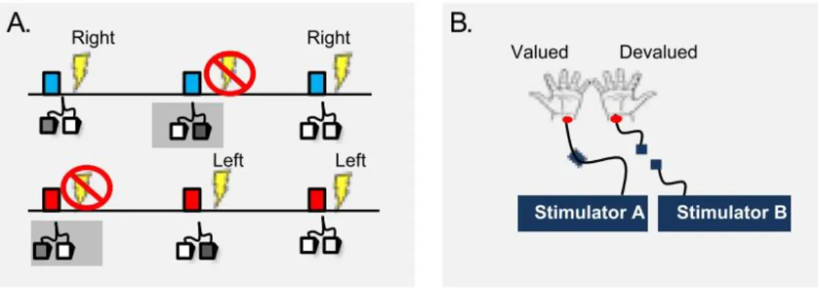
There is no doubt that habits take an important part in human life, their dominance over behavior has been hypothesized to manifest when the stimulus-response link become very strong, or/and by a diminution of goal-directed control over action (a reduction in of the ability to take control over habits) (Gillan et al., 2016). There is a significant interest in investigating the role of habits in human compulsivity, especially in the case of OCD. One hypothesis suggested that habit biases in OCD patients could be explained by a selective deficit in goal-directed control over the action. Another point of view indicates that compulsions result from failure of habitual inhibition in motor and non-motor domains (Jahanshahi, Obeso, Rothwell, & Obeso, 2015). Specifically, there is an interest to reveal if compulsivity could result from deficits in goal-directed control or an excessive build-up of stimulus-response habits (Gillan et al., 2014). A series of human studies showed that OCD patients had a shift in balance away from goal-directed control and towards habits (Gillan et al., 2011). OCD patients showed a deficit in their ability to take control over Stimulus-Response behavior considering the change in the value of outcomes (Gillan et al., 2014; Voon et al., 2015), which means that patients, compared to controls, were more likely to continue to perform an action even though this action no longer served its original purpose. For instance, Gillan and colleagues demonstrated with a goal devaluation procedure (Figure 3) that OCD patients had greater avoidance habits than control subjects, and these habits were associated with a subjective urge to respond. In this study the authors suggested that excessive habit formation may be a contributor reflected in compulsivity in OCD(Gillan et al., 2014).
Figure 3. Goal devaluation procedure to test if OCD patients have a bias toward habits. (A) The blue stimulus predicts
a right shock and the red one a left shock. If the correct avoidance response is executed on time, subjects avoid shock (left pedal to avoid left shock). In this shock avoidance task, habits were induced through overtraining which were identified using goal devaluation. (B) In the devaluation procedure the electrodes on one side are disconnected (devaluated) while the electrodes on the other side are unchanged (valued). Figure from (Gillan et al., 2011)
Habits have become a popular model of compulsivity, in part because how relatively well characterized are the supportive processes at the neurobiological level. Indeed, there is evidence of a neurobiological overlap regarding neural substrates of habit formation and the pathophysiology of OCD (discussed in Chapter 2). Notably, emerging evidence points to the striatum as a critical structure involved to the establishment of ritualized sequences of actions (Barnes, Kubota, Hu, Jin, & Graybiel, 2005; Ann M Graybiel, 2008; Yin et al., 2009). Given the number and diversity of disorders related to repetitive behaviors (RB), the obvious question arises as to whether they share a common etiology. The disorders mentioned before, share high frequency of comorbidity, symptom domains and associated mechanisms, resulting in overlapping diagnostic criteria. Concerning cognitive correlates, deficits in executive function3 are often reported in individuals with persistent
RB (Benzina, Mallet, Burguiere, N’Diaye, & Pelissolo, 2000). Although the etiology and pathophysiology of compulsive related disorders are still under investigation, converging evidence from functional neuroimaging studies, analysis of the lesions that result in for example OCD and the observations regarding neurosurgical interventions that can improve compulsive symptoms implicate the prefrontal cortical-striatal circuits in its pathogenesis. These circuits appear to be involved in habit formation and repetitive behaviors and will be explained in the next Chapter.
M
AINP
OINT SUMMARY• For optimal everyday life, we rely on habits, i.e., actions that we have acquired and automatized. In order to obtain a specific outcome, the activity of a goal-directed system drives us to maintain certain behaviors. After repetition of this goal-directed behavior, a shift to the habit mediating system allows us to optimize our behavior for greater efficiency.
• When over-expressed, RB can become symptoms that characterized some neuropsychiatric disorders such as OCD, TS, and ASD.
• RB-related neuropsychiatric disorders have high rates of comorbidity, and are often diagnosed together in the same patient.
• It has been proposed that compulsive behavior may result from dysfunction in the goal-directed system, increasing the dependence on the habitual responding system.
Chapter 2. N
EUROPHYSIOLOGY OF
COMPULSIVE BEHAVIOR
This chapter aims to review the neurobiological mechanisms that are involved in compulsive behaviors, highlighting the role of the cortical-basal ganglia circuitry and its importance as a putative doorstep to be used in a symptom preventing strategy. Here I summarize the anatomical-functional organization of the basal ganglia, and review the data implicating the cortico-basal ganglia circuits in a variety of compulsive-related disorders including OCD. The center of the attention will be the striatum with a particular interest in its different functional domains and its microcircuitry in the context of compulsive symptoms. This chapter includes a small review of the animal model studies related to compulsive behaviors and the relevance of rodent self-grooming behavior for translational research.
2.1
T
HE ANATOMICAL-
FUNCTIONAL ORGANIZATION OF THE BASAL GANGLIATo understand the underlying neural correlates of pathologies involving compulsive behavior, it is necessary to know how the cortico-basal-ganglia-thalamocortical loops (CBGTC) are engaged in the normal sensorimotor function. Indeed, to make advances in understanding the clinical aspects of RB, investigators have been studying the basic brain circuits that underlie habit formation and internally guided motor control. Progress has been made particularly studying the multisynaptic neural loops that link the cerebral cortex with several subcortical regions. Research into the underlying mechanisms of RB has focused on microcircuits and the role of different cell types, especially interneurons, suggesting that the same neural cell-types are likely to underlie the emergence of pathological RB in a range of disorders.
The basal ganglia (BG) is a group of forebrain nuclei that is present in all vertebrates, and that interconnects with neural systems that affect behavior such as the cerebral cortex, the thalamus, and the brainstem. At the beginning of the ’90s the BG was assigned a role in motor function, and the cortex more a cognitive role. Now we know that the role of the BG is more elaborate than that, it also plays an essential role in many other cognitive functions such as decision-making (B. W. Balleine, Delgado, & Hikosaka, 2007) or learning (Hélie, Ell, & Ashby, 2015). The anatomy of the BG is complex, not to mention that the terminology has changed over the years. Several component nuclei compose the BG at various levels in the brain, and two of them (the globus pallidus (GP) and the substantia nigra (SN)) are divided into anatomically (in terms of connectivity) different elements. The BG include the striatum, the subthalamic nucleus (STN), the globus pallidus (internal segment (GPi), external segment (GPe), and ventral pallidum), and the substantia nigra (pars compacta (SNc) and pars reticulata (SNr)) (Figure 4). The ventral BG consists of the
nucleus accumbens, the ventral pallidum and the medial parts of the STN and SN and is primarily involved with limbic or emotional functions, while the dorsal aspect is primarily involved in motor and associative functions(J. M. Tepper, Abercrombie, & Bolam, 2007).
Figure 4. Location of basal ganglia nuclei in the human and rat brain. (A) Diagram of a coronal section of the human
brain. Figure from (Squire, Larry; Berg, Darwin; Bloom, Floyd; du Lac, Sascha; Ghosh, Anirvan; Spitzer, 2008). (B) Black arrows show the primary input and output connections of the basal ganglia on a sagittal diagram of the rat brain. Figure modified from (Gerfen & Bolam, 2010).
The striatum and the STN are primary receivers of information from outside the basal ganglia. Most of those inputs are excitatory cortical inputs, but thalamic nuclei also provide a broad spectrum of inputs to the striatum. The cortical input uses glutamate as a neurotransmitter and terminates mostly on the heads of the dendritic spines of spiny projection neurons (SPNs). Distinct cortical input can overlap to the same local regions within the striatum (Flaherty & Graybiel, 2013) and inputs from a single cortical region project to multiple striatal zones. The information received is then processed by the BG circuitry through different pathways, generating output to frontal lobe and brain stem areas that are involved in the planning and execution of movements. The basal ganglia output (GPi and SNr) is inhibitory and projects to motor areas in the brain steam and thalamus; thus an increase in the BG outputs leads to a reduction in the activity of its targets. The fact that the BG output is inhibitory to thalamocortical and brainstem targets are relevant to understand its normal motor function. The spinal or brainstem sensory or motor systems do not project directly to the basal ganglia, and there are no direct outputs from BG to spinal motor circuitry. The GPi and the SNr are the primary inhibitory outputs to thalamic nuclei and brain stem.
2.1.1STRUCTURE
The striatum is in the forebrain and includes the caudate nucleus, the putamen (CPu) and
nucleus accumbens. It is named striatum because the axons fibers that pass through it give it a striped appearance. It sends inhibitory projections to the BG output nuclei, the GPi, and the SNr (Figure 5). The striatum is one of the regions that is densely innervated by cortical afferents. It is a crucial region for motor programming, habit formation, and social behavior(Hélie et al., 2015). The striatum does not have a highly organized structure like the cortex or the hippocampus but it does present a functional segregation (section 2.1.3). It is compartmentalized on an overall level in the dorsal and ventral striatum and neurochemically into the matrix/ striosome system (A. M. Graybiel & Ragsdale, 1978).
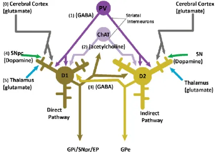
Figure 5. A simplified view of the basal ganglia circuitry showing the direct, indirect and hyperdirect pathways.
Glutamatergic inputs are represented in red, gabaergic in blue and dopaminergic in violet. Figure modified from (Fino &
Venance, 2010)
The external segment of the pallidum (GPe) is a relatively large nucleus located
caudomedial to the striatum(Hitoshi Kita, 2007)(H. Kita & Jaeger, 2017). GPe together with the SNc can be viewed as a central nuclei of the BG because they both receive inputs from BG and send outputs to other BG nuclei. GPe receives excitatory projections from the
STN and inhibitory projections from the striatum (GABA and enkephalin). It projects in the majority to the STN, and there is an inhibitory GABAergic monosynaptic output directly to GPi and SNr and a GABAergic projection back to the striatum (Squire et al., 2008). These organization means that GPe’s neurons can provide feedback inhibition to the striatum and STN, but also feed-forward inhibition to neurons in GPi and SNr, suggesting that the GPe may act to restrain the effect of the striatum and STN projections to the GPi and SNr.
The globus pallidus internal segment (GPi) is the primary BG output for limb
movements. Indeed, the GPi has large neurons that project outside the BG. Most of these outputs sent via collaterals to the thalamus and the brain stem. The principal inputs to GPi come from inhibitory neurons projecting from the striatum and the STN, indeed GPi cells have dendrites that can span up to 1 mm in diameter allowing them to receive many converging inputs (Squire et al., 2008). GPi and the substantia nigra pars reticulata (SNr) are rather consider similar since they both receive inputs from the GPe, striatum and subthalamic nucleus, they both project to the cerebral cortex through the thalamus, and are separated by dense fiber bundles of the internal capsule. The GPi has strong interactions with motor cortices and is believed to control somato-motor behaviors. On the other hand the SNr has interactions with the prefrontal cortex and is considered to control associative functions(Nambu, 2007).
The substantia nigra pars reticulata (SNr) is the segment of the SN that is more sparsely
cellular (compared to the SNc). Like the GPi, the SNr contains large neurons that projects outside the BG (Squire et al., 2008). It receives inhibitory inputs from the striatum, and excitatory inputs from the STN. It provides inhibitory outputs to the thalamus (ventro-lateral and ventral anterior thalamus). These thalamic areas in turn project to the premotor and prefrontal cortex(Deniau, Mailly, Maurice, & Charpier, 2007).
The substantia nigra pars compacta (SNc) contains large DA-containing cells, DA
neurons contain a substance called neuromelanin which gives a dark pigmentation to the SNc therefore it gets its name “substantia nigra”(Squire et al., 2008). SNc receives input from the striatum a sparse input from the prefrontal cortex. In return, the SNc DA neurons project to the CPu in a topographic organization. Nigral DA neurons receive inputs from one striatal circuit and project back to the same or adjacent circuit, modulating activity across circuits (Haber, Fudge, & McFarland, 2000). The action of the DA cells depends on the receptors located on target neurons.
The subthalamic nucleus (STN) has been considered as an important modulator of the
BG output. It receives major excitatory glutamatergic inputs from the frontal cortex with significant contribution from motor areas (frontal lobe only) especially form the motor, premotor and supplementary motor cortex and from the frontal eye field. It also receives inhibitory inputs from the GPe. The STN projects to both segments of the globus pallidus (GPi and GPe) and the SNr. Its outputs are excitatory and is essentially composed of projection glutamatergic neurons (Hamani, Saint-Cyr, Fraser, Kaplitt, & Lozano, 2004).
Most details of the neuroanatomical organization of the basal ganglia circuits come from rodent studies. The most notable differences between primates and rodents are the anatomy of the basal ganglia nuclei. The striatum in primates is subdivided into the caudate nucleus and putamen by the internal capsule. This separation results in functional segregation in which we can distinguish that the caudate nucleus is mainly the target of prefrontal cortical inputs and that the putamen is the target of motor and somatosensory inputs. However, the internal capsule is not a strict divider of functional regions, and there is some overlap of prefrontal cortex’s inputs to the putamen (Gerfen & Bolam, 2010). In the rodent, we can still find striatum’s regional differences that are comparable to those of primate’s and that are determined by the distribution of cortical inputs. In primates, the internal segment of the globus pallidus is adjacent to the external segment, in rodents the equivalent nucleus is known as the entopeduncular nucleus is separated from the GPe and embedded in the fiber tract of the internal capsule (Figure
4-B). Despite these differences, the major connectional organization of the basal ganglia in
rodents and primates is highly similar.
The basal ganglia participate in control movement and behavior. Indeed the most substantial portion of basal ganglia inputs and outputs relate to motor areas. In the history of basal ganglia study, it has been shown that basal ganglia lesions cause severe movement anomalies. In addition to its essential role in motor control, the basal ganglia have an essential role in cognitive function. A considerable portion of the cortical areas provides inputs to the basal ganglia, which reciprocally provide outputs to brain systems involved in the generation of behavior (Gerfen & Bolam, 2010). Indeed, frontal cortical areas involved in the planning and execution of movement behavior provide direct projections to the spinal cord that are responsible for the generation of movement, and the cerebral cortex and its connection to the basal ganglia are known to influence or affect behavior. Activity in the basal ganglia does not cause movement directly. Instead, it influences activity in other brain regions like the motor cortex that affects movement. This motor execution has been hypothesized to be via different circuits in the basal ganglia that promote and inhibit movement respectively. Following a top-down description, these neural circuits continue from cortical areas and pass through the basal ganglia through the direct, indirect or hyper direct pathways with a final projection through the thalamus.
2.1.2BASAL GANGLIA TRIPLE-CIRCUIT MODEL
The direct and indirect basal ganglia pathways have, via glutamatergic and GABAergic neurotransmission, opposing actions on the thalamus and therefore on the frontal cortex. They appear to work oppositely to modulate the thalamic and cortical activation. Activation of the direct pathway projections to the GPi and SNr exerts a powerful inhibition effect over these regions that in return relieves the inhibitory brake on the thalamus. The thalamus then sends glutamatergic projections that activate the frontal cortex. In contrast, the indirect pathway results in the inhibition of the thalamus and the frontal cortex. Activating the indirect pathway results in inhibition of the GPe which
over the GPi/SNr. When this occurs, the GPi/SNr then increases its inhibitory drive over the thalamus, resulting in a reduced glutamatergic output to the frontal cortex (Rapanelli, Frick, & Pittenger, 2017). The hyperdirect pathway is the pathway from cerebral cortex to STN, which in turn sends outputs to the GPi and SNr. This pathway passes on powerful excitation effects from motor-related cortical areas to the pallidum, bypassing the striatum, and therefore having shorter conduction time than the direct and indirect pathways(Squire et al., 2008). Nambu and colleagues baptized it the hyperdirect pathway precisely because it is the fastest route for information flow from cerebral cortex to the basal ganglia output (Figure 5). A simplified center-model of the BG to control voluntary limb movements that include these three pathways would start when a voluntary movement is about to be initiated by cortical mechanisms. The hyperdirect pathway would first inhibit large areas of the thalamus and cerebral cortex that are involved in the selected motor program and other competing programs, this, because it is the fastest route for information flow from the cortex of the BG output. Then disinhibition of specific targets and release of the selected motor program would be possible via an intervention through the direct pathway. Finally, the indirect pathway would allow extensive inhibition of their targets (Nambu, Tokuno, & Takada, 2002). This is a simplified sequential view of information processing where only the selected motor program would be initiated and executed at specific timing whereas other competing programs are canceled.
2.1.3FUNCTIONAL SEGREGATION OF THE CORTICO-BASAL GANGLIA LOOPS
A general organization plan of the CBGTC is represented in Figure 6. The components of this system include the prefrontal cortex, the striatum (composed of the caudate and the putamen) and the thalamus. Cortical and striatal circuits are integrated in various ways. Starting from a broad perspective, CSTC circuits are functionally distinct neural “loops” originating from cortical regions and these regions are as well the site of feedback projections from the thalamus. In these circuits, the striatum does not have direct reciprocal projections to cortical regions, instead, its effects are indirect as they are sent back to the cortex through thalamic projections.
Figure 6. A simplified representation of a CSTC circuit in a sagittal view of the human brain. Figure from (A
The view on the BG has changed over the years from being principally motor-based to include the cognitive and emotional domains. In addition to the BG involvement in the expression of behaviors through movement, the BG is also involved in the process that leads to movement such as the elements that drive actions, including motivation, cognition, and emotions (Haber, 2016). The first models were “box and arrow” models that considered each nucleus of the basal ganglia as a unique and homogeneous structure (a box) that communicates with one or several nuclei by connections characterized either as excitatory or inhibitory (the arrows) (Albin, Young, & Penney, 1989). Nowadays we know this view to be more complicated than that, the basal ganglia not only connect to motor areas of the cortex (motor cortex, supplementary motor cortex, premotor cortex, cingulate motor area, and frontal eye fields) but also to more cognitive associated areas, with converging properties at several levels (Yelnik, 2008). An alternative model of the basal ganglia organization is the five-circuit model proposed by Alexander and Delong (G E Alexander, M R DeLong, 1986; Mahlon R. DeLong & Thomas Wichmann, 2007) (Figure
7). In this model, the cortical projections from the frontal cortex comprise five different
circuits: one limbic, two associative, and two motor circuits. It comprises, an oculomotor circuit with origins in the frontal eye field, a motor circuit originating in the frontal supplementary motor area, two associative circuits originating in the dorsolateral prefrontal cortex (DLC) and the lateral orbitofrontal cortex (lOFC), and one limbic circuit originating in the anterior cingulate cortex. Within each of these circuits, there are the indirect and direct pathways, both projecting to the thalamus (Jeffrey L. Cummings, 1993) and sharing the common structures described. In the five circuit model, the cortical projections to the striatum arise only from the frontal cortex, whereas the other cortices (temporal, parietal and occipital) are not integrated into this model.
Figure 7. The five circuit model of the BG. It includes parallel circuits connecting the basal ganglia, thalamus and
cerebral cortex. ACA, anterior cingulate area; APA, arcuate premotor area; CAUD, caudate; b, body; h, head; DLC, dorsolateral prefrontal cortex; EC, entorhinal cortex; FEF, frontal eye fields; GPi, internal segment of globus pallidus; HC, hippocampal cortex; ITG, inferior temporal gyrus; LOF, lateral orbitofrontal cortex; MC, motor cortex; MDpl, medialis dorsalis pars paralarnellaris; MDme, medialis dorsalis pars magnocellularis; MDpc, medialis dorsalis pars parvocellularis; PPC, posterior parietal cortex; PUT, putamen; SC, somatosensory cortex; SMA, supplementary motor area; SNr, substantia nigra pars reticulate; STG, superior temporal gyrus; VAmc, ventralis anterior pars magnocellularis; Vapc, ventralis anterior pars parvocellularis; VLm, ventralis lateralis pars medialis; VLo, ventralis lateralis pars oralis; VP, ventral pallidum; VS, ventral stria- tum; cl, caudolateral; cdm, caudal dorsomedial; d1, dorsolateral; 1, lateral; 1 dm, lateral dorsomedial; m, medial; mdm, medial dorsomedial; pm, posteromedial; rd, rostrodorsal; rl, rostrolateral; rm, rostromedial; vm, ventromedial; vl, ventrolateral. Figure from (Squire et al., 2008)
Since the entire cerebral cortex projects to the basal ganglia (Yelnik, 2008) there is a more global subdivision of cortical activity including three functional territories (Figure 8) (Parent, 1990). Indeed, the five circuit model can be simplified into three circuits when the motor and oculomotor pathways are grouped into one motor pathway, and the DLC and lOFC are grouped into one cognitive/associative circuit, leaving the limbic circuit involving the projections form the ACC (Squire et al., 2008). These subdivisions take into account the fact that terminals from the cortex in the striatum are organized in a topographic manner (Figure 8). The sensorimotor territory in the dorsolateral part, the limbic territory in the ventromedial part and the associative territory in the central intermediate part of the striatum. Nevertheless, when terminals from various cortical regions are examined closely, there is a convergence of inputs from different functional regions (Jahanshahi et al., 2015). In a more detailed manner, the sensorimotor territory includes the primary motor, the somesthetic cortices, the premotor cortex, supplementary motor area, and the oculomotor areas. It processes motor and somesthetic information. Following the somatotopic organization, the dorsolateral striatum is therefore linked to motor function. This projections were some of the first to be identified (McFarland & Haber, 2000) and supported by physiological studies demonstrating somatotopic maps and neural responses to specific movements (Kimura, 1986). The associative (or sometimes called cognitive) territory includes the prefrontal dorsolateral and lateral orbitofrontal cortices as well as the parietal, temporal and occipital cortices. It processes cognitive information such as the ability to attend external stimuli or internal motivation, to identify the significance of such stimuli, and to plan meaningful responses to them (Purves et al., 2001). The limbic (or sometimes called emotional) territory comprises the hippocampus, the anterior cingulate and medial orbitofrontal cortices. It processes emotional and motivational information. These three functional regions can be mapped in the striatal regions according to the received cortical inputs.
Figure 8. Principal functional subdivisions in the cortico-basal ganglia circuits within the human brain. These
include (a) the motor loop, (b) the associative loop and (c) the limbic circuit sensorimotor. Figure from (Jahanshahi et al.,
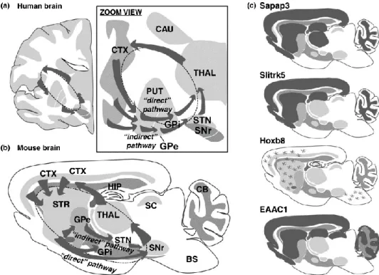
The pattern of cortical inputs helps to define the corresponding striatal regions between humans and rodents (Figure 9). The human dorsolateral putamen and dorsolateral caudate (sensorimotor territory) correspond to the lateral portion of the dorsal striatum in rodents. The large parts of the rostral putamen and most of the head, body and tail of the caudate (associative territory) correspond to the medial portion of the dorsal striatum in rodents. The ventral striatum in humans comprises the nucleus accumbens (NAc) and ventral parts of the caudate and putamen (the limbic territory). In rodents the ventral striatum corresponds to the NAc and the striatal portion of the olfactory tubercle. In rodents the NAc is subdivided into core and shell regions with different connectivity and function, while this division is not clear in primates (Chuhma, Mingote, Kalmbach, Yetnikoff, & Rayport, 2017).
à
Figure 9. Striatal functional subdivisions between humans and mice. The homologous functional territories in the
striatum in humans and rodents can be determined by cortical inputs mediating each function. (Cd: caudate, Pu: putamen, NAc: nucleus accumbens, dStr: dorsal striatum, OT: olfactory tubercle). Figure from (Chuhma et al., 2017).
Interesting to add is that the basal ganglia has unique integrative properties, it seems to have beautifully evolved to optimally receive a great sample of three specific functional aspects of cortical information. These characteristics include its anatomical volume configuration, the number of neurons at different levels and the geometry of its projections (Figure 10). First, the volumes of the successive nuclei that each circuit cross decrease in proportions (the subthalamic nucleus is 3000 times smaller than the emitting cortex). Because of volume and cell density, the number of neurons in each nucleus also decreases (subthalamic nucleus neurons are 4000 times fewer than in cortex). Third, the pallidal dendritic arborizations are arranged perpendicularly to striatal afferent axons, maximizing the number of possible connections (Yelnik, 2008).
Figure 10. Schematic representation of the integrative properties of the basal ganglia. The basal ganglia receive
overlapping information from three functional territories of the cerebral cortex. Volume and cell density decrease in each level. In the globus pallidus, even if neurons are fewer than in the striatum, their flattened and large dendritic arborizations make transmission of striatal information onto pallidal neurons highly converging. In the subthalamic nucleus, information comes not only from the three functional territories but also directly from motor cortices, which makes it a convergence node in the circuit. Figure from (Yelnik, 2008).
Within the associative territory, the existence of a lateral orbitofrontal loop was proposed, involving projections from the orbitofrontal cortex (OFC) to the head of the caudate and ventral striatum, following to the mediodorsal thalamus via the internal pallidus and finally returning to the OFC. From an anatomical perspective, the OFC is a prefrontal brain area with an abundance of reciprocal connections to other brain structures, including the striatum, amygdala and cingulate cortex. The OFC is a focus center for sensory inputs of all five traditionally recognized senses (gustatory, auditory, olfactory, somatosensory and visual). It is composed of several functionally distinct areas that make it highly heterogeneous. So, what exactly is its role? Evidence from lesion studies in animals and humans suggest that the OFC has a crucial role in the emotional and motivational aspects of behavior. Patients with orbitofrontal lesions show behavioral changes related to inappropriate affect, poor decision-making and disinhibition (Namiki et al., 2008). Moreover, functional imaging studies showed that the OFC has a role in monitoring changes in reward value (including anticipation of expected rewards and the probability that rewards will occur) (Gorka, Phan, & Shankman, 2015). Further work dissociate medial and lateral regions of the OFC into different functions, with the lateral OFC (lOFC) being likely to be activated when a response (previously associated with a reward) has to be supresses, indicating that the lOFC may play an inhibitory role(Elliott, Frith, & Dolan, 1997). In coherence with these findings, orbitfrontal lesions in animals and humans lead to reward-related learning deficits in tasks such as reversal learning4 and this could be
reflected in inability to detect changes in the motivational value of stimuli and to then modify behavior accordingly(Rudebeck & Murray, 2009) Therefore the OFC is known to
4 In a reversal learning task the individual first learns to make a discrimination and then is supposed to learn to reverse
be implicated in decision-making, response inhibition, sensory integration, emotional operations, and learning. The utility of investigating the cortical function in the segregated view of the frontal-subcortical circuits has is its value as a unifying framework for understanding human behavioral disorders. Indeed, a wide range of behavioral alterations, including OCD can be linked to dysfunction of frontal-subcortical circuits. For example, animal and human imaging studies have shown that behavior controlled by a goal-directed system and a habitual system are mediated via the associative and motor circuits respectively (Bernard W. Balleine & O’Doherty, 2010).
2.2
T
HE MICROCIRCUITRY OF THES
TRIATUMThe striatum is strategically located in the forebrain, it receives inputs from all cortical areas and has an essential role in processing convergent inputs through different pathways. It plays a central role in motor learning, motor control and cognitive process such as decision making in motivated behaviors. The most abundant and studied neuron in the striatum is the medium spiny projection neuron (SPN, but found in the literature also as MSN) because they are the primary and only output neurons of the striatum. Nevertheless, recent studies have shown that striatal interneurons, even though being very few, may play a dominant role for the regulation of the striatal microcircuitry. In this section, we review the basic neurocytology and micro circuitry of the striatum with a focus on parvalbumin-positive interneurons.
2.2.1MEDIUM SPINY PROJECTION NEURONS
The first and by far most numerous neuron in the striatum is the medium spiny projection neuron (MSN). They make up as much as 80-95% of the total number of striatal neurons, depending on the species (Kemp & Powell, 1971). They are homogeneously distributed in the striatum, and they constitute the output of the striatum via axonal projections to the GP and SN, so they can be categorized as either striatopallidal (indirect pathway) or striatonigral (direct pathway). Cortical input to the striatum targets mainly MSNs through monosynaptic contact, making them the major input target and the major output neuron of the striatum. MSNs have also local axon collaterals within the striatum that form synapses with other spiny neurons (Somogyi, Bolam, & Smith, 1981) (Figure 11). In terms of morphology they have large dendritic trees that stretch over 200-500 µm from the cell body of origin (Wilson, 1980), this property makes them perfect receivers from adjacent projections coming from multiple areas of the cortex. They have a cell body of approximately 12-20 µm and from this cell bodies expand 7-10 dendrites that are moderately branched but densely charged with spines.
Besides cortical inputs that make contact primarily with the head of dendritic spines, MSNs receive inputs from a number of afferents from outside the striatum and from within (Figure 11), specifically: (1) Inhibitory GABA inputs from small striatal interneurons. This correspond to a subpopulation that is positive for the calcium-binding protein parvalbumin (H. Kita, Kosaka, & Heizmann, 1990). (2) Cholinergic inputs from large aspiny neurons (striatal interneurons). (3) Inhibitory GABA, neuropeptide substance P and enkephalin inputs from adjacent MSNs. (4) A broad input from dopamine (DA) neurons from the SNc and VTA. Five type of G protein DA receptors have been found (D1-D5) and they are classified into two families based on their response to agonist; D1 family (D1 and D5 receptors) increase the effect of cortical input to striatal neurons and D2 family (D2,D3,D4 receptors) have the opposite effect. (5) Glutamatergic excitatory inputs from the thalamus (intralaminar and ventrolateral nuclei). These form asymmetric synaptic contacts and have strong excitatory effects on the MSNs. Regarding cortical