HAL Id: tel-02947230
https://tel.archives-ouvertes.fr/tel-02947230
Submitted on 23 Sep 2020HAL is a multi-disciplinary open access
archive for the deposit and dissemination of sci-entific research documents, whether they are pub-lished or not. The documents may come from teaching and research institutions in France or abroad, or from public or private research centers.
L’archive ouverte pluridisciplinaire HAL, est destinée au dépôt et à la diffusion de documents scientifiques de niveau recherche, publiés ou non, émanant des établissements d’enseignement et de recherche français ou étrangers, des laboratoires publics ou privés.
Biosensor imaging of dopamine and glutamate signaling
in striatal projection neurons in a mouse model of
dopamine depletion
Louise-Laure Mariani
To cite this version:
Louise-Laure Mariani. Biosensor imaging of dopamine and glutamate signaling in striatal projection neurons in a mouse model of dopamine depletion. Neurons and Cognition [q-bio.NC]. Sorbonne Université, 2018. English. �NNT : 2018SORUS511�. �tel-02947230�
Sorbonne Université
Ecole doctorale Cerveau-Cognition-Comportement
Laboratoire Institut du Fer à Moulin, UMR S-839 / Equipe Neurotransmission et Signalisation
Biosensor imaging of dopamine and glutamate signaling in striatal
projection neurons in a mouse model of dopamine denervation
Par Louise-Laure MARIANI
Thèse de doctorat de Neurosciences
Dirigée par Jean-Antoine GIRAULT et Denis HERVE
Présentée et soutenue publiquement le 14/12/2018
Devant un jury composé de :
Pr. Angela CENCI, Professeur des universités
Rapportrice
Pr. Avrama BLACKWELL, Professeur des universités
Rapportrice
Dr Peter VANHOUTTE, Directeur de recherche
Examinateur
Pr. Emmanuel ROZE, PU-PH
Membre invité
2 Table of contents
List of Figures and Tables ... 5
Acknowledgments ... 8
Abbreviations ... 9
Summary/Context of the study ... 12
Introduction ... 14
1- The dorsal striatum and the basal ganglia-thalamo-cortical motor circuit ... 15
1.1 General organization of the striatum ... 15
1.2 Striatal projection neurons and main efferent pathways of the dorsal striatum ... 16
1.3 Cortical and thalamic inputs to the dorsal striatum ... 19
1.4 Striatal dopamine inputs ... 20
2- Neurotransmitter receptors and signaling pathways in the dorsal striatum ... 22
2.1 DA receptors ... 22
2.2 Glutamate receptors ... 24
2.3 cAMP production and actions ... 26
2.4 Phosphodiesterases ... 27
2.5 DARPP-32 ... 29
2.6 The ERK Cascade ... 30
2.7 Integration of multiple signaling pathways activated by DA, roles of D1R and D2R ... 32
2.8 Signaling crosstalk between glutamate and DA ... 34
2.9 Adenosine receptors and adenosine signaling in SPNs ... 35
2.10 Calcium signaling ... 38
3- PD and striatal alterations in the absence of dopamine ... 42
3.1 Animal models of PD ... 42
3.2 Striatal alterations in PD and dopamine deficiency ... 43
3.3 Physiological and signaling alterations in SPNs in the absence of dopamine ... 48
4- L-DOPA-induced dyskinesia ... 52
4.1 Clinical features ... 52
4.2 Model of basal ganglia circuit alterations in the dyskinetic state ... 53
4.3 Postsynaptic and presynaptic mechanisms of LIDs ... 54
4.4 Changes in structural and synaptic plasticity in LIDs ... 54
4.5 Molecular bases of LID ... 55
4.6 Role of glutamate transmission in LID ... 58
3
4.8 Role of adenosine in LIDs ... 62
5- Introduction to biosensor live imaging ... 63
5.1 FRET principles ... 64
5.2 An historical perspective on fluorescent probes ... 65
5.3 cAMP sensors ... 66
5.4 ERK sensors ... 68
5.5 Genetically encoded Ca2+ indicators (GECIs) ... 69
Results ... 74
1- Cell-specific up-regulation of signaling pathways in the dopamine-depleted striatum ... 74
Main results of the article ... 74
Article ... 76
2- Setting up the experimental protocol (Additional results I) ... 121
2.1. Slicing protocol, slice preparation for imaging and pharmacological applications in brain slices... 121
2.2. Expression of the probes via viral vectors ... 123
2.3. Combined intrastriatal microinjection of biosensor-expressing AAV and 6-OHDA ... 123
2.4. Activation of ERK by a D1 DA agonist in acute brain slices ... 125
2.5. Activation of biosensors in acute brain slices ... 126
2.6. Dose-dependence of Ca2+ responses to NMDA or AMPA application ... 127
3- Upregulation of ERK activation and Ca2+ responses to glutamate receptors stimulations in SPNs after dopamine depletion by 6-OHDA lesion (Additional results II) ... 129
4- Upregulation of Ca2+ responses to D1-type receptor stimulation occurs in iSPNs after DA depletion possibly via a A2AR-dependent pathway (Additional results III) ... 132
4.1. Ca2+ responses induced by the D1 receptor agonist SKF81297 are increased in SPNs after 6-OHDA lesion ... 132
4.2. Ca2+ responses induced by the D1 receptor agonist SKF81297 in dSPNs after 6-OHDA lesion ... 133
4.3. Ca2+ responses induced by the D1 receptor agonist SKF81297 are upregulated in iSPNs after 6-OHDA lesion ... 135
Discussion ... 139
1- Methodology discussion ... 139
2- Cell-specific up-regulation of signaling pathways in the dopamine-depleted striatum (Manuscript in preparation) ... 140
2.1. ERK phosphorylation is upregulated after 6-OHDA lesion ... 140
2.2. D1R-Gαolf-PKA pathway is upregulated in dSPNs after 6-OHDA lesion ... 142
2.3 Differences in D1R-PKA signaling in SPNs of young and adult mice ... 143
3- Upregulation of ERK activation and Ca2+ signaling after glutamate receptors stimulations in SPNs in the dopamine-depleted striatum (Manuscript in preparation and Additional Results II) ... 144
4
4- Up-regulation of dopamine-induced Ca2+ signaling in the dopamine-depleted striatum (Additional Results III)
... 147
5. Increased spontaneous intracellular Ca2+ transients in SPNs in the dopamine-depleted striatum (Aditional Results IV) ... 148
Concluding remarks ... 149
References ... 151
Appendix: Supplementary Methods ... 202
5
List of Figures and Tables
Figures
Figure Intro 1. Schematic of the connections between the cortex, thalamus, and basal ganglia.
Figure Intro 2. Striatal regions
Figure intro 3. Model of the basal ganglia-thalamo-cortical motor circuit
Figure Intro 4. Fluorescence imaging of D1-SPN and D2-SPN in the dorsal striatum,
corresponding to the direct and indirect pathway neurons.
Figure Intro 5. Proportions of targets for corticostriatal (A) and thalamostriatal (B) synapses in
mice.
Figure intro 6. Distribution of DA neuron cell groups in the adult rodent brain
Figure intro 7. Localization of mGluR in the basal ganglia
Figure Intro 8. Roles of PDEs in the control of basal ganglia–thalamocortical circuitry
Figure Intro 9. Multisite phosphorylation of DARPP-32
Figure Intro 10. The D1 receptor signaling cascades in striatonigral/direct pathway SPNs
Figure Intro 11. Signaling networks regulated by D2-class DA receptor
Figure Intro 12. Major signaling pathway of A2A adenosine receptor (A2AR)
Figure Intro 13. Intracellular Calcium (Ca
2+) homeostasis and signaling
Figure Intro 14. Model of alterations in the basal ganglia-thalamo-cortical motor circuit in the
parkinsonian state
Figure Intro 15. Spine density in striatal SPNs
Figure Intro 16. Motor complications in PD associated with levodopa
Figure Intro 17. Model of the basal ganglia-thalamo-cortical motor circuit in the dyskinetic state
Figure Intro 18: Some of the glutamatergic mechanisms involved in LID
Figure Intro 19. Basic Designs of Fluorescent Indicators Employing GFP-Based FRET
Figure Intro 20. The PKA FRET biosensor AKAR3
6
Figure Intro 21. The ERK FRET biosensor EKAR-EV
Figure Intro 22. Models of the classes of GECIs
Figure Intro 23. Crystal structure of Ca
2+-bound GCaMP in two orthogonal views
Figure Intro 24. Comparisons of GCaMP6 indicators
Results
Article:
Figure 1. Quantitative real-time analysis of ERK activity dynamics with single cell resolution in
neurons in culture and brain slices.
Figure 2. ERK responses are increased after dopamine depletion induced by 6-OHDA lesion
Figure 3. PKA responses to D1R stimulation are increased in dSPN after 6-OHDA lesion in the
striatum
Figure 4. Role of Gαolf and PDEs in the upregulation of PKA response to D1R agonist in the
6-OHDA-lesioned striatum
Figure 5. Spontaneous Ca
2+transients are increased in D1R-expressing neurons of
6-OHDA-lesioned striatum
Figure 6. Specific upregulation of AMPA-induced intracellular Ca
2+dynamics in A2AR-expressing
neurons in 6-OHDA-lesioned striatum
Figure R.7. Different slicing angles
Figure R.8. Comparison of AKAR3 expression in the striatum using Sindbis virus and AAV
Figure R.9. Lesion and AAV infection checking
Figure R.10. GCaMP6S expression driven by AAV in the striatum
Figure R.11. ERK activation by stimulation of D1 DA receptors
Figure R.12. Ca
2+responses after local electrical stimulation in the striatum of mice injected with
GcAMP6S-expressing AAV
7
Figure R.14. Effects of NMDA on Ca
2+monitored by GcAMP6s
Figure R.15 Effects of AMPA on Ca
2+monitored by GcAMP6s
Figure R.16. Effects of Group I mGluR stimulation on phospho-ERK immunostaining in the
striatum following 6-OHDA lesion
Figure R.17. Ca
2+responses induced by the group I mGluR agonist DHPG are increased after
6-OHDA lesion
Figure R.18. Ca
2+responses induced by the D1 receptor agonist SKF81297 are increased in SPNs
after 6-OHDA-lesion
Figure R.19. Ca
2+responses induced by SKF81297 are not increased in dSPN and are only
partially D1R-dependent
Figure R.20. Ca
2+responses induced by SKF81297 are increased in iSPN in an A2AR-dependent
manner after dopamine depletion.
Figure R.21. Quantification of cells potentially co-expressing D1R and D2R in the striata of
6-OHDA-lesioned animals
Discussion
Figure D.1: PKA activity is reduced in both types of SPNs in adult wild-type mice compared to
young mice
Tables
Table 1: Major differences between the two types of SPNs found with BAC transgenic mice
Table 2: Basic characteristics of DA receptors
Table 3: Cellular distribution of DA receptors in the cortex and striatum of rodents
Table 4: Spine changes in SPN in PD
8
Acknowledgments
9
Abbreviations
6-OHDA : 6-hydroxydopamine
α-syn: α-synuclein
AAV: adeno-associated virus
AC: adenylate cyclase
ACh: Acetylcholine
AKAR: A kinase activity reporter
AMP: Adenosine monophosphate
AMPA: α-amino-3-hydroxy-5-methyl-4-isoxazolepropionic acid
AMPAR: AMPA receptor
ATP: Adenosine triphosphate
Ca
2+: Calcium
CaM: Ca
2+/calmodulin
CaMK: Ca
2+/calmodulin-dependent protein kinase
cAMP: cyclic AMP
CFP: blue variant of GFP
CICR: Ca
2+-induced Ca
2+-release
CREB: cAMP-response element binding protein D1R: Dopamine D1 receptor
D2R: Dopamine D2 receptor
DA: Dopamine
DAG: diacylglycerol
DARPP-32: 32-kDa DA and cAMP-regulated phosphoprotein
dSPN: SPN of the direct pathway
EGFP: enhanced green fluorescent protein
EKAR: ERK activity reporter
ER: endoplasmic reticulum
ERK: Extracellular signal-Regulated Kinases
FRET: fluorescence resonance energy transfer
GABA: γ-aminobutyric acid
GECIs: genetically encoded calcium indicator
GFP: green fluorescent protein
GluA: subunit of AMPAR
GluN: subunit of NMDAR
GPCR: guanine nucleotide binding protein-coupled receptors
GPe: globus pallidus pars externa
GPi: globus pallidus pars interna
10
IF: Immunofluorescence
iGluR: ionotropic glutamate receptors
IHC: Immunohistochemistry
IP3: inositol trisphosphate
IP3R: inositol trisphosphate receptor
iSPN: SPN of the indirect pathway
KO: knockout
LIDs: Levodopa-induced dyskinesia
LTD: Long-term depression
LTP: Long-term potentiation
MEK: MAP-kinase and ERK-kinase
mGluR: metabotropic glutamate receptors
MSK1: mitogen- and stress-activated kinase 1
NHP: Non-human primates
NMDA:
N-methyl-d-aspartateNMDAR:
NMDA receptorMPTP: 1-methyl-4-phenyl-1,2,3,6-tetrahydropyridine
PD: Parkinson’s Disease
PDE: phosphodiesterase
PET: positron emission tomography
PFA: paraformaldehyde
PIP2: phosphatidylinositol-4,5-bisphosphate
PKA: cAMP-dependent protein kinase
PKC: protein kinase C
PLA: proximity ligation assay
PLC: phospholipase C
PMCA: Plasma membrane Ca
2+-ATPase
PP1: protein phosphatase 1
PSD: post-synaptic density
RyRs: ryanodine receptors
SERCA: Sarco/endoplasmic reticulum Ca
2+-ATPase
SNr: Substantia Nigra pars reticulate
SNc: Substantia Nigra pars compacta
SOCE: store-operated Ca
2+entry
SPN: striatal projection neurons
STEP: striatal enriched tyrosine phosphatase
STN: subthalamic nucleus
11
VTA:
ventral tegmental areaWB: Western Blot
12
Summary/Context of the study
Parkinson’s disease (PD) is the second most common neurodegenerative disorder after Alzheimer’s disease. There is currently no cure for this disease. Symptomatic drug therapy essentially relies on dopamine (DA) replacement therapy. The spectacular antiparkinsonian effect of L-DOPA in PD is however hampered by long-term complications. One of the most difficult complications to treat is dyskinesia, which occurs in virtually all patients during the course of the disease. L-DOPA-induced dyskinesia (LID) results from maladaptive striatal plasticity whose mechanisms are not fully understood yet. The aim of this project is to address a question of therapeutic importance: what are the dysregulations of signaling pathways in striatal projection neurons (SPNs) that will ultimately lead to the development of LID?
To address this question, we explored in striatal neurons the changes of various signaling pathways produced by DA depletion, using genetically encoded protein sensors for two-photon imaging in identified living neurons in mouse striatal slices. We tracked the activity of neuronal populations with genetically encoded Ca2+ indicator GCaMP6S. We used fluorescence resonance energy transfer (FRET)-based biosensors to monitor protein kinase activities with a high temporal resolution in living neurons to address signaling integration. We monitored protein kinase A (PKA) activity with AKAR-3 and extracellular signal-regulated kinase (ERK) activity with EKAR-EV. We compared the effects of various pharmacological manipulations of glutamate, DA and adenosine receptors as well as phosphodiesterase and kinase activities in striatal slices of intact and 6-hydroxydopamine (6-OHDA)-lesioned mice. These latter mice correspond to a model of striatal DA depletion occurring in PD. Using Cre-Lox system we targeted biosensor expression specifically in striatal projection neurons of the direct pathway (dSPNs) bearing D1 DA receptors and/or striatal projection neurons of the indirect pathway (iSPNs) bearing DA D2 receptors and adenosine A2A receptors.
We first demonstrate that two-photon imaging with various biosensors allows monitoring the signaling pathway dynamics in specific populations of SPN in DA-intact and depleted striatum. We show increased spontaneous activity of SPNs and up-regulation of Ca2+, cAMP/PKA and ERK signaling in SPNs following DA depletion. In the 6-OHDA-lesioned striatum, cAMP/PKA and ERK responses to D1 receptor stimulation are increased in dSPNs whereas cAMP/PKA signaling downstream of adenosine A2A receptors is not affected in iSPNs. Interestingly, the upregulation of cAMP/PKA signaling is associated with an apparent loss of phosphodiesterase activity in the dSPNs of DA-depleted striatum, an effect not observed in the iSPNs. Upregulation of Ca2+ and ERK signaling is observed in response to stimulation of AMPA-type glutamate ionotropic receptors (AMPAR) selectively in the iSPNs. DA depletion also amplifies the activation of Ca2+ signaling downstream metabotropic glutamate receptor stimulation in SPNs.
Our results studying the DA- or glutamate-dependent pathways in different subpopulations of SPNs suggest that the increased ERK activation following DA D1R activation relies at least partly on the upregulation of PKA signaling in dSPNs and not iSPNs. Glutamate released from corticostriatal afferent also participate in the upregulation of ERK signaling but Ca2+ entry induced by cell depolarization after AMPAR stimulation is only increased in iSPN and not dSPNs. Hence DA/D1R and glutamate/AMPAR signaling pathways seem to be affected differentially in the two populations of SPNs rather than being jointly modified to enhance ERK activation in the same population.
13
14
Introduction
Parkinson’s disease (PD) is the second most common neurodegenerative disorder after Alzheimer’s disease. The number of PD patients will increase by ~65% between 2010 (n=155,000) and 2030 (n~260,000) (Wanneveich et al., 2018). There is currently no cure for PD, which means that there is no therapeutic possibility to reverse the neurodegenerative processes of dopamine (DA) neurons and other types of neurons. Symptomatic dopaminergic replacement therapy does not prevent debilitating complications occurring during disease course. Development of new disease modifying drugs remains a major challenge in PD.
Symptomatic drug therapy essentially relies on DA replacement therapy. The characteristic triad of motor symptoms of PD—akinesia, rigidity and tremor (Hughes et al., 1992) — related to the loss of dopaminergic neurons is indeed spectacularly improved by DA replacement therapy. The gold standard remains the DA precursor, levodopa, whose efficacy was demonstrated more than 50 years ago (Hornykiewicz, 1966; Cotzias et al., 1967; Rascol et al., 2011; Fahn, 2015, 2018; Lees et al., 2015). This spectacular antiparkinsonian effect of levodopa in PD is however balanced by major limitations. The dopatherapy is hampered by long-term complications, motor fluctuations and dyskinesia (You, Mariani
et al., 2018). Apart from the classical dopa-responsive motor symptom triad, PD course is also
characterized by the occurrence of motor symptoms resistant to levodopa like postural instability, falls, freezing of gait and non-motor features affecting cognition, sleep, mood, behavior, and autonomic functions (Mariani et al submitted 2018 under review). L-DOPA causes motor fluctuations and L-DOPA-induced dyskinesia (LIDs) in 40% of the patients after 4–6 years and up to 90% after 10 years (Ahlskog and Muenter, 2001; Manson et al., 2012). Thus, due to the high frequency of LID, and L-DOPA remaining the gold standard for PD symptomatic treatment, understanding the molecular processes leading to LID occurrence is of major importance.
The majority of movement alterations observed in PD is linked to the lack of DA in the dorsal striatum (DS) (Bernheimer et al., 1973; Price et al., 1978; Dauer and Przedborski, 2003) and LID are induced by altered synaptic plasticity in the DS challenged by intermittent sharp variations of DA (Carta
et al., 2006; Cenci and Lundblad, 2006; Lindgren et al., 2007; Iravani and Jenner, 2011; Iravani et al.,
2012; Antonini and Jenner, 2018). To progress in the understanding of PD pathophysiology and find new ways to decrease the incidence or severity of DOPA-therapy negative effects, it is critical to identify signaling alterations in DS neurons in the absence of DA. This thesis work aims at addressing this question using for the first time live biosensor imaging in striatal neurons in a mouse model of DA deficiency. In the bibliographic introduction we will review the organization and function of the DS and basal ganglia, the signaling pathways in striatal neurons, the pathophysiology of PD and LID, and the bases of biosensor imaging.
15
1- The dorsal striatum and the basal ganglia-thalamo-cortical motor
circuit
1.1 General organization of the striatum
The basal ganglia have a key role in action selection; it has been proposed that the basal ganglia carry out the task of devoting most brain resources to a single movement or behavior at a time, adapted to the context (Mink, 1996; Redgrave et al., 1999; Grillner et al., 2005).
The striatum is the main input structure of the basal ganglia (Kincaid et al., 1998; Bolam et al., 2000) (Figure Intro 1). The striatum is a convergence point for glutamatergic inputs from cortex and thalamus into specific downstream pathways (Alexander et al., 1986; Alexander and Crutcher, 1990; Berendse and Groenewegen, 1990; Gerfen, 1992; Smith et al., 2011; Kress et al., 2013; Huerta-Ocampo et al., 2014), as well as dopaminergic afferents from the midbrain (Smith and Bolam, 1990; Bolam et al., 2000; Björklund and Dunnett, 2007; Surmeier et al., 2010; Gerfen and Surmeier, 2011a). The striatal projection neurons (SPNs) integrate these convergent glutamatergic inputs predominantly on their spines (Kemp and Powell, 1971; Kincaid et al., 1998; Carter et al., 2007; Huerta-Ocampo et al., 2014; Hunnicutt et al., 2016).
[Figure intro 1: Schematic of the connections between the
cortex, thalamus, and basal ganglia. The indicated output
nuclei of the basal ganglia are the globus pallidus internal segment (GPi) and the substantia nigra pars reticulata (SNr) (gray box indicates the basal ganglia). adapted from
Hunnicutt et al 2016]
The striatum is classically divided into three functionally and anatomically distinct circuits: the dorsolateral, dorsomedial, and ventral striatum respectively accounting for the sensorimotor, associative, and limbic domains (Yin and Knowlton, 2006; Balleine et al., 2009; Belin et al., 2009; Kreitzer, 2009; Thorn et al., 2010; Gruber and McDonald, 2012) (Figure intro 2). In rodents, motor descending axonal bundles perforate the striatum. In primates and carnivores, the dorsal striatum is divided by the internal capsule into: on the medial side the caudate nucleus and on the lateral side, the putamen. In both rodents and primates, even if there is no clear division between dorsomedial and dorsolateral striatum, these striatal regions are anatomically and functionally distinct (Joel and Weiner, 1994; Parent and Hazrati, 1995; Yin and Knowlton, 2006). The dorsolateral striatum receives inputs from sensorimotor cortex (Künzle, 1975; Liles and Updyke, 1985; McGeorge and Faull, 1989). The dorsomedial striatum receives inputs from associative cortices (Goldman and Nauta, 1977; Ragsdale and Graybiel, 1981; McGeorge and Faull, 1989). The ventral striatum, the nucleus accumbens subdivided into core and shell, has distinct properties from the dorsal striatal regions (Nicola, 2007; Taha et al., 2007). It receives glutamatergic inputs from the frontal cortex and limbic regions (Brog et al., 1993). The dopaminergic
16
innervation of the ventral striatum comes from a midbrain nucleus adjacent to the SNc, the ventral tegmental area (VTA), which is less affected in PD and explains why denervation predominates in the dorsal striatum in PD (Price et al., 1978; Joel and Weiner, 2000; Dauer and Przedborski, 2003).
The dorsal striatum seems more involved in action selection, goal-directed behaviour and the emergence of habits (Yager et al., 2015; Howard et al., 2017; Volkow et al., 2017). The nucleus accumbens is more likely an interface between the limbic and motor systems implicated in reward processing and motivation (Bolam et al., 2000; Joel and Weiner, 2000; Morgane et al., 2005; Wise, 2009; Volkow et al., 2011).
[Figure intro 2: Striatal regions. Coronal schematic hemi-section of the mouse
forebrain. The dorsolateral (DLS), dorsomedial (DMS), and ventral divisions (nucleus
accumbens, NAc) of the striatum are schematically illustrated in the left hemisphere.
adapted from Kreitzer et al 2009.]
In addition to these regional differences, the dorsal striatum is divided in two intermingled compartments, the matrix and the striosomes (or patches) (Joel and Weiner, 2000; Crittenden and Graybiel, 2011). These two compartments are distinguished by neurochemical markers and have a different organization of inputs and outputs (Prensa and Parent, 2001; Fujiyama et al., 2006; Matsuda et
al., 2009). The proper balance between their activities appears to be critical for the function of the dorsal
striatum (Crittenden and Graybiel, 2011), although their exact respective role in the overall function is still not fully clarified.
1.2 Striatal projection neurons and main efferent pathways of the dorsal striatum
The striatum is mainly composed by GABAergic medium-sized spiny neurons also known as striatal spiny projection neurons (SPN), which represent 95% (in rodents) of the neurons of the striatum. SPN are characterized by the presence of spines on their dendrites. These spines are absent from the most proximal dendrites. Their peak density (1–2 per μm) is 50–60 μm away from the soma. Then their density gradually decreases down to the tips of the sparsely branching dendrites (250–400 μm). SPNs form the main or sole output to downstream basal ganglia nuclei. Activation of GABAergic SPNs inhibits their primary targets (Chevalier et al., 1985; Deniau and Chevalier, 1985; Chevalier and Deniau, 1990).
The role of the striatum in the control of many aspects of movement and motivation depends on the balance between two trans-striatal circuits, involving pathways called the direct and indirect pathways (Figure intro 3). The striatonigral SPNs (dSPN), of the direct pathway facilitating movement execution, directly project to the GABAergic output nuclei of the basal ganglia, the globus pallidus pars
interna (GPi) and the substantia nigra pars reticulata (SNr). They modulate the thalamus and brainstem
outputs (Figure intro 3), with the GPi involved in axial and limb movements and the SNr involved in head and eye movements. The substantia nigra pars reticulata (SNr) neurons are tonically active and maintain
17
a strong inhibition of their targets including the thalamus. Thus they inhibit action. Activation of the direct pathway inhibits SNr neurons (Chevalier et al., 1985; Deniau and Chevalier, 1985) and thus facilitates action (e.g., a movement due to the selective activation of appropriate muscle groups).
The striatopallidal SPNs (iSPN) of the indirect pathway, a multisynaptic circuit between the striatum and basal ganglia output nuclei inhibiting movement, project to and inhibit the GABAergic neurons of the globus pallidus pars externa (GPe). The latter in turn project to and modulate the glutamatergic neurons of the subthalamic nucleus (STN) that project to the GPi/SNr, resulting in the inhibition of thalamocortical projection neurons thus inhibiting movement (Bateup et al., 2010; Kravitz et
al., 2010; Gerfen and Surmeier, 2011b; Cui et al., 2013). Activation of the indirect pathway results in
increased activity of subthalamic neurons and further activates SNr neurons, thereby reinforcing action inhibition (Figure intro 3 left panel).
[Figure intro 3:
Left Panel. Model of the basal ganglia-thalamo-cortical motor circuit. Arrows represent excitatory projections; «T»
represent inhibitory projections. The striatum receives excitatory cortical and thalamic inputs. Dopaminergic nigrostriatal projections to dSPN activate the direct pathway and those to iSPN inhibit the indirect pathway. Outputs of the basal ganglia from GPi and SNr are directed to the thalamus, superior colliculus, and pedunculopontine nucleus (PPN). from You, Mariani et al 2017
Right Panel. Schematic illustration of putative action selection mechanism. D1 : DA receptors of D1 type situated
on the dSPN, D2: D2 receptors of the D2 type situated on the iSPN. Adapted by Girault after Mink, Prog Neurobiol,
18
The function of the basal ganglia depends on the balance between the two pathways exerting opposing influences on motor function (Alexander et al., 1986; Albin et al., 1989; DeLong, 1990; Kravitz
et al., 2010). These two pathways are oppositely modulated by DA. Tonic release of DA inhibits the
indirect pathway. Phasic release of DA activates the direct pathway. Recent models propose coordinated activation of both pathways during action selection (Cui et al., 2013; Alcacer et al., 2017). Activation of the direct pathway could facilitate output of the desired motor programs but activation of the indirect pathway would inhibit competing motor programs (Hikosaka et al., 2000; Mink, 2003; Nambu, 2008; Alcacer et al., 2017) (Figure 3 right panel).
Histochemically, striatonigral dSPNs express high levels of DA D1 (D1R) and muscarinic M4 receptors, as well as dynorphin and substance P (Gerfen et al., 1990; Gerfen, 1992; Yung et al., 1995; Ince et al., 1997). Striatopallidal iSPNs have a high expression of D2R and adenosine A2A receptors, and immunoreactivity for enkephalin (Gerfen et al., 1990; Schiffmann et al., 1991; Fink et al., 1992; Gerfen, 1992; Yung et al., 1995; Bertran-Gonzalez et al., 2009). Morphologically, striatopallidal iSPNs seem to have more primary dendrites than striatonigral dSPNs (Gertler et al., 2008) but it is not always the case in other studies (Huerta-Ocampo et al., 2014). Physiologically, dSPNs and iSPNs exhibit similar properties, characterized by their hyperpolarized resting membrane potential at ∼ –90mV (Wilson and Kawaguchi, 1996; Kawaguchi, 1997; Stern et al., 1997; Blackwell et al., 2003), low input resistance, inward rectification, and a long delay to initial spiking (Kreitzer, 2009). Although iSPNs exhibit increased excitability with an action potential discharge rate twice bigger (Kreitzer and Malenka, 2007; Cepeda et
al., 2008; Gertler et al., 2008; Valjent et al., 2009). Conditional expression of fluorescent proteins under
the control of D1R or D2R gene promoter (Figure intro 4) allowed further characterization of the differences between direct- and indirect-pathway SPNs (Lobo et al., 2006; Heiman et al., 2008, 2014b; Matamales et al., 2009). Some of these differences are summarized in Table 1 and intracellular signaling aspects are further detailed below.
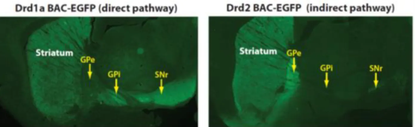
Figure Intro 4: Fluorescence imaging of D1-SPN and D2-SPN in the dorsal striatum, corresponding to the direct and indirect pathway neurons. Left panel: Brain section from a BAC-transgenic mouse expressing enhanced green
fluorescent protein (EGFP) under regulation of the Drd1a promoter (Drd1a BAC-EGFP) shows D1-expressing SPNs in the striatum that project axons which terminate in the GPi and GPe. Right panel. Fluorescent imaging of a Drd2-EGFP mouse (BAC-transgenic mouse expressing Drd2-EGFP under regulation of the Drd2 promoter) shows that D2-expressing SPNs project to the GPe but not to the GPi or SNr. From Gerfen and Surmeier 2011]
19
[Table 1: Major differences between the two types of SPNs found with BAC transgenic mice. From Valjent et al
2009]
In addition to the SPNs, the dorsal striatum contains several types of interneurons which represent the remaining 5% of the total number of neurons in rodents (Kawaguchi, 1997; Tepper and Bolam, 2004; Tepper et al., 2004, 2010; Ibáñez-Sandoval et al., 2011; Gittis and Kreitzer, 2012; Assous and Tepper, 2018). They include the large size cholinergic interneurons and several types of GABAergic interneurons like the parvalbumin Fast-spiking interneurons (FSIs) inhibiting both dSPNs and iSPNs, persistent and low-threshold spike (PLTS) interneurons which release neuropeptides such as neuropeptide Y (NPY) and somatostatin (SOM) and the neuromodulator nitric oxide (NO), Neuropeptide Y-neurogliaform interneurons and TH positive interneurons.
1.3 Cortical and thalamic inputs to the dorsal striatum
The corticostriatal input is overall topographically organized. Corticostriatal pyramidal cells have multiple projections sites including ipsi- but also contralateral striatum (Shepherd, 2013). Recent studies have focused on systematically mapping the precise excitatory projection patterns to the entire striatum (Hunnicutt et al., 2016), revealing striatal subregions and their boundaries by systematic analysis of these input patterns from individual cortical and thalamic subregions (Oh et al., 2014; Hunnicutt et al., 2016). They found clear boundaries separating the three traditional striatal domains and uncovered a fourth subdivision in the posterior striatum. Interestingly, the posterior striatum has also been reported to be innervated by a specific dopamine neurons population (Menegas et al., 2015). The dorsomedial striatum seem to show the highest degree of cortical input heterogeneity, suggesting it serves as an information hub. In addition, thalamic subregions receiving basal ganglia outputs are preferentially interconnected with motor-related cortical subregions.
Thalamic subregions are defined by cytoarchitectural boundaries to delineate ~40 nuclei. The organization of thalamostriatal inputs, which provide ~1/3 of all glutamatergic synapses in the striatum (Huerta-Ocampo et al., 2014), particularly originating from the intralaminar thalamic nuclei (Smith et al., 2004; Doig et al., 2010), has been less studied than corticostriatal inputs.
Cortical and thalamic neurons regulate the activity of SPNs in the striatum through long-range glutamatergic excitatory projections (Landry et al., 1984; Wilson, 1987; Graybiel et al., 1994; Lovinger and Tyler, 1996; Reiner et al., 2003; Kress et al., 2013; Hunnicutt et al., 2016), while inhibition is mediated by feed-forward and feed-back circuits (Tepper et al., 2008). The main feed-forward circuit is characterized by GABAergic striatal interneurons receiving excitatory inputs from the cortex and monosynaptically inhibiting SPNs. The feed-back circuit comprises SPNs interconnections via local axon collaterals (Calabresi et al., 1991; Kawaguchi, 1993; Kita, 1993, 1996; Kawaguchi et al., 1995; Tunstall et
20
al., 2002; Planert et al., 2010). SPN to SPN synapses are typically unidirectional, predominantly localized
onto distal dendrites of other SPNs (Tunstall et al., 2002; Plenz, 2003; Koos et al., 2004; Taverna et al., 2004, 2005). dSPNs preferentially innervate other dSPNs, whereas iSPNs innervate both subtypes equally (Taverna et al., 2008). More recent work using optogenetic and electrophysiology approaches to activate SPNs has inferred an even higher degree of connectivity (Chuhma et al., 2011; Wei et al., 2017).
Each SPN receives several thousands of corticostriatal synapses on its dendrites, but each individual corticostriatal axon makes only a few contacts with each SPN, typically making one or two en passant synapses (Kincaid et al., 1998; Parent and Parent, 2006). Corticostriatal and thalamostriatal
glutamatergic synapses are similar morphologically and intermingled along the dendrites of SPNs (Smith
et al., 2004; Raju et al., 2006). The synapses they form on SPNs are nearly equal in number,
corticostriatal synapses being slightly more numerous (Smith et al., 2004). The vast majority of synapses target spines rather than dendritic shafts and the proportion seems more elevated in corticostriatal than thalamostriatal synapses (Fujiyama et al., 2006; Raju et al., 2006, 2008; Moss and Bolam, 2008; Doig et
al., 2010; Lei et al., 2013; Zhang et al., 2013; Huerta-Ocampo et al., 2014) (Figure Intro 5).
[Figure Intro 5: Pie charts
illustrate the proportions of targets for corticostriatal (A) and thalamostriatal (B) synapses in mice. Axospinous,
synapses onto spines; Axodendritic, contacts onto dendrites; Axo?, contacts onto undetermined targets. From
Zhang et al 2013].
A study from Wall et al 2013, using a monosynaptic rabies virus system, also showed that innervation of D1-SPN and D2-SPN seems anatomically biased towards specific brain regions (Wall et al., 2013). They found that thalamostriatal input and dopaminergic input seem similar onto both pathways. Motor cortex preferentially targets the indirect pathway while sensory cortical and limbic structures preferentially innervate the direct pathway. Others find that pyramidal tract axons innervate both type of SPN (Kress et al., 2013).
1.4 Striatal dopamine inputs
There are ten DA-producing nuclei in the mammalian brain (Björklund and Dunnett, 2007; Tritsch and Sabatini, 2012) designated A8 to A17 (Figure intro 6). Projections from a given subset of DA neurons target one region of the brain (Yetnikoff et al., 2014), but projections to a given subset of DA neurons arise from many different regions as shown by studies using combinations of techniques using CLARITY and Rabies virus injections (Watabe-Uchida et al., 2012; Menegas et al., 2015). The substantia nigra pars compacta (SNc; field A9) and VTA (field A10), which project to the dorsal and ventral striatum, and form the nigrostriatal and mesocorticolimbic pathways respectively; each contain in the rodent ≈20,000 – 30,000 neurons bilaterally (German and Manaye, 1993; Zaborszky and Vadasz, 2001), and a total of neurons between 160 000–320 000 in monkeys and 400 000–600 000 in humans, with >70% of the neurons located in the SN (Björklund and Dunnett, 2007). These neurons form widely spread highly
21
dense axonal arborizations in the striatum (Prensa and Parent, 2001; Matsuda et al., 2009). Individual SNc neurons extend highly branched axons of half a meter in total length that densely ramify throughout up to 1 mm3 of tissue (Matsuda et al., 2009). Dopaminergic synaptic boutons represent ≈10% of all synapses in the striatum (Groves et al., 1994). The closest distance in between each dopaminergic bouton is only ∼1.18 μm (Arbuthnott and Wickens, 2007). Some of these terminals are found at spine necks abutting cortical synapses (Smith et al., 1994; Moss and Bolam, 2008), but many dopaminergic terminals have been found against dendritic shafts with no detectable electron-dense postsynaptic structure (Groves et al., 1994; Hanley and Bolam, 1997).
[Figure intro 6: Distribution of DA
neuron cell groups in the adult rodent brain as illustrated schematically on a sagittal view. The numbering of cell groups, from A8 to A16, was introduced in the classic study of Dahlström and Fuxe in 1964. A17 Retinal dopaminergic neurons are not shown on this drawing. The principal projections of the DA cell groups are illustrated by arrows. Adapted from Bjorklund
and Dunnett 2007]
Midbrain DA neurons are autonomous pacemakers spontaneously active at low frequencies (1–8 Hz) in vivo (Guzman et al., 2009; Kreitzer, 2009), suggesting that each neuron provides a basal DA tone to many target neurons, adjusted by either phasic bursts or transient pauses of activity, critical for normal striatal function (Schultz, 2007a). This basal DA tone most likely activates high-affinity DA receptors of the D2-type (D2–D4) (Richfield et al., 1989). Bursts of action potentials that briefly elevate striatal extracellular DA are fired by DA neurons in response to behaviorally relevant stimuli (Schultz, 2007b). These phasic spikes of DA are activating both the high-affinity D2 type receptors and the lower-affinity DA receptors of the D1 type (D1, D5) (Richfield et al., 1989). The phasic and tonic firings of DA neurons allow encoding transient responses, for instance to reward prediction error, or longer timescale responses like uncertainty (Fiorillo et al., 2003; Schultz, 2007b). Dopamine thus plays a crucial role in the control of motor, cognitive and emotional behaviors and their adaptation by learning in response to reward.
22
2- Neurotransmitter receptors and signaling pathways in the dorsal
striatum
2.1 DA receptors
DA receptors belong to the large superfamily of guanine nucleotide binding protein-coupled receptors (GPCRs). These metabotropic receptors share interaction with G-proteins, structure with seven alpha-helices transmembrane domains that are interconnected by alternating intracellular and extracellular loops. The heterotrimeric G-proteins are formed by a combination of an α-subunit and a βγ dimer that can each lead to activation of signaling effectors. These receptors do not signal exclusively through heterotrimeric G proteins and may also engage in G protein-independent signaling events (Luttrell, 2014; Peterson and Luttrell, 2017; Luttrell et al., 2018; Pack et al., 2018).
DA binds and activates two families of GPCRs (Kebabian and Calne, 1979; Andersen et al., 1990; Sibley and Monsma, 1992; Greengard et al., 1999; Beaulieu and Gainetdinov, 2011; Tritsch and Sabatini, 2012): the D1 family (D1 and D5 subtypes) (Tiberi et al., 1991) and the D2 family (D2, D3 and D4 subtypes) with different affinities for DA ranging from nanomolar to micromolar range, but less different between individual subtypes within a family. The affinity of D2-like receptors for DA is generally reported to be 10- to 100-fold greater than that of D1-like receptors, with D3 and D4 receptors displaying the highest sensitivity for DA and D1 receptors the lowest (Beaulieu and Gainetdinov, 2011) (Table 2). But D1 and D2 receptors can exist in both high and low affinity states.
[Table 2: Basic characteristics of DA receptors. from Tritsch and Sabatini 2012]
The organization of the genes of D1- and D2-class DA receptors is also different. The D1 and D5 DA receptor genes do not contain introns in their coding regions, so do not generate splice variants. The genes encoding the D2-class receptors have several introns, with six introns in the gene that encodes the D2R receptor, five in the gene for the D3R, and three in the gene for the D4R (Gingrich and Caron, 1993). The alternative splicing of an 87-base-pair exon between introns 4 and 5 lead to different isoforms. The two main isoforms are the short and long variants of D2 receptors (D2S and D2L, respectively mediating pre- and post- synaptic signaling (De Mei et al., 2009). D2S and D2L isoforms differ in the presence of an additional 29 amino acids in the third intracellular loop. Variants of D3 and D4 receptors have also been described (Callier et al., 2003). The D3 splice variants encode proteins essentially nonfunctional (Giros et
al., 1991). The D4 polymorphic variants have a 48 bp repeat sequence in the third cytoplasmic loop, with
23
The D1 and D5 DA receptors are 80% identical in their transmembrane domains, whereas the D3 and D4 DA receptors are 75% and 53% identical, respectively, with the D2R. Whereas the NH2-terminal domain has a similar number of amino acids in all of the DA receptors, the COOH-terminal for the D1-class receptors is seven times longer than for the D2-D1-class receptors (Gingrich and Caron, 1993).
DA receptors are broadly expressed in the CNS, their distribution matching the density of innervation by DA fibers. D1- and D2-like receptors are expressed in both SPNs and interneurons in the striatum, and in subpopulations of pyramidal neurons, interneurons, and glial cells in cortex (Tritsch and Sabatini, 2012) (Table 2). DA receptors are located both synaptically and extrasynaptically (Gerfen and Surmeier, 2011b).
As mentioned above, D1 receptors (D1R) are expressed by striatonigral neurons of the direct pathway (D1-dSPNs), whereas D2 receptors (D2R) are expressed by striatopallidal neurons of the indirect pathway (D2-iSPNs) (Figure intro 3). The segregation in two distinct populations is high but not complete (Le Moine and Bloch, 1996; Bertran-Gonzalez et al., 2008, 2010; Matamales et al., 2009) and changes in technical approaches have allowed to further detail these repartitions (Valjent et al., 2009; Ade et al., 2011; Durieux et al., 2011). In the dorsal striatum, only 5% of the striato-pallidal iSPN express D1R and 5% of the striato-nigral dSPN express D2R (Le Moine and Bloch, 1995; Ince et al., 1997; Valjent et al., 2009) (Table 3). In the nucleus accumbens (Le Moine and Bloch, 1995), a higher proportion of iSPN, mainly originating from the shell of the nucleus accumbens rather than from the core, express D1-like receptors (Robertson and Jian, 1995; Bertran-Gonzalez et al., 2010; Gangarossa et al., 2013a). The D3R are also mostly present in the ventral regions of the striatum. Along the rostro-caudal axis of the mouse dorsal striatum, D1R- and D2R-expressing SPNs are randomly distributed in most of the dorsal striatum, except a specific region in the caudal striatum, adjacent to the GPe. This region exclusively comprises D1R-expressing dSPNs and lacks neurons expressing markers for iSPN, and especially D2R (Gangarossa et
al., 2013b).
[Table 3: Cellular distribution of DA receptors in the cortex and striatum of rodents. This table reports semiquantitative expression levels of various DA receptor subtypes (+++, highest expression; +, low expression; -, mRNA not detected) and their relative cellular distribution (in parentheses) within defined cortical and striatal neuronal populations. *For the most part, PV+ interneurons. from Tritsch and Sabatini 2012]
As mentioned above, D2R are also expressed at the presynaptic level, on dopaminergic neurons where they act as autoreceptors. D2R have also been reported on non-dopaminergic afferent fibers to the striatum among which the glutamatergic cortical and thalamic afferents and that innervate SPNs and interneurons (Sesack et al., 1994; Wang and Pickel, 2002). D1R have also been observed in a small
24
number of presynaptic glutamatergic terminals in the striatum (Huang et al., 1992; Dumartin et al., 2007). Some interneurons have also been reported to bare DA receptors: D2 and D5 receptors on cholinergic interneurons, D5 receptors on somatostatin/neuropeptide Y interneurons, on parvalbumin and on calretinin GABAergic interneurons (Yan and Surmeier, 1997; Rivera et al., 2002) (Table 3).
2.2 Glutamate receptors
Glutamate is the major excitatory neurotransmitter in the vertebrate central nervous system responsible for the fast excitatory transmission. Glutamate acts on two types of glutamate receptors: the ionotropic (iGluR), which are ligand-gated ion channels, and the metabotropic glutamate receptors (mGluR).
2.2.1 Striatal ionotropic glutamate receptors (iGluRs)
The iGluRs comprise two major groups termed after their selective synthetic ligands: the N-methyl-d-aspartate (NMDA) receptors (NMDAR) and the α-amino-3-hydroxy-5-methyl-4-isoxazolepropionic acid (AMPA)/kainate receptors (AMPAR) (Dingledine et al., 1999). NMDAR and AMPAR are heterotetramers and their subunit composition controls their properties (Seeburg and Hartner, 2003; Traynelis et al., 2010; Paoletti et al., 2013).
Localization
NMDA- and AMPA/kainate iGluR are expressed presynaptically in dopaminergic and glutamatergic terminals, but also postsynaptically in SPNs where they produce postsynaptic excitatory currents (Bernard et al., 1997; Bernard and Bolam, 1998; Tarazi et al., 1998; Tarazi and Baldessarini, 1999; Gardoni and Bellone, 2015). A study has shown that AMPAR, but not NMDAR, are located on glutamatergic corticostriatal and thalamostriatal terminals (Fujiyama et al., 2004). AMPA, NMDA and kainate receptors are also expressed on cholinergic and GABAergic striatal interneurons (Tallaksen-Greene and Albin, 1994; Deng et al., 2007, 2010).
Function
AMPARs, heteromers composed of GluA1-4 subunits, are the main type of glutamate receptor mediating the fast excitatory response of neurons to glutamate (Traynelis et al., 2010). They are permeable to cations and, at normal resting membrane potential, depolarize the cell by letting Na+ flow in and K+ flow out. AMPAR lacking the GluA2 subunit can also let Ca2+ enter the cell. GluA2 subunits impair Ca2+ permeability of AMPAR. RNA editing and alternative splicing generate sequence variants, and those variants, as in GluA2-4 AMPA receptor subunits, generally show different properties (Seeburg and Hartner, 2003; Penn et al., 2012; Wright and Vissel, 2012; La Via et al., 2013; Wen et al., 2017).
NMDAR are heterotetramers composed of GluN1 subunits and GluN2 (A-D) and/or GluN3 (A-B) subunits encoded by 7 different genes (Stroebel et al., 2018). Subunit composition determines receptor properties. The GluN2 regulatory subunits are responsible for glutamate binding (Laube et al., 1997) and exist as four different isoforms, mostly GluN2A and GluN2B in the striatum (formerly known as NR2A and NR2B, respectively). To open NMDAR requires binding not only of glutamate but also a coagonist, namely glycine (Johnson and Ascher, 1987; Kleckner and Dingledine, 1988) or D-serine (Mothet et al., 2000) at GluN1 or GluN3, the obligatory subunit to form the ion channel. Importantly, at negative membrane potential (i.e., resting potential or partial depolarization), NMDA channels are blocked by extracellular Mg2+ ions (Nowak et al., 1984). This obstruction is lifted at positive membrane potential (Mayer et al., 1984) and the channels then become permeable to Na+, K+ and Ca2+ ions; providing a major entry point for Ca2+-dependent intracellular pathways. NMDA channels are coincidence detectors that
25
are activated when the presynaptic terminal releases glutamate and when the postsynaptic element is depolarized.
Regarding synaptic plasticity, long term potentiation (LTP) usually corresponds to an increased function of AMPAR,with first an increased permeability of existing channels followed by an increased number of channels at the postsynaptic side, with possible changes in subunit composition. LTP is usually triggered by a massive postsynaptic Ca2+ influx resulting from the opening of NMDAR. Long-term depression (LTD), in contrast, often results from a decreased permeability and mostly a decreased number of AMPAR in the synaptic region and is triggered by a small increase in postsynaptic Ca2+.
2.2.2 Striatal metabotropopic glutamate receptor (mGluRs)
The mGluR are GPCR. Eight mGluRs are classified into three groups, namely the group I mGlu receptors (that includes the mGlu1 and mGlu5 receptors), the group II mGlu receptors (that includes the mGluR2 and mGluR3 receptors) and the group III mGlu receptors (that includes the mGluR4, mGluR6, mGluR7, and mGluR8 receptors)(David et al., 2005; Ferraguti and Shigemoto, 2006).
Localization
Histological studies have revealed that mGlu receptors are densely distributed within the striatum (Ferraguti and Shigemoto, 2006). Although the vast majority (90%) of mGlu receptors is thought to be preferentially located postsynaptically and less in presynaptic level, it has been discussed wether mGluR, notably group I, were located at the presynaptic level which seems to be the case (Pittaluga, 2016). Postsynaptic mGluR group I receptors appear to be concentrated in perisynaptic and extrasynaptic area (Nicoletti et al., 2011). Main localizations of mGluR in the basal ganglia circuits are summarized in Figure
intro 7 (Conn et al., 2005).
26
Function
mGluR1 and mGluR5 are Gq protein-coupled and stimulate phosphoinositide hydrolysis. Their activation induces mobilization of intracellular Ca2+ stores and activation of phospholipase C (PLC)(Conn et al., 2005). Activation of PLC catalyzes the cleavage of phosphatidylinositol-4,5-bisphosphate to inositol trisphosphate (IP3) and diacylglycerol (DAG). The latter activates protein kinase C (PKC) in the presence of Ca2+. mGluR5 regulates IP3-induced and indirectly Ca2+-induced Ca2+-release (CICR) from the endoplasmic reticulum (ER) via IP3 and ensuing stimulation of IP3 receptors. mGluR5 also modulates L-type VGCC in a PLC/PKC dependent way (Fieblinger et al., 2014b). The activation of the presynaptic Group II mGluRs inhibits cyclic AMP (cAMP) and cAMP-dependent protein kinase A (PKA) signaling since they are coupled to Gi/o proteins. Group III, similarly to Group II mGluRs, are negatively coupled to adenylate cyclase (AC) activity and found presynaptically in the glutamatergic terminals of the striatum. mGluR exist as either homo or heterodimers (Nicoletti et al., 2011). When they are presynaptic, they regulate the release of glutamate and of various transmitters, including GABA, dopamine, noradrenaline, and acetylcholine (Musante et al., 2008; Vergassola et al., 2018).
2.3 cAMP production and actions
In the intact striatum, DA regulates the cAMP pathway in opposite directions in dSPN and iSPN. In D1-dSPNs, DA activates D1R, which increase cAMP production by activating the AC. In D2-iSPNs, DA activates D2R, which decrease cAMP production by inhibiting the AC (Greengard et al., 1999). D1R in dSPNs and A2AR in iSPNs are coupled to Golf proteins, whereas D2R to Go and Gi proteins (Neve et al., 2004; Beaulieu and Gainetdinov, 2011). Gs and Golf proteins stimulate AC. Golf is a heterotrimeric G protein involved in olfaction, very closely related to Gs (88% amino acid homology) (Jones and Reed, 1989). In the neostriatum, expression of Gαs is very low, whereas Gαolf is abundantly expressed (Hervé
et al., 1993). The heterotrimeric olfactory type G-protein Golf comprising the Gαolf, Gβ2 and Gγ7
subunits is required for DA-activated AC in the striatum, expressed in the two types of SPNs. Its alpha subunit, Gαolf, is necessary to couple D1 receptors in D1R expressing-SPNs but also A2A receptors in D2R expressing-SPNs, to the AC (Zhuang et al., 2000; Corvol et al., 2001). The G-protein γ subunit, Gγ7, associates with Gαolf to couple the receptors to the AC (Wang et al., 2001). In mutant KO mice for Gαolf or Gγ7, the activation of AC by DA or adenosine is severely impaired (Zhuang et al., 2000; Corvol et al., 2001; Hervé et al., 2001; Schwindinger et al., 2003). The main AC isoform in SPNs is AC5 (Visel et al., 2006; Hervé, 2011), whose deletion has severe functional consequences (Kheirbek et al., 2009). AC5 is inhibited by Ca2+ at concentration < 1 µM (Hanoune and Defer, 2001).
The major target of cAMP in neurons is the regulatory subunit (R) of PKA, a heterotetramer kinase containing two regulatory and two catalytic (C) subunits (Girault, 2012). The binding releases the C subunits, which become fully active, phosphorylate membrane-bound and cytosolic substrates, and can penetrate the nucleus to phosphorylate nuclear targets. R subunits interact with a family of partners called A-kinase-associated proteins (AKAPs), which enrich PKA at specific subcellular locations such as postsynaptic sites or perinuclear region (Logue and Scott, 2010). cAMP can also activate directly guanine nucleotide exchange factors (cAMP-GEF or EPACs) highly enriched in the striatum (Gloerich and Bos, 2010). The importance of these targets is increasing but not fully understood yet.
In the striatum, several protein substrates of PKA were identified as highly enriched in DA-innervated regions including 32-kDa DA and cAMP-regulated phosphoprotein (DARPP-32) (Walaas et al.,
27
1983), and a number of other PKA substrates (Girault et al., 1990; Girault, 2012). Recent approaches using phosphoproteomics have extended the early work of Greengard and colleagues and identified numerous proteins phosphorylated in response to stimulation of D1R (Nagai et al., 2016b).
2.4 Phosphodiesterases
In SPN, cAMP is degraded by several phosphodiesterases (PDE) highly enriched in SPNs (Polli and Kincaid, 1994; Fujishige et al., 1999; Coskran et al., 2006; Xie et al., 2006) where they play a key role in regulating cAMP signaling (Reed et al., 2002; Menniti et al., 2006; Siuciak et al., 2006; Nishi et al., 2008; Sharma et
al., 2013). PDEs are enzymes cleaving the phosphodiester bond in the second messenger molecules
cAMP and/or cGMP. PDEs are encoded by 21 genes and subdivided into 11 families according to structural and functional properties (Conti and Beavo, 2007; Heckman et al., 2018), each gene containing several splice variants and isoforms making up to more than one hundred specific PDEs in human (Bender and Beavo, 2006). PDE isoforms share a common structural organization, with a conserved catalytic domain (250–300 amino acids) in the terminal portion, followed by a short hydrophilic C-terminal tail and preceded by an N-C-terminal regulatory region (Sharma et al., 2013). Catalytic domains contain family-specific sequences that determine differences in substrate affinities, catalytic properties, and sensitivities to specific effectors and inhibitors, as well as common structural determinants involved in cleavage of cyclic phosphate bonds. N-terminal portions of PDEs contain domains involved in selective responses of individual PDEs to specific signals that regulate catalytic activity, protein–protein interactions, or targeting to specific subcellular locations. These regulatory domains contain autoinhibitory modules, dimerization domains, GAF domains, sites for phosphorylation by PKA, protein kinase G (PKG), Ca2+/calmodulin-dependent protein kinases (CaMK), protein kinase B (PKB/Akt), protein kinase C, etc (Sharma et al 2013). PDE10A (Fujishige et al., 1999; Coskran et al., 2006; Xie et al., 2006), PDE1B (Polli and Kincaid, 1994; Lakics et al., 2010), and PDE7A and B (Miró et al., 2001; Sasaki et al., 2002; Reyes-Irisarri et al., 2005) are enriched in the striatum. PDE4 (A, B and D isoforms) (Conti et al., 2003), PDE2A and PDE9A, which are widely distributed in the brain, are also expressed in the striatum (Menniti et al., 2006; Lakics et al., 2010; Sharma et al., 2013; Nthenge-Ngumbau and Mohanakumar, 2018). PDE4B and to a lesser extent PDE4A and PDE4D have been reported in the caudate–putamen expression level being higher in iSPNs than in dSPNs (Pérez-Torres et al., 2000; Ramirez and Smith, 2014).
PDE1B. PDE1B may have a predominant role in dSPN. Mice lacking PDE1B exhibit increased
phosphorylation of DARPP-32 at Thr34 and GluR1 at PKA sites, indicating that PDE1B normally downregulates cAMP/PKA signaling in striatal neurons. These mice exhibited increased spontaneous locomotor activity (Reed et al., 2002). PDE1B expression correlates with D1R regional expression (Polli and Kincaid, 1994).
PDE7B1. In the D1-dSPNs, the expression of PDE7B1 is reported to increase with D1R activation
(Sasaki and Omori, 2004).
PDE4. PDE4 is a cAMP-specific PDE with high affinity for cAMP. Interestingly, Extracellular
signal-Regulated Kinase (ERK) signaling can modulate PDE activity. Dopamine-induced ERK activation phosphorylates at the C-terminus at Ser579 and inhibits PDE4 basal activity by 80% (Hoffmann et al., 1999). This effect is enhanced by increased cAMP levels and PKA-mediated phosphorylation of DARPP-32 (Song et al., 2013). PDE4 inhibitors predominantly regulate TH phosphorylation and stimulate dopamine
28
synthesis at dopaminergic terminals. PDE4 inhibitors also facilitate adenosine A2AR-activated cAMP/PKA signaling in iSPN (Nishi et al., 2008; Nishi and Snyder, 2010).
PDE10A. PDE10A is the major cAMP-hydrolyzing enzyme in striatum, abundant in the SPNs
across all their cellular compartments, including cell body, axon terminals and dendrites. Inhibition of PDE10 produces the greatest effect on cAMP hydrolysis (60 to 70%) as compared to inhibition of other PDEs. Electron microscopy studies further support that PDE10A is located close to the post-synaptic density (PSD) in synaptic spines (Xie et al., 2006). More recently, PDE10A has been reported to be targeted into a signaling complex containing the scaffolding proteins AKAP150 and PSD95, as well as the NMDAR (Russwurm et al., 2015) (Russwurm et al 2015). The complex to which PDE10A is targeted might put PDE10A into the position of a “gate keeper” that limits cAMP accumulation at postsynaptic sites, prevents spreading of synaptic signals into the cell body, and ensures precisely timed phosphorylation and thereby regulation of NMDAR for instance.
Nishi et al reported distinct predominant roles of PDE4 and PDE10A in striatal dopamine neurotransmission in relation to their predominant cellular localization: in dopaminergic terminals (PDE4) and SPNs (PDE10A). PDE10A inhibitors counteract D2R signaling in iSPNs. They potentiate D1R signaling in dSPNs mainly via cAMP-mediated effects, increasing for instance phosphorylation of DARPP-32 at Thr34, GluR1 at Ser845 and CREB at PKA sites (Nishi et al., 2008; Grauer et al., 2009)(Figure Intro
8). PDE10A inhibitors have a predominant action in D2-iSPN. They regulate DARPP-32 phosphorylation
and induction of c-Fos in iSPNs (Nishi et al., 2008; Wilson et al., 2015). They increase PKA activity only in A2A-R SPN (Polito et al., 2015). Their effect on behavior and cAMP increase can be reversed via A2A antagonism (Bleickardt et al., 2014). The phosphorylation of ERK1/2 that they induce is potentiated when combined with a D2R-antagonist and to a lesser degree with a D1R-agonist (Hsu et al., 2011). It was hypothesized that PDE10A and PDE1B have complementary functions in striatal neurons, since knockout (KO) mice for each of these phosphodiesterases show opposing phenotypes: PDE10A KO mice show decreased exploratory activity whereas PDE1B KO mice show a hyperlocomotor phenotype (Reed
et al., 2002; Siuciak et al., 2006).
[Figure Intro 8: Roles of PDEs in the
control of basal ganglia– thalamocortical circuitry. The inhibition of PDEs increases cAMP/PKA signaling in both dSPNs and iSPNs. PDE inhibitors predominantly acting in dSPNs work like D1R agonists and activate motor function. PDE inhibitors predominantly acting in iSPNs work like D2R antagonists and inhibit motor function. Relative size of each PDE in different SPNs subtypes reflects its predominant action. from
Nishi and Snyder 2010, Nishi et al 2011]
29
2.5
DARPP-32DARPP-32 (dopamine- and cAMP-regulated phosphoprotein, Mr 32 000) is the most studied striatal-enriched PKA-regulated phosphoproteins. DARPP-32 is a small unstructured protein of about 202 amino acids (Yger and Girault, 2011)(Figure Intro 9). The phosphorylation levels of DARPP-32 are low at Thr34 and high at Thr75, Ser97, and Ser130 under basal conditions. Activation of PKA induces the phosphorylation of DARPP-32 at Thr34 and the dephosphorylation of DARPP-32 at Thr75 and Ser97 by PP-2A/B56δ complex. When Thr-34 is phosphorylated by PKA or PKG, DARPP-32 becomes a powerful inhibitor of PP1 (IC50 in the nM range). Thus, a major function of DARPP-32 is to provide a feed-forward amplification of PKA signaling by inhibiting dephosphorylation of substrates of this kinase that are dephosphorylated by PP1 (Figure Intro 10). It is also a potential source of cross talk with other signaling pathways by preventing dephosphorylation by PP1 of substrates of other kinases. pThr-34 is dephosphorylated by two phosphatases, Ca2+/calmodulin-activated calcineurin (also known as PP2B or PPP3) and PP2A (or PPP2) thereby allowing PP1 activity (Nishi et al., 1999).
[Figure Intro 9: Multisite phosphorylation of DARPP-32.
Schematic representation of mouse DARPP-32 with the residues found phosphor-rylated in vivo (Thr-34, Ser-45, Thr-75, Ser-97, Ser-130, Ser-192). The identified kinases are indicated with green arrows (phosphorylation), and protein phosphatases by the red arrows (dephosphorylation). The role of the phosphorylation is given for each residue. P, phosphate.
From Yger et Girault 2011]
DARPP-32 is also phosphorylated by cyclin-dependent protein kinase 5 (CDK5) on Thr-75. When DARPP-32 is phosphorylated on Thr-75, it becomes a powerful inhibitor of PKA (Bibb et al., 1999). DARPP-32 is both a PKA-regulated inhibitor of PP1 and a CDK5-regulated inhibitor of PKA, providing the theoretical possibility to switch on and switch off the PKA pathway in striatal neurons.
DARPP-32 is expressed in both dSPNs and iSPNs (Bertran-Gonzalez et al., 2008). In BAC transgenic mice where tagged DARPP-32 is expressed under the control of the D1R and D2R promoters, respectively, D1R stimulation results in an increased phosphorylation of DARPP-32 at Thr34 in response to PKA activation in dSPNs, whereas D2R stimulation reduces this phosphorylation, presumably as a consequence of a reduction in PKA activaty (Bateup et al., 2008) and/or activation of the calmodulin-dependent PP2B-calcineurin resulting from the increased intracellular Ca2+ after activation of D2R (Nishi
et al., 1997). DARPP-32 plays an important role in the regulation of ion channels activity by controlling
directly or indirectly their phosphorylation state through inhibition of PP1. Thus, the D1R–cAMP–PKA– DARPP-32 pathway is strongly involved in DA-enhanced function of AMPAR, NMDAR, voltage-gated Na, and L-type Ca2+ channels, as well as in DA-decreased function of GABA-A channels and Ca2+ channels