RESEARCH OUTPUTS / RÉSULTATS DE RECHERCHE
Author(s) - Auteur(s) :
Publication date - Date de publication :
Permanent link - Permalien :
Rights / License - Licence de droit d’auteur :
Institutional Repository - Research Portal
Dépôt Institutionnel - Portail de la Recherche
researchportal.unamur.be
University of Namur
D1, but not D2, dopamine receptor regulates steroid levels during the final stages of
pikeperch gametogenesis
Roche, Jennifer; Zarski, D.; Khendek, Amine; Ben Ammar, I.; Broquard, C.; Depp, A.; Ledoré,
Y.; Policar, T.; Fontaine, P.; Milla, S.
Published in: Animal DOI: 10.1017/s1751731118000824 Publication date: 2018 Document Version
Publisher's PDF, also known as Version of record Link to publication
Citation for pulished version (HARVARD):
Roche, J, Zarski, D, Khendek, A, Ben Ammar, I, Broquard, C, Depp, A, Ledoré, Y, Policar, T, Fontaine, P & Milla, S 2018, 'D1, but not D2, dopamine receptor regulates steroid levels during the final stages of pikeperch gametogenesis', Animal, vol. 12, no. 12, pp. 2587-2597. https://doi.org/10.1017/s1751731118000824
General rights
Copyright and moral rights for the publications made accessible in the public portal are retained by the authors and/or other copyright owners and it is a condition of accessing publications that users recognise and abide by the legal requirements associated with these rights. • Users may download and print one copy of any publication from the public portal for the purpose of private study or research. • You may not further distribute the material or use it for any profit-making activity or commercial gain
• You may freely distribute the URL identifying the publication in the public portal ? Take down policy
If you believe that this document breaches copyright please contact us providing details, and we will remove access to the work immediately and investigate your claim.
D1, but not D2, dopamine receptor regulates steroid levels during
the
final stages of pikeperch gametogenesis
J. Roche
1, D.
Żarski
1a, A. Khendek
1, I. Ben Ammar
1, C. Broquard
1, A. Depp
1, Y. Ledoré
1,
T. Policar
2, P. Fontaine
1and S. Milla
1†1UR AFPA, USC INRA 340, Université de Lorraine, Boulevard des Aiguillettes, 54506, Vandoeuvre-lès-Nancy, France;2South Bohemian Research Centre of Aquaculture and
Biodiversity of Hydrocenoses, Faculty of Fisheries and Protection of Waters, University of South Bohemia, Zatisi 728/II, 389 25, Vodnany, Czech Republic
(Received 6 September 2017; Accepted 9 March 2018; First published online 22 April 2018)
In pikeperch,Sander lucioperca, aquaculture hormonal treatment is usually applied to synchronize ovulation. However, the effect of dopamine (DA) receptor antagonists, in particular those blocking the D1 DA receptors, remains unknown. Thus, the aim of the present study was to investigate and compare the effects of D1 and D2 DA receptor antagonists on the sex-steroid production and reproductive performance of the species. Two experiments were performed during which mature pikeperch females were injected with different molecules: NaCl 0.9% (negative control) or human chorionic gonadotropin 500 IU/kg (positive control) in both experiments, metoclopramide (a D2 receptor antagonist; 4 mg/kg or 20 mg/kg) or SCH23390 (a D1 receptor antagonist; 0.8 mg/kg or 4 mg/kg) alone (experiment 1) or in combination with a salmon gonadotropin-releasing hormone analogue (sGnRHa at 25µg/kg; experiment 2). In experiment 2,fish were also injected with sGnRHa (25 µg/kg) as positive control. Samplings of oocytes and blood were performed on the day of injection and after 24 h (both experiments), after 48 h (experiment 2) and at the time of ovulation (both experiments). In non-ovulatingfish, samplings were performed 7 days (experiment 1) or 14 days (experiment 2) after injection. In experiment 2, various zootechnical parameters of fertilized eggs were recorded (survival, hatching and malformation rates). The two antagonists alone were ineffective in inducing thefinal stages and regulating sex-steroid (testosterone,
11 ketotestosterone, 17βestradiol and 17,20β-dihydroxy-4-pregnen-3-one) production. When administered with sGnRHa, both SCH23390 and metoclopramide induced thefinal stages. However, only SCH23390 stimulated testosterone (4 mg/kg) and 17βestradiol (0.8 mg/kg) production compared with sGnRHa alone. None of the treatments affected the survival, hatching or malformation rates. This is thefirst report suggesting that in pikeperch the D1, but not the D2, DA receptor antagonist would be involved in the testosterone and 17βestradiol production as a potentiator of the sGnRHa effect.
Keywords: pikeperch, ovulation, dopamine, antagonist, sex steroid
Implications
This work provides information about the effects of some hormonal treatments on the induction of pikeperch reproduction in order to produce high-quality offspring in a synchronous and predictable way for aquaculture. This work will allow optimizing pikeperch spawning and therefore improving production. Introduction
Pikeperch, Sander lucioperca, is a species of interest for aquaculture diversification. It is a highly valuable economic and commercial species partly thanks to itsflesh quality and
recreational value (Kestemont et al., 2015). Despite an increase in inland aquaculture production over the past decades, declining captures remain today the main supply to the increasing market demand (Food and Agriculture Organization of the United Nations, 2017). Consequently, great efforts are made to develop pikeperch aquaculture in order to respond to consumer demand. To achieve this objective, controlling the production of high-quality offspring in a synchronous and predictable way, year after year, is a crucial step in the culture process (Zarskiet al., 2015).
Controlledfish spawning is usually supported by the appli-cation of hormonal treatment, which allows synchronizing the ovulation process and optimizing egg collection while mini-mizing the handling and stress to thefish (Zohar and Mylonas, 2001). In pikeperch, various hormonal treatments have already been tested. The most widely used, human chorionic
†E-mail: sylvain.milla@univ-lorraine.fr
aPresent address: Department of Ichthyology, Faculty of Environmental Sciences,
University of Warmia and Mazury, ul. Oczapowskiego 2, 10-719, Olsztyn, Poland. doi:10.1017/S1751731118000824
gonadotropin (hCG), was found to be highly effective in trig-gering ovulation (Zarskiet al., 2015). However, the spawning effectiveness observed in this species was highly variable, with frequently reported ovulation rates between 75% and 100% and embryonic survival rates between 50% and 90% (Zarski
et al., 2015). Besides, some authors demonstrated that appli-cation of hCG induces immune (Zohar and Mylonas, 2001) and stress responses (Falahatkar and Poursaeid, 2014), which could alter the subsequent reproductive operations (i.e. need to use higher doses of hCG or ineffectiveness of the treatment) (Zohar and Mylonas, 2001). For these reasons, analogues of the gonadotropin-releasing hormone (GnRHa) were applied as an alternative treatment in finfish reproduction (Mylonaset al., 2010; Zarski et al., 2015). Gonadotropin-releasing hormone acts directly at the pituitary level of the hypothalamic– pituitary–gonadal axis stimulating the release of gonadotropin (LH), sex-steroid secretion andfinally progression of the final stages of gametogenesis (oocyte meiotic maturation and ovulation). However, both basal and GnRH-stimulated LH secretion are under dopaminergic inhibition (Yaron and Levavi-Sivan, 2011). Thus, the combination of dopamine (DA) receptor antagonists with GnRHa is usually used (Mylonas
et al., 2010) partly because antagonists would enhance the reproductive effectiveness of GnRHa therapy (Zarski et al., 2015). Usually, the D2 DA receptor antagonists (metoclo-pramide (MCP), domperidone or pimozide) have been used. The application of D2 DA receptor antagonists alone had positive effects on spawning in crucian carp, Carassius carassius (Cejko and Kucharczyk, 2015). By contrast, in Senegalese sole,Solea senegalensis, these treatments were found to be ineffective (Guzmanet al., 2011). In some perci-forms, the effects of these antagonists on the reproductive effectiveness are highly contradictory and reduce the sig-nificance of the DA effect in this fish order (Dufouret al., 2010; Zarski et al., 2015). Thus, the effectiveness of DA antagonists seems to be species-specific and its use should be verified for each species separately.
In vertebrates, DA effects are mediated through the binding to two receptor families: D1 and D2 receptor families (Cardinaudet al., 1997; Dufour et al., 2010). Interestingly, unlike D2 receptors, there is a lack of data concerning the existence and the role of D1 receptors infish reproduction, although they were suggested to be involved in the control of GnRH release (Yu and Peter, 1992; Kapsimaliet al., 2000). In addition, in vivo studies showed that the DA/D1 receptor complex regulates the decrease in serum LH levels and aromatase B transcript levels in the hypothalamus of the goldfish,Carassius auratus(Popeskuet al., 2010 and 2012). These data suggest that species specificity of DA may be associated with different involvement of D1 and D2 receptors in the overall DA-related processes. The use of DA antago-nists specific to D1 or D2 receptor family alone has never been tested in pikeperch. In addition, the combination of the D1 receptor antagonist with GnRHa has never been studied to date. This, together with the unclear role of DA in perci-form reproduction (Dufouret al., 2010; Zarskiet al., 2015), creates the need for a detailed investigation of this
mechanism which could form the basis for pikeperch-specific hormonal treatment protocols.
Given the lack of data about the effects of DA receptor antagonists alone (D1 and D2) or in combination with GnRHa (D1) and the unknown involvement of receptors D1 in pike-perch reproduction, we aimed to investigate the in vivo
physiological (sex steroid) and zootechnical responses (gonado-somatic index (GSI), progress of the oocyte meiotic maturation, ovulation rate, latency time, survival, hatching and malformation rates) to D1 and D2 DA receptor antago-nists in pikeperch.
Material and methods
This study was split into two independent in vivo experi-ments. Thefirst experiment was performed as a preliminary study to describe the effects of two DA antagonists specific to D1 or D2 receptor, SCH23390 (SCH) or MCP, respectively, applied alone on pikeperch reproduction. The second experiment was dedicated to point out the effects of the two antagonists combined with a GnRHa on pikeperch reproduction. Both experiments were performed according to the European and French legislation for fish welfare and approved by the institutional Ethics Committee (APA-FIS3073-2016022913149909). Fish were handled after anaesthesia by immersion in a bath containing 150 mg/l of ethyl 3-aminobenzoate (MS-222; Sigma-Aldrich, Lyon, France).
Broodstock management
Experiment 1. On 1 May 2015, 36 mature females (origin: production pond, Fishery Nove Hrady Ltd, Czech Republic; age: 3 to 4 years old; mean BW: 1.07 ± 0.06 kg) were transported to an outdoor recirculating system (La Bouzule, Laneuvelotte, France). On 4 May 2015, all the fish were individually tagged (ID-100A Microtransponder; Dorset Group BV, Aalten, The Netherlands). Fish were maintained in sub-squared tanks (3000 l, 1-m deep), fed to satiation with forage fish and exposed to natural photoperiod (Nancy, France) and temperature conditions (mean temperature: 17.3 ± 2.7°C) throughout the 2 weeks of experiment. Once a week, pH, and ammonia and nitrite concentrations in the water were measured using a WTW 340i pH meter and a CARY I spectrophotometer, respectively. All values remained above 7.5 for pH and below 1 mg/l for nitrites and ammonia.
Experiment 2. On 15 January 2016, 47 mature females reared in captivity (origin: Czech Republic; age: 4 to 5 years old; mean BW: 2.39 ± 0.48 kg) were used in the facilities of the fish farm Asialor (Pierrevillers, France). Tagged fish (FDX-B transponder; Biolog-ID, Bernay, France), were maintained in two 8000 l tanks in a recirculating aquaculture system under 20 lux of light intensity at the water surface. Before the experiment, allfish were subjected to an increase in photo-period and temperature to reach 14 h light–10 h dark and 12.5°C, respectively. Throughout the experiment, fish were kept under automatically controlled photoperiod (from 14 h
light–10 h dark to 15 h light–9 h dark) and temperature (12.9 ± 0.14°C) mimicking the environmental conditions prevailing during spawning. Dissolved oxygen (>6 mg/l) and pH (7.8 ± 0.2) were monitored daily. Ammonia and nitrite concentrations in the water were measured using a colouri-metric method once a week and remained below 0.5 mg/l.
Evaluation of oocyte maturation stages
For both experiments, each female was catheterized for evaluation of the oocyte maturation stages at different sampling times (as described in the sampling strategy) according to the classification by Zarskiet al. (2012). In brief, oocytes were sampled using a catheter (CH06; 1.2 mm internal and 2 mm external diameter) and placed in Serra’s solution (ethanol/formalin/glacial acetic acid, 6 : 3 : 1 v/v/v). After mixing slowly oocytes in Serra’s solution and waiting (about 5 min) until the cytoplasm of the oocyte will become clarified, the oocyte maturation stage was evaluated under binocular microscope, magnification 4× (Motic® SFC-11 Series, Motic Asia, Hong Kong, China). In pikeperch, thefinal stages of maturation were divided into seven morphological stages, from stage I to stage VII (ovulation; Zarski et al., 2012). This allowed following the progression of the oocyte meiotic maturation (hereinafter termedfinal oocyte matura-tion (FOM)) until ovulamatura-tion.
Hormonal treatments
Experiment 1. On 4 May 2015, oocyte maturation stages were evaluated for each female. Females between stages II and IV were randomized, sampled for blood (0 h) and injec-ted intraperitoneally with one of the following treatments: (1) negative control with saline solution, the vehicle of all molecules (NaCl 0.9%,n= 5); (2) positive control with hCG
(500 IU/kg,n= 5; Sigma-Aldrich); a D2 DA receptor
antago-nist; (3) MCP 4 (4 mg/kg,n= 6; Sigma-Aldrich) or (4) MCP 20
(20 mg/kg,n= 7); a D1 DA receptor antagonist, (5) SCH 0.8
(0.8 mg/kg, n= 6; Abcam, Paris, France) or (6) SCH 4
(4 mg/kg, n= 7). The doses applied for hCG and MCP 20
were the most commonly used in controlled reproduction of percids (Zarskiet al., 2015). For SCH, the doses applied were chosen to be consistent with the literature and to get a common dose between MCP and SCH.
Experiment 2. On 15 January 2016, after determination of the oocyte maturation stage, all the females at stage I were randomized and injected intraperitoneally with one of the following treatments: (1) negative control with NaCl (0.9%,
n= 9); (2) a first positive control with hCG (500 IU/kg,n= 7);
(3) a second positive control with a salmon-GnRH analogue ((sGnRHa), 25µg/kg, n= 7;
Pyr-His-Trp-Ser-Tyr-D-Arg-Trp-Leu-Pro-NHEt acetate salt; Syndel Laboratories Ltd, Nanaimo, Canada); (4) MCP 4 or (5) MCP 20 in combination with sGnRHa (25µg/kg, n= 6 and n= 5, respectively);
(6) SCH 0.8 or (7) SCH 4 in combination with sGnRHa (25µg/kg, n= 7 and n= 6, respectively). Before injection,
10 females were randomly sampled for blood (0 h). The doses applied in both positive control groups were the most
commonly used in controlled reproduction of percids (Zarski
et al., 2015).
Sampling strategy
Experiment 1. Blood and oocytes were sampled 0 and 24 h after injection and at the time of ovulation or 7 days after injection (final sampling time, if the female did not ovulate). Blood was sampled from the caudal vein and placed in tubes containing heparin (28 mg/ml; 100 kU Sigma-Aldrich). Plasma was obtained by centrifugation (15 min at 10 000 r.p.m.) and stored at−80°C until further steroid hormone analysis. At thefinal sampling time, the fish were killed by overexposure to anaesthetics MS-222 (240 mg/l) and the whole gonads were cut out and weighed.
The following parameters were recorded: the progression of FOM (= number of oocyte meiotic maturation stages between injection and the time of ovulation (or the end of the experiment)); the GSI (= 100 × gonad weight/total fish weight); the ovulation rate for each treatment group (= 100 × number of ovulating females/total number of females); and the latency time (= time interval between injection and ovulation).
Experiment 2. Blood and oocytes were sampled 0, 24 and 48 h after injection and at the time of ovulation or 14 days after injection (final sampling time). Then, plasma was recovered and stored at −80°C until further steroid hormone analysis. From 48 h, if the females did not reach stage VI, oocyte maturation stages were determined every 2 days. At stage VI, the genital papilla was sewn (as described by Zarski et al., 2015) in order to prevent spontaneous releasing of eggs into the tank. From stage VI, ovulation control was performed every 6 h by gentle massage of the abdomen. At the time of ovulation, eggs were collected in dry plastic containers, weighed and then kept tightly covered at 11°C for no longer than 30 min until fertilization.
For in vitro fertilization, sperm was collected from 30 males of pikeperch (origin: Czech Republic; age: 4 to 5 years old; mean BW: 2.39 ± 0.36 kg). For each spawn, sperm from three males, injected with hCG (250 IU/kg) at 0 h, was collected in a dry syringe. For each fertilization procedure, only freshly collected sperm (15 min maximum before fertilization), with a motility rate above 80% evaluated under a light microscope (magnification 400×; Motic® B3 Series, Motic,
Hong Kong, China) (Cejko et al., 2010) was used. For each female, three egg samples (~50 to 100 eggs each) were placed in three glass Petri dishes containing 5 ml of hatchery water. Simultaneously, 50µl of pooled sperm was added in each dish. After vigorous agitation for 15 s, each dish was incubated in plastic cups containing 500 ml of water at 12°C. The progression of FOM, the ovulation rate and the latency time were recorded. The GSI was not calculated due to the necessity to keep the fish alive. Additional zootechnical parameters were recorded: the embryo survival rate at 72 h post fertilization (= 100 × number of viable eggs/total number of eggs), the hatching rate (= 100 × number of larvae/total
number of viable eggs) and the malformation rate (= 100 × number of larvae showing malformations (lordosis, cardiac oedema, kyphosis, fragmentation of oil droplet, yolk sac oedema, spinal curvature, scoliosis, C-shaped larvae)/total number of larvae).
Levels of sex-steroid hormones in blood plasma
For both experiments, sex-steroid hormones (17βestradiol (E2), testosterone (T), 11 ketotestosterone (11KT) and
17,20β-dihydroxy-4-pregnen-3-one (DHP)) were measured in plasma using commercially available competitive ELISA kits. The kits for E2(KAP0621) and T (KAPD1559) were obtained
from Diasource (Louvain-La-Neuve, Belgium), the kit for 11KT (582751) from Cayman Chemical (Ann Arbor, MI, USA) and the kit for DHP (MBS2602842) from MyBiosource (San Diego, CA, USA). The sensitivity limit, and the intra- and interassay CV were, respectively, 0.005 ng/ml (range: 0 to 0.935 ng/ml), <4% and <5% for E2; 0.083 ng/ml (range:
0 to 16 ng/ml),<10% and <9% for T; 1.3 pg/ml (range: 0.78 to 100 pg/ml), <9% and <13% for 11KT; and 0.06 ng/ml (range: 0.312 to 20 ng/ml),<9% and <13% for DHP.
Statistical analysis
Statistical analyses were performed using the free software R version 3.3.1. For all dependent variables, homogeneity of variances was tested using Levene test (leveneTest, package ‘car’, Fox and Weisberg, 2011). For sex steroids, GSI, latency time, survival rate, hatching rate and malformation rate, data were analyzed by a linear mixed model (lmer, package ‘lme4,’ Bates et al., 2015) with hormonal treatment and sampling time as fixed effects, and either the fish and the maturation stage at P0 (experiment 1) or only the fish (experiment 2) as random effects: model = lmer(Y~ treatment× sampling_time + (1|fish) + (1|maturation_ stage) with Y: dependent variable. For model validation, residuals were tested for homogeneity and normality using residual v. fitted value and sample v. theoretical quantile (Q-Q) plots, respectively (plotresid, package‘RVAideMemoire,’ Hervé, 2016). If necessary, data were log transformed, root square transformed or arcsin root square transformed (only
for data expressed in percentage). When the model was validated, an ANOVA table was performed to calculate
F-tests (Anova, package‘car,’ Fox and Weisberg, 2011) fol-lowed by a least-squares means (predicted marginal means) multiple comparison between treatments, sampling times and/or their interaction aspost hoctest (lsmeans, package ‘lsmeans’, Lenth, 2016). When data, even transformed, did not meet the assumptions for the linear mixed model, we used the aligned rank transformation for non-parametric factorial analysis (aligned.rank.transform, package ‘ART,’ Villacorta, 2015) followed by a pairwise comparison using Dunn test (posthoc.kruskal.dunn.test, package ‘PMCMR,’ Pohlert, 2016). For the ovulation rate, data were analyzed with a χ2 test (chisq.test, package ‘MASS,’ Venables and Ripley, 2002). Data are expressed as mean ±SEM. The level of significance used in all tests wasP< 0.05.
Results
Experiment 1
Effect of dopamine receptor antagonists alone on reproductive performance. Infish treated with hCG, 100% of ovulation was recorded associated with higher GSI and progression of FOM (3 to 5 stages) than in the other groups. In the latter, similar GSI and progression of FOM (0 to 3) were noted. Consequently, MCP and SCH failed to induce ovulation and did not trigger a significant progression of FOM (Table 1).
Effect of dopamine receptor antagonists alone on plasma steroid concentrations. Plasma T and 11KT levels varied significantly as a function of the interaction between hormonal treatments and sampling times (P< 0.05;
Figure 1). Only females injected with hCG showed a sig-nificant decrease in T and 11KT concentrations between 24 h and the final sampling time (P< 0.001). However, no
sig-nificant difference in plasma androgen levels was observed between the groups at each sampling time.
Only a time effect was monitored for E2 with a drop
observed at thefinal sampling time compared with 0 h and 24 h (P< 0.01; Figure 2a).
Table 1Effect of hormonal treatments on reproductive performance1in pikeperch females
Treatments
Controls2 Antagonist groups3
Parameters NaCl SEM hCG SEM MCP 4 SEM MCP 20 SEM SCH 0.8 SEM SCH 4 SEM P-value GSI (%) 13.5A 0.3 19.5B 1.3 11.8A 0.8 11.6A 1.4 11.5A 0.8 10.4A 0.4 <0.001 FOM prog 0 to 1 3 to 5 0 to 1 0 to 3 0 to 1 0 to 1
OR (%) 0A 100B 0A 17A 0A 0A <0.001 LT (h) 53.4 6.2 47.1
GSI= gonado-somatic index; FOM prog = final oocyte maturation progression; OR = ovulation rate; LT = latency time (time between injection and ovulation); NaCl = saline solution; hCG= human chorionic gonadotropin; MCP 4 or MCP 20 = metoclopramide at 4 mg/kg or 20 mg/kg; SCH 0.8 or SCH 4 = SCH23390 at 0.8 mg/kg or 4 mg/kg.
1
Reproductive performance parameters are GSI, FOM prog, OR and LT.
2Controls include negative control (NaCl) and positive control (hCG).
3Antagonist groups are treatments with one dopamine receptor antagonist (SCH or MCP). A,BMeans within the same row with different superscripts differ significantly atP< 0.001.
No significant difference related to hormonal treatments and sampling times was obtained for plasma DHP levels (Figure 2b).
For all steroid levels, high variability was observed.
Experiment 2
Effect of dopamine receptor antagonists in combination with salmon gonadotropin-releasing hormone analogue on reproductive performance. Only the negative control group did not promote the progression of FOM and ovulation. All the other treatments triggered 100% of ovulation. The latency time and zootechnical parameters (survival rate, hatching rate, malformation rate) were similar in all the groups (Table 2).
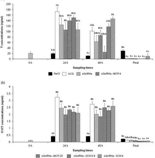
Effect of dopamine receptor antagonists in combination with salmon gonadotropin-releasing hormone analogue on plasma steroid concentrations. Plasma T concentrations
showed significant differences related to the interaction between hormonal treatments and sampling times (P< 0.001; Figure 3a). All hormonal treatments induced a marked increase in T compared with the negative control at 24 h (P< 0.05). At 48 h, the SCH 0.8, SCH 4, MCP 4 and hCG
treatments were still effective in maintaining higher T levels compared with the negative control (P< 0.05). At the
final sampling time, all hormonal treatments reduced the T concentrations (P< 0.05). The comparison between
sGnRHa alone and the combination of DA receptor antago-nists with sGnRHa showed that T levels increased with SCH 4 at 48 h (P< 0.01).
Concerning 11KT, significant differences were observed related to the interaction between hormonal treatments and sampling times (P< 0.001; Figure 3b). All hormonal
treatments induced an increase in 11KT levels at 24 h and Figure 1 Effect of hormonal treatments on plasmatic concentrations of (a) testosterone (T) and (b) 11 ketotestosterone (11KT) in pikeperch. Fish were injected with NaCl (0.9%,n= 5), human chorionic gonadotropin (hCG) (500 IU/kg, n = 5), MCP 4 or MCP 20 (metoclopramide at 4 mg/kg (n = 6) or 20 mg/kg (n= 7), respectively) and SCH 0.8 or SCH 4 (SCH23390 at 0.8 mg/kg (n = 6) or 4 mg/kg (n = 7), respectively). Blood was sampled at three sampling times: 0 and 24 h after injection and at the time of ovulation or 7 days after injection (if ovulation did not occur;final). Values are means ± SEM.a,b,c,dDifferent
48 h (P< 0.001), while at the final sampling time they were significantly abolished but only after SCH and MCP 20 treatments (P< 0.001). However, for each sampling time,
the DA receptor antagonist failed to change the 11KT level compared with the sGnRHa treatment alone.
For E2, concentrations varied significantly as a function
of the interaction between hormonal treatments and sampling times (P< 0.001; Figure 4a). All hormonal
treatments induced an increase in E2 levels at 24 h and
48 h (P< 0.05) except for MCP 4 and SCH 4 at 24 h, and
sGnRHa and MCP 4 at 48 h. At the final sampling time, all treatments decreased E2 levels (P< 0.01). Here also,
there was not any significant difference between sGnRHa alone and the combination of DA receptor antagonists
with sGnRHa except with SCH 0.8 at 48 h which increased E2 levels (P< 0.01).
No significant difference related to hormonal treatments and sampling times was obtained for plasma DHP levels (Figure 4b).
Mild variability was observed for all steroid levels. Discussion
This study investigated, for the first time, the potential regulatory effects of DA receptor antagonists alone on repro-ductive mechanisms in pikeperch. Our resultsfirst confirmed the high effectiveness of hCG in inducing the progression of FOM and ovulation in this species (Zarski et al., 2015). Figure 2 Effect of hormonal treatments on plasmatic concentrations of (a) 17β estradiol (E2) and (b) 17,20β-dihydroxy-4-pregnen-3-one (DHP) in
pikeperch. Fish were injected with NaCl (0.9%,n= 5), human chorionic gonadotropin (hCG) (500 IU/kg, n = 5), MCP 4 or MCP 20 (metoclopramide at 4 mg/kg (n= 6) or 20 mg/kg (n = 7), respectively) and SCH 0.8 or SCH 4 (SCH23390 at 0.8 mg/kg (n = 6) or 4 mg/kg (n = 7), respectively). Blood was sampled at three sampling times: 0 h and 24 h after injection and at the time of ovulation or 7 days after injection (if ovulation did not occur;final). Values are means ± SEM.a,bDifferent letters indicate significant differences between sampling times (P < 0.05).
Figure 3 Effect of hormonal treatments on plasmatic concentrations of (a) testosterone (T) and (b) 11 ketotestosterone (11KT) in pikeperch. Fish were injected with NaCl (0.9%,n= 9), human chorionic gonadotropin (hCG) (500 IU/kg, n = 7), salmon gonadotropin-releasing hormone analogue (sGnRHa) (25µg/kg, n = 7), sGnRHa (25 µg/kg) in combination with MCP 4 or MCP 20 (metoclopramide at 4 mg/kg (n = 6) or 20 mg/kg (n = 5), respectively) or with SCH 0.8 or SCH 4 (SCH23390 at 0.8 mg/kg (n= 7) or 4 mg/kg (n = 6), respectively). Blood was sampled at four sampling times: 0, 24 and 48 h after injection and at the time of ovulation or 14 days after injection (if ovulation did not occur;final). Values are means ± SEM.A,B,C,DDifferent capital letters
indicate significant differences between hormonal treatments for the same sampling time (P < 0.05). a,bDifferent lowercase letters indicate significant differences between sampling times for the same hormonal treatment (P< 0.05).
Table 2Effect of hormonal treatments on reproductive performance1in pikeperch females
Treatments
Controls2 Antagonist groups with GnRH3
Parameters NaCl hCG SEM sGnRHa SEM MCP 4 SEM MCP 20 SEM SCH 0.8 SEM SCH 4 SEM P-value
FOM prog 0 to 1 6 6 6 6 6 6 OR (%) 0A 100B 100B 100B 100B 100B 100B < 0.001 LT (h) 129.7 5.4 136.9 20.0 168.5 32.6 208.3 17.0 187.8 27.1 159.7 37.6 > 0.05 SR (%) 46.8 8.1 15.0 10.7 28.3 7.7 44.0 15.2 41.5 10.2 24.7 18.0 > 0.05 HR (%) 32.0 8.3 11.8 10.5 21.0 7.0 37.0 13.4 36.5 12.8 9.0 5.5 > 0.05 MR (%) 22.6 4.1 40.7 26.9 34.5 15.5 14.3 3.2 35.5 3.9 38.5 28.5 > 0.05
FOM prog= final oocyte maturation progression; OR = ovulation rate; LT = latency time (time between injection and ovulation); SR = survival rate; HR = hatching rate; MR= malformation rate; NaCl = saline solution; hCG = human chorionic gonadotropin; sGnRHa = analogue of gonadotropin-releasing hormone; MCP 4 or MCP 20= metoclopramide at 4 mg/kg or 20 mg/kg; SCH 0.8 or SCH 4 = SCH23390 at 0.8 mg/kg or 4 mg/kg.
1
Reproductive performance parameters are FOM prog, OR, LT, SR, HR and MR.
2Controls include negative control (NaCl) and positive controls (hCG and sGnRHa). 3
Antagonist groups with sGnRHa are treatments combining GnRH with one dopamine receptor antagonist (SCH or MCP).
During FOM, oocytes undergo a phenomenon of hydration inducing, in turn, an increase in follicle weight (Mañanóset al., 2008). After hCG treatment, the rise in GSI may thus stem from the hydration process even if the latter remains to be demon-strated in pikeperch. Contrary to this positive control, no antagonist treatments were found to induce either GSI increase or ovulation. The latterfinding is in accordance with previous reports on the Senegalese sole (Guzmanet al., 2011) and the common tench,Tinca tinca(Podhorecet al., 2016). In contrast, Cejko and Kucharczyk (2015) showed that the injection of MCP induces ovulation in the crucian carp. This inter-species differ-ence may be due to variable potency of the DA inhibition as already suggested (Mañanóset al., 2008; Zarskiet al., 2015). Consequently, we hypothesize that DA receptor antagonists applied alone would be ineffective in inducing ovulation in pikeperch because of a weakness in the DA inhibition during
the spontaneous progression of FOM. Alternatively, we might speculate that the doses chosen were not sufficient to generate a GnRH and/or LH endogenous surge. However, when com-bined with sGnRHa, these DA receptor antagonists do not prevent sGnRHa from triggering ovulation. They do not thus appear as inhibitors of the final stages of gametogenesis in pikeperch. Conversely, DA receptor antagonists would not by themselves allow the reproductive performance to be improved or impaired.
The DA receptor antagonists alone did not induce sig-nificant changes in the sex-steroid levels. These results are in accordance with prior studies in striped bass, Morone saxatilis(Kinget al., 1994), and in Senegalese sole (Guzman
et al., 2011) in which application of the D2 DA receptor antagonist did not modify the T and E2levels in plasma. The
attempt to block the D1 receptor family did not succeed in Figure 4 Effect of hormonal treatments on plasmatic concentrations of (a) 17β estradiol (E2) and (b) 17,20β-dihydroxy-4-pregnen-3-one (DHP) in
pikeperch. Fish were injected with NaCl (0.9%,n= 9), human chorionic gonadotropin (hCG) (500 IU/kg, n = 7), salmon gonadotropin-releasing hormone analogue (sGnRHa) (25µg/kg, n = 7), sGnRHa (25 µg/kg) in combination with MCP 4 or MCP 20 (metoclopramide at 4 mg/kg (n = 6) or 20 mg/kg (n = 5), respectively) or with SCH 0.8 or SCH 4 (SCH23390 at 0.8 mg/kg (n= 7) or 4 mg/kg (n = 6), respectively). Blood was sampled at four sampling times: 0, 24 and 48 h after injection and at the time of ovulation or 14 days after injection (if ovulation did not occur;final). Values are means ± SEM.A,B,CDifferent
capital letters indicate significant differences between hormonal treatments for the same sampling time (P < 0.05).a,bDifferent lowercase letters indicate significant differences between sampling times for the same hormonal treatment (P < 0.05)
altering those hormonal levels as well. The ineffectiveness of DA receptor antagonists alone in changing the sex-steroid secretion is consistent with the absence of effect on the ovulation and progression in oocyte maturation. In addition, this would indicate absence of or weakness in the dopami-nergic inhibition by the application of D1 or D2 receptor antagonist at the tested doses during the non-hormonally manipulated oocyte maturation process in pikeperch.
Interestingly, we observed that the hCG treatment was unequally effective in inducing changes in the sex-steroid levels. These results are quite surprising considering that this molecule is a common substitute for natural (endogenous) fish LH, which induces fish spawning by direct action on gonads and sex-steroid levels (Mylonaset al., 2010). Con-sidering our results from experiment 1, we noticed that at the same sampling time (e.g. 24 h), fish exhibited different stages of oocyte maturation, contrary to the synchronous stages in experiment 2. This high inter-individual variability may have induced a larger range in steroid concentrations among thefish, which could potentially explain statistically irrelevant endocrine response following hCG treatment. However, other differences between the two populations (e.g. environmental conditions, stress status,final number of individuals) may also explain this difference in sensitivity to hCG. However, some features of the experimental setup, notably the realization of two independent experiments, the low number of hormonal treatments but repeated over time, the usual number of individuals per group (Kinget al., 1994; Barryet al., 1995; Guzmanet al., 2011), lead us to think that the statistical power of the tests were sufficient to detect some potential effects.
The sGnRHa treatment stimulated the production of sex-steroids confirming the widely observed activation of the gonadotropic axis infinfishes after such a treatment (Yaron and Levavi-Sivan, 2011). Interestingly, T and E2production
were stimulated when sGnRHa was complemented with SCH depending on the dose. These results lead us to hypothesize that SCH would boost the sGnRHa effect on T and E2
secre-tion. To our knowledge, this is thefirst report of plasma sex-steroid change after exposure to D1 receptor family antagonist infish. Either this antagonist would directly block the D1 receptors at the gonad level as shown in rats (Vene-gas-Meneseset al., 2015), which in turn would stimulate the ovarian steroidogenesis. Or, the blockage of those receptors in the brain would disrupt the aromatase activity and the further metabolism of T and E2(Marshet al., 2006; Popesku
et al., 2012). Also, the blockage of D1 receptors could induce a surge in blood LH (Popesku et al., 2010) which would stimulate the sex-steroid production. Finally, we cannot rule out some indirect effects of SCH through other metabolic factors. Further studies would be needed to check the pre-sence of those receptors in the ovary and to test these regulations by investigating the aromatase expression and activity in brain and oocytes as well as LH in the blood plasma.
Plasma DHP concentrations remained basal and stable over time in all the treatments. In many teleosts, the steroid
DHP is the maturation-inducing steroid (MIS) (Nagahama and Yamashita, 2008). Progression of FOM was found to be linked to a significant increase in DHP levels in walleye,
Sander vitreus (Barry et al., 1995), and in striped bass (Mylonas et al., 1997). However, in our study, even after application of hCG or sGnRHa with which all thefish ovu-lated, no peak of DHP concentration was observed. Surpris-ingly, these results are not consistent with GnRH commonly known effects on the gonadotrope axis (Yaron and Levavi-Sivan, 2011) and with a prior study in which hCG induced an increase in plasma DHP levels during the progression of FOM in white perch,Morone americana, and white bass,Morone chrysops(Kinget al., 1995). Several hypotheses may explain thesefindings. First, as reported in walleye, the DHP could be rapidly removed from the plasma after its conjugation to a non-immunodetectable molecule such as 17,20-P-sulphate or 17,20-P-glucuronate (Scott and Canario, 1992). Second, in a closely related species, the Eurasian perch,Percafluviatilis, Migaud et al. (2003) found very low DHP levels in plasma and suggested that these levels may be higher in the early morning than at other times. Due to experimental conditions, our sampling times were performed at the beginning of the afternoon, so we may have missed the peak of detectable DHP in the blood plasma. Third, a close hormone, the 17α,20β, 21-trihydroxy-4-pregnen-3-one (20β-S or 17,20, 21-P), identified as MIS in other perciforms (Nagahama and Yamashita, 2008) could also play this role in pikeperch. That would explain the mild level of DHP in this species, even after injection with hCG or sGnRHa. Nevertheless, although Barry
et al. (1995) demonstrated that 20β-S was not detectable in walleye, others studies conducted with an other perciform, the European sea bass,Dicentrarchus labrax, suggested that DHP and 20β-S could be both considered as MIS (Sorbera
et al., 1999; Asturianoet al., 2000). From this latter study, it appears that DHP would be involved in the initiation of the oocyte maturation, just before our first sampling time, potentially explaining the absence of DHP detection in our study. From a general point of view, the absence of progestin peaks in the plasma can be due to their local and transitory actions into the gonad rendering them difficult to detect using the blood analyses. This indicates that both methodical and physiological studies on MIS in pikeperch should be reconsidered in the future. In any case, by considering DHP as the primary MIS in pikeperch, the lack of DA receptor antagonist effects (positive or negative) on FOM progression would support the absence of DHP regulation by these treatments.
In conclusion, this study, in the current experimental conditions, showed the ineffectiveness of DA receptor antagonist treatments alone, whatever the receptor family (D1 or D2), in inducing sex-steroid changes, FOM and ovu-lation in pikeperch. Combined with sGnRHa, these DA receptor antagonists did not prevent sGnRHa from triggering ovulation. However, in this combination with sGnRHa, SCH but not MCP proved efficiency to increase sGnRHa-stimulated steroid levels. Thus, only SCH would be involved in the regulation of sex-steroids indicating a putative
potentiator effect of sGnRHa through D1 DA receptor blockage. The use ofin vitrobiological tests of organ culture (e.g. brain, pituitary and ovary) could be useful in the future to pinpoint these endocrine mechanisms.
Acknowledgements
This study was partly supported by the Eurostars project (E!9390 TRANSANDER), the Lorraine region, and the Ministry of Education, Youth and Sports of the Czech Republic, projects CENAKVA (No. CZ.1.05/2.1.00/01.0024) and CENAKVA II (No. LO1205 under the NPU I programme).
Declaration of interest None.
Ethics statement
Fish were handled according to the European and French legislation for fish welfare and approved by the institutional Ethics Committee (APAFIS-2016022913149909).
Software and data repository resources Data are not deposited in an official repository.
References
Asturiano JF, Sorbera LA, Ramos J, Kime DE, Carillo M and Zanuy S 2000. Hormonal regulation of the European sea bass (Dicentrarchus labrax, L.) reproductive cycle: an individualized female approach. Journal of Fish Biology 56, 1155–1172.
Barry TP, Malison JA, Lapp AF and Procarione LS 1995. Effects of selected hormones and male cohorts on final oocyte maturation, ovulation, and steroid production in walleye (Stizostedion vitreum). Aquaculture 138, 331–347.
Bates D, Machler M, Bolker B and Walker S 2015. Fitting linear mixed-effects models using lme4. Journal of Statistical Software 67, 1–48.
Cardinaud B, Sugamori KS, Coudouel S, Vincent JD, Niznik HB and Vernier P 1997. Early emergence of three dopamine D1 receptor subtypes in vertebrates. The Journal of Biology Chemistry 272, 2778–2787.
Cejko BI, Kowalski RK, Kucharczyk D, Targonska K, Krejszeff S, Zarski D and Glogowski J 2010. Influence of the length of time after hormonal stimulation on selected parameters of milt of ideLeuciscus idusL. Aquaculture Research 41, 804–813.
Cejko BI and Kucharczyk D 2015. Application of dopaminergic antagonist: metoclopramide, in reproduction of crucian carpCarassius carassius(L.) under controlled conditions. Animal Reproduction Science 160, 74–81.
Dufour S, Sebert ME, Weltzien FA, Rousseau K and Pasqualini C 2010. Neuroendocrine control by dopamine of teleost reproduction. Journal of Fish Biology 76, 129–160.
Falahatkar B and Poursaeid S 2014. Effects of hormonal manipulation on stress responses in male and female broodstocks of pikeperchSander lucioperca. Aquaculture International 22, 235–244.
Food and Agriculture Organization of the United Nations 2017. Cultured aquatic species information programme.Sander lucioperca(Linnaeus, 1758). Retrieved on 10 July 2017 from http://www.fao.org/fishery/culturedspecies/ Sander_lucioperca/en
Fox J and Weisberg S 2011. An R companion to applied regression, 2nd edition. Sage Publications, Inc, Los Angeles, CA, USA.
Guzman JM, Cal R, Garcia-Lopez A, Chereguini O, Kight K, Olmedo M, Sarasquete C, Mylonas CC, Peleteiro JB, Zohar Y and Mañanós EL 2011. Effects ofin vivotreatment with the dopamine antagonist pimozide and gonadotropin-releasing hormone agonist (GnRHa) on the reproductive axis of Senegalese sole (Solea senegalensis). Comparative Biochemistry and Physiology, Part A 158, 235–245.
Hervé M 2016. RVAideMemoire: diverse basic statistical and graphical functions. R package version 0.9-61.
Kapsimali M, Vidal B, Gonzalez A, Dufour S and Vernier P 2000. Distribution of the mRNA encoding the four dopamine D1 receptor subtypes in the brain of the European eel (Anguilla anguilla): comparative approach to the function of D1 receptors in vertebrates. The Journal of Comparative Neurology 419, 320–343.
Kestemont P, Dabrowski K and Summerfelt RC 2015. Biology and culture of percidfishes: principles and practices. Springer, Dordrecht, The Netherlands. King W, Berlinsky DL and Sullivan CV 1995. Involvement of gonadal steroids infinal oocyte maturation of white perch (Morone americana) and white bass (M. chrysops):in vivoandin vitrostudies. Fish Physiology and Biochemistry 14, 489–500.
King W, Thomas P, Harrell RM, Hodson RG and Sullivan CV 1994. Plasma levels of gonadal steroids duringfinal oocyte maturation of striped bass, Morone saxatilisL. General and Comparative Endocrinology 95, 178–191.
Lenth R 2016. Least-squares means: the R package lsmeans. Journal of Statis-tical Software 69, 1–33.
Mañanós EL, Duncan N and Mylonas CC 2008. Reproduction and control of ovulation, spermiation and spawning in culturedfish. In Methods in reproduc-tive aquaculture: marine and freshwater species (ed. E Cabrita, V Robles and MP Herraez), pp. 3–80. CRC Press, Taylor and Francis Group, Boca Raton, FL, USA.
Marsh KE, Creutz LM, Hawkins MB and Godwin J 2006. Aromatase immunoreactivity in the bluehead wrasse brain, Thalassoma bifasciatum: immunolocalization and co-regionalization with arginine vasotocin and tyrosine hydroxylase. Brain Research 1126, 91–101.
Migaud H, Mandiki R, Gardeur JN, Fostier A, Kestemont P and Fontaine P 2003. Synthesis of sex steroids in final oocyte maturation and induced ovulation in female Eurasian perch, Perca fluviatilis. Aquatic Living Resources 16, 380–388.
Mylonas CC, Fostier A and Zanuy S 2010. Broodstock management and hormonal manipulations offish reproduction. General and Comparative Endo-crinology 165, 516–534.
Mylonas CC, Scott AP and Zohar Y 1997. Plasma gonadotropin II, sex steroids, and thyroid hormones in wild striped bass (Morone saxatilis)during spermiation and final oocyte maturation. General and Comparative Endocrinology 108, 223–236.
Nagahama Y and Yamashita M 2008. Regulation of oocyte maturation infish. Development Growth and Differentiation 50, 195–219.
Podhorec P, Socha M, Ben Ammar I, Sokolowska-Mikolajczyk M, Brzuska E, Milla S, Gosiewski G, Stejskal V, Simko M and Kouril J 2016. The effects of GnRHa with and without dopamine antagonist on reproductive hormone levels and ovum viability in tenchTinca tinca. Aquaculture 465, 158–163.
Pohlert T 2016. The Pairwise Multiple Comparisons of Mean Ranks package (PMCMR), R package.
Popesku JT, Martyniuk CJ, Denslow ND and Trudeau VL 2010. Rapid dopami-nergic modulation of thefish hypothalamic transcriptome and proteome. PLoS ONE 5, e12338.
Popesku JT, Martyniuk CJ and Trudeau VL 2012. Meta-type analysis of dopa-minergic effects on gene expression in the neuroendocrine brain of female goldfish. Frontiers in Endocrinology 3, 1–24.
Scott AP and Canario AVM 1992. 17α,20β-Dihydroxy-4-pregnen-3-one 20-sulphate: a major new metabolite of the teleost oocyte maturation-inducing steroid. General and Comparative Endocrinology 85, 91–100.
Sorbera LA, Asturiano JF, Carillo M, Cerda J, Kime DE and Zanuy S 1999.In vitro
oocyte maturation in the sea bass: effects of hCG, pituitary extract and steroids. Journal of Fish Biology 55, 9–25.
Venables WN and Ripley BD 2002. Modern applied statistics with S, 4th edition. Springer, New York, NY, USA.
Venegas-Meneses B, Padilla JF, Juarez CE, Moran JL, Moran C, Rosas-Murrieta NH, Handal A and Dominguez R 2015. Effects of ovarian dopaminergic receptors on ovulation. Endocrine 50, 783–796.
Villacorta PJ 2015. ART: Aligned rank transform for nonparametric factorial analysis, R package version 1.0. 1–5.
Yaron Z and Levavi-Sivan B 2011. Endocrine regulation offish reproduction. In Encyclopedia offish physiology: from genome to environment (ed. AP Farrell), pp. 1500–1508. Elsevier Inc, San Diego, CA, USA.
Yu KL and Peter RE 1992. Adrenergic and dopaminergic regulation of gonadotropin-releasing hormone release from goldfish preoptic-anterior hypothalamus and pituitaryin vitro. General and Comparative Endocrinology 85, 138–146. Zarski D, Horvath A, Held JA and Kucharczyk D 2015. Artificial reproduction of percidfishes. In Biology and culture of percid fishes (ed. P Kestemont, K Dabrowski and RC Summerfelt), pp. 123–161. Springer, Dordrecht, The Netherlands.
Zarski D, Kucharczyk D, Targonska K, Palinska K, Kupren K, Fontaine P and Kestemont P 2012. A new classification of pre-ovulatory oocyte maturation stages in pikeperch,Sander lucioperca(L.), and its application during artificial reproduction. Aquaculture Research 43, 713–721.
Zohar Y and Mylonas CC 2001. Endocrine manipulations of spawning in cultured fish: from hormones to genes. Aquaculture 197, 99–136.