HYPNOTIC AUTOMATICITY IN THE BRAIN AT REST: An Arterial Spin Labelling Study
PIERRE RAINVILLE
University of Montréal and University Institute of Geriatrics of Montréal, Québec, Canada
ANOUK STREFF
University Institute of Geriatrics of Montréal, Québec, Canada
JEN-ICHEN
University of Montréal and University Institute of Geriatrics of Montréal, Québec, BÉRENGÈRE HOUZÉ
University Institute of Geriatrics of Montréal, Québec, Canada
CAROLANE DESMARTEAUX
University Institute of Geriatrics of Montréal and University of Montréal Québec, Canada
MATHIEU PICHÉ
University of Québec in Trois-Rivières, Canada
Manuscript submitted January 3, 2019; final revision accepted February 11, 2019. Address correspondence to
de Montréal, 4565, ch. Queen-Mary, Montreal, Qué, H3W 1W5 Canada. Email: [email protected]
To cite this article:
Pierre Rainville, Anouk Streff, Jen-I Chen, Bérengère Houzé, Carolane Desmarteaux & Mathieu Piché (2019). Hypnotic Automaticity in the Brain at Rest: An Arterial Spin Labelling Study. International Journal of Clinical and Experimental Hypnosis, 67:4,
Abstract:
The feeling of automaticity reported by individuals undergoing a hypnotic procedure is an essential dimension of hypnosis phenomenology. In the present study, healthy participants rated their
subjective experience of automaticity and resting-state arterial spin labelling (ASL) scans were acquired before and after a standard hypnotic induction (i.e.,
perceived automaticity was positively associated with activity in the parietal operculum (PO) and seed-based coactivation analysis revealed additional associations in the anterior part of the
supracallosal cingulate cortex (aMCC). This is consistent with the role of these regions in perceived self-agency and volition and demonstrates that these effects can be evidenced at rest, in the absence of overt motor challenges. Future studies should further examine if/how these changes in brain
activity associated with automaticity might facilitate the responses to suggestions and contribute to clinical benefits of hypnosis.
The altered sense of agency is recognized as a hallmark of hypnosis phenomenology based on the idea that responses to suggestions must have a novoluntary quality, an effect known as the
(Weitzenhoffer, 1974)
(Weitzenhoffer, 2002) creating changes in perception, motivation, emotion, cognition or behavior, with a reduced sense of self-agency (e.g., Price & Barrell, 1990). The feeling of automaticity has been described by two constructs, involuntariness and effortlessness, that correlate with standard
measures of hypnotizability (Polito, Barnier, & Woody, 2013). In parallel with the investigation of this essential phenomenon, clinical effects of hypnosis are becoming more and more recognized for a variety of conditions, including pain management (Jensen, Day, & Miró, 2014; Jensen & Patterson, 2014), headaches and migraines (Hammond, 2007), irritable bowel syndrome (Schaefert, Klose, Moser, & Hauser, 2014), and in the treatment of stress and anxiety (Nunns et al., 2018; Provencal, Bond, Rizkallah, & El-Baalbaki, 2018), including symptoms of posttraumatic stress disorders (Rotaru & Rusu, 2016). Training can further be devised to enhance the feeling of automaticity (Schweiger Gallo, Pfau, & Gollwitzer, 2012), and clinical suggestion of automaticity may in turn improve
therapeutic effect of hypnosis (Kirsch & Lynn, 1999). This emphasizes how basic research on this key construct may help improve the clinical impact of hypnosis.
AUTOMATICITY IN HYPNOSIS PHENOMENOLOGY
While automaticity is increasingly recognized as a key factor, research on hypnosis has relied largely on the assessment of hypnotizability or hypnotic susceptibility/suggestibility in response to
suggested alterations in physio
hypnotic behavior since the early 19th century, but they may overlook important aspects of the
construct of hypnotizability (Council, 2002). Furthermore, the contribution of hypnosis to the response to suggestions remains a matter of debate (e.g., Kirsch, 1997; Kirsch, Mazzoni, & Montgomery, 2007; Montgomery, DuHamel, & Redd, 2000; Wagstaff, 2010; Wagstaff, Cole, & Brunas-Wagstaff, 2008). The fact that automaticity, suggestibility, and hypnotizability are not considered equivalent, coupled
facilitate the experience of automaticity (Weitzenhoffer, 1974), makes their separation difficult. This calls for a more direct within-study comparison of effects associated with dependent variables reflecting these different aspects.
Another approach to measuring hypnotizability is based on the notion that hypnosis is characterized by changes in phenomenological experience that can be assessed using self-report measurements (e.g., Pekala & Kumar, 2000; Price & Barrell, 1990; Wagstaff et al., 2008). Given that the felt sense of automaticity in response to suggestions is a cardinal feature of hypnosis, a measure of perceived automaticity appears essential. Consistent with this link, positive associations have been documented between standardized measures of hypnotizability and the degree of automaticity or involuntariness reported in response to hypnotic suggestions (K. S. Bowers, 1981; P. Bowers, 1982; Cunningham & Ramos, 2012; Dufresne et al., 2010). Combining subjective reports of automaticity with standard behavioral measures of hypnosis responsiveness may help assess these related but distinctive aspects of hypnosis phenomenology.
In addition to measuring the self-reported experience of automaticity and the behavioral responses to hypnotic suggestions, self-reported hypnotic depth scores have been used to characterize hypnotic experiences (e.g., Lecron, 1953; Price & Barrell, 1990) and have been
validated in the context of fMRI research (Oakley, Deeley, & Halligan, 2007). The construct of depth Cardeña, Jonsson, Terhune, & Marcusson-Clavertz, 2013, p.2). This construct can also be conceived as a response to
hypnotic induction and is tied to state theories of hypnosis implying that such a state could be experienced outside of a formal induction procedure and independent from the assessment of suggestion-related effects inherent to hypnotizability testing (Wagstaff et al., 2008). Reports of hypnotic depth have been associated with hypnotic automaticity (Price & Barrell, 1990) and with hypnotizability scores on standard tests (Perry & Laurence, 1980; Tart, 1970). However, these separate aspects of hypnosis responsiveness are potentially dissociable and may reflect partly distinct changes in brain activity (McGeown, Mazzoni, Vannucci, & Venneri, 2015).
The concurrent consideration of automaticity, hypnotic depth, and individual hypnotizability scores may further help attain a more comprehensive characterization of brain mechanisms underlying hypnosis phenomenology.
BRAIN IMAGING OF AUTOMATICITY
Consistent with the notion that the effects of hypnosis on brain activity largely depend on the specific suggestions tested, distinctive changes in brain activity have been reported in brain imaging studies examining the modulation of pain, auditory and visual perception, cognitive processes, and motor responses (Landry, Lifshitz, & Raz, 2017). Responses to hypnotic suggestions are generally described as automatic and/or involuntary across those domains. This may imply a common process, independent from the domain-specific target of the suggestions and possibly related to the general hypnotic induction process.
The first brain imaging study examining more directly the brain correlates of hypnotic
involuntariness showed robust parietal activity while subjects moved their arm in response to hypnotic suggestions that their arm would be moved passively (Blakemore, Oakley, & Frith, 2003). Importantly, the study included control passive and active movements in a nonhypnotic condition to allow
comparing brain responses associated with normal sensory feedback alone (passive condition) and executive motor processes (active condition). In the normal passive condition, the afferent sensory
signal conveyed through the somatosensory system activated the parietal cortex in the region of the inferior parietal lobule (parietal operculum/supramarginal gyrus). In the active condition, motor
cortices were also activated to produce the motor command. In this condition, there was less parietal activity, consistent with the feedforward model of motor control (Wolpert & Ghahramani, 2000). Indeed, during voluntary actions, a motor command is sent to the motoneurons while a copy of this efferent signal is sent to sensory areas of the parietal cortex to monitor the correspondence between the sensory feedback and the expected effect of the action that was prescribed (i.e., prediction signal). Importantly, when the feedback matches the expectations, activity is reduced in the parietal cortex. However, when there is a mismatch (i.e., prediction error) or during passive movement (i.e., no prediction signal), the parietal cortex is strongly activated.
Stronger parietal activation found in the hypnotic condition compared to the active condition suggests that the automaticity experienced during hypnosis may reflect a perturbation in the
generation or transmission of the efferent copy or in the comparison with the sensory feedback. This
movement was external (Blakemore & Frith, 2003; Blakemore et al., 2003; Frith, Blakemore, & Wolpert, 2000). Interestingly, a reduction in posterior parietal activity (supramarginal gyrus) has also been associated with a reduction in awareness of involuntary movements, consistent with the
possibility that activity in this area reflects the subjective experience of involuntariness (Deeley, Walsh, et al., 2013). However, in these studies, it is not clear if the parietal response is a motor-specific effect or if it might reflect a nonmotor-specific phenomenon associated with the general feeling of automaticity associated with hypnosis responding.
In the present study, we assessed resting-state brain activity using arterial spin labelling (ASL), a functional magnetic resonance imaging method sensitive to brain perfusion (van Osch et al., 2018). This approach allows direct comparisons of regional cerebral blood flow between states and
(i.e. without any stimulus or task) are positively associated with activity in the parietal operculum (Blakemore et al., 2003), and are related to a fronto-parietal network involved in phenomenological aspects of self-agency and volition (Darby, Joutsa, Burke, & Fox, 2018). In order to verify that automaticity-related effects did not simply reflect nonspecific aspects of hypnosis phenomenology, brain activity was also examined in relation to hypnotic depth and hypnotizability scores to highlight putative distinctive neural associations. Importantly, participants were not selected a priori based on their scores to allow conducting regression analyses examining individual differences in hypnosis-related effect across a representative range of hypnotizability (see Jensen et al., 2017).
METHOD
Participants
Thirty-three healthy participants (f =17) between the ages of 19 and 45 (mean = 27.38 years; SD = 6.81) were recruited and scanned in this study. Volunteers were recruited through the research participant registry at the Research Center of the Institut universitaire de gériatrie de Montréal
(CRIUGM) and through advertisement on the campus of Université de Montréal. Exclusion criteria included self-reported history of chronic pain, psychiatric and neurological disorders, as well as recent (2 weeks prior to the experiment) consumption of pain medication or medication that could alter pain perception and modulation (e.g., antihypertensive, anxiolytic, antidepressant, and other psychotropic agents). During the screening phone call, participants were asked to stop consuming alcohol at least 1 day before the experiment, and to refrain from consuming tea and coffee on the day of the scan. All participants were part of a separate psychophysiological experiment involving similar experimental conditions prior to the present fMRI study. Of the 33 subjects scanned, 2 were excluded from the analysis due to incomplete imaging data (n=1) and high Beck depression score (n=1). Of the remaining 31 subjects, 3 subjects could not contribute to the analysis of automaticity and 2 to the analysis of hypnotic depth due to technical issues with the recording of self-report data.
All experimental procedures met the guidelines of the latest revision of the Declaration of Helsinki and were approved by the Ethics committee of the
universitaire de gériatrie de Montréal. All participants provided written informed consent and received monetary compensation for their participation.
Experimental Procedures
This study consisted of two experimental sessions carried out on separate days. The first session served to determine hypnotizability and familiarize participants to the pain protocol included in this study.
The second part of the experiment consisted of a brain imaging session in which a structural scan, resting-state ASL perfusion, and pain-related blood-oxygen-level-dependent (BOLD) fMRI data were acquired. For the purpose of this paper, we focus solely on the results of the resting-state ASL data in which we examined brain correlates of self-reported automaticity, hypnotic depth and
individual hypnotizability following hypnotic induction. Procedures related to the pain protocol will be described in a separate article.
Prescanning Session
Hypnotizability was assessed with the French version of the Stanford Hypnotic Susceptibility Scale, Form A (SHSS:A; Bourassa & Leclerc, 1991). The SHSS:A is administered individually in 30 to 45 minutes, starting with an induction phase with suggestions of relaxation and absorption, and
comprising 12 test items involving ideomotor (e.g., hand lowering) and cognitive (e.g., amnesia) suggestions. Behavioral responses to these items provided the hypnotizability score.
Brain Imaging Session
Scanning procedure. Imaging data were collected at the Unité de neuroimagerie fonctionnelle of the CRIUGM using a 3T Siemens Magneton TIM Trio magnetic resonance imaging (MRI) system
with a 12-channel head coil. Participants were positioned comfortably in the scanner and stabilized with a pelvic strap as well as foam pads to immobilize the head. MRI compatible earphones were used to communicate with the participants and deliver hypnotic suggestion while reducing the noise from the scanner. The entire scanning session lasted approximately 75 minutes.
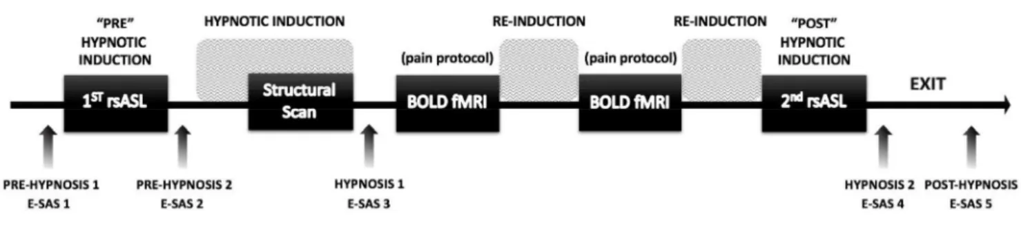
During the scanning session, a T1-weighted structural MRI scan and two resting-state whole-brain perfusion scans were acquired using a pseudo-continuous ASL (pCASL) sequence (van Osch et al., 2018). Figure 1 shows the detailed experimental protocol during the imaging session. Briefly,
the session started with the first resting- utes), followed by
hypnotic induction (14 minutes). The anatomical scan was then launched 8 minutes into the induction, such that the induction was completed at the end of the anatomical scan. Then two BOLD fMRI scans were acquired as part of the pain protocol (not discussed further here). The imaging session
utes) followed by suggestions to end hypnosis and recover normal alertness.
Figure 1. Experimental design. In the brain imaging session, we performed five scans. The two resting-state ASL scans described in the present report were performed at the beginning of the session, before the induction of hypnosis, and at the end of the session, while subjects were still under hypnosis. The instructions for hypnotic induction were given after the first ASL scan and throughout the structural scan. Two BOLD-fMRI scans were then performed involving painful
electrical stimulation and suggestions for pain modulation (not reported here). Instructions were given after these scans to maintain or deepen hypnosis (Re-Induction). Instructions to come out of hypnosis and recover normal alertness were given after the second ASL scan (Exit). Experiential
self-assessment scales (E-SAS) were used between scans to obtain self-ratings of automaticity and
hypnotic depth at five time points, before (E-SAS 1-2), during (E-SAS 3-4), and after hypnosis (E-SAS 5).
Structural images. High-resolution (1 mm isotropic voxels) anatomical images were acquired using T1-weighted multi-echo MPRAGE sequence (ME-MPRAGE) with the following parameters: 176 slices per whole brain volume, repetition time = 2530 ms, 4 echo times = 1.64, 3.50, 5.36, 7.22, 13, and 15 ms combined to form one root mean squared volume, flip angle = 7°, field of view (FOV) = 256 mm, matrix = 256 × 256, parallel imaging with GRAPPA 2, and a bandwidth of 651 Hz/Px. The anatomical scan lasted 6.3 minutes.
Arterial Spin Labeling (ASL) Image Acquisition. Images from two resting-state ASL runs were acquired with eyes closed using a 2D single-echo pseudo-continuous arterial spin labeling (pCASL) sequence with the following parameters: 16 axial slices of 6 mm thickness; 4 mm x 4 mm in-plane resolution; FOV = 256 mm; TR = 3000 ms; TE = 10 ms; FA = 90°; 20 pulses; 82 RF blocks; label offset = 100 mm; labeling time =1476 ms; post label delay = 900ms. The duration of each resting state ASL scan was 6.12 minutes, producing 120 acquisition volumes (60 pairs of control and labeled images).
Hypnotic Induction
A prerecorded verbatim hypnotic induction based on the SHSS:A was adapted to the scanner environment and administered via earphones before and during the anatomical scan. The length of the induction was approximately 15 minutes. The aim of the hypnotic induction is to reach a state of deep relaxation without falling asleep. It starts with a fixation cross displayed on the scanner screen that participants are asked to focus on. Then follows a brief psychoeducation about what hypnosis is and is not, before participants are mainly told to slowly enter a relaxed state where they are
encouraged to let their body become heavy and comfortable. It is further suggested that participants may notice that their thoughts might come and go and that they can concentrate on the present moment by simply being attentive to and curious about what is happening. It is then suggested that they should concentrate on the voice and let themselves be guided through the protocol by simply
paying attention to the suggestions and not letting themselves be bothered by the scanner noise. A count (1 to 20) is also used to deepen the hypnotic state. Additional suggestions taken from the induction procedure (e.g., count) were given after each fMRI scan for the maintenance and/or deepening of the hypnotic state (see Figure 1).
Experiential Assessment
Ratings were collected throughout the scanning session to document the subjective
experience of participants using experiential self-assessment scales (E-SAS). A visual analog scale (VAS) format was used for all measurements to simplify the instructions and procedures. Ratings were obtained by asking subjects to open their eyes and use a hand-held MRI-compatible response key to move a cursor on a VAS displayed on a computer screen projected in the scanner room and viewed by the participant through a mirror mounted on the head coil. Scales were explained in the training session and reexplained immediately before the scanning session. All hypnosis-related ratings were converted linearly to values between 0 and 100.
The experience of automaticity and hypnotic depth was assessed at five time points: prior to the first ASL scan (prehypnosis 1); immediately after the ASL scan and before the hypnotic induction (prehypnosis 2); at the end of the hypnotic induction and anatomical scan (hypnosis 1); at the end of
the 2nd posthypnosis).
Low and high automaticity/involuntariness were defined as having perfect control over actions and thoughts (0/100) versus being a passive witness of actions and thoughts (100/100) (Price & Barrell, 1990). The instructions and anchors of the hypnotic depth VAS were adapted in an attempt to reduce the typical positive skewness of the distribution (see Wagstaff et al., 2008). Subjects were instructed
as follows: from not at all to deeply hypnotized
ratings excluded any explicit reference to suggestions and did not use numerical anchors (i.e., VASs were used for all ratings).
Behavioral measures were analyzed using SPSS v. 25. The normality of distributions across subjects was first tested at each time point and on the hypnosis change scores (post- vs preinduction; see below) using the
Kolmogorov-automaticity and hypnotic depth were assessed using
repeated-with the Greenhouse-Geisser adjustment of dfs when applicable. A hypnosis change score of automaticity and hypnotic depth was then computed for each subject by subtracting the mean prehypnosis ratings from the mean hypnosis ratings. Associations between hypnotizability scores (SHSS:A) and hypnosis-related change scores were tested using partial correlation controlling for prehypnosis ratings.
Participants also rated their level of mental relaxation (calm, relaxed versus active, agitated), attention stability (stable versus unstable), and attentional focus (focal versus global perception). Relaxation increased from the prehypnosis to the hypnosis condition, but ratings did not change significantly from hypnosis to posthypnotic measurements, so we could not exclude a temporal confound independent from hypnosis (i.e., order effect). Attentional stability and focus did not vary significantly with hypnosis. For the sake of parsimony, these additional variables are not discussed further.
ASL Data Processing & Analysis
Data preprocessing and CBF quantification. ASL data were processed using the ASLtbx (Wang et al., 2008; https://cfn.upenn.edu/~zewang/ASLtbx.php) and SPM8
(http://www.fil.ion.ucl.ac.uk/spm/software/spm8/) in MATLAB R2015a (https://www.mathworks.com). Custom wrapper script made available by Chris Rorden
ASL scan was preprocessed independently. Briefly, each dataset was first motion-corrected to produce a mean image that was then coregistered to the T1-weighted anatomical scan. The ASL images were then resliced to match the coregistered mean image, and spatially smoothed with a 6 mm FWHM Gaussian kernel. Cerebral blood flow (CBF) was then estimated by subtraction of the tag and control images to create a series of 60 CBF volumes (quantified as ml/100g/min; Wang et al., 2008). These 60 successive volumes were then averaged to obtain one mean CBF image before hypnosis and one during hypnosis for each subject. The mean CBF and the T1 images were
normalized CBF images to remove out-of-brain voxels.
CBF statistical analysis. Voxel-by-voxel statistical analysis of the mean CBF volumes was performed using SPM 8. Data from 5 subjects were excluded from statistical analysis due to
incomplete dataset (4 subjects) or outlier bias (1 subject), leaving a total of 28 datasets (50% female). To address our questions, we performed the following analyses to assess changes in rCBF following hypnosis and examine how those changes relate to individual increases in automaticity and hypnotic depth, and to individual hypnotizability scores. Paired t-tests were computed at each brain voxel to assess changes in mean CBF maps from the pre- to postinduction scan (POST vs PRE). Note that this basic comparison is confounded with temporal effects (i.e. prehypnosis data was always acquired before hypnosis; see Figure 1) and was computed as a preliminary step before the more informative between-subject regression analyses. Regressions tested the hypothesis that individual changes in rCBF following hypnotic induction were related to the magnitude of changes reported in automaticity and hypnosis depth ratings. All regression analyses included the individual mean prehypnosis scores as a covariate of no interest to ensure that effects associated with hypnotic induction were not
confounded with baseline individual differences (note that the prehypnosis covariates did not show significant effects on rCBF; not reported).
Regression on hypnotic automaticity. A directed search was performed on the parietal operculum (PO) based on the previously reported activation of this region in relation to automatic responding (Blakemore et al., 2003) and awareness of involuntary actions (Deeley, Walsh, et al., 2013). A small-volume correction thresholded at p < .05 (FWE correction) was used to assess significance using a 15 mm-radius sphere centered on the peak coordinates reported by Blakemore et al. (2003): x=58, y=-32, z=24. A global search was also conducted on the rest of the brain using a permissive threshold of p-uncorrected < .001 to reduce type II error and provide exploratory findings. Similar regression analyses were conducted on hypnotic depth and hypnotizability scores.
Coactivation analysis. A follow-up analysis was performed on the peak PO response to determine if the observed change was associated with brain networks involving the mid/anterior cingulate cortex recently shown to contribute to perceived self-agency and volition (Darby et al., 2018). The coactivation analysis was performed using the first eigenvariate of this region, extracted from a 15 mm-radius sphere centered on the PO response observed in the regression on
automaticity.
RESULTS
Experiential Ratings
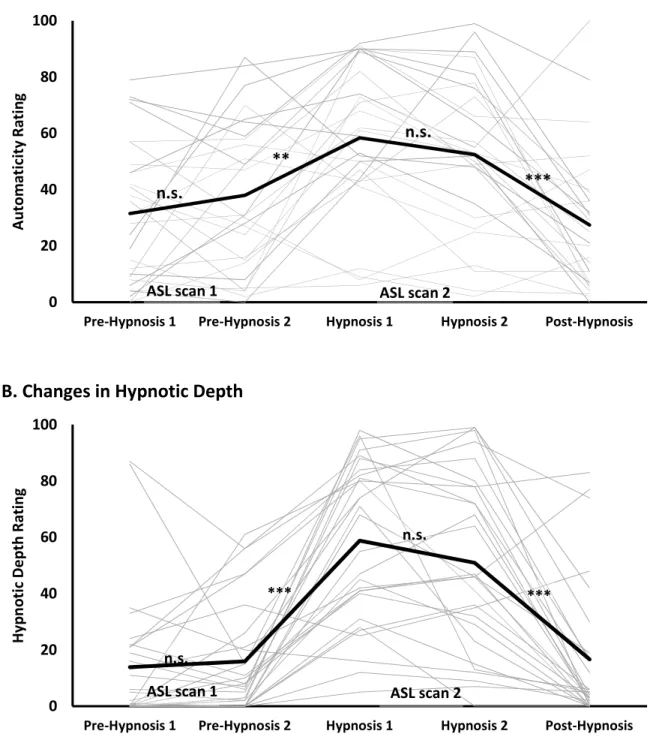
Individual ratings of automaticity and hypnotic depth across the five successive measurements are shown in Figures 2A and 2B, respectively. The distribution of ratings of automaticity did not depart significantly from normality at any time point (p ngs also did not differ significantly from normality in the hypnosis condition (p
normally distributed at the pre- and posthypnotic time points (p
change scores (i.e., hypnosis minus prehypnosis) were considered normal for both automaticity and hypnotic depth (p , and these indices were used in the analysis of imaging data.
Figure 2. Perceived automaticity (A) and hypnotic depth (B) reported throughout the scanning
session. Ratings increased significantly on both scales during hypnosis and returned to pre-hypnosis levels at the post-hypnosis measure. The thick black lines represent the group means and the thin grey lines represent individual subjects. ASL scans were acquired in the pre-hypnosis and the hypnosis phase (see Figure 1). ** p < .01, *** p < .001; n.s. : not significant (p > .05).
0 20 40 60 80 100
Pre-Hypnosis 1 Pre-Hypnosis 2 Hypnosis 1 Hypnosis 2 Post-Hypnosis
Au to m at ic ity R at in g
A. Changes in Automaticity
** *** n.s. n.s.ASL scan 1 ASL scan 2
0 20 40 60 80 100
Pre-Hypnosis 1 Pre-Hypnosis 2 Hypnosis 1 Hypnosis 2 Post-Hypnosis
Hy pn ot ic De pt h Ra tin g
B. Changes in Hypnotic Depth
*** ***
n.s.
n.s.
Participants reported changes in their subjective experience following the induction of hypnosis (Figure 2A). Across the five successive measurements, significant effects were found for both
automaticity, F(2.91,72.84) = 12.21, p p2 = .328, and hypnotic depth, F(2.69,70.01) = 35.61, p p2 = .578. Successive within-subject contrasts confirmed a significant increase in automaticity immediately after hypnotic induction, F(1,26) = 13.13, p p2 = .344, and a significant decrease after the end of hypnosis, F(1,26) = 18.63, p p2 = .427. Contrast effects on hypnotic depth confirmed a similar increase immediately after hypnotic induction, F(1,26) = 57.59, p p2= .689, and decrease after the end of hypnosis, F(1,26) = 35.20, p p2 = .575. Importantly, the mean automaticity and depth returned to the prehypnotic levels following the suggestions to come out of hypnosis (posthypnosis) at the end of the scanning session (post- vs prehypnotic measures: p ns). This confirms that the increase in automaticity and hypnotic depth reported following hypnotic induction was not confounded with nonspecific effects of time or order of conditions.
The increase in automaticity reported following hypnotic induction was significantly correlated to the increase in hypnotic depth (r = .53, p = .007). In turn, the increase in hypnotic depth was positively associated with hypnotizability (SHSS:A; r = .52, p = .008). However, no significant
correlation was observed between the increase in automaticity and hypnotizability (r = .07, p = .73). Similar results were obtained with nonparametric tests.
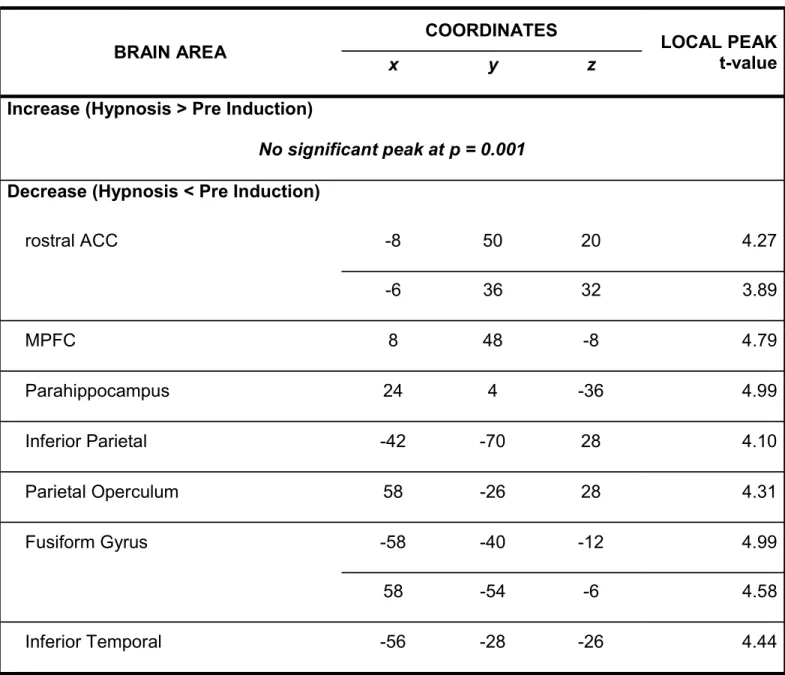
Changes in Resting-State ASL
A decrease in rCBF was observed following hypnotic induction in the following areas: rostral anterior cingulate cortex (ACC), medial prefrontal cortex (MPFC), the inferior parietal gyrus, the PO, fusiform gyrus, inferior temporal gyrus, and parahippocampal gyrus (coordinates reported in Table 11). No significant increase in rCBF was observed. Note that these effects are reported at a
permissive statistical threshold and are confounded with time. This contrast was computed prior to
1 Tables are at the end of this document
the follow-up regression analyses to show baseline changes in rCBF before testing the effects associated with automaticity.
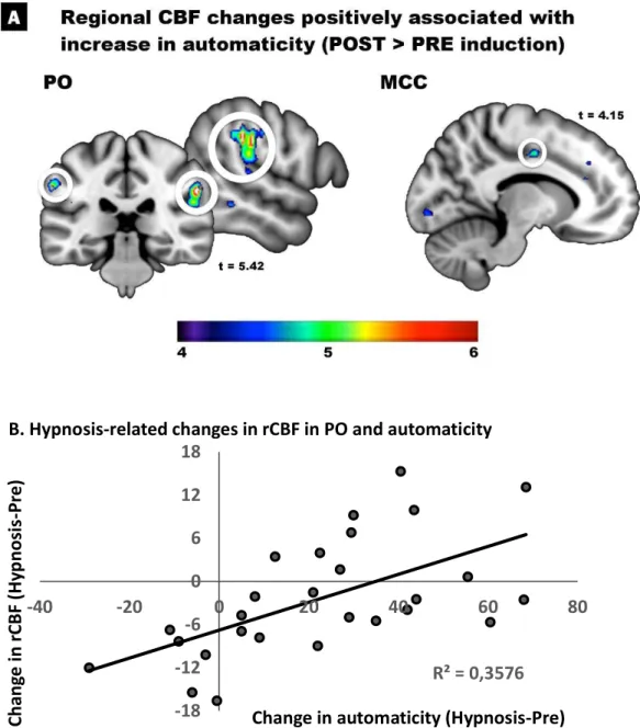
Regression on hypnotic automaticity. Between-subject regression were conducted on the mean change reported by each participant in experiential ratings. The magnitude of the increase in automaticity predicted changes in rCBF in the right PO at the location defined based on the results of Blakemore et al. (2003; Figure 3A). The coordinates of the highest peak was just a few mm away from the center of the a priori ROI (Table 2), and the next highest peak across the whole brain was found in the homologous area of the contralateral hemisphere. Another peak was found in the anterior part of the mid cingulate cortex. Plotting the peak effect of automaticity in the PO revealed that most participants displayed a decrease in rCBF at that location following hypnotic induction, consistent with the results of the main contrast in this region (Table 1). However, subjects reporting an increase in automaticity generally showed an increase or a smaller decrease in rCBF in this area (Figure 3B).
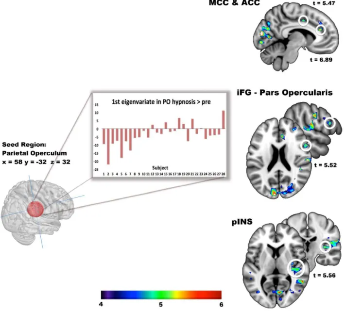
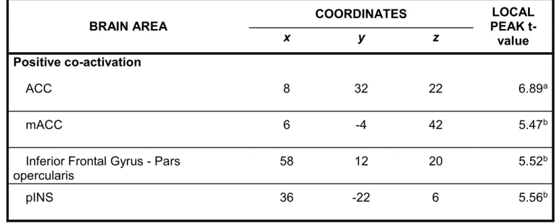
Coactivation analysis. A coactivation analysis based on the PO seed further revealed positive peaks in the anterior cingulate cortex (ACC), anterior mid-cingulate cortex (aMCC), inferior frontal gyrus (iFG) pars opercularis, and posterior insula (pINS) (Figure 4; Table 3). No significant negative coactivation was found.
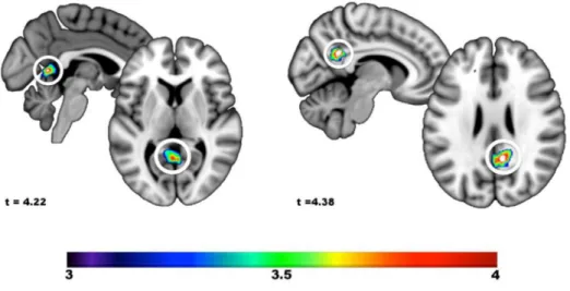
Regression on hypnotic depth and individual hypnotizability. Between-subject regression analyses were also performed using individual changes in hypnotic depth and the individual hypnotizability scores as predictors of hypnosis-related rCBF changes (Figure 5). The analysis of hypnotic depth revealed a single positive peak at x= -2, y = -56, z = 4 (t = 4.22, p < .001). This effect was difficult to interpret anatomically as the peak fell in the midline, within cerbrospinal fluid (CSF), below the most caudal part of the posterior cingulate cortex and above the cerebellum. The
= 26 (t = 4.38, p < .001). This peak was located in the retrosplenial part of the posterior cingulate cortex. No negative regression peak was found on hypnotic depth and hypnotizability.
Figure 3. Changes in rCBF from pre-hypnosis to hypnosis positively associated with the increase in self-reported automaticity. (A) A significant peak response is confirmed in the right parietal operculum (PO) based on a priori hypotheses and using a small-volume correction (FEW corrected p < .05; see text). Additional peaks are found at more permissive thresholds in the left PO and in the middle cingulate cortex (aMCC). Peak coordinates are reported in Table 2. (B) The peak found in the right PO is illustrated graphically to show that the few participants reporting a decrease in automaticity during hypnosis (i.e. x < 0) displayed a reduction in rCBF in the PO (i.e. y < 0) while participants reporting an increase in automaticity generally showed a smaller decrease or an increase in rCBF at the PO peak. R² = 0,3576 -18 -12 -6 0 6 12 18 -40 -20 0 20 40 60 80 Ch an ge in rC BF (Hy pn os is -P re )
Change in automaticity (Hypnosis-Pre) B. Hypnosis-related changes in rCBF in PO and automaticity
Figure 4. Co-activation of automaticity-related peak in the PO. A sphere of 15mm diameter centered on the peak coordinates of automaticity observed in the PO was used as the seed for the
co-activation analysis. The first eigenvariate of the automaticity effect across voxels was extracted for each subject. The bar graph shows these values for each subject, ranked as a function of the change scores in automaticity. Peak co-activation sites are observed in the middle (MCC) and anterior parts (ACC) of the supracallosal cingulate cortex. Additional peaks are shown in the frontal operculum (iFG) and posterior insula (pINS). Peak coordinates are reported in Table 3.
Figure 5. Changes in rCBF from pre-hypnosis to hypnosis positively associated with the increase in self-reported hypnotic depth and individual hypnotic susceptibility scores. (A) The peak response observed in the retrosplenial area with hypnotic depth fell into the cerebrospinal fluid making its interpretation difficult. (B) The effect of hypnotic susceptibility was observed in the posterior cingulate area, within a region generally associated with the posterior default mode network. No other peak was found.
DISCUSSION
The objective of this report was to explore resting-state brain activity in relation to the increase in automaticity reported following a standard hypnotic induction. Results confirmed the hypothesized involvement of the PO in the increased feelings of automaticity reported during hypnosis. Activity in PO was also associated with frontal executive areas related to perceived self-agency and volition. Corollary analyses suggested that activity in the retrosplenial cingulate cortex, a posterior node of the default-mode network (DMN; Raichle, 2015), was positively related to individual hypnotizability
scores. These results are interpreted in relation to the neurocognitive literature on self-agency and volition, followed by a discussion of strengths and limitations of the present study, and a consideration of distinctive aspects of ASL methodology.
Understanding brain mechanisms underlying hypnosis requires an assessment of basic effects associated with standard hypnotic induction procedures separately from specific suggestions
administered to modify perception (e.g., analgesia) or behavior (e.g., paralysis). Consistent with the present results, three early positron emission tomography studies demonstrated hypnosis-related activation of fronto-parietal networks, including the anterior cingulate cortex and the parietal cortex (PO and/or inferior parietal lobule; Maquet et al., 1999; Rainville, Hofbauer, Bushnell, Duncan, & Price, 2002; Rainville et al., 1999). However, these effects were assessed in conditions involving the recall of pleasant autobiographical memories and color hallucinations (Maquet et al., 1999), or
thermal stimuli applied to the hand (Rainville, Hofbauer, Bushnell, Duncan, & Price, 2002; Rainville et al., 1999). This implies that changes associated with hypnotic induction in these studies might reflect a modification of brain activity related to memory, visual or somatosensory processes rather than, or in addition to, the isolated effects of hypnotic induction (i.e., -state. The present study overcomes this limitation and suggests that changes in activity in the PO and
the aMCC relate to the experience of automaticity following the induction of neutral hypnosis in the absence of any overt confounding factor. We also observed a positive association between
hypnosis-hypnotizability score.
Automaticity and the Parietal Operculum (PO)
Increased activity in the PO has been previously reported during a typical motor challenge used to test hypnotizability (Blakemore et al., 2003). In this previous study, activation of the parietal cortex was associated with the attribution of a movement to an external cause, whether the
movement was truly passive or produced actively in response to hypnotic suggestions and perceived as passive. This effect is consistent with the involvement of this region in perceived self-agency as shown in other experimental and clinical contexts (Blakemore & Frith, 2003). A more recent study further demonstrated that parietal activity (supramarginal g. : x=-48, y=-36, z=30), at a location very close to the peak observed in the present study (see Table 2), is related to the awareness of
involuntary actions (Deeley, Walsh, et al., 2013). The present results further demonstrate that activity in this area may also be associated with a feeling of automaticity denoting an altered sense of self-agency experienced at rest, in the absence of any overt behavioral response or challenge. This is discussed further in the next subsection.
Importantly, the coactivation analysis further supports the notion that changes in PO activity relate to a brain network involving the mid and anterior cingulate cortices and the inferior frontal cortex (Table 3). These areas are part of the executive network of the brain broadly construed.
Interestingly, coactivation of the anterior cingulate cortex and the inferior parietal cortex has also been reported at very similar locations during hypnosis-induced limb paralysis, with unsuccessful efforts to move the paralyzed arm associated with increased activity in the anterior cingulate cortex and
adjacent supplementary motor area (see Tables 2a and 3b in Deeley, Oakley, et al., 2013; also see ACC activation in Burgmer et al., 2013). Using a lesion mapping method, a recent report showed that
the dorsal anterior cingulate region constitutes a core of functional networks including the parietal operculum and underlying the experience of volition (Darby et al., 2018). Lesions to the dorsal anterior cingulate area produce deficits in self-generated goal-directed actions (Cohen, Kaplan, Moser, Jenkins, & Wilkinson, 1999), while electrical stimulation of this area during neurosurgical procedures may produce subjective experiences of urges to act or persevere (Parvizi, Rangarajan, Shirer, Desai, & Greicius, 2013). Theoretical accounts of the function of the dorsal ACC have
suggested a key role in conflict monitoring, evaluative processes, and cognitive control (e.g., Carter, Botvinick, & Cohen, 1999; Shackman et al., 2011). Recent models further integrate a motivational perspective, suggesting that the ACC regulates control processes based on the cost-benefit analysis (i.e. value) of allocating executive resources (Shenhav, Cohen, & Botvinick, 2016). Interestingly, this value-based process of executive engagement is also suggested to translate into a subjective
experience of mental effort (Shenhav et al., 2017), another experiential dimension relevant to hypnosis phenomenology (Polito et al., 2013). These findings have direct implication for the current models of hypnosis and reinforce the proposition that hypnosis is a relevant experimental model to study and test neuro-cognitive/phenomenological theories of consciousness and brain function (Rainville & Price, 2003; Raz & Shapiro, 2002; Terhune, Cleeremans, Raz, & Lynn, 2017).
Neurocognitive Models of Hypnotic Automaticity
Modern theories highlight neurocognitive mechanisms underlying these effects. In the dissociated control theory, automaticity is explained by a disconnection between executive and supervisory or monitoring processes (Egner, Jamieson, & Gruzelier, 2005; Jamieson & Woody, 2007). In contrast, the cold control theory suggests that executive control processes are monitored but operate without conscious awareness (Dienes, 2007). Related to this view, a metacognitive perspective further suggests that highly hypnotizable individuals may be aware of executive
representation of self-agency (Terhune & Hedman, 2017). These propositions consistently involve a modulation in the interplay between executive processes and self-monitoring or the meta-cognitive representation of these processes. In the present study, activity in PO is consistent with an altered sense of agency and the positive coactivation with the anterior cingulate regions suggests that hypnotic automaticity reflects a concomitant engagement of frontal executive networks. This implies that both executive and monitoring processes are active in individuals reporting high levels of
hypnotic automaticity but that their interaction or representation may be modified such that executive engagement is experienced with an altered sense of agency.
Automaticity in the brain at rest. Importantly, coactivation of the PO and aMCC were observed here during a resting state. This demonstrates that feelings of automaticity reported following hypnotic induction reflect changes in brain activity that are not necessarily tied to overt behavioral responses to suggestions. In the present context, subjects were asked to rate automaticity in relation to behaviors and thoughts so we may assume that the reports of increased automaticity at rest might reflect the
perceived self- -agency over behavioral
responses. However, given that the loss of control may not be reported spontaneously during neutral hypnosis (Cardeñåa et al., 2013), further experiential analysis would be indicated to document the underlying meaning conveyed by automaticity ratings at rest. We speculate that the changes in brain activity produced during neutral hypnosis leads to covert alterations of perceived self-agency that affect the experienced automaticity over thoughts and/or behavior and is revealed explicitly during the motor challenges typically employed to assess hypnotic responsiveness.
Expectancy effects at rest. The present findings appear difficult to reconcile with a strict interpretation involving only expectancy, demand characteristics, report biases, or a post hoc misattributions of volition. Nevertheless, these results are not incompatible with expectancy-related effects based on socio-cultural representations (schemas) of hypnosis that might be reinforced by the suggestions used in the induction protocol to create response sets that incorporate the experience of
automaticity (see Kirsch & Lynn, 1997; Lynn, 1997). This is in line with modern proposals integrating socio-cognitive factors to understand how hypnotic involuntariness comes about efficient cause and reflects changes in brain activity material cause (Lynn, Laurence, & Kirsch, 2015; see a discussion of the Aristotelian causal analysis of hypnosis in Killeen & Nash, 2003). The present results may help reconcile the socio-cognitive perspective with state theories of hypnosis as changes produced by hypnotic induction on brain activity may be viewed at least in part as the actualization of expectancy-induced effects affecting fundamental aspects of self-representation to produce a
subjective experience of involuntariness. Consistent with this integrative perspective, the coactivation of the PO-aMCC observed at rest following hypnotic induction may reflect such preparatory response set inherent to the experience of involuntariness, involving executive and self-monitoring processes, or their meta-representation, and installed prior to and independently from the typical behavioral challenges used to test hypnosis responsiveness.
Hypnosis and the Brain Default Mode Network (DMN)
In addition to the reports of automaticity, hypnotic depth ratings and individual hypnotizability scores were used as predictors of changes in brain activity following hypnotic induction. Analysis of hypnotic depth revealed a single activation site that could not be readily attributed to brain activity as its peak fell in CSF (see Figure 5a), while individuals with higher hypnotizability scores showed a stronger hypnosis-related increase in the posterior cingulate region, a part of the posterior DMN (Figure 5b). This result should be considered with caution as the statistical threshold applied in these analyses may not adequately protect against type I error. However, these results provide yet another case demonstrating the importance of considering the multidimensionality of hypnosis
neurophenomenology.
Two previous studies examined changes in BOLD-signal in the DMN following a classical block-design paradigm involving visual stimuli alternating with baseline visual fixation during hypnosis
and nonhypnotic conditions. McGeown et al. focused on the baseline period (visual fixation) between visual stimuli viewed passively or under suggestions for hallucinations (McGeown, Mazzoni, Venneri, & Kirsch, 2009). The typical positive BOLD-response during the fixation epoch in the medial frontal region of the DMN was reduced during hypnosis in highly hypnotizable individuals. This suggested that highs were possibly less prone to engage in self-oriented mind wandering typically associated with DMN activity (Raichle, 2015). In contrast, Deeley et al. used a similar block-design paradigm but looked at brain activity during the visual stimuli (Deeley et al., 2012). In this study, the typical negative BOLD-response evoked by the stimuli in the medial nodes of the DMN was increased in participants reporting more hypnotic depth. Intriguingly, results of McGeown et al. (2009) and Deeley et al. (2012) appear contradictory given the expected inverse colinearity of the on/off stimulus periods in a block-design. Indeed, BOLD-fMRI provides relative measures and a larger decrease in the DMN during the stimuli implies a relatively larger increase during fixation. These results appear difficult to reconcile without a more refined analysis of the dynamic shifts in brain activity at the onset and offset of the visual stimuli.
A follow-up study confirmed that highs display reduced anterior DMN connectivity in a more -state condition comparable to the present study (i.e., with no stimulus or task confound; McGeown et al., 2015). These findings are supported by an independent study suggesting a
reduction in connectivity within both the anterior and posterior sectors of the DMN as well as in the -parietal systems during pleasant autobiographical recall (Demertzi et al., 2011). Similarly, decreased connectivity was found between the posterior DMN and dorsolateral frontal executive regions during hypnotic conditions involving autobiographical recall of pleasant events compared to rest (Jiang et al., 2017). These results are generally consistent with the notion that hypnotic induction modifies the brain dynamical interplay between competing internal/external representations (Rainville & Price, 2003). However, specific convergences and mechanisms are
difficult to extract given the diversity of methodologies involving possible stimuli- and tasks-related confounds in most of the available studies.
In contrast to these effects suggesting decrease DMN connectivity, increased resting-state connectivity has also been reported in the medial posterior node of the default-brain network (precuneus) and in both the dorsolateral prefrontal and the posterior parietal regions in highly hypnotizable individuals (Pyka et al., 2011). Although the analysis of hypnotic induction was
confounded with the suggestion of arm paralysis in this study, the resting state scans were performed without any motor activity or explicit motor challenges. This might be more readily comparable to the present results showing a positive association between activity in the posterior cingulate cortex (part of the posterior DMN) and individual hypnotizability. Such observation implies that the changes in connectivity in the posterior DMN may not be strictly tied to the motor challenge and that hypnotic induction may be sufficient to modify the dynamics of this system in highly hypnotizable individuals. Taken together, these results call for further delineation of the roles of the anterior and posterior components of the DMN in hypnosis across a variety of experimental conditions and in relation to hypnosis-relevant experiential dimensions.
Strengths and Limitations
The present study has several strengths with its share of limitations.
Neutral hypnosis and automaticity. One important aspect of this study is the assessment of the effects of hypnotic induction using a standardized method with no confounding effect associated with concurrent stimuli or tasks, and with no suggestions to alter specific aspects of perception of
behavior. This allowed testing basic associations between brain activity and neutral hypnosis (see Mazzoni, Venneri, McGeown, & Kirsch, 2013; also, Jensen et al., 2015; Landry et al., 2017; Oakley & Halligan, 2013). However, this may yet introduce another confound as distinctive spontaneous mental contents have been reported as a function of hypnotizability (Cardeña et al., 2013). It is not clear
whether this mental activity should be considered inherent to hypnotic states and some research groups have explicitly avoided using a neutral hypnosis condition by comparing brain activity
associated with a relevant task (e.g., pleasant autobiographical memory) performed with and without hypnosis (e.g., Demertzi et al., 2011; Jiang et al., 2017; Maquet et al., 1999). As discussed above, such design introduces a reciprocal limitation as the assessment of hypnosis-related effects may not generalize to other conditions involving different stimuli or tasks.
Another limitation of the present design is the constant order of the conditions where the
nonhypnosis control always preceded the hypnosis condition (see Figure 1). This was imposed by the other constraints of the study and therefore a temporal effect cannot be excluded. However, since the subjective reports of automaticity and hypnotic depth did return to baseline after the instruction to recover a normal state after hypnosis, consistent with previous reports (e.g., Oakley et al., 2007), we may thus assume that automaticity-related effects on brain activity reflect more than nonspecific time-related changes in alertness or vigilance. These phenomena may nevertheless be functionally time-related in many contexts (e.g., Paus et al., 1997) such that a clear separation may be difficult to achieve without counterbalancing conditions.
Interestingly, the increase reported in automaticity during hypnosis was correlated with the increase in hypnotic depth but not with the individual hypnotizability assessed in a separate session. Such observation may have helped distinguish changes in brain activity associated with these
variables. However, it may also question the interpretation that the increase in automaticity reported here reflected a hypnosis-related phenomenon. Yet another possibility is that hypnotic automaticity may be experienced at rest in individuals that do not respond readily to the behavioral challenges used in the standard hypnotizability tests. This problem relates to classic issues regarding the definitions of hypnosis, hypnotizability, and suggestibility, the central role of
automaticity/involuntariness in hypnosis responsiveness, and the pervasiveness of this experience in a variety of conditions not typically described as hypnosis (e.g., Kirsch & Lynn, 1997; Lynn, Maxwell,
& Green, 2017; Wagstaff, 2010; Woody & Sadler, 2016). We submit that the present results are relevant to understanding hypnosis to the extent that automaticity is a major feature of hypnosis phenomenology that may be experienced in a variety of contexts and reflect changes in activity within brain networks involved in self-agency.
Arterial-spin-labelling. One unique aspect of the present study is the use of ASL. The
acquisition of resting-state data using ASL was motivated by the possibility to compare hypnosis and nonhypnosis conditions directly. ASL provides an indirect quantitative measure of brain activity and allows for a direct voxel-by-voxel comparison of mean absolute rCBF between conditions acquired in independent scans (van Osch et al., 2018). This assessment of state-related brain activity is not directly possible with the most common BOLD-signal fMRI which only allows measuring relative changes induced by stimuli or tasks. However, this advantage of the ASL method is obtained at the cost of sensitivity, a major limitation inherent to the relatively low signal-to-noise ratio. This implies that ASL studies may miss some effects observed with more sensitive methods. The directed-search of automaticity-related effects in the PO using a small volume correction was based on previous findings of Blakemore et al. (2003) and planned explicitly to mitigate this potential power issue. The validity of the findings is therefore directly dependent upon the validity of the proposed justification to focus on this region. The inclusion of whole-brain results at a more permissive statistical criterion was offered here as a compromise to reduce the risk of type II error but these additional findings should be considered with caution.
Finally, we must underscore that the present report did not include the typical connectivity analyses conducted on resting-state brain imaging data, as performed in a few previous BOLD-fMRI studies of neutral hypnosis (Demertzi et al., 2011; Jiang et al., 2017; McGeown et al., 2015). The coactivation analysis performed in the present study was based on a between-subject regression using the difference in mean rCBF measured in each participant at the PO and across the full duration of the prehypnosis and hypnosis scans. In contrast, resting-state connectivity analysis of
BOLD-fMRI data typically involves spontaneous fluctuations in signal across regions within each scan. These methods should be considered complementary.
CONCLUSION
Medical imaging provides a unique window to study hypnosis neurophenomenology by using an integrative approach based on experiential reports and compatible with neurocognitive models of consciousness and brain function. The present study demonstrates that the subjective experience of automaticity reported during hypnosis reflects activity within brain networks underlying the sense of self-agency. Importantly, these effects were observed during neutral hypnosis at rest and cannot be readily explained by a modification of the mechanisms underlying overt behavioral responses. This is consistent with the notion that the induction of hypnosis produces changes in brain activity and subjective experience that precede and may facilitate responses to suggestions. Accordingly, these automaticity-related effects may constitute more valid and powerful proxies to assess the effects of hypnosis on the responsiveness to suggestions in both experimental and clinical contexts.
AUTHORS NOTE
Acknowledgements: We thank Audrey-Anne Dubé, Alexandre Lehmann, Mathieu Roy, and Etienne Vachon-Presseau for their constructive comments and their help in the planning phase of the study, and Rick Hoge and Pierre Bellec for their recommendations regarding the acquisition of
resting-state brain imaging data using arterial-spin labelling. Carollyn Hurst and André Cyr helped acquire data at the Unité de Neuroimagerie Fonctionnelle (UNF) of the CRIUGM. We also thank Ze Wang for his help in the analysis of brain imaging data with the ASLtbx.
Contribution of authors: PR, AS, BH, and MP conceived the study. AS planned the
experiment, performed the hypnotic induction, and assessed the hypnotizability of each participant individually using the SHSS-A. AS acquired data with BH and JIC. BH, AS, and PR analyzed the behavioral data. JIC and PR analyzed the brain imaging data. CD updated the literature review and
contributed to the interpretation of the results. PR, JIC, and CD wrote the paper. All authors provided feedback on or approved the final version of the manuscript.
REFERENCES
Blakemore, S. J., & Frith, C. (2003). Self-awareness and action. Current Opinion in Neurobiology, 13(2), 219-224.
Blakemore, S. J., Oakley, D. A., & Frith, C. D. (2003). Delusions of alien control in the normal brain. Neuropsychologia, 41(8), 1058-1067.
Bourassa, M., & Leclerc, C. (1991). L'hypnose clinique en médecine dentaire [Hypnosis in dental medicine]. Montréal, Canada: Méridien.
Bowers, K. S. (1981). Do the Stanford Scales tap the "classic suggestion effect"? International Journal of Clinical and Experimental Hypnosis, 29, 42-53. doi: 10.1080/00207148108409142 Bowers, P. (1982). The classic suggestion effect: relationships with scales of hypnotizability,
effortless experiencing, and imagery vividness. International Journal of Clinical and Experimental Hypnosis, 30, 270-279. doi:10.1080/00207148208407264
Burgmer, M., Kugel, H., Pfleiderer, B., Ewert, A., Lenzen, T., Pioch, R., . . . Konrad, C. (2013). The mirror neuron system under hypnosis - Brain substrates of voluntary and involuntary motor activation in hypnotic paralysis. Cortex, 49(2), 437-445. doi: 10.1016/j.cortex.2012.05.023 Cardeña, E., Jonsson, P., Terhune, D. B., & Marcusson-Clavertz, D. (2013). The
neurophenomenology of neutral hypnosis. Cortex, 49(2), 375-385. doi: 10.1016/j.cortex.2012.04.001
Carter, C. S., Botvinick, M. M., & Cohen, J. D. (1999). The contribution of the anterior cingulate cortex to executive processes in cognition. Reviews in the Neurosciences, 10(1), 49-57.
Cohen, R. A., Kaplan, R. F., Moser, D. J., Jenkins, M. A., & Wilkinson, H. (1999). Impairments of attention after cingulotomy. Neurology, 53(4), 819-824.
Council, J. R. (2002). A historical overview of hypnotizability assessment. American Journal of Clinical Hypnosis, 44(3-4), 199-208. doi: 10.1080/00029157.2002.10403480
Cunningham, P. F., & Ramos, P. (2012). Involuntary experiencing and the performance of hypnotic test suggestions. International Journal of Clinical and Experimental Hypnosis, 60, 416-431. doi: 10.1080/00207144.2012.701090
Darby, R. R., Joutsa, J., Burke, M. J., & Fox, M. D. (2018). Lesion network localization of free will. Proceedings of the National Academy of Sciences USA, 115(42), 10792-10797. doi: 10.1073/pnas.1814117115
Deeley, Q., Oakley, D. A., Toone, B., Bell, V., Walsh, E., Marquand, A. F., ... Halligan, P. W. (2013). The functional anatomy of suggested limb paralysis. Cortex, 49(2), 411-422. doi:
10.1016/j.cortex.2012.09.016
Deeley, Q., Oakley, D. A., Toone, B., Giampietro, V., Brammer, M. J., Williams, S. C., & Halligan, P. W. (2012). Modulating the default mode network using hypnosis. International Journal of Clinical and Experimental Hypnosis, 60(2), 206-228. doi: 10.1080/00207144.2012.648070 Deeley, Q., Walsh, E., Oakley, D. A., Bell, V., Koppel, C., Mehta, M. A., & Halligan, P. W. (2013).
Using hypnotic suggestion to model loss of control and awareness of movements: An exploratory FMRI study. PLoS One, 8(10), e78324. doi: 10.1371/journal.pone.0078324 Demertzi, A., Soddu, A., Faymonville, M. E., Bahri, M. A., Gosseries, O., Vanhaudenhuyse, A., ...
network connectivity. Progress in Brain Research, 193, 309-322. doi: 10.1016/B978-0-444-53839-0.00020-X
Dienes, Z. P., J. (2007). Executive control without conscious awareness: The cold control theory. In J. A. Jamieson (Ed.), Hypnosis and conscious states: The cognitive neuroscience perspective (pp. 293 314.). New York, NY: Oxford University Press.
Dufresne, A., Rainville, P., Dodin, S., Barre, P., Masse, B., Verreault, R., & Marc, I. (2010).
Hypnotizability and opinions about hypnosis in a clinical trial for the hypnotic control of pain and anxiety during pregnancy termination. International Journal of Clinical and Experimental Hypnosis, 58, 82-101. doi: 10.1080/00207140903310865
Egner, T., Jamieson, G., & Gruzelier, J. (2005). Hypnosis decouples cognitive control from conflict monitoring processes of the frontal lobe. NeuroImage, 27(4), 969-978.
Elkins, G. R., Barabasz, A. F., Council, J. R., & Spiegel, D. (2015). Advancing research and practice: the revised APA Division 30 definition of hypnosis. International Journal of Clinical and Experimental Hypnosis, 63(1), 1-9. doi: 10.1080/00207144.2014.961870
Frith, C. D., Blakemore, S. J., & Wolpert, D. M. (2000). Abnormalities in the awareness and control of action. Philosophical Transactions of the Royal Society of London. Series B, Biological
Sciences, 355(1404), 1771-1788. doi: 10.1098/rstb.2000.0734
Hammond, D. C. (2007). Review of the efficacy of clinical hypnosis with headaches and migraines. International Journal of Clinical and Experimental Hypnosis, 55(2), 207-219. doi:
10.1080/00207140601177921
Jamieson, G. A., & Woody, E. (2007). Dissociated control as a paradigm for the
cognitive-neuroscience research and theorising in hypnosis. In G. A. Jamieson (Ed.), Hypnosis and conscious states: The cognitive-neuroscience perspective (pp. 111-129). Oxford, UK: Oxford University Press.
Jensen, M. P., Adachi, T., Tome-Pires, C., Lee, J., Osman, Z. J., & Miro, J. (2015). Mechanisms of hypnosis: toward the development of a biopsychosocial model. International Journal of Clinical and Experimental Hypnosis, 63, 34-75. doi: 10.1080/00207144.2014.961875 Jensen, M. P., Day, M. A., & Miró, J. (2014). Neuromodulatory treatments for chronic pain: efficacy
and mechanisms. Nature Reviews. Neurology, 10(3), 167-178. doi:10.1038/nrneurol.2014.12 Jensen, M. P., Jamieson, G. A., Lutz, A., Mazzoni, G., McGeown, W. J., Santarcangelo, E. L., ..
Terhune, D. B. (2017). New directions in hypnosis research: strategies for advancing the cognitive and clinical neuroscience of hypnosis. Neuroscience of Consciousness, 3(1). doi: 10.1093/nc/nix004
Jensen, M. P., & Patterson, D. R. (2014). Hypnotic approaches for chronic pain management: clinical implications of recent research findings. The American Psychologist, 69(2), 167-177.
doi:10.1037/a0035644
Jiang, H., White, M. P., Greicius, M. D., Waelde, L. C., & Spiegel, D. (2017). Brain Activity and Functional Connectivity Associated with Hypnosis. Cerebral Cortex, 27(8), 4083-4093. doi:10.1093/cercor/bhw220
Killeen, P. R., & Nash, M. R. (2003). The four causes of hypnosis. International Journal of Clinical and Experimental Hypnosis, 51, 195-231.
Kirsch, I. (1997). Suggestibility or hypnosis: What do our scales really measure? International Journal of Clinical and Experimental Hypnosis, 45, 212-225. doi: 10.1080/00207149708416124 Kirsch, I., & Lynn, S. J. (1997). Hypnotic involuntariness and the automaticity of everyday life.
American Journal of Clinical Hypnosis, 40(1), 329-348.
Kirsch, I., & Lynn, S. J. (1999). Automaticity in clinical psychology. American Psychologist, 54, 504-515.
Kirsch, I., Mazzoni, G., & Montgomery, G. H. (2007). Remembrance of hypnosis past. American Journal of Clinical Hypnosis, 49(3), 171-178.
Landry, M., Lifshitz, M., & Raz, A. (2017). Brain correlates of hypnosis: A systematic review and meta-analytic exploration. Neuroscience and Biobehavioral Reviews, 81(Pt A), 75-98. doi: 10.1016/j.neubiorev.2017.02.020
Lecron, L. M. (1953). A method of measuring the depth of hypnosis. Journal of Clinical and Experimental Hypnosis, 1(2), 4-7. doi:10.1080/00207145308409812
Lynn, S. J. (1997). Automaticity and hypnosis: A sociocognitive account. International Journal of Clinical and Experimental Hypnosis, 45(3), 239-250.
Lynn, S. J., Laurence, J.-R., & Kirsch, I. (2015). Hypnosis, suggestion, and suggestibility: An integrative model. American Journal of Clinical Hypnosis, 57(3), 314-329.
doi:10.1080/00029157.2014.976783
Lynn, S. J., Maxwell, R., & Green, J. P. (2017). The hypnotic induction in the broad scheme of hypnosis: A sociocognitive perspective. American Journal of Clinical Hypnosis, 59(4), 363-384. doi:10.1080/00029157.2016.1233093
Maquet, P., Faymonville, M. E., Degueldre, C., Delfiore, G., Franck, G., Luxen, A., & Lamy, M. (1999). Functional neuroanatomy of hypnotic state. Biological Psychiatry, 45(3), 327-333. Mazzoni, G., Venneri, A., McGeown, W. J., & Kirsch, I. (2013). Neuroimaging resolution of the altered
state hypothesis. Cortex, 49(2), 400-410. doi: 10.1016/j.cortex.2012.08.005
McGeown, W. J., Mazzoni, G., Vannucci, M., & Venneri, A. (2015). Structural and functional
correlates of hypnotic depth and suggestibility. Psychiatry Research, 231(2), 151-159. doi: 10.1016/j.pscychresns.2014.11.015
McGeown, W. J., Mazzoni, G., Venneri, A., & Kirsch, I. (2009). Hypnotic induction decreases anterior default mode activity. Consciousness and Cognition, 18(4), 848-855. doi:
10.1016/j.concog.2009.09.001
Montgomery, G. H., DuHamel, K. N., & Redd, W. H. (2000). A meta-analysis of hypnotically induced analgesia: how effective is hypnosis? International Journal of Clinical and Experimental Hypnosis, 48, 138-153.
Nunns, M., Mayhew, D., Ford, T., Rogers, M., Curle, C., Logan, S., & Moore, D. (2018). Effectiveness of nonpharmacological interventions to reduce procedural anxiety in children and adolescents undergoing treatment for cancer: A systematic review and meta-analysis. Psychooncology, 27(8), 1889-1899. doi: 10.1002/pon.4749
Oakley, D. A., Deeley, Q., & Halligan, P. W. (2007). Hypnotic depth and response to suggestion under standardized conditions and during fMRI scanning. International Journal of Clinical and Experimental Hypnosis, 55, 32-58. doi: 10.1080/00207140600995844
Oakley, D. A., & Halligan, P. W. (2013). Hypnotic suggestion: Opportunities for cognitive neuroscience. Nature Reviews. Neuroscience, 14(8), 565-576. doi: 10.1038/nrn3538 Parvizi, J., Rangarajan, V., Shirer, W. R., Desai, N., & Greicius, M. D. (2013). The will to persevere
induced by electrical stimulation of the human cingulate gyrus. Neuron, 80(6), 1359-1367. doi: 10.1016/j.neuron.2013.10.057
Paus, T., Zatorre, R. J., Hofle, N., Caramanos, Z., Gotman, J., Petrides, M., & Evans, A. C. (1997). Time-related changes in neural systems underlying attention and arousal during the
performance of an auditory vigilance task. Journal of Cognitive Neuroscience, 9(3), 392-408. Pekala, R. J., & Kumar, V. K. (2000). Operationalizing "trance". I: Rationale and research using a
psychophenomenological approach. American Journal of Clinical Hypnosis, 43(2), 107-135. doi: 10.1080/00029157.2000.10404265
Perry, C., & Laurence, J. -R. (1980). Hypnotic depth and hypnotic susceptibility: A replicated finding. International Journal of Clinical and Experimental Hypnosis, 28, 272-280. doi:
Polito, V., Barnier, A. J., & Woody, E. Z. (2013). Developing the Sense of Agency Rating Scale (SOARS): An empirical measure of agency disruption in hypnosis. Consciousness and Cognition, 22(3), 684-696. doi:10.1016/j.concog.2013.04.003
Price, D. D., & Barrell, J. J. (1990). The structure of the hypnotic state: A self-directed experiential study. In J. J. Barrell (Ed.), The experiential method: Exploring the human experience (pp. 85-97). Acton, MA, USA: Copely Publishing Group.
Provencal, S. C., Bond, S., Rizkallah, E., & El-Baalbaki, G. (2018). Hypnosis for burn wound care pain and anxiety: A systematic review and meta-analysis. Burns, 44(8), 1870-1881. doi: 10.1016/j.burns.2018.04.017
Pyka, M., Burgmer, M., Lenzen, T., Pioch, R., Dannlowski, U., Pfleiderer, B., ... Konrad, C. (2011). Brain correlates of hypnotic paralysis-a resting-state fMRI study. NeuroImage, 56(4), 2173-2182. doi: 10.1016/j.neuroimage.2011.03.078
Raichle, M. E. (2015). The brain's default mode network. Annual Review of Neuroscience, 38, 433-447. doi: 10.1146/annurev-neuro-071013-014030 [doi]
Rainville, P., Hofbauer, R. K., Bushnell, M. C., Duncan, G. H., & Price, D. D. (2002). Hypnosis modulates activity in brain structures involved in the regulation of consciousness. Journal of Cognitive Neuroscience, 14(6), 887-901.
Rainville, P., Hofbauer, R. K., Paus, T., Duncan, G. H., Bushnell, M. C., & Price, D. D. (1999). Cerebral mechanisms of hypnotic induction and suggestion. Journal of Cognitive Neuroscience, 11(1), 110-125.
Rainville, P., & Price, D. D. (2003). Hypnosis phenomenology and the neurobiology of consciousness. International Journal of Clinical and Experimental Hypnosis, 51, 105-129.
Raz, A., & Shapiro, T. (2002). Hypnosis and neuroscience: A cross talk between clinical and cognitive research. Archives of General Psychiatry, 59(1), 85-90.
Rotaru, T. S., & Rusu, A. (2016). A meta-analysis for the efficacy of hypnotherapy in alleviating ptsd symptoms. International Journal of Clinical and Experimental Hypnosis, 64, 116-136. doi: 10.1080/00207144.2015.1099406
Schaefert, R., Klose, P., Moser, G., & Hauser, W. (2014). Efficacy, tolerability, and safety of hypnosis in adult irritable bowel syndrome: Systematic review and meta-analysis. Psychosomatic Medicine, 76(5), 389-398. doi: 10.1097/PSY.0000000000000039
Schweiger Gallo, I., Pfau, F., & Gollwitzer, P. M. (2012). Furnishing hypnotic instructions with
implementation intentions enhances hypnotic responsiveness. Consciousness and Cognition, 21(2), 1023-1030. doi: 10.1016/j.concog.2012.03.007
Shackman, A. J., Salomons, T. V., Slagter, H. A., Fox, A. S., Winter, J. J., & Davidson, R. J. (2011). The integration of negative affect, pain and cognitive control in the cingulate cortex. Nature Reviews. Neuroscience, 12(3), 154-167. doi: 10.1038/nrn2994
Shenhav, A., Cohen, J. D., & Botvinick, M. M. (2016). Dorsal anterior cingulate cortex and the value of control. Nature Neuroscience, 19(10), 1286-1291. doi: 10.1038/nn.4384
Shenhav, A., Musslick, S., Lieder, F., Kool, W., Griffiths, T. L., Cohen, J. D., & Botvinick, M. M. (2017). Toward a rational and mechanistic account of mental effort. Annual Review of Neuroscience, 40, 99-124. doi: 10.1146/annurev-neuro-072116-031526
Tart, C. T. (1970). Self-report scales of hypnotic depth. International Journal of Clinical and Experimental Hypnosis, 18, 105-125. doi: 10.1080/00207147008415909
Terhune, D. B., Cleeremans, A., Raz, A., & Lynn, S. J. (2017). Hypnosis and top-down regulation of consciousness. Neuroscience and Biobehavioral Reviews, 81(Pt A), 59-74.
doi:10.1016/j.neubiorev.2017.02.002
Terhune, D. B., & Hedman, L. R. A. (2017). Metacognition of agency is reduced in high hypnotic suggestibility. Cognition, 168, 176-181. doi: 10.1016/j.cognition.2017.06.026
van Osch, M. J., Teeuwisse, W. M., Chen, Z., Suzuki, Y., Helle, M., & Schmid, S. (2018). Advances in arterial spin labelling MRI methods for measuring perfusion and collateral flow. Journal of Cerebral Blood Flow and Metabolism, 38(9), 1461-1480. doi: 10.1177/0271678X17713434 Wagstaff, G. F. (2010). Hypnosis and the relationship between trance, suggestion, expectancy and
depth: some semantic and conceptual issues. American Journal of Clinical Hypnosis, 53, 47-59. doi: 10.1080/00029157.2010.10401746
Wagstaff, G. F., Cole, J. C., & Brunas-Wagstaff, J. (2008). Measuring hypnotizability: The case for self-report depth scales and normative data for the long Stanford scale. International Journal of Clinical and Experimental Hypnosis, 56, 119-142. doi: 10.1080/00207140701849452 Wang, Z., Aguirre, G. K., Rao, H., Wang, J., Fernández-Seara, M. A., Childress, A. R., & Detre, J. A.
(2008). Empirical optimization of ASL data analysis using an ASL data processing toolbox: ASLtbx. Magnetic resonance imaging, 26(2), 261 269. doi:10.1016/j.mri.2007.07.003
Weitzenhoffer, A. M. (1974). When is an "instruction" an "instruction"? International Journal of Clinical and Experimental Hypnosis, 22, 258-269. doi: 10.1080/00207147408413005
Weitzenhoffer, A. M. (2002). Scales, scales and more scales. American Journal of Clinical Hypnosis, 44, 209-219. doi: 10.1080/00029157.2002.10403481
Wolpert, D. M., & Ghahramani, Z. (2000). Computational principles of movement neuroscience. Nature Neuroscience, 3(11s), 1212-1217. doi: 10.1038/81497
Woody, E., & Sadler, P. (2016). What can a hypnotic induction do? American Journal of Clinical Hypnosis, 59, 138-154. doi: 10.1080/00029157.2016.1185004
Table 1. Changes in rCBF following hypnotic induction. BRAIN AREA COORDINATES LOCAL PEAK t-value x y z
Increase (Hypnosis > Pre Induction)
No significant peak at p = 0.001 Decrease (Hypnosis < Pre Induction)
rostral ACC -8 50 20 4.27 -6 36 32 3.89 MPFC 8 48 -8 4.79 Parahippocampus 24 4 -36 4.99 Inferior Parietal -42 -70 28 4.10 Parietal Operculum 58 -26 28 4.31 Fusiform Gyrus -58 -40 -12 4.99 58 -54 -6 4.58 Inferior Temporal -56 -28 -26 4.44