HAL Id: hal-02424854
https://hal-ens.archives-ouvertes.fr/hal-02424854v2
Submitted on 3 Jan 2021
HAL is a multi-disciplinary open access
archive for the deposit and dissemination of sci-entific research documents, whether they are pub-lished or not. The documents may come from teaching and research institutions in France or abroad, or from public or private research centers.
L’archive ouverte pluridisciplinaire HAL, est destinée au dépôt et à la diffusion de documents scientifiques de niveau recherche, publiés ou non, émanant des établissements d’enseignement et de recherche français ou étrangers, des laboratoires publics ou privés.
intravacuolar proliferation of Listeria monocytogenes in
epithelial cells
Caroline Peron Cane, José Carlos Fernandez, Julien Leblanc, Laure
Wingertsmann, Arnaud Gautier, Nicolas Desprat, Alice Lebreton
To cite this version:
Caroline Peron Cane, José Carlos Fernandez, Julien Leblanc, Laure Wingertsmann, Arnaud Gautier, et al.. Fluorescent secreted bacterial effectors reveal active intravacuolar proliferation of Listeria mono-cytogenes in epithelial cells. PLoS Pathogens, Public Library of Science, 2020, 16 (10), pp.e1009001. �10.1371/journal.ppat.1009001�. �hal-02424854v2�
RESEARCH ARTICLE
Fluorescent secreted bacterial effectors reveal
active intravacuolar proliferation of Listeria
monocytogenes in epithelial cells
Caroline Peron-CaneID1,2, Jose´-Carlos FernandezID2, Julien Leblanc2,
Laure Wingertsmann2, Arnaud GautierID3,4, Nicolas DespratID1,2,5*, Alice LebretonID2,6* 1 Laboratoire de Physique de l’E´ cole normale supe´rieure, ENS, Universite´ PSL, CNRS, Sorbonne Universite´, Universite´ de Paris, Paris, France, 2 Institut de biologie de l’ENS (IBENS), E´ cole normale supe´rieure, CNRS, INSERM, Universite´ PSL, Paris, France, 3 Sorbonne Universite´, E´ cole normale supe´rieure, Universite´ PSL, CNRS, Laboratoire des Biomole´cules, LBM, Paris, France, 4 Institut Universitaire de France, 5 UFR de Physique, Universite´ Paris-Diderot, Universite´ de Paris, Paris, France, 6 INRAE, IBENS, Paris, France
*nicolas.desprat@ens.psl.eu(ND);alice.lebreton@ens.psl.eu(AL)
Abstract
Real-time imaging of bacterial virulence factor dynamics is hampered by the limited number of fluorescent tools suitable for tagging secreted effectors. Here, we demonstrated that the fluorogenic reporter FAST could be used to tag secreted proteins, and we implemented it to monitor infection dynamics in epithelial cells exposed to the human pathogen Listeria
mono-cytogenes (Lm). By tracking individual FAST-labelled vacuoles after Lm internalisation into
cells, we unveiled the heterogeneity of residence time inside entry vacuoles. Although half of the bacterial population escaped within 13 minutes after entry, 12% of bacteria remained entrapped over an hour inside long term vacuoles, and sometimes much longer, regardless of the secretion of the pore-forming toxin listeriolysin O (LLO). We imaged LLO-FAST in these long-term vacuoles, and showed that LLO enabled Lm to proliferate inside these com-partments, reminiscent of what had been previously observed for Spacious Listeria-contain-ing phagosomes (SLAPs). Unexpectedly, inside epithelial SLAP-like vacuoles (eSLAPs),
Lm proliferated as fast as in the host cytosol. eSLAPs thus constitute an alternative
replica-tion niche in epithelial cells that might promote the colonizareplica-tion of host tissues.
Author summary
Bacterial pathogens secrete virulence factors to subvert their hosts; however, monitoring bacterial secretion in real-time remains challenging. Here, we developed a convenient method that enabled fluorescent imaging of secreted proteins in live microscopy, and applied it to the human pathogenListeria monocytogenes. Listeria has been described to
invade cells and proliferate in their cytosol; it is first internalized inside vacuoles, from where it escapes thanks to the secretion of virulence factors that disrupt membranes. Our work revealed the existence, in human epithelial cells, of a population ofListeria that failed
to escape vacuoles but instead multiplied efficiently therein, despite—and in fact, thanks
a1111111111 a1111111111 a1111111111 a1111111111 a1111111111 OPEN ACCESS
Citation: Peron-Cane C, Fernandez J-C, Leblanc J,
Wingertsmann L, Gautier A, Desprat N, et al. (2020) Fluorescent secreted bacterial effectors reveal active intravacuolar proliferation of Listeria
monocytogenes in epithelial cells. PLoS Pathog
16(10): e1009001.https://doi.org/10.1371/journal. ppat.1009001
Editor: Rene´e M. Tsolis, University of California,
Davis, UNITED STATES
Received: May 27, 2020 Accepted: September 21, 2020 Published: October 12, 2020
Peer Review History: PLOS recognizes the
benefits of transparency in the peer review process; therefore, we enable the publication of all of the content of peer review and author responses alongside final, published articles. The editorial history of this article is available here:
https://doi.org/10.1371/journal.ppat.1009001
Copyright:© 2020 Peron-Cane et al. This is an open access article distributed under the terms of theCreative Commons Attribution License, which permits unrestricted use, distribution, and reproduction in any medium, provided the original author and source are credited.
Data Availability Statement: All relevant data are
within the manuscript and its Supporting Information files.
to—the active secretion of a toxin that permeates membranes. This intravacuolar niche may provideListeria with an alternative strategy to colonize its host.
Introduction
Bacterial pathogens harness distinct colonization strategies to take advantage of their host resources. While some remain extracellular, others adopt an intracellular lifestyle. Internalisa-tion into host cells provides invasive bacteria with multiple abilities, such as that of crossing cellular barriers, escaping humoral immune surveillance, or disseminating throughout the organism as cargo of circulating cells. After internalisation, bacteria are entrapped inside pri-mary vacuoles from where they can follow two distinct routes: either subverting endomem-brane compartments, or leaving them. For instanceChlamydia trachomatis, Brucella abortus
orLegionella pneumophila perturb the maturation and rearrange the properties of vacuoles,
thereby creating a compartment prone to their replication [1]. Others, such asShigella flexneri
orListeria monocytogenes, typically do not grow inside endomembrane compartments, but
rather escape from entry vacuoles and gain access to the host cell cytoplasm, where they can replicate as well as exploit the host actin cytoskeleton for intracellular motility and cell-to-cell spread [2].
The foodborne pathogenListeria monocytogenes (hereafter, Lm) is the causative agent of
lis-teriosis, and has emerged as a model facultative intracellular bacterium [3,4]. This pathogen can cross the protective barriers of its host and colonize tissues and organs by promoting its internalisation into non-phagocytic cells. The classical scheme ofLm intracellular life cycle
implies that, both in professional phagocytes and in epithelial cells,Lm rapidly escapes from
entry vacuoles due to the combined action of a potent pore-forming toxin, listeriolysin O (LLO), and of two phospholipases C (PlcA and PlcB), before replicating in the cytosol [5]. All three genes (hlyA that encodes LLO, plcA and plcB) are part of Lm LIPI-I virulence gene cluster
and are transcriptionally induced by PrfA, the main regulator ofLm virulence gene, in
intra-cellular bacteria [6].
LLO is a cholesterol-dependent pore-forming toxin secreted byLm via the general secretion
system (Sec) [7]. LLO assembles into oligomers on biological membranes, forming arcs and pores of several tens of nm that disrupt membrane integrity [8]. Its activity is optimal at acidic pH representative of the acidification occurring during the maturation of phagosomes (pH = 4.9 to 6.7), which has been proposed to facilitate the escape of bacteria from entry vacu-oles while avoiding damages to the host plasma membranes at neutral pH [9,10]. Whereas LLO-deficientLm cannot gain access to the host cytosol in many cell types, the activity of the
phospholipases PlcA and PlcB and the influence of host factors render LLO dispensable for vacuole escape in several human epithelial cell lines [11]. In phagocytes, it has been shown that bacteria secreting reduced amounts of LLO could remain entrapped in long-term compart-ments named SpaciousListeria-containing Phagosomes (SLAPs) and even replicate extremely
slowly therein, with a doubling time in the range of 8 h [12].
The escape dynamics from the entry vacuole has been experimentally addressed using sev-eral distinct strategies. One of them consisted in using medium containing a membrane-impermeant fluorescent dye during infection [9]. Upon encapsulation into the internalisation vacuoles together with invading bacteria, the fluorescent dye stained the intravacuolar space until it broke down. However, this method required a washing step after bacterial entry in order to remove the unwanted background due to the high extracellular amount of the dye. This washing step thus prevented the observation of the first 10 minutes of the infection
Funding: This project received support from ANR
(LiVaLife-CE15-PRC-2020) for AL’s and ND’s groups. Work in the group of AL also received support under the program “Investissements d’Avenir” implemented by ANR (ANR-10-LABX-54 MemoLife and ANR-10-IDEX-0001-02 PSL University), Fondation pour la Recherche Me´dicale (FRM-AJE20131128944), Inserm ATIP-Avenir and Mairie de Paris (programme E´mergences – Recherche me´dicale). The group of ND contributes to the IdEx Universite´; de Paris (ANR-18-IDEX-0001) and is part of “Institut Pierre-Gilles de Gennes” (“Investissements d’Avenir” program ANR-10-IDEX-0001-02 PSL and ANR-10-LABX-31) and the Qlife Institute of Convergence (PSL). CPC received a doctoral fellowship from programme Interface pour le Vivant from Sorbonne University. The funders had no role in study design, data collection and analysis, decision to publish, or preparation of the manuscript.
Competing interests: I have read the journal’s
policy and the authors of this manuscript have the following competing interests: AG is co-founder and holds equity in Twinkle Bioscience/The Twinkle Factory, a company commercializing the FAST technology. FAST was patented by AG and L. Jullien (Patent Publication# WO/2016/001437, International Application# PCT/EP2015/065267).
dynamics. Alternative strategies were based on the assessment of vacuole rupture events and bacterial access to the host cytosol using fluorescent sensors. For instance, galectin-3 was shown to label membrane remnants of damaged vacuoles and thereby allowed the spotting of vacuole lysis [13]. Likewise, actin or the Cell-wall Binding Domain CBD (a domain from the
Lm phage endolysin Ply118) are recruited to the bacterial surface only once Lm has escaped
the vacuole [5,14]. Cytoplasmic FRET probes that are cleaved by aβ-lactamase secreted by invasive bacteria have also been described as efficient reporters of average vacuole rupture time at a cellular scale [15,16]. Although these approaches yielded the order of magnitude of the time lapse between bacterial entry and vacuole escape in various cell types (between 15 min and 1 h), they did not allow a precise recording of the onset of entry events, which pre-vented their use for establishing the distribution ofLm residence time in entry vacuoles with
accuracy. These constraints have limited the possibilities of refined quantitative comparisons of variations in intravacuolar residence times between different conditions.
In order to measure the heterogeneity ofLm residence time in entry vacuoles and to assess
the role played by LLO in the dynamics of bacterial escape from these compartments, we developed live imaging assays allowing an accurate measurement of the time elapsed between the moment when individual bacteria were internalised into cells and the moment when the integrity of the vacuole membrane was disrupted. We devised a strategy based on tagging pro-teins secreted by bacteria with the FAST reporter system [17]. FAST is a 14-kDa protein tag which displays fluorescence upon binding with a synthetic fluorogenic probe supplied in the medium. The fluorogen is membrane permeant, non-toxic, and has very little fluorescence by itself. The small size of FAST, its rapid folding kinetics, the reversible binding of fluorogens together with good brightness and photostability made this system an ideal candidate for tag-ging secreted proteins such as LLO and imatag-ging them in real time.
Using live imaging of FAST-tagged proteins, we quantified the distribution ofLm residence
times in primary vacuoles in the LoVo intestinal epithelial cell line. We observed that a fraction of the population of entry vacuoles lasted for several hours and were reminiscent of SLAPs. However, in contrast with SLAPs, the prolonged residence ofLm inside vacuoles was observed
in cells infected with wild type (WT)Lm as well as with a hlyA deletion strain. The secretion of
LLO allowedLm to proliferate actively inside these compartments, suggesting that besides its
role in vacuole escape, LLO may contribute to setting up an intravacuolar niche permissive for
Lm replication in epithelial cells.
Results
Fluorescent tagging with FAST of proteins secreted by
Listeria
monocytogenes
With the aim of detecting proteins that were secreted by intracellular bacteria into their host cells in live-cell microscopy experiments, we explored the possibilities offered by the FAST reporter system for the fluorescent tagging ofLm secreted bacterial effectors. A set of
integra-tive plasmids harbouring gene fusions under control of thePHYPERpromoter were designed
(Fig 1A) and introduced in the genome ofLm strain LL195. These plasmids drove the
constitu-tive production of either FAST or eGFP, either for intrabacterial localisation, or fused in their N-terminus to the secretion signal peptide (SP) of listeriolysin O (LLO) (FAST and SP-eGFP constructs), or to full-length LLO (LLO-FAST, LLO-SP-eGFP and untagged LLO con-structs), a classical Sec substrate. A Myc tag in the C-terminus of all constructs allowed detec-tion by immunoblotting. Protein producdetec-tion and secredetec-tion by each one of these seven strains was assessed by in-gel colloidal Coomassie staining and immunoblotting against the Myc tag, on bacterial total extracts and culture supernatant fractions from 16-h cultures in brain heart
infusion (BHI) (S1 Fig). All transgenes were efficiently expressed byLm, even though in
vary-ing amounts. As expected, constructs harbourvary-ing either the LLO SP, or full-length LLO, were recovered in bacterial culture supernatants, indicating that the SP of LLO promoted Sec-dependent export of FAST or FAST-tagged proteins, as well as eGFP-fusion proteins albeit to a lesser extent (S1C and S1D Fig). Constructs devoid of signal peptides were not detected in supernatant fractions, arguing against the release of proteins into the medium due to bacterial lysis. FAST-tagged Sec substrates can thus efficiently undergo secretion through the general secretion pathway.
To assess whether the FAST reporter system remained fluorescent after secretion, we quan-tified the fluorescence signals in the filtered culture medium of bacteria grown for 6 h in Lis-teria synthetic medium (LSM) (Fig 1B). In presence of 5μM of HBR-3,5DM, fluorescence was detected in the culture supernatant of strains secreting SP-FAST or LLO-FAST. By calibrating fluorescence measurements with a standard curve of known FAST concentrations diluted in the same medium, we estimated the concentration of secreted tagged proteins; that of SP-FAST reached 325± 55 nM, and that of LLO-FAST was 28 ± 6 nM. In contrast, fluores-cence levels in the culture medium of strains producing non-secreted FAST remained low, indicating that the release of fluorescent proteins in the culture medium due to bacterial lysis was minor. We conclude that FAST-labelled proteins retained their fluorescent properties after undergoing secretion through Sec.
Diverse attempts by others in Gram–negative bacteria [18] and our own observations using taggedLm virulence factors suggested that the Sec-dependent secretion and subsequent
matu-ration of an eGFP tag into its active, fluorescent fold was inefficient. Surprisingly, the secretion of SP-eGFP—but not that of LLO-eGFP—also gave rise to fluorescent signals in culture super-natants, even though in a range 10-fold lower than that obtained for the secretion of SP-FAST
Fig 1. FAST-tagged proteins retain fluorescent properties after secretion into bacterial culture media. (A) Diagram of constructs in the pAD
vector for constitutive expression inLm. (B) Lm strains expressing FAST-tagged proteins were cultured in LSM, then fluorescence intensities were
measured on the filtered supernatants of each culture in presence of 5μM HBR-3,5DM. Concentrations of FAST-labelled proteins were calculated by reference to a standard curve of purified FAST in LSM. Residual fluorescence measured for the strain producing non-secreted FAST represents bacterial lysis. (C) Diagram of constructs in the pSU2.1 vector for expression inSf. (D) WT or ΔipaD Sf strains expressing FAST-tagged OspF or
IpaB were cultured in M9 medium, then fluorescence intensities were measured on the filtered supernatants of each culture in presence of 5μM HBR-3,5DM. Concentrations of FAST-labelled proteins were calculated by reference to a standard curve of purified FAST in M9 medium. WhereasΔipaD strains secrete proteins constitutively, T3SS secretion is not activated in WT strains, thus the fluorescent signals measured for these strains (blue dots) reflect bacterial lysis and/or leakage of the T3SS. (B, D) Normalized values, means and standard deviations from three independent experiments were plotted.p-values represent the results of two-tailed Student’s t-tests with equal variance assumption. Source data
are provided inS3 Table.
(S2 Fig). A consistent proportion of eGFP undergoing Sec-dependent secretion was thus able to acquire its mature fold in bacterial culture medium, at least in conditions where it was not fused to a bacterial effector and in LSM.
Fluorescent tagging with FAST of
Shigella effectors secreted through the
type III secretion system
To evaluate the versatility of FAST as a reporter of bacterial secretion, we next asked if FAST was suitable for fluorescent tagging of effectors secreted through the syringe of the type III secretion system (T3SS) from a Gram-negative pathogen,Shigella flexneri (Sf) strain M90T. As
model T3SS substrates, we tagged the C-terminal ends of the effectors OspF and IpaB with FAST-Myc (Fig 1C), which are translocated upon adhesion ofSf to host cells [19]. Bacterial total extracts and culture supernatant fractions were recovered from 16-h cultures in M9 medium, with or without stimulation of type-III dependent secretion by addition of Congo red. By immunoblotting these fractions against the Myc epitope, we observed that tagged OspF and IpaB were secreted into the bacterial culture medium upon Congo red induction (S3A Fig). The secretion of both tagged effectors was constitutive when using aΔipaD mutant strain for which translocation lacks gating controls [20] (S3B Fig). We then assessed whether the fusion proteins secreted by theΔipaD strain had retained their fluorescent properties, by measuring fluorescence intensities in the supernatants of 16-h bacterial cultures in M9 medium (Fig 1D). Fluorescence levels were consistently higher with this constitutively secret-ing strainΔipaD than the fluorescence leakage measured for the WT strain when the T3SS was not induced. The concentration of OspF-FAST by theΔipaD strain was estimated to be 3.8± 0.3 nM, that of IpaB-FAST of 9.4 ± 1.7 nM. Like Sec substrates, FAST-tagged T3SS sub-strates can thus pass through the needle of the TS33, and keep fluorescent properties after secretion at least when gating controls are lacking.
FAST-tagging of secreted
Listeria effectors for live fluorescence microscopy
We next investigated whether the FAST reporter system was suited for detecting proteins secreted into the cytoplasm of host cells by real-time microscopy during infection. To this end, we monitored FAST signals in LoVo cells infected withLm producing SP-FAST by confocal
spinning disk microscopy over an infection time course (Fig 2andS1 Movie). FAST fluores-cence labelled uniformly the cytoplasm of infected cells and increased over time (Fig 2A). At 562 nm (the emission wavelength specific for FAST:HBR-3,5DM), fluorescent signals accumu-lated in cells infected with a strain producing SP-FAST, and not with a control isogenic strain that constitutively expressed mCherry (Fig 2B). In infected cells, measured fluorescence inten-sities—which reflects the intracellular concentration of SP-FAST—increased exponentially with time (Fig 2C), likely mirroring the exponential growth ofLm in the host cytosol. After
several hours of signal accumulation, the intracellular fluorescence dropped abruptly. This cor-responded to a sudden shrinkage of infected cells, probably resulting from their death and from the concomitant permeation of their membranes. For each cell, we fitted the dynamics of fluorescent signals to an exponential curve as shown inFig 2C(black curve), measured the rates of fluorescence increaser for each exponential curve, calculated fluorescence doubling
times (t ¼ln2
r), and then plotted their distribution (Fig 2D). The mean doubling time of FAST
signals was 106.7± 41.9 min (n = 39). This value, which represents the characteristic time for SP-FAST accumulation in infected cells, was comparable to the mean doubling time of bacte-ria (94.7± 7.9 min, n = 4) measured for mCherry-labelled bacteria in similar conditions of infection and illumination (S4 Fig). Altogether, the secretion of FAST into host cells allowed a quantitative monitoring of infection progression by live imaging of individual cells.
Residence time of
Listeria monocytogenes in internalisation vacuoles
When FAST-tagged proteins were secreted into the large volume of the host cell cytoplasm, fluorescent signals were diluted and therefore only became clearly visible after several hours of
Fig 2. Secreted FAST accumulates exponentially in the cytoplasm of infected cells. (A) Spinning disk fluorescence microscopy images of LoVo cells
infected withLm secreting SP-FAST (cyan) at different time-points post-infection (h:min). The actin cytoskeleton (purple) was labelled with SiR-actin. The
area where FAST fluorescence intensity was measured for graph (C) is boxed in orange. Scale bars, 5μm. (B) Dispersion of fluorescence intensities. Fluorescence emission at 562 nm (FAST:HBR-3,5DM channel) was quantified over time within a region of fixed area in cells infected byLm strains
expressing either SP-FAST (in green, n = 127) or mCherry as a negative control (in blue, n = 35). As an indicator of the amplitude of fluorescence
accumulation, the standard deviation of fluorescence intensity over time was plotted for each cell. A.U., arbitrary units. Thep-value represents the result of a
two-tailed Mann-Whitney non-parametric test. (C) Intensity of FAST signals measured over time in the region boxed in yellow in (A). The black line displays an exponential fit obtained over the ascending part of the curve (green dots). (D) Distribution of the doubling time of FAST fluorescence signals among the population of infected cells (n = 39). Source data are provided inS4 Table.
infection, once secreted FAST had accumulated sufficiently to be significantly discriminated from non-specific signals. Meanwhile, we reasoned that ifLm was confined inside
micron-sized internalisation vacuoles, the higher concentration of secreted FAST molecules in a reduced volume would allow their detection and tracking until the disruption of vacuole mem-branes, thereby providing an accurate measurement of individual vacuole lifetimes (Fig 3A). Indeed, we observed that secreted FAST signals were enhanced in compartments that co-local-ized with mCherry-expressing bacteria within minutes after bacterial adhesion to cells, until these signals suddenly dropped, most likely when vacuoles ruptured (Fig 3BandS2 Movie).
We used SP-FAST secretion to track intravacuolar fluorescent signals and compare the resi-dence time of WT orΔhlyA Lm strains inside internalisation vacuoles formed in LoVo cells (Fig 3C). ThehlyA deletion strain used in this experiment was generated by in-frame allelic
replacement of thehlyA open reading frame with SP-FAST (ΔhlyA::SP-FAST,S5A Fig) and also expressed mCherry constitutively. The growth rates of these two strains in BHI were undistinguishable (S5B Fig). The median value for the residence time of the WT strain was 12.7± 0.7 min (Fig 3C). When using theΔhlyA::SP-FAST strain, the distribution of residence times was significantly shifted compared to the WT (p = 0.0191). The median residence time
was longer (21.1± 1.4 min) but remained of the same order of magnitude as for a strain pro-ducing LLO, confirming previous observations thatLm gained efficient access to the
cyto-plasm independently of LLO in epithelial cells [11]. Unexpectedly, a consistent proportion of the entry vacuoles lasted for more than one hour (12.0% for the WT strain; 14.8% for the ΔhlyA mutant), and intact vacuoles were still observed 3 h p.i. (4.6% for the WT strain; 6.2% for theΔhlyA mutant) (Fig 3C). The fact that the WT strain remained entrapped in vacuoles in proportions nearly identical to that of theΔhlyA strain could either suggest that a sub-popula-tion of WTLm failed to escape primary vacuoles in spite of LLO secretion, or that LLO was
not produced by this sub-population of intravacuolar bacteria. To discriminate between these two hypotheses, we investigated whether LLO fused to a FAST tag was detected in vacuoles from whichLm had failed to escape.
Long-term residence and rapid replication of
Listeria inside LLO-decorated
vacuoles
To examine whether LLO was produced and secreted by bacteria that remained entrapped in vacuoles, we engineered aLm strain where LLO was C-terminally fused with FAST at the
endogenoushlyA locus (S5A Fig). In this strain, the fluorescence of FAST reported not only for LLO secretion and localisation, but also forhlyA expression under its natural promoter. In
order to be relevant for monitoring the dynamics ofLm intracellular infection, the 15-kDa
FAST-Myc tag should not interfere with the function of the protein it reports for. We con-trolled that the haemolytic properties of the strain expressinghlyA-FAST did not differ from
that of the WT strain (S5C Fig); the production, secretion and activity as a cytolysin of LLO are thus quantitatively and qualitatively preserved after C-terminal fusion with FAST.
The strain producing the LLO-FAST fusion also constitutively expressed mCherry, which allowed us to segment and track bacteria in 3D during infection. When imaging mCherry-labelled bacteria and LLO-FAST from 2 h post-infection (p.i.) in LoVo cells, we observed that
Lm could remain entrapped inside vacuoles for several hours before the enclosed structure of
LLO-labelled membranes eventually disrupted and bacteria dispersed into the cytosol (Fig 4A
andS3 Movie). OnFig 4A, the two vacuoles indicated with arrowheads ruptured after 4 h 25 min and 7 h of infection, respectively. A number of these vacuoles lasted even longer, and up to 9 h p.i., when observations were interrupted. Strikingly, the size of these compartments and the number of mCherry-labelled bacteria they contained increased over time, revealing that some
bacteria not only inhabited vacuoles for a long time, but also efficiently multiplied therein. The ability ofLm to grow inside LLO-FAST-labelled vacuoles was observed for both the LL195
genetic background (Lm lineage I, ST1) (Fig 4AandS3 Movie) and the EuropeanLm reference
strain EGD-e (lineage II, ST9) (S6A FigandS4 Movie), indicating that this property was not specific to the hypervirulent clone LL195. Likewise, the proliferation ofLm inside long-term
vacuoles decorated with LLO-FAST was observed in Caco-2 cells, suggesting that LoVo cells were not the only epithelial niche allowingLm to replicate inside endomembrane
compart-ments (S6B Fig). Because these vacuoles were reminiscent of the SLAPs previously described in macrophages [12], we will refer to them as eSLAPs (for epithelial SLAP-like vacuoles) hereafter.
By tracking vacuoles and segmenting the mCherry fluorescence signals, we computed the volume occupied by intravacuolar bacteria, which is proportional to their number, and thereby
Fig 3. Secreted FAST reveals the heterogeneity ofListeria residence time in internalisation vacuoles. (A) Expected profile
of fluorescence accumulation in internalisation vacuoles forLm secreting SP-FAST. After bacterial adhesion, Lm enters
epithelial cells via a zipper mechanism. Secreted FAST should start accumulating in vacuoles upon their closure, and then remain visible until vacuole rupture. In each vacuole, the level of fluorescence reflects the equilibrium between bacterial secretion of FAST and its leakage in case of membrane permeation. (B) Spinning disk microscopy images of LoVo cells infected withLm ΔhlyA expressing SP-FAST (cyan) and mCherry (orange) for 35 min after entry. The actin cytoskeleton
(purple) was labelled with SiR-actin. Scale bars, 5μm; timescale, h:min. (C) Distribution of Lm residence times in internalisation vacuoles in LoVo cells. Green, WT strain carrying an integrated pAD-SP-FAST plasmid (n = 284); orange,
ΔhlyA::SP-FAST strain carrying an integrated pHpPL3-mCherry plasmid (n = 306). The interpolated median lifetime of SP-FAST-labelled vacuoles, calculated from the raw distributions, are displayed in dark green and dark orange dashed lines for the WT andΔhlyA strains, respectively. The p-value indicates the result of a two-tailed Student’s t-test on the distributions, assuming equal variance. Source data are provided inS5 Table.
determined the growth rate of WTLm in eSLAPs (S7A Fig). Segmenting mCherry-labelled bacteria also allowed the measurement of growth rates of free bacteria in the cytosol (S8 Fig). Like cytosolic growth, intravacuolar growth was exponential, with a mean doubling time (102.2± 36.3, n = 18) similar to that of cytosolic bacteria (96.3 ± 13.0, n = 7) (Fig 4B). This
Fig 4.Listeria monocytogenes replicates inside long-term vacuoles decorated with LLO. (A) Spinning disk microscopy images of LoVo
cells infected withLm expressing both LLO-FAST (in cyan) and mCherry (in orange) at several time-points post-infection. SiR-actin
staining is shown in purple. eSLAPs are indicated with solid orange arrowheads; their past location is pointed with open arrowheads after their rupture. Scale bars, 5μm; timescale, h:min. (B) Doubling times of Lm expressing mCherry in the cytoplasm (grey, n = 7) or in eSLAPs (green, n = 18) in infected LoVo cells. (C) Quantification of the increase in volume of eSLAPs, and thus of the growth of the bacteria they contain, for WT (green),prfA�(blue) orΔhlyA (orange) Lm strains in infected LoVo cells. (D) Proportion of intracellular Lm that multiplied
inside eSLAPs during a time-course of 8 h. Plotted values represent the ratio of the number of eSLAPs that had at least doubled in volume over the time course to the number of segmented mCherry objects (i.e. cytoplasmic or intravacuolar bacteria, isolated or in clusters) at the
beginning of the observation (2 h p.i). Green, WT strain (n = 134); blue,prfA�strain (n = 33); orange (null),ΔhlyA strain (n = 113). Source
data are provided inS6 Table.
doubling time was consistently faster than that previously described in SLAPS, which was in the range of 8 h [12].
Role of listeriolysin O in the long-term intravacuolar residence and
replication of
Listeria
Our above-mentioned results showed that LLO-FAST was secreted byLm and functional, and
also that it was present in eSLAPs, which is likely to permeate their membranes. However, despite the presence of LLO, the integrity of eSLAPs was preserved over several hours, and intravacuolar replication ofLm occurred without vacuole rupture. To determine whether LLO
concentration influencedLm residence time in eSLAPs, we took advantage of the LLO-FAST
reporter strain in order to measure the variability in LLO abundance in vacuoles. LLO-FAST signals measured in eSLAPs displayed a broad spectrum of dynamics, indicating that eSLAP formation and duration were independent of the amounts of secreted LLO (S7B Fig). In some vacuoles, LLO-FAST accumulated linearly over time, while others displayed large-scale fluctu-ations in signals that may reflect varifluctu-ations in LLO synthesis, secretion, degradation, leakage from vacuole or membrane repair. Some eSLAPs yielded a strong signal while others displayed low levels of decoration by LLO. The lifetime of eSLAPs was correlated with neither the aver-age nor the maximal level of LLO-FAST concentration in eSLAPs (S7C and S7D Fig), suggest-ing that LLO concentration had a limited influence on the probability ofLm escape from these
structures.
To further assess the effects of LLO concentration on the stability of eSLAPS, we tracked intravacuolar bacteria for WT,ΔhlyA and prfA�mutant strains (Figs 4CandS9). When using thehlyA deletion strain, lasting vacuoles were observed (Figs3C,4CandS9). However,ΔhlyA bacteria were unable to proliferate inside these long-lived vacuoles (Figs4CandS9). Similar to SLAPs, eSLAPs thus required that bacteria secreted LLO to allow intravacuolar growth.
To examine whether the activity of LLO as a pore-forming toxin was required for bacterial growth in eSLAPs, we carried out the same experiment using a strain with a W492A point mutation in LLO that had been previously described to almost abolish haemolytic activity [21]. The introduction of this mutation in chromosomalhlyA-FAST in the LL195 background
resulted in a strain that retained ~1 to 3% of the haemolytic titre of the isogenic WT strain (S5C Fig). These bacteria were still able to grow in vacuoles (S9 Fig), suggesting that either the pore-forming toxin activity of LLO was itself dispensable for allowing intravacuolar growth, or the residual activity of the toxin was enough to support growth.
Conversely, to assess the effects of increased LLO production, we used aprfA�mutant strain
to investigate the outcome of LLO overexpression. TheprfA�allele encodes a PrfA variant
with a G145S substitution that has been previously described to be constitutively active, and to lead to the strong overexpression of PrfA-dependent virulence genes, including that ofhlyA
[22]. Accordingly, thein-vitro haemolytic titre of the LL195 prfA�strain was fifty-fold higher
than that of the isogenic WT strain, indicative of LLO hyperproduction (S5C Fig). Using WT orprfA�reporter strains where eGFP was inserted by allelic replacement under control of the
endogenousPhlyApromoter, we measured that eGFP expression was on average 6.7-fold higher
in theprfA�strain than in the WT strain at 1 h p.i., and 2.7-fold higher at 3 h p.i. (S10 Fig). In our experimental model, theprfA�mutation thus also led to overexpression ofhlyA by
intra-cellular bacteria, at least in the first hours of infection when eSLAPs start being formed by intravacuolar bacteria. Despite higher levels ofhlyA expression, eSLAPs were still detectable
for theprfA�strain, indicating that increased secretion of the pore-forming toxin did not ham-per the ability ofLm to reside and multiply inside vacuoles during several hours. This feature
moderate [12]. LLO quantity did not influence intravacuolar bacterial growth, since theprfA�
strain replicated at a similar rate as the WT strain in eSLAPs (Fig 4C). Consistently, the growth rate of LLO-FAST-secreting bacteria in eSLAPs was correlated with neither the average nor the maximal level of LLO-FAST fluorescence intensity (S7E–S7F Fig). Nevertheless, we observed by live-cell imaging that the escape of theprfA�strain from eSLAPs occurred earlier
than for the WT strain (Fig 4CandS9). In agreement with this observation, the ratio of eSLAPs to the initial number of entry events was lower when cells were infected with theprfA�
strain than with the WT strain (Fig 4D). This higher probability of vacuole escape for theprfA�
strain suggests that a high concentration of LLO exerts a mild destabilising effect on the integ-rity and duration of eSLAPs, though it does not preclude their formation.
Altogether, our results suggest that the secretion of LLO is required for proliferation ofLm
in eSLAPs, but that its concentration and overall activity exert only a minor influence upon eSLAP lifetime and on the ability of bacteria to grow inside.
Origin and properties of
Listeria long residence vacuoles in epithelial cells
The eSLAPs in whichLm replicated (Fig 4andS3andS4 Movies) likely originated from inter-nalisation vacuoles from which bacteria had failed to escape (Fig 3C), unless they derived from secondary vacuoles produced by cell-to-cell spread, or by autophagy vacuoles where bacteria would have been entrapped after a first exposure to the host cytoplasm. To assess whether eSLAPs resulted from primary vacuoles, we monitored the intravacuolar stages of mCherry-expressing bacteria in LoVo cells transfected with the YFP-CBD fusion protein reporter [14]. This reporter has been previously described to specifically label the surface of bacteria once exposed to the host cytosol, because the cell wall-binding domain (CBD) from theLm phage
endolysin Ply118 binds the peptidoglycan ofLm with high affinity. Bacteria that replicated
within eSLAPs remained unlabelled with YFP-CBD until the vacuole ruptured and bacteria dispersed throughout the cell (Fig 5AandS5 MovieandS11A Fig), indicating that they had not been in prior contact with the host cytosol. This result ruled out the possibility that bacteria became entrapped into secondary vacuoles by canonical autophagy or cell-to-cell spread after a first exposure to the host cell cytosol, and thereby confirmed that eSLAPs whereLm
repli-cated originated from internalisation vacuoles.
Because the replication compartments we observed were reminiscent of SLAPs, we hypothesized that they could originate from a process analogous to LC3-associated phago-cytosis (LAP), except it would occur in epithelial cells rather than in phagocytes. We thus carried out a molecular characterization of this intravacuolar replication niche in order to analyse whether it had typical features of endosomal, lysosomal and/or noncanonical autop-hagy-derived compartments. By immunofluorescence staining of LoVo cells infected with mCherry-expressingLm for 3 hours, we observed that the vacuoles containing several
bacte-ria were negative for the early endosomal marker Rab5 (9% of colocalisation, n = 22), while they were positive for the late endosomal marker Rab7 (88.5%, n = 26), LC3 (100%, n = 63), as well as the lysosomal marker LAMP1 (80.5%, n = 41) (Figs5BandS11B). These are typi-cal markers of SLAPs, suggesting that, similar to what occurs in phagocytes, LC3 is lipidated and the noncanonical autophagy machinery recruited to entry vacuoles in epithelial cells. Also, as in SLAPs the pH inside eSLAPs remained neutral, which is revealed by their absence of staining when using the acidophilic fluorescent probe LysoTracker Deep Red (0%, n = 17) (Figs5BandS11B). Altogether, we conclude that eSLAPs display molecular characteristics highly reminiscent of SLAPs, although they allow a faster replication ofLm,
and their maturation and rupture is less sensitive to the concentration of secreted LLO than the compartments observed in phagocytes.
Fig 5.Listeria eSLAPs derive from internalisation vacuoles and display typical markers of LC3-associated phagocytosis. (A) Differential
labelling by YFP-CBD of the cytosolicversus intravacuolar populations of intracellular bacteria. LoVo cells were transfected with
pEYFP-CBD (in cyan) 24 h before being infected withLm expressing mCherry (in orange), then imaged at different time-points post
infection. eSLAPs are indicated with solid orange arrowheads; their past location is pointed with open arrowheads after their rupture. The cell outline is indicated with purple dashed lines. Note that a strong non-specific YFP-CBD signal is also detected in the cell nucleus. Timescale, h:min. (B) Rab5, Rab7, LC3 and LAMP1 (in green) were detected by immunofluorescence in LoVo cells infected for 3 h with mCherry-expressing bacteria (in red). For acidity staining, LoVo cells infected for 2 h with eGFP-expressing bacteria (in red) were stained
Discussion
Exploring the dynamics of secreted virulence factors at the (sub-)cellular scale constitutes one of the main challenges for real-time microscopy of infectious processes. Here, we bring evi-dence that FAST offers a versatile, convenient opportunity for tackling this challenge. We took advantage of this system to measure the lifetime ofLm internalisation vacuoles, and to monitor
the endomembrane localisation of the secretedLm virulence factor LLO in live cells. As a
result, we uncovered an intravacuolar replication niche forLm in epithelial cells.
Real-time imaging of LLO during infection
On fixed samples, observing the localisation of LLO in infected cells has often constituted a hurdle, due to the poor quality of the labelling allowed by existing anti-LLO antibodies in immunofluorescence assays [14]. LLO localisation at vacuole membranes, or more recently in bacterial-derived membrane vesicles, was first observed by electron microscopy using immu-nogold labelling [23,24]. However, the precise dynamics of infectious processes cannot accu-rately be caught by fixed-cell studies. Besides, the high spatial resolution gained by electron microscopy compromises the observation of events at a cellular scale. As a complementary approach, LLO-eGFP fusions that were ectopically-expressed in host cells have enabled live imaging, yielding precious insight into the dynamics of LLO localisation at membranes and its turnover [25]. Nevertheless, ectopic expression by host cells cannot mimic the concentrations, location, and insertion into membranes from the inside of the vacuole obtained with bacterial secretion. Moreover, host cell signalling pathways and membrane dynamics differ between non-infected and infected cells. Here, we report that (a) the FAST system can be used to tag
LLO without loss of function, (b) the LLO-FAST fusion, expressed from its endogenous
pro-moter, is secreted byLm in infected cells, (c) the vacuoles it decorates can be imaged with
accu-racy, and (d) some of these vacuoles unexpectedly last for several hours.
FAST, a versatile fluorescent reporter of bacterial secretion
Beyond the live detection of LLO secreted byL. monocytogenes through the general Sec
secre-tion system, FAST opens new perspectives for real-time imaging of bacterial proteins secreted by a broader range of bacterial models and secretion systems. For instance, we provide data supporting that FAST-tagged effectors can also be efficiently secreted through the T3SS ofS. flexneri.
In recent years, several strategies have emerged for fluorescent labelling of Sec–or T3SS– dependent substrates [26]. Tagging bacterial effectors with Split-GFP provides a possible solu-tion that has been successfully applied for live detecsolu-tion ofSalmonella T3SS-dependent effectors
orListeria Sec-dependent secreted substrates [27,28]; however, the reconstitution process is slow compared with microbial growth, and requires the stable expression of GFP1-10in recipient
cells, which limits its application in most biological systems. Superfolder GFP (sfGFP) or its derivative rsFolder have been successfully used for labellingE. coli periplasmic proteins
exported through the Sec pathway [18,29], but to our knowledge has not been applied yet for other bacterial systems or in the context of host-pathogen interactions. Other fluorescent tags such as FlAsH and phiLOV were successfully used for monitoring the secretion ofSf
T3SS-dependent effectors [30,31]. Nevertheless, the toxicity in eukaryotic cells of the biarsenite dye used for FlAsH labelling and the rather modest brightness of phiLOV hamper their general use.
with LysoTracker Deep Red (in green), and observed 1 h afterwards on an inverted spinning disk microscope. Orange arrowheads point to representative eSLAPs. (A, B) Scale bars, 5μm. Quantitative analyses of these experiments are provided asS11 Fig.
FAST compares with previously existing tools, while broadening the possible range of appli-cations, due to (a) its ease of implementation (compared with Split-GFP); (b) its low toxicity
(compared with FlAsH); (c) its independence to oxygen allowing studies in anaerobes [32,33] as well as (d) its rapid and reversible folding dynamics allowing transport through the T3SS
(com-pared with GFP-derived probes); (e) its reasonable brightness and fast maturation time
(com-pared with phiLOV). In addition, FAST-labelled proteins can be imaged at different
wavelengths between 540 and 600 nm by selecting the appropriate fluorogen [34], thereby pro-viding users with flexibility in the choice of other fluorescent reporters in co-localisation studies. Red-shifted fluorogens also limit the toxicity of certain wavelength for bacteria when perform-ing long-term imagperform-ing, and membrane-impermeant fluorogens offer the possibility to discrimi-nate between intracellular and extracellular proteins [35], for instance when addressing the localisation of bacterial effectors that anchor to the bacterial cell wall or to membranes [36].
Hence, FAST expands the panel of fluorescent reporters for monitoring secreted virulence factors and offers a wealth of opportunities to accurately seize the spatiotemporal aspects of infectious mechanisms.
eSLAPs, an alternative replication niche for
Listeria monocytogenes in
epithelial cells
We document that in LoVo and Caco-2 epithelial cells, a consistent proportion ofLm fails to
escape from internalisation vacuoles, but instead replicates efficiently inside epithelial SLAP-like vacuoles (eSLAPs), which are positively labelled by LLO-FAST (Fig 6). After several hours of intravacuolar residence and growth, eSLAPs eventually break open and bacteria resume a canonical cytosolic lifestyle.
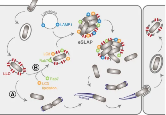
Fig 6. Extended model of the intracellular life cycle ofListeria monocytogenes in colon adenocarcinoma epithelial
cell lines. (A) In the classical scenario, after receptor-mediated entry,Lm evades the vacuole thanks to the combined
action of LLO and phospholipases. (B) Here we identified a population ofLm that can remain for several hours and
multiply inside vacuoles in LoVo cells. These compartments (eSLAPs) are neutral, positive for Rab7, LC3 and LAMP1, and decorated with LLO. This second population of bacteria finally escapes into the cytoplasm at later time points.
The decoration of eSLAPs by LC3, Rab7 and LAMP1 as well as their neutral pH are reminis-cent of the SLAPs (SpaciousListeria-containing Phagosomes) previously described in phagocytes
[12], and which derive from LAP (LC3-associated phagocytosis) [37]. Upon infection byLm, we
propose a model for the formation of replicative eSLAPs, analogous to the current model of SLAP formation (Fig 6). The entrapment ofLm inside internalisation vacuoles could result in
two distinct fates. (A) In the classically-described pathway, the coordinated actions of LLO, PlcA and PlcB result in a rapid disruption of the vacuole and escape of bacteria into the cytoplasm, where they can start replicating and polymerising actin. (B) In the second scenario, a proportion of internalisation vacuoles would undergo LC3 lipidation in addition to their maturation attested by decoration with Rab7, as well as fusion with lysosomes as suggested by LAMP1 labelling.
Whereas the eSLAPs observed in LoVo cells display similarities with SLAPs, they are nota-bly distinct from LisCVs, which are an intravacuolar persistence niche ofLm recently
described in human hepatocytes and trophoblast cells [38]. Contrary to SLAPs and eSLAPs, LisCVs do not derive from primary vacuoles. Instead, they form late in the intracellular cycle ofLm by recapture of bacteria that have lost ActA-dependent motility. Indeed, bacteria found
in LisCVs are labelled with YFP-CBD, while the bacteria we observe in eSLAPs are not. More-over, whereas eSLAPs are lipidated by LC3, LisCVs are not. Last,Lm replicates in eSLAPs,
whereas it adopts a viable but non-culturable state in LisCVs and does not grow. Altogether, though occurring in epithelial cells, the features we describe for eSLAPs are in agreement with compartments similar to SLAPs, and distinct from LisCVs. Our observations that even low LLO activity allowsLm residence and replication in vacuoles is consistent with the previous
report by Birminghamet al. showing that reduced hlyA expression allowed slow replication in
macrophage SLAPs [12]. However, the replication ofLm inside eSLAPs is significantly faster
than the 8 hours of doubling time reported in SLAPs [12], perhaps due to a lower bactericidal capacity of the epithelial niche compared with phagocytes. Membrane permeation by LLO might also attenuate the bactericidal properties of eSLAPs, and/or allow nutrient uptake through the permeated membrane, thereby promoting bacterial replication.
Conclusion
Together with LisCVs and SLAPs, eSLAPs enrich the palette ofLm intravacuolar lifestyles that
can establish in various cells types. Apprehending the importance of eSLAPs in the context of
in vivo infections prompts future investigation. Indeed, whilst intravacuolar lifestyles impose
constraints on motility or nutrient uptake, these compartments might provide shelter from cytosolic surveillance mechanisms such as autophagy and RIG-I-dependent activation of type-I interferon signalling, or favour chronic forms of infections by dampening cell-to-cell spread within tissues. Conversely, delayed residence within vacuoles could promote recognition of
Listeria pathogen-associated molecular patterns by endosomal Toll-like receptors and activate
NF-κB-dependent inflammatory pathways. Prolonged exposure to the intravacuolar environ-ment could also tune the expression ofLm virulence genes. Deciphering the extent to which
these intravacuolar niches influence the balance between bacterial fitness and host defences becomes critical to better appreciate long-term relationships betweenLm and its host.
Materials and methods
Bacterial strains, plasmids and culture conditions
The bacterial source strains used in this work wereEscherichia coli NEB5α (New England
Bio-labs) for plasmid constructions, Rosetta(DE3)pLysS (Novagen) for recombinant protein pro-duction, the clinical isolate ofListeria monocytogenes LL195 (lineage I, ST1) [39] for most of the experiments involvingLm, and Shigella flexneri M90T [40] for experiments onSf
T3SS-dependent secretion.Lm reference strain EGD-e (lineage 2, ST9) [41] (lineage II, ST9) was also used as a control that the observed eSLAPs were not specific to LL195. All strains were grown at 37˚C under shaking at 190 rpm in Luria Bertani (LB) medium forE. coli, in LB or
tryptic soy broth (TSB) forSf, in brain hear infusion (BHI) or Listeria synthetic medium
(LSM) [42] forLm. Whenever required, media were supplemented with antibiotics for plasmid
selection (chloramphenicol, 35μg/ml for E. coli; 20 μg/ml for Sf; 7 μg/ml for Lm or Ampicillin, 100μg/ml), or Congo red (1 mg/ml) for activation of the Sf T3SS.
In order to favour the expression of transgenes, the DNA coding sequence for FAST, fused with a Myc-tag, was codon-optimized forLm or Sf using the online Optimizer application
(http://genomes.urv.es/OPTIMIZER/) in guided random mode (S1 Text). The optimized sequences were obtained as synthetic Gene Fragments (Eurofins genomics). The
Lm-opti-mized sequence additionally contained the 5’-untranslated (5’-UTR) of thehlyA gene, and the
sequence encoding the signal peptide (SP) of LLO in its N-terminal part.
For plasmid constructions in the pAD vector derived from the pPL2 backbone [43,44], the 5’-UTRhlyA-SPhlyA-FAST-Myc fusion was amplified with primers oAL543-4, the sequence of
theLm hlyA gene encoding LLO was amplified from the EGD-e genomic DNA with primers
oAL549-50b, and the coding sequence for eGFP was amplified from pAD-cGFP (BUG2479)
[44] with primers oAL543-7. The UTRhlyA-SP-FAST-Myc amplicon was inserted instead of
UTRhlyA-eGFP into theEagI-SalI restriction sites of cGFP, thus generating pAD-SP-FAST, where FAST is under control of the PHYPERconstitutive promoter (Fig 1A). pAD-FAST, pAD-eGFP, pAD-SP-eGFP, pAD-hlyA, pAD-hlyA-FAST and pAD-hlyA-eGFP, all
con-taining the 5’-UTR ofhlyA and a Myc tag, were likewise generated by inserting the cognate
DNA amplicons into the same restriction sites (Fig 1A). After cloning inE. coli NEB5α, these
plasmids were integrated in the genome ofL. monocytogenes strains LL195 at the tRNAArg
locus by electroporation as previously described [43]. The pHpPL3-mCherry plasmid was
introduced at thetRNAArglocus by conjugation [45].
For allelic replacement at thehlyA locus (S5A Fig), pMAD-ΔhlyA::FAST and pMAD-ΔhlyA::eGFP were created by amplifying three partially overlapping fragments by PCR: one
thousand base pairs (bp) upstream (plcA gene) and downstream (mpl gene) of the hlyA open
reading frame in the EGD-e genome were amplified, respectively, with oAL981-2 and oAL976-7, while the FAST-Myc or eGFP-Myc open reading frames were amplified from pAD-FAST with oAL983-75 and from pAD-eGFP with oAL987-75, respectively. These frag-ments were inserted into the pMAD vector [46], between theSalI and BglII restriction sites by
Gibson Assembly, using the NEBuilder HiFi DNA Assembly Cloning Kit (New England Bio-Labs). pMAD-hlyA-FAST (S5A Fig) containing the last 1000 bp ofhlyA fused with the FAST
sequence, a Myc tag and one thousand bp downstream ofhlyA was likewise generated by
inserting the cognate DNA amplicons into the same restriction sites in pMAD. Site-directed mutagenesis to obtain pMAD-hlyAW492A-FAST was carried out by PCR using oAL1073-74 as
primers. Allelic replacements of thehlyA open reading frame by these constructs in the
genomes ofL. monocytogenes strains LL195 and EGD-e were obtained as previously described
[46]. For complementation purposes in haemolysis assays, a simple in-frame deletion mutant of thehlyA gene was also created using the pMAD backbone.
ForSf constructs, ipaB and ospF were amplified from M90T genomic DNA with primers
oAL703-4 and 707–8 respectively, and the optimized FAST-Myc was amplified with primers oAL705-6. pSU2.1-ospF-FAST (Fig 1C) was obtained by inserting an oAL707-6 amplicon over-lappingospF and FAST-Myc, with a BamHI restriction linker, in place of mCherry into the KpnI-XbaI restriction sites of pSU2.1rp-mCherry [47]. pSU2.1-ipaB-FAST was generated by
replacingospF with ipaB (oAL703-4) at the KpnI-BamHI sites (Fig 1C). After cloning inE. coli
The complete lists of bacterial strains and oligonucleotides used in this work are supplied as
S1andS2Tables, respectively.
Analysis of protein contents in bacterial total extracts or culture media
Bacterial total extracts or culture supernatants were recovered from 1 ml ofLm strains grownto an OD600nmof 2.0 in BHI at 37˚C as previously described [48].
Total bacterial extracts ofSf were prepared by boiling for 2 × 10 min at 95˚C in 100 μl of
Laemmli sample buffer (SB 1X) the bacterial pellets obtained by centrifugation of 1 ml of each strain grown to an OD600nmof 2.0 in TCS medium at 37˚C. For assessment of secretion
leak-age prior to T3SS induction, 2 ml ofSf culture supernatants were collected, precipitated with
16% trichloroacetic acid (TCA), and centrifuged for 30 min at 16,000× g at 4˚C. Protein pellets were washed twice in acetone before resuspension in 50μl of SB 1X. For induction of secre-tion,Sf were resuspended in 0.6 ml phosphate buffered saline (PBS) containing 1 mg/ml of
Congo red at a final OD600nmof 40, and incubated at 37˚C for 45 min. Bacteria were
elimi-nated by centrifugation; 100μl of supernatant were collected and mixed with 33 μl of SB 4X for SDS-PAGE separation. The remainder supernatant was TCA-precipitated and resus-pended in 50μl SB 1X.
10μl of each sample were separated on 4–15% Mini-Protean TGX gels (Bio-Rad) by sodium dodecyl sulfate-polyacrylamide gel electrophoresis (SDS-PAGE), and then revealed by staining with colloidal Coomassie Brilliant blue G-250 or by immunoblotting. For immunoblots, after transfer on nitrocellulose membrane (Amersham) using PierceG2 Fast Blotter, proteins were probed with anti-Myc mouse monoclonal antibody #9E10 (sc-40, Santa Cruz Biotechnology) at a 1:400 dilution in PBS supplemented with 0.05% tween-20 and 5% skimmed milk powder, followed by secondary hybridization with anti-Mouse IgG-heavy and light chain Antibody (Bethyl) at a 1:50 000 dilution in the same buffer. Signals were detected using Pierce ECL Plus Western Blotting Substrate and a Las4000 imager (GE Healthcare).
Fluorescence measurements on culture supernatants
Lm were grown for 16 h in BHI, washed and diluted to 1:10 in Listeria synthetic medium (LSM),
and then grown for 6 h at 37˚C, 180 rpm. Likewise, for secretion bySf a culture in TSB was diluted
to 1:10 in M9 medium supplemented with 0.2% glucose and 10μg/ml nicotinic acid. From 1 ml of culture, bacterial pellets were collected by centrifugation of the cultures at 6,000× g, then washed in PBS and resuspended in 1 ml of PBS. The culture supernatants were filtered (0.2μm pores). For fluorescence measurements of FAST-tagged fusions, 180μl of each sample was mixed with 20μl of PBS containing 50 μM HBR-3,5DM ((Z)-5-(4-Hydroxy-3,5-dimethylbenzylidene)-2-thioxothiazolidin-4-one) to obtain a final concentration of 5μM of fluorogen. Fluorescence intensity of technical triplicates was measured on a Spark 10M multimode microplate reader (Tecan), with excitation/emission wavelength set to 499/562 nm for FAST:HBR-3,5DM; 488/ 507nm for eGFP. Background fluorescence was measured on culture media from negative control strains (Lm LL195 [pAD-LLO] or Sf M90T ΔipaD) and subtracted to each sample. The standard
curve linking FAST fluorescence to its concentration was performed by diluting, in control medium corresponding to a culture of the corresponding negative control strain, known amounts of recombinant FAST produced inE. coli Rosetta(DE3)pLysS as previously described [17]. This enabled the calculation of fluorescent FAST-tagged proteins released in each culture.
As a negative control that the fluorescence was due to the formation of the FAST-HBR-3,5DM complexes, the fluorescence was also measured on samples mixed with 20μl of PBS instead of PBS containing HBR-3,5DM. No fluorescence was detected above that of the control culture media.
For normalisation between measurements for FAST- and eGFP-tagged proteins, cence intensities measured in filtered culture media were expressed relatively to the fluores-cence measured for a suspension of OD600nm= 1 ofLm constitutively expressing either
non-secreted FAST, or eGFP. The fluorescence intensities emitted by each one of these reference suspensions were fixed arbitrarily to 100 A.U.
Each experiment was reproduced three times independently. Statistical significance was assessed by two-tailed Student’st-tests with equal variance assumption, on the results of three
independent experiments.
Haemolysis assay
The supernatants of 16-h cultures ofLm in BHI were recovered by centrifugation for 1 min at
6,000× g followed by filtration through 0.2-μm pore filters, in order to eliminate bacteria. Serial two-fold dilutions of these supernatants were performed in round-bottom, clear, 96-well plates (100μl final volume per well) using as a diluent PBS, the pH of which was adjusted to 5.6, and supplemented with 0.1% bovine serum albumin (BSA). Erythrocytes from defibrin-ated mice blood were washed twice in PBS pH 6.4 and diluted 1:10 in PBS pH 5.6. 50μl of this suspension was added to each one of the wells containing diluted culture supernatants. After 30 min of incubation at 37˚C, the plates were centrifuged for 10 min at 430× g and haemolytic titres were calculated as the reciprocal of the dilution for which 50% of haemolysis was observed [49]. Two-way ANOVA on log2-transformed haemolytic titres followed by post-hoc
Tukey test was used for statistical testing between conditions.
Infection and transfection of epithelial cells
Infections of intestinal epithelial cells were performed in the LoVo cell line originating from colon adenocarcinoma (ATCC Cat# CCL-229, RRID: CVCL_0399). The Caco-2 epithelial cell line (ATCC Cat# HTB-37, RRID: CVCL_0025), also from colon adenocarcinoma, was used as a control that eSLAPs were not a specificity of LoVo cells. All cells were cultured in Dulbecco0s Modified Eagle’s medium (D-MEM) supplemented with 10% FBS, at 37˚C in a humidified atmosphere containing 5% CO2. For live microscopy, cells were seeded on Ibidiμslides 72 h
prior to infection at a density of 105cells/ml, in 300 ml of culture medium. When needed, cells were transfected 24 h before infection with pEYFP-C1-CBD expressing YFP-CBD [14], using Lipofectamine LTX (Invitrogen) and 1μg/ml of plasmid, according to the manufacturer’s specifications.
Lm strains were grown in BHI until they reached early stationary phase (OD600of 2 to 3),
washed in pre-warmed D-MEM, and then diluted in culture medium without serum to achieve a multiplicity of infection (MOI) of 5 (for long-term infections) to 30 (for short-term infections). Except for short-term imaging when bacterial entry was monitored under the microscope, after 1 h of bacteria-cell contact the inoculum was washed away twice with serum-free medium containing 40μg/ml gentamicin, then the medium was replaced by com-plete culture medium without phenol red containing 25μg/ml gentamicin in order to kill extracellular bacteria.
Live fluorescence microscopy of infected cells
Infected cells were observed in D-MEM without phenol red supplemented with 5μM of HBR-3,5DM for fluorescence detection, 250 nM of the fluorogenic probe SiR-actin for actin detec-tion, and 25μg/ml of gentamicin for long-term infections. For experiments where early events were monitored, the labelling of actin by SiR-actin was initiated 2 h prior to infection by add-ing 250 nM of SiR-actin to the medium.
For live cell imaging, preparations were observed with a Nikon Ti PFS microscope coupled to a spinning disk confocal device (CSU-XI-A1, Yokogawa), connected to a cooled EM-CCD camera (Evolve, Photometrics), and equipped with a cube for temperature control and a brick gas mixed for CO2and humidity control (Life Imaging Services). Image acquisition and
microscope control were actuated with the MetaMorph software (Molecular Devices, RRID: SCR_002368). Fluorescent illumination was driven by three lasers, of wavelength 491 nm for eGFP, YFP or FAST, 561 nm for mCherry, and 635 nm for SiR-actin. Images were acquired with apochromat 63x objective lenses (NA 1.4) in 1μm step-z-stacks. Acquisition parameters were similar for all samples of an experiment. For snapshot display, maximum intensity signals from 16 successive stacks (i.e. 16 μm slices) were integrated with Fiji (RRID:SCR_002285).
Each picture or video is representative of the population observed in three independent experiments.
Quantification of FAST accumulation in infected cells
For measurements of the accumulation of secreted FAST in cells, images were first z-projected by maximum intensity. The accumulation of fluorescence in a given cell was measured as the mean value of pixel intensities in a Region Of Interest (ROI) of 30 by 30 pixels. Statistical sig-nificance of the difference between conditions in the dispersion of fluorescence intensities over time was assessed by Two-tailed Mann-Whitney non-parametric test. The dynamics of FAST accumulationI(t) was fitted to an exponential curve I(t) = I0ert+Ibg, withr the rate of
accumulation,I0the initial fluorescence andIbgthe fluorescence of the background. The
dou-bling timeτ was then computed according to the following formula: t ¼ln2
r. Image computing
was done with Fiji.
In-situ growth measurements of mCherry-labelled bacteria
We performed for each time point an Otsu-thresholding on the entire z-stack of mCherry images in order to obtain the 3D segmentation of bacteria. Although the segmentation was not able to isolate individual bacteria in dense regions, it yielded the total volume occupied by bac-teria in the field of view for each frame. The growth rate of mCherry bacbac-teria was measured by fitting the total volume of segmented objectsV(t) to an exponential curve V(t) = V0ert, withr
the growth rate andV0the initial volume. The doubling timeτ was then computed according
to the following formula: t ¼ln2
r. Image computing was performed using MatLab (RRID:
SCR_001622).
Tracking of primary vacuoles in short term infection assays
The slices of the z-stacks obtained from spinning confocal imaging were projected onto a sin-gle plane (maximal projection). Fluorescent vacuoles were tracked using the plugin TrackMate in Fiji. The time at which tracks began during the infection was used to compute the time of
Lm entry into LoVo cells. The distribution of residence times in primary vacuoles was
com-puted from the statistics of track lengths. Statistical significance was assessed by two-tailed Stu-dent’st-test on the two distributions, with equal variance assumption. The interpolated
median (IM) lifetime of SP-FAST-labelled vacuoles was calculated from the raw distributions
as follows:IM ¼ M þðng nlÞ
ð2�neÞdt, where M is the median, dt is the time interval, and ng,nlandne
are the number of events greater, lower or equal toM, respectively. The accuracy of the
inter-polated median estimate was assessed by bootstrapping analysis (100 random samplings for each distribution, yielding 100 estimates ofIM for each condition) followed by two-tailed
Tracking of eSLAPs in long-term infection assays
At 2 h p.i., Ibidiμslides were mounted on a confocal spinning disk microscope for time-lapse observations. Pixel size was 250 x 250 nm and step size inz was 1 μm. For each time point taken
every 5 or 15 minutes, we recorded a z-stack of fluorescent images in two channels for FAST signals and mCherry labelled bacteria. Given the good signal-to-noise ratio of mCherry images, we applied for each time point Otsu’s thresholding algorithm on the entire z-stack in order to obtain a 3D segmentation of bacteria. The Otsu segmentation did not allow to isolate bacteria when they were too dense, for instance in eSLAPs, or after prolonged infection when cells were full of bacteria. Hence, we detected objects that could either be single bacteria or clusters of bac-teria. We tracked each segmented object from frame to frame based on their similarities in size and location. Our method took benefit from the fact that cytosolic bacteria were moving very fast compared to those encapsulated in eSLAPs. The growth rates of bacteria inside eSLAPs were computed by fitting the dynamics of segmented mCherry volumes to an exponential func-tionV(t) = V0ert, withr the growth rate and V0the initial volume of the vacuole. The doubling
timeτ was then computed according to the following formula: t ¼ln2
r. No growth was observed
for theΔhlyA strain in long-term vacuoles tracked by the co-occurrence of FAST and mCherry signals (based on the spherical shape of the object). For the WT and theprfA�strains, an object
was classified as a growing eSLAP if it met the two following criteria: (a) the initial size was
equal to or larger than the size a of single bacterium (32 voxels) (S12 Fig), and (b) the size of the
object at least doubled over the whole track. The fraction of the objects in whichLm grew was
then computed as the ratio of the number of tracked vacuoles that at least doubled their size during the 8-h course of the movie (thus matchinga and b) to the initial number of objects
big-ger than 32 voxels on the first frame of the movie (S12 Fig), which is a good proxy for the initial number of entry events. To quantify LLO-FAST signals in a given vacuole, we used the binary mask of mCherry labelled objects to measure the average intensity in the corresponding region of the FAST image. Image computing was performed using MatLab.
Quantification of YFP-CBD signals
Images were first z-projected by maximum intensity. For eSLAPs we measured YFP fluores-cence intensity per pixel in ROIs with an area of 16 pixels that were manually positioned at each time point according to the mCherry signal of bacteria. For cytosolic bacteria, the mCherry images were segmented to get the masks of the individual bacteria. Segmented objects were scored as cytosolic if their size was below 40 pixels. At each time point, we mea-sured the average value of fluorescence intensity per pixel in the population of bacteria that were cytosolic. The average background YFP-CBD signals per pixel measured in the cytosol was subtracted to each value for normalization.
Immunofluorescence or LysoTracker staining of infected cells
LoVo cells were seeded 48 h before infection in 24-well plates containing 12 mm diameter cov-erslips. Infection with bacteria expressing mCherry (for immunofluorescence experiments) or eGFP (for LysoTracker staining) was performed as described above, using a MOI of 30, except that plates were centrifuged for 1 min at 200× g after addition of the inoculum in order to syn-chronise bacteria-cell contacts. 3 h p.i., cells were washed in pre-warmed PBS, fixed 20 min with 4% paraformaldehyde in PBS, then permeabilized for 5 min at room temperature with 0.5% Triton X-100 in PBS, and blocked for 5 min in PBS buffer containing 2% bovine serum albumin (BSA, Sigma). Incubation with primary antibodies in PBS buffer, 1% BSA was per-formed for 1 h, followed by three PBS washes, and incubation with the Alexa Fluor