HAL Id: cea-01591840
https://hal-cea.archives-ouvertes.fr/cea-01591840
Submitted on 22 Sep 2017
HAL is a multi-disciplinary open access
archive for the deposit and dissemination of
sci-entific research documents, whether they are
pub-lished or not. The documents may come from
teaching and research institutions in France or
abroad, or from public or private research centers.
L’archive ouverte pluridisciplinaire HAL, est
destinée au dépôt et à la diffusion de documents
scientifiques de niveau recherche, publiés ou non,
émanant des établissements d’enseignement et de
recherche français ou étrangers, des laboratoires
publics ou privés.
The crystalline alpha,omega-dicarboxylate metal
complex with the longest aliphatic chain to date: uranyl
1,15-pentadecanedioate
Pierre Thuéry, Jack Harrowfield
To cite this version:
Pierre Thuéry, Jack Harrowfield. The crystalline alpha,omega-dicarboxylate metal complex with the
longest aliphatic chain to date: uranyl 1,15-pentadecanedioate. Dalton Transactions, Royal Society of
Chemistry, 2017, 46, pp.13677-13680. �10.1039/C7DT03273K�. �cea-01591840�
The crystalline
αααα
,ω
ω
ω-dicarboxylate metal complex with the longest
ω
aliphatic chain to date: uranyl 1,15-pentadecanedioate
†
Pierre Thuéry*a and Jack Harrowfieldb
Under solvo-hydrothermal conditions, 1,15-pentadecanedioic acid (H2C15) reacts with uranyl ions to form the complex [H2NMe2]2[(UO2)2(C15)3] (1), in which the uranyl ions are tris-chelated by carboxylate groups and the well-ordered aliphatic chains are either all-trans or kinked. 1 crystallizes as a two-dimensional network of the bilayer type and with the {82.10} point symbol, the packing displaying alternate sheets containing either the ionic parts or stacks of aliphatic chains, at variance with the helicates or honeycomb networks previously obtained with other long-chain HOOC–(CH2)n–2–COOH dicarboxylic acids (n = 9–13) and bulkier counterions.
The potential of long-chain α,ω-dicarboxylic acids of general formula HOOC–(CH2)n–2–COOH (H2Cn) for the building of
uranyl–organic coordination polymers or frameworks (UOFs1)
was demonstrated some years ago by Cahill’s group,2 who
reported several one- to three-dimensional assemblies based on the acids with n = 5–10. The role of organic counter-cations containing pyridyl groups as structure-directing agents was specifically investigated and enabled the synthesis of one of the first polycatenated species in uranyl chemistry.2c The presence
of additional coordinated chloride anions was later shown to lead to dinuclear complexes in the cases with n = 4 and 6,3 and the
association of uranyl dicarboxylate (n = 6–10) complexes with cucurbit[6]uril molecules was also studied.4 More recently, the
uranyl complexes formed by the acids with n = 7–10, 12 and 13 in the presence of various 3d-block metal-containing cations, in particular those of the form [M(bipy/phen)n]+/2+ (n = 2 or 3, bipy
= 2,2ʹ-bipyridine, phen = 1,10-phenanthroline), or in the presence of bipy or phen as co-ligands for uranyl were investigated.5 This work provided the first examples of
uranyl-based triple-stranded helicates (n = 9 and 12)5a and Borromean
entanglement (n = 13),5b as well as a case of 2D → 3D parallel
polycatenation.5d In many cases, and particularly for n = 13, the
crystal structures of these complexes are marred by disorder affecting the aliphatic chains, and complexes with higher members in the dicarboxylate series (n = 14, 15 and 20) proved extremely resistant to crystallization.
More generally, a search of the Cambridge Structural Database (CSD, Version 5.38)6 shows that, although there are a
great many crystal structures involving monocarboxylic acids of the form HOOC–(CH2)n–2–CH3, with n often as large as 17–20,7
and even up to 23,8 which most often crystallize in the all-trans
conformation, studies of their dicarboxylic counterparts are scarce. A remarkable early study of the structures of dicarboxylic fatty acids was extended to n = 18,9 and more recent reports
include investigations of the association of H2C13 with
1,1ʹ-di-pyridyl-ferrocene,10 and H
2C14 with bis-amidopyridines11 and
β-cyclodextrin.12 Concerning dicarboxylate metal complexes,
and apart from those with uranyl ions cited above, the only ones to be found with the highest members of the series are a diosmium(I) complex in which C142– makes an intramolecular
loop,13 and several complexes with SrII, BaII, CoII, CuII, RhII and
PbII with C12–/2–,14 which have been characterized by diffraction
on single crystals, or powders when no single crystal of sufficient quality could be obtained.14b,d
After many failed attempts at synthesis of uranyl ion complexes with diacids for which n > 13 under varying experimental conditions, single crystals of the complex [H2NMe2]2[(UO2)2(C15)3] (1), involving 1,15-pentadecanedioic
acid (H2C15), were finally obtained under solvo-hydrothermal
conditions in a 70:30 % v/v mixture of water and N,N-dimethylformamide (DMF)‡, which were of suitable quality for crystal structure determination.§ The dimethylammonium counterions are formed in situ from DMF hydrolysis, which is a frequent occurrence,15 but which may be of particular
significance in the generation of nicely crystalline material in the present case because of its slow generation of the precipitating cation. Complex 1 crystallizes in the monoclinic space group
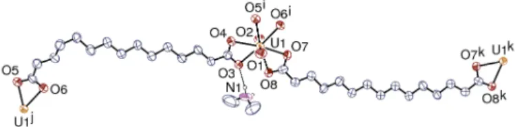
Fig. 1 View of complex 1. Displacement ellipsoids are drawn at the 50% probability level and carbon-bound hydrogen atoms are omitted. Only the strongest hydrogen bond is shown as a dashed line. Symmetry codes: i = x – 1/2, y – 1/2, z – 1; j = x + 1/2, y + 1/2, z + 1; k = –x, y, 1/2 – z. Selected bond lengths (Å) and angles (°): U1–O1 1.758(4), U1–O2 1.757(4), U1–O3 2.439(4), U1–O4 2.487(4), U1–O5i 2.468(4), U1–O6i 2.463(4), U1–O7 2.465(4), U1–O8 2.474(4), O1–U1–O2 178.93(19), O3–U1–O4 52.98(13), O4–U1–O5i 70.33(13), O5i–U1–O6i 52.90(14), O6i–U1–O7 66.26(15), O7– U1–O8 52.29(14), O8–U1–O3 65.85(14).
C2/c with an asymmetric unit containing one uranyl cation, one H2NMe2+ cation and two fully deprotonated C152– anions, one of
them having twofold rotation symmetry (Fig. 1, Table S1). The uranium atom is in a hexagonal bipyramidal environment, being chelated by three carboxylate groups from three different anions. The two independent ligands, which show no sign of disorder, have different conformations; the symmetrical one is in the all-trans, nearly planar geometry, with only a slight rotation of the carboxylate group [O–C–C–C torsion angle 167.0(7)°], while the unsymmetrical one is kinked due to one C–C–C–C torsion angle being gauche [56.8(8)° around the fourth C–C bond from the
O5/O6 end] and the carboxylate group nearer to the curved part being also slightly rotated [O–C–C–C 161.0(6)°].
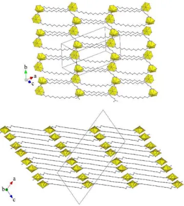
The uranium atoms are threefold nodes, and the ligands are simple links, in the two-dimensional network parallel to (2 0 ī) which is formed (Fig. 2). This assembly has the point (Schläfli)
Fig. 2 Top: View of the 2D network. Bottom: Packing with four bilayers viewed edge-on. Uranium coordination polyhedra are yellow and hydrogen atoms are omitted.
Fig. 3 Simplified view of the 2D network showing the 1D subunits in red and green, and the linkers in blue. Uranium atoms are yellow and the centroids of the ligands are shown as blue dots.
symbol {82.10} and corresponds to the plane SP KIa topological
type of the topos&RCSR database.16 This arrangement can be
viewed as formed of two families of non-intersecting one-dimensional subunits running at an angle from each other, and connected by the ligand with twofold rotation symmetry, as shown in the simplified representation given in Fig. 3. The overall shape is that of a slightly serrated chiral bilayer with internal links, as shown in the view of the packing given in Fig. 2, the chirality changing from one bilayer to the next (the crystal being centrosymmetric). An arrangement with the same topology was found with the much shorter C52– ligand and
4,4ʹ-bipyridinium cations,2c thus establishing an unexpected
connection between two widely separated members of this
dicarboxylate family. The counterions are hydrogen bonded to carboxylate oxygen atoms [N1⋅⋅⋅O3 2.818(8) Å, N1–H⋅⋅⋅O3 149°; N1⋅⋅⋅O6l 3.117(10) Å, N1–H⋅⋅⋅O6l 156°; symmetry code: l
= 1 – x, y, 5/2 – z], and they are thus located close to the uranyl cations. As a consequence, the packing displays sheets parallel to (1 0 0) containing the ionic components, separated by large regions occupied by nearly parallel aliphatic chains. The presence of voids in the structure and the moderate value of 0.56 for the Kitaigorodski packing index estimated with PLATON17
indicate that unresolved solvent molecules may be present. It is notable that the metal/ligand ratio of 2:3 and the ligand elongated shape and flexibility would be suitable for the formation of a two-dimensional network with {63} honeycomb topology, as
observed (with significant distortion from planarity) in [Mn(phen)3][(UO2)2(C10)3]· 6H2O,
[Ni(bipy)3][(UO2)2(C12)3][UO2(C12)(H2O)2]· H2O,5c and
[Mn(phen)3][(UO2)2(C13)3],5b or of a triple-stranded helicate as
in [(M)(bipy)3][(UO2)2(C9)3] (M = Co, Ni) and
[M(phen)3][(UO2)2(C12)3] (M = Mn, Co).5a However, all these
latter species possess bulky counterions that have been shown to play a structure-directing role, particularly in the case of helicates, while the small counterions in 1, located very near the metal centres, do not interact significantly with the aliphatic chains, which are then left to organize in the parallel fashion commonly found with fatty acids. Unfortunately, no crystal could be obtained with [M(bipy/phen)3]2+ counterions in the case
of C152– for the sake of comparison.
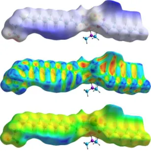
The interactions present in the lattice can be visualized through calculation of Hirshfeld surfaces (HSs)18 with CrystalExplorer.19 The
HS of the anionic part, mapped with dnorm and calculated on the
asymmetric unit (Fig. 4), evidences the hydrogen bonds made by the counter-cations (not only the strong ones indicated above, but also much weaker, bifurcated ones). The red dot located on the HS near the oxo atom (O1) and that just to the left of the hydrogen bonded carboxylate group correspond to a CH⋅⋅⋅O(oxo) hydrogen bond, not shown for clarity [C⋅⋅⋅O1m 3.315(7) Å, C–H⋅⋅⋅O1m 143°; symmetry
code: m = 1/2 – x, 1/2 – y, 2 – z]. One CH⋅⋅⋅O(carboxylate) hydrogen bond may also be present [C⋅⋅⋅O 3.626(8) Å, C–H⋅⋅⋅O 163°], but several short contacts between methyl hydrogen atoms of the counter-cation and carboxylate oxygen atoms are more dubious due to the somewhat uncertain location of these hydrogen atoms. Interactions involving the aliphatic chains can be compared to those described in the series of n-alkanes,20 and they can best be visualized with different
mappings of the HS (Figs. 4 and S1). It appears that interactions do not exceed dispersion on one side of the chains, as shown by the dark blue parts in the dnorm mapping representation, corresponding to
regions of large inter-chain separations (it is notable however that large voids in the lattice may indicate the presence of unresolved, and probably very disordered, solvent molecules). The chains are involved only in H⋅⋅⋅H and C⋅⋅⋅H interactions, apart from the two CH⋅⋅⋅O hydrogen bonds indicated above. The alternating pattern of red/yellow (concave) and blue (convex) regions in the shape index mapping representation is considered characteristic of C⋅⋅⋅H contacts, but these appear to represent a negligible percentage of the surface area here, while the yellow/orange dots in the de representation indicate the
Fig. 4 Hirshfeld surface calculated on the anionic part of the asymmetric unit and mapped (from top to bottom) with dnorm, shape index, and de. Hydrogen bonds with the dimethylammonium cation are shown as dashed lines. Anomalous parts in the vicinity of uranium are due to truncation of the polymer chain.
the symmetrical chain on the right side of the HS, which overall represent 55% of the surface area. The pattern of contacts is much less regular than that which is found in n-alkanes such as hexane– nonane,20 the packing of chains being here disturbed by the presence
of the ionic moieties and the ensuing kinked shape of the asymmetric ligand, although the chains are for the most part parallel to one another.
The uranyl emission spectrum of 1 measured in the solid state, at room temperature and with an excitation wavelength of 420 nm (Fig. S2) is of weak intensity, but it displays three well-resolved peaks at 488, 508 and 529 nm which are in the upper range of values generally measured for complexes in which uranyl is chelated by three carboxylate groups,21 and it is red-shifted with respect to the spectrum
of the complex with C52– displaying the same topology (but with
different counter-ions).2c The infrared spectrum (Fig. S3) shows the
usual absorption band at 911 cm–1 corresponding to the uranyl
antisymmetric stretching mode frequency ν3.22
In conclusion, we succeeded in obtaining single crystals, of sufficient quality for structure determination, of a metal complex with the longest α,ω-dicarboxylate reported to date in such a study. The C152– ligands show no sign of disorder, and the two-dimensional
chiral bilayers generated pack so as to create alternate planes containing the ionic parts and large regions dominated by H⋅⋅⋅H contacts between aliphatic chains. The difference between the connectivity observed and that in honeycomb networks or helicates with the same metal/ligand ratio and α,ω-dicarboxylates with n in the range 9–13 is ascribed to the variation in counterions, much less bulky in the present case.
Conflicts of interest
There are no conflicts of interest to declare.
Notes and References
‡ Synthesis of complex 1. H2C15 (14 mg, 0.05 mmol),
UO2(NO3)2·6H2O (25 mg, 0.05 mmol), formic acid (8 mg, 0.17
mmol), N,N-dimethylformamide (0.3 mL), and demineralized water (0.7 mL) were placed in a 10 mL tightly closed glass vessel and heated at 140 °C under autogenous pressure, giving light yellow crystals of complex 1 in low yield within one week.
§ Crystallographic data were collected at 150(2) K on a Nonius Kappa-CCD area-detector diffractometer23 using
graphite-monochromated Mo Kα radiation (λ = 0.71073 Å). The data were processed with HKL2000,24 and absorption effects were corrected
with SCALEPACK.24 The structure was solved by intrinsic phasing
with SHELXT25 and refined by full-matrix least-squares on F2with
SHELXL-2014.26 All non-hydrogen atoms were refined with
anisotropic displacement parameters. All hydrogen atoms were introduced at calculated positions and were treated as riding atoms with an isotropic displacement parameter equal to 1.2 times that of the parent atom (1.5 for CH3). Large voids in the lattice indicate the
possible presence of unresolved solvent molecules, and the corresponding electron density has been subtracted with the SQUEEZE software.17 The drawings were done with ORTEP-3,27
VESTA,28 and TOPOS.16 Crystal data for 1: C
49H94N2O16U2, M =
1443.32, monoclinic, space group C2/c, a = 33.683(2), b = 13.1767(9), c = 16.5993(8) Å, β = 108.896(4)°, V = 6970.2(7) Å3, Z
= 4. Refinement of 314 parameters on 6593 independent reflections out of 158636 measured reflections (Rint = 0.036) led to R1 = 0.038,
wR2 = 0.105, ∆ρmin = –0.87, ∆ρmax = 0.76 e Å–3.
1 (a) M. B. Andrews and C. L. Cahill, Chem. Rev., 2013, 113, 1121; (b) T. Loiseau, I. Mihalcea, N. Henry and C. Volkringer, Coord. Chem. Rev., 2014, 266–267, 69; (c) W. Yang, T. G. Parker and Z. M. Sun, Coord. Chem. Rev., 2015, 303, 86; (d) J. Su and J. S. Chen, Struct. Bond., 2015, 163, 265.
2 (a) L. A. Borkowski and C. L. Cahill, Inorg. Chem., 2003, 42, 7041; (b) L. A. Borkowski and C. L. Cahill, Cryst. Growth Des., 2006, 6, 2241; (c) L. A. Borkowski and C. L. Cahill, Cryst. Growth Des., 2006, 6, 2248; (d) A. T. Kerr and C. L. Cahill, Cryst. Growth Des., 2011, 11, 5634.
3 I. Mihalcea, C. Falaise, C. Volkringer, N. Henry and T. Loiseau, Inorg. Chem. Commun., 2014, 44, 63.
4 P. Thuéry, Cryst. Growth Des., 2011, 11, 2606.
5 (a) P. Thuéry and J. Harrowfield, Inorg. Chem., 2015, 54, 10539; (b) P. Thuéry, Cryst. Growth Des., 2016, 16, 546; (c) P. Thuéry and J. Harrowfield, Inorg. Chem., 2016, 55, 2133; (d) P. Thuéry, E. Rivière and J. Harrowfield, Cryst. Growth Des., 2016, 16, 2826; (e) P. Thuéry and J. Harrowfield, CrystEngComm, 2016, 18, 3905.
6 C. R. Groom, I. J. Bruno, M. P. Lightfoot and S. C. Ward, Acta Crystallogr., Sect. B, 2016, 72, 171.
7 See, for example: (a) D. R. Whitcomb and R. D. Rogers, J. Chem. Cryst., 1996, 26, 99; (b) R. W. Corkery and D. C. R. Hockless, Acta Crystallogr., Sect. C, 1997, 53, 840; (c) M. Amai, T. Endo, H. Nagase, H. Ueda and M. Nakagaki, Acta Crystallogr., Sect. C, 1998, 54, 1367; (d) J. M. Rueff, N. Masciocchi, P. Rabu, A. Sironi and A. Skoulios, Chem. – Eur. J., 2002, 8, 1813; (e) M. Amai, M. Kamijo, H. Nagase, T. Endo and H. Ueda, Anal. Sci., 2005, 21, x9; (f) D. R. Trivedi and P. Dastidar, Cryst. Growth Des., 2006, 6, 1022; (g) H. Basit, A. Pal, S. Sen and S. Bhattacharya, Chem. – Eur. J., 2008, 14, 6534; (h) E. Moreno-Calvo, G. Gbabode, R. Cordobilla, T.
Calvet, M. A. Cuevas-Diarte, P. Negrier and D. Mondieig, Chem. – Eur. J., 2009, 15, 13141.
8 G. Gbabode, P. Negrier, D. Mondieig, E. Moreno Calvo, T. Calvet and M. A. Cuevas-Diarte, Chem. – Eur. J., 2007, 13, 3150.
9 W. A. Caspari, J. Chem. Soc., 1928, 3235.
10 D. Braga, S. L. Giaffreda, F. Grepioni, G. Palladino and M. Polito, New J. Chem., 2008, 32, 820.
11 F. Garcia-Tellado, S. J. Geib, S. Goswami and A. D. Hamilton, J. Am. Chem. Soc., 1991, 113, 9265.
12 S. Makedonopoulou and I. M. Mavridis, Carbohyd. Res., 2001, 335, 213.
13 N. Gwini, D. M. Marolf, S. H. Yoon, A. G. Fikes, A. C. Dugan, G. L. Powell, V. M. Lynch, V. N. Nesterov and G. T. McCandless, J. Organomet. Chem., 2017, DOI: 10.1016/j.jorganchem.2017.01.026.
14 (a) R. P. Bonar-Law, T. D. McGrath, N. Singh, J. F. Bickley, C. Femoni and A. Steiner, J. Chem. Soc., Dalton Trans., 2000, 4343; (b) J. M. Rueff, N. Masciocchi, P. Rabu, A. Sironi and A. Skoulios, Eur. J. Inorg. Chem., 2001, 2843; (c) B. Finkel, L. Pan, X. Huang and J. Li, C. R. Chimie, 2005, 8, 1670; (d) M. Grzesiak, W. Nitek, A. Rafalska-Łasocha and W. Łasocha, Z. Kristallogr., 2012, 227, 629; (e) D. J. Price, S. J. Coles and M. B. Hursthouse, J. Struct. Chem., 2013, 54, 474; (f) M. Grzesiak-Nowak, W. Nitek, A. Rafalska-Łasocha and W. Łasocha, Z. Kristallogr., 2013, 228, 590; (g) X. Y. Li, L. Gao, G. B. Che, Y. S. Yan and Q. F. Guan, Chin. J. Inorg. Chem., 2014, 30, 2669; (h) X. Y. Li, B. Hu, G. B. Che, Y. S. Yan, Q. F. Guan, C. X. Li, Chin. J. Inorg. Chem., 2014, 30, 2818. 15 P. Thuéry and J. Harrowfield, Inorg. Chem., 2016, 55, 6799
and references therein.
16 V. A. Blatov, TOPOS, Samara State University, 2004. 17 A. L. Spek, Acta Crystallogr., Sect. D, 2009, 65, 148. 18 M. A. Spackman and D. Jayatilaka, CrystEngComm, 2009, 11,
19.
19 S. K. Wolff, D. J. Grimwood, J. J. McKinnon, M. J. Turner, D. Jayatilaka and M. A. Spackman, CrystalExplorer 3.1, University of Western Australia, 2012.
20 J. J. McKinnon, M. A. Spackman and A. S. Mitchell, Acta Crystallogr., Sect. B, 2004, 60, 627.
21 P. Thuéry and J. Harrowfield, Inorg. Chem., submitted. 22 E. Faulques, N. Kalashnyk, F. Massuyeau and D. L. Perry, RSC
Advances, 2015, 5, 71219.
23 R. W. W. Hooft, COLLECT, Nonius BV: Delft, The Netherlands, 1998.
24 Z. Otwinowski and W. Minor, Methods Enzymol., 1997, 276, 307.
25 G. M. Sheldrick, Acta Crystallogr., Sect. A, 2015, 71, 3. 26 G. M. Sheldrick, Acta Crystallogr., Sect. C, 2015, 71, 3. 27 L. J. Farrugia, J. Appl. Crystallogr., 1997, 30, 565.
28 K. Momma and F. Izumi, J. Appl. Crystallogr., 2008, 41, 653.
Table of Contents:
A bilayer 2D network is formed in uranyl 1,15-pentadecanedioate, different from the species obtained with related ligands and bulkier counterions.