Review Article
Ann Nutr Metab 2012;60:6–16 DOI: 10.1159/000334879The Evaluation of Body Composition:
A Useful Tool for Clinical Practice
Ronan Thibault
a
Claude Pichard
b
a Centre de Recherche en Nutrition Humaine Auvergne, UMR 1019 Nutrition Humaine, INRA, Clermont Université,
Service de Nutrition Clinique, CHU de Clermont-Ferrand, Clermont-Ferrand , France; b Nutrition Unit, Geneva
University Hospital, Geneva , Switzerland
Introduction
Chronic undernutrition is characterized by a progres-sive reduction of the fat-free mass (FFM) and fat mass (FM) which has deleterious consequences on health. Un-dernutrition is insufficiently screened and treated in hos-pitalized or at-risk patients despite its high prevalence and negative impact on mortality, morbidity, length of stay (LOS), quality of life, and costs [1–4] . The risk of un-derestimating hospital undernutrition is likely to worsen in the next decades because of the increasing prevalence of overweight, obesity, and chronic diseases and the in-creased number of elderly subjects. These clinical condi-tions are associated with FFM loss (sarcopenia). There-fore, an increased number of patients with FFM loss and sarcopenic obesity will be seen in the future.
Sarcopenic obesity is associated with decreased sur-vival and increased therapy toxicity in cancer patients [5–10] , whereas FFM loss is related to decreased survival, a negative clinical outcome, increased health care costs [2] , and impaired overall health, functional capacities, and quality of life [4–11] . Therefore, the detection and treatment of FFM loss is a major issue of public health and health costs [12] .
Weight loss and the body mass index (BMI) lack sen-sitivity to detect FFM loss [13] . In this review, we support the systematic assessment of FFM with a method of body composition evaluation in order to improve the
Key Words
Fat-free mass ⴢ Fat mass ⴢ Undernutrition ⴢ Bioelectrical impedance analysis ⴢ Sarcopenic obesity ⴢ Drug toxicity
Abstract
Undernutrition is insufficiently detected in in- and outpa-tients, and this is likely to worsen during the next decades. The increased prevalence of obesity together with chronic illnesses associated with fat-free mass (FFM) loss will result in an increased prevalence of sarcopenic obesity. In patients with sarcopenic obesity, weight loss and the body mass in-dex lack accuracy to detect FFM loss. FFM loss is related to increasing mortality, worse clinical outcomes, and impaired quality of life. In sarcopenic obesity and chronic diseases, body composition measurement with dual-energy X-ray ab-sorptiometry, bioelectrical impedance analysis, or comput-erized tomography quantifies the loss of FFM. It allows tai-lored nutritional support and disease-specific therapy and reduces the risk of drug toxicity. Body composition evalua-tion should be integrated into routine clinical practice for the initial assessment and sequential follow-up of nutritional status. It could allow objective, systematic, and early screen-ing of undernutrition and promote the rational and early ini-tiation of optimal nutritional support, thereby contributing to reducing malnutrition-induced morbidity, mortality, worsening of the quality of life, and global health care costs.
Copyright © 2011 S. Karger AG, Basel
Received: October 21, 2011
Accepted after revision: November 7, 2011 Published online: December 16, 2011
Dr. Ronan Thibault
Centre de Recherche en Nutrition Humaine Auvergne © 2011 S. Karger AG, Basel
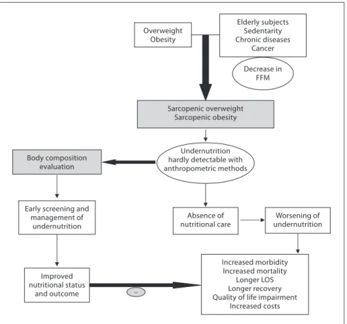
tion, management, and follow-up of undernutrition. Such an approach should in turn reduce the clinical and func-tional consequences of diseases in the setting of a cost-effective medico-economic approach ( fig. 1 ). We discuss the main applications of body composition evaluation in clinical practice ( fig. 2 ).
Rationale for a New Strategy for the Screening of Undernutrition
Screening of Undernutrition Is Insufficient
Academic societies encourage systematic screening of undernutrition at hospital admission and during the hos-pital stay [14] . The detection of undernutrition is generally based on measurements of weight and height, calculations of BMI, and the percentage of weight loss. Nevertheless, screening of undernutrition is infrequent in hospitalized or nutritionally at-risk ambulatory patients. For example, in France, surveys performed by the French Health Au-thority [15] indicate that: (i) weight alone, (ii) weight with BMI or percentage of weight loss, and (iii) weight, BMI,
and percentage of weight loss are reported in only 55, 30, and 8% of the hospitalized patients’ rec ords, respectively. Several issues, which could be improved by specific educa-tional programs, explain the lack of implementation of nu-tritional screening in hospitals ( table 1 ). In addition, the accuracy of the clinical screening of undernutrition could be limited at hospital admission. Indeed, patients with un-dernutrition may have the same BMI as sex- and age-matched healthy controls but a significantly decreased FFM hidden by an expansion of the FM and the total body water which can be measured by bioelectrical impedance analysis (BIA) [13] . This example illustrates that body composition evaluation allows a more accurate identifica-tion of FFM loss than body weight loss or BMI decrease. The lack of sensitivity and specificity of weight, BMI, and percentage of weight loss argue for the need for other methods to evaluate the nutritional status.
Changes in Patients’ Profiles
In 2008, twelve and thirty percent of the worldwide adult population was obese or overweight; this is two times higher than in 1980 [16] . The prevalence of
over-Increased morbidity Increased mortality
Longer LOS Longer recovery Quality of life impairment
Increased costs Absence of nutritional care Elderly subjects Sedentarity Chronic diseases Cancer Body composition evaluation Sarcopenic overweight Sarcopenic obesity Overweight Obesity Undernutrition hardly detectable with anthropometric methods Decrease in FFM Worsening of undernutrition Improved nutritional status and outcome Early screening and
management of undernutrition
_
Fig. 1. Conceptualization of the expected
impact of early use of body composition for the screening of fat-free loss and under-nutrition in sarcopenic overweight and obese subjects. An increased prevalence of overweight and obesity is observed in all Western and emerging countries. Simulta-neously, the ageing of the population, the reduction of the level of physical activity, and the higher prevalence of chronic dis-eases and cancer increased the number of patients with or at risk of FFM impair-ment, i.e. sarcopenia. Thus, more patients are presenting with ‘sarcopenic over-weight or obesity’. In these patients, evalu-ation of nutritional status using anthropo-metric methods, i.e. weight loss and calcu-lation of BMI, is not sensitive enough to detect FFM impairment. As a result, un-dernutrition is not detected, worsens, and negatively impacts morbidity, mortality, LOS, length of recovery, quality of life, and health care costs. On the contrary, in pa-tients with ‘sarcopenic overweight or obe-sity’, early screening of undernutrition with a dedicated method of body composi-tion evaluacomposi-tion would allow early initia-tion of nutriinitia-tional support and, in turn, improvements of nutritional status and clinical outcome.
weight and obesity is also increasing in hospitalized pa-tients. A 10-year comparative survey performed in a Eu-ropean hospital showed an increase in patients’ BMI, to-gether with a shorter LOS [17] . The BMI increase masks undernutrition and FFM loss at hospital admission. The increased prevalence of obesity in an ageing population has led to the recognition of a new nutritional entity: ‘sar-copenic obesity’ [18] . Sar‘sar-copenic obesity is characterized by increased FM and reduced FFM with a normal or high body weight. The emergence of the concept of sarcopenic obesity will increase the number of situations associated with a lack of sensitivity of the calculations of BMI and
body weight change for the early detection of FFM loss. This supports a larger use of body composition evalua-tion for the assessment and follow-up of nutrievalua-tional status in clinical practice ( fig. 1 ).
Body Composition Evaluation for the Assessment of Nutritional Status
Body composition evaluation is a valuable technique to assess nutritional status. Firstly, it gives an evaluation of nutritional status through the assessment of FFM.
Sec-Tailoring of therapy (e.g. cancer patients) Evaluation of nutritional status Measurement of fluid changes - Calculation of energy needs - Tailoring of nutritional support Measurement of bone mineral density
BIA DEXA
BIA, L3-targeted CT, DEXA Better clinical
outcome
Diagnosis and management of osteoporosis Assessment of fluid balance? Better tolerance and efficacy of chemotherapy Better patient follow-up
- Better recognition of nutritional care by the medical community, health care
facilities, and health authorities - Increased medico-economic benefits
of nutritional evaluation BIA, L3-targeted CT, DEXA
BIA, L3-targeted CT, DEXA Assessment of
body composition
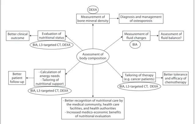
Fig. 2. Current and potential applications of body composition
evaluation in clinical practice. The applications are indicated in the boxes, and the body composition methods that could be used for each application are indicated inside the circles. The most used application of body composition evaluation is the measurement of bone mineral density by DEXA for the diagnosis and manage-ment of osteoporosis. Although a low FFM is associated with worse clinical outcomes, FFM evaluation is not yet implemented enough in clinical practice. However, by allowing early detection of undernutrition, body composition evaluation could improve the clinical outcome. Body composition evaluation could also be used to follow up nutritional status, calculate energy needs, tailor nutritional support, and assess fluid changes during periopera-tive period and renal insufficiency. Recent evidence indicates that
a low FFM is associated with a higher toxicity of some chemo-therapy drugs in cancer patients. Thus, by allowing tailoring of the chemotherapy doses to the FFM in cancer patients, body com-position evaluation should improve the tolerance and the efficacy of chemotherapy. BIA, L3-targeted CT, and DEXA could be used for the assessment of nutritional status, the calculation of energy needs, and the tailoring of nutritional support and therapy. Fur-ther studies are warranted to validate BIA as an accurate method for fluid balance measurement. By integrating body composition evaluation into the management of different clinical conditions, all of these potential applications would lead to a better recogni-tion of nutrirecogni-tional care by the medical community, the health care facilities, and the health authorities, as well as to an increase in the medico-economic benefits of the nutritional evaluation.
ondly, by measuring FFM and phase angle with BIA, it allows evaluation of the disease prognosis and outcome.
Body Composition Techniques for FFM Measurement
Body composition evaluation allows measurement of the major body compartments: FFM (including bone mineral tissue), FM, and total body water. Table 2 shows indicative values of the body composition of a healthy subject weighing 70 kg. In several clinical situations, i.e. hospital admission, chronic obstructive pulmonary dis-ease (COPD) [21–23] , dialysis [24–26] , chronic heart fail-ure [27] , amyotrophic lateral sclerosis [28] , cancer [5, 29] , liver transplantation [30] , nursing home residence [31] , and Alzheimer’s disease [32] , changes in body
compart-ments are detected with the techniques of body composi-tion evaluacomposi-tion. At hospital admission, body composicomposi-tion evaluation could be used for the detection of FFM loss and undernutrition. Indeed, FFM and the FFM index
(FFMI) [FFM (kg)/height (m 2 )] measured by BIA are
sig-nificantly lower in hospitalized patients (n = 995) than in age-, height-, and sex-matched controls (n = 995) [3] . Con-versely, clinical tools of nutritional status assessment, such as BMI, subjective global assessment, or mini-nutri-tional assessment, are not accurate enough to estimate FFM loss and nutritional status [30, 32–34] . In 441 pa-tients with non-small cell lung cancer, FFM loss deter-mined by computerized tomography (CT) was observed in each BMI category [7] , and in young adults with all
Table 1. Main reasons for the lack of nutritional screening at hospitals
Parameters Impossible measurement for medical reasons
Difficult interpretation Organizational reasons
Weight Impossibility to stand up: mandatory bedridden position, subject confined to bed, impaired physical function.
Overhydration Dehydration
Miscalibrated weigh machine Overweight and obesity
Lack of time
Lack of knowledge of health carers, including physicians, about the impact of undernutrition on clinical outcome. Lack of manpower
Lack of weigh machines Body mass
index
Undeterminable height: mandatory bedridden position, subject confined to bed, impaired physical function.
Unknown height: memory dysfunction, dementia, coma.
Overhydration Dehydration
Miscalibrated weigh machine Overweight and obesity
Lack of time
Lack of knowledge of health carers about the impact of undernutrition on clinical outcome
Lack of manpower Lack of height gauge Percentage of
weight loss
Undeterminable usual weight: unknown, memory dysfunction, dementia, coma.
Overhydration Dehydration
Miscalibrated weigh machine Overweight and obesity
Lack of time
Lack of knowledge towards undernutrition Lack of health carers
Table 2. Mean values of body composition compartments (adapted from Pichard and Kyle [19] and Wang et al. [20])
Whole-body compartments
Specific compartments Compartment levels, according to Wang et al. [20]
Percentage of total body weight
Absolute values in a 70-kg human subject, kg
FFM Body proteins molecular 13 9
(including TBW) ICW cellular 36 25
ECW cellular 24 17
Bone tissue tissular 7 5
ACM = body proteins + ICW cellular 49 34
TBW = ICW + ECW molecular 60 42
Total FFM (= body proteins + ICW + ECW + bone tissue)
whole-body 80 56
FM – 20 14
types of cancer, an increase in FM together with a de-crease in FFM were reported [29] . These findings reveal the lack of sensitivity of BMI to detect FFM loss. More-over, the FFMI is a more sensitive determinant of LOS than a weight loss over 10% or a BMI below 20 [3] . In COPD, the assessment of FFM by BIA is a more sensitive method to detect undernutrition than anthropometry [33, 35] . BIA is also more accurate at assessing nutrition-al status in children with severe neurologic impairment than the measurement of skinfold thickness [36] .
Body Composition for the Evaluation of Prognosis and Clinical Outcome
FFM loss is correlated with survival in different clini-cal settings [5, 21–28, 37] . In patients with amyotrophic lateral sclerosis, an FM increase, but not an FFM in-crease, measured by BIA, was correlated with survival during the course of the disease [28] . The relation be-tween body composition and mortality has not yet been demonstrated in the intensive care unit. The relation be-tween body composition and mortality has been demon-strated with anthropometric methods, BIA, and CT. Measurement of the mid-arm muscle circumference is an easy tool to diagnose sarcopenia [38] . The mid-arm muscle circumference has been shown to be correlated with survival in patients with cirrhosis [39, 40] , HIV in-fection [41] , and COPD in a stronger way than BMI [42] . The relation between FFM loss and mortality has been extensively shown with BIA [21–28, 31, 37] , which is the most used method. Recently, very interesting data sug-gest that CT could evaluate the disease prognosis in rela-tion to muscle wasting. In obese cancer patients, sarco-penia as assessed by CT measurement of the total skeletal muscle cross-sectional area is an independent predictor of the survival of patients with bronchopulmonary [5, 7] , gastrointestinal [5] , and pancreatic cancers [6] . FFM as-sessed by measurement of the mid-thigh muscle cross-sectional area by CT is also predictive of mortality in COPD patients with severe chronic respiratory insuffi-ciency [43] . In addition to mortality, a low FFMI at hos-pital admission is significantly associated with an in-creased LOS [3, 44] . A bicentric controlled population study performed in 1,717 hospitalized patients indicates that both loss of FFM and excess of FM negatively affect the LOS [44] . Patients with sarcopenic obesity are most at risk of increased LOS. This study also found that ex-cess FM reduces the sensitivity of BMI to detect nutri-tional depletion [44] . Together with the observation that the BMI of hospitalized patients has increased during the last decade [17] , these findings suggest that FFM and
FFMI measurement should be used to evaluate nutri-tional status in hospitalized patients.
BIA measures the phase angle [45] . A low phase angle is related to survival in oncology [46–50] , HIV infection/ AIDS [51] , amyotrophic lateral sclerosis [52] , geriatrics [53] , peritoneal dialysis [54] , and cirrhosis [55] . The phase angle threshold associated with reduced survival is vari-able: less than 2.5 degrees in amyotrophic lateral sclerosis patients [52] , 3.5 degrees in geriatric patients [53] , from less than 1.65 to 5.6 degrees in oncology patients [47–50] , and 5.4 degrees in cirrhotic patients [55] . The phase angle is also associated with the severity of lymphopenia in AIDS [56] , and with the risk of postoperative complica-tions among gastrointestinal surgical patients [57] . The relation of phase angle with prognosis and disease sever-ity reinforces the interest in using BIA for the clinical management of patients with chronic diseases at high risk of undernutrition and FFM loss.
In summary, FFM loss or a low phase angle is related to mortality in patients with chronic diseases, cancer (in-cluding obesity cancer patients), and elderly patients in long-stay facilities. A low FFM and an increased FM are associated with an increased LOS in adult hospitalized patients. The relation between FFM loss and clinical out-come is clearly shown in patients with sarcopenic obesity. In these patients, as the sensitivity of BMI for detecting FFM loss is strongly reduced, body composition evalua-tion appears to be the method of choice to detect under-nutrition in routine practice. Overall, the association between body composition, phase angle, and clinical outcome reinforces the pertinence of using a body com-position evaluation in clinical practice.
Which Technique of Body Composition Evaluation Should Be Used for the Assessment of Nutritional Status?
Numerous methods of body composition evaluation have been developed: anthropometry, including the 4-skinfold method [58] , hydrodensitometry [58] , in vivo neutron activation analysis [59] , anthropogammametry from total body potassium-40 [60] , nuclear magnetic res-onance [61] , dual-energy X-ray absorptiometry (DEXA) [62, 63] , BIA [45, 64–66] , and more recently CT [7, 43, 67] . DEXA, BIA, and CT appear to be the most convenient methods for clinical practice ( fig. 2 ), while the other methods are reserved for scientific use.
Compared with other techniques of body composition evaluation, the lack of reproducibility and sensitivity of the 4-skinfold method limits its use for the accurate mea-surement of body composition in clinical practice [33,
34] . However, in patients with cirrhosis [39, 40] , COPD [34] , and HIV infection [41] , measurement of the mid-arm muscle circumference could be used to assess sarco-penia and disease-related prognosis. DEXA allows non-invasive direct measurement of the three major compo-nents of body composition. The measurement of bone mineral tissue by DEXA is used in clinical practice for the diagnosis and follow-up of osteoporosis. As the clinical conditions complicated by osteoporosis are often associ-ated with undernutrition, i.e. elderly women, patients with organ insufficiencies, COPD [68] , inflammatory bowel diseases, and celiac disease, DEXA could be of the utmost interest for the follow-up of both osteoporosis and nutritional status. However, the combined evaluation of bone mineral density and nutritional status is difficult to implement in clinical practice because the reduced acces-sibility of DEXA makes it impossible to be performed in all nutritionally at-risk or malnourished patients. The principles and clinical utilization of BIA have been large-ly described in two ESPEN position papers [45, 66] . BIA is based on the capacity of hydrated tissues to conduct electrical energy. The measurement of total body imped-ance allows estimation of total body water by assuming that total body water is constant. From total body water, validated equations allow the calculation of FFM and FM [69] , which are interpreted according to reference values [70] . BIA is the only technique which allows calculation of the phase angle, which is correlated with the prognosis of various diseases. BIA equations are valid for: COPD [65] ; AIDS wasting [71] ; heart, lung, and liver transplan-tation [72] ; anorexia nervosa [73] patients, and elderly subjects [74] . However, no BIA-specific equations have been validated in patients with extreme BMI (less than 17 and higher than 33.8) and dehydration or fluid overload [45, 66] . Nevertheless, because of its simplicity, low cost, quickness of use at bedside, and high interoperator repro-ducibility, BIA appears to be the technique of choice for the systematic and repeated evaluation of FFM in clinical practice, particularly at hospital admission and in chron-ic diseases. Finally, through written and objective re-ports, the wider use of BIA should allow improvement of the traceability of nutritional evaluation and an increase in the recognition of nutritional care by the health au-thorities. Recently, several data have suggested that CT images targeted on the 3rd lumbar vertebra (L3) could strongly predict whole-body fat and FFM in cancer pa-tients, as compared with DEXA [7, 67] . Interestingly, the evaluation of body composition by CT presents great practical significance due to its routine use in patient di-agnosis, staging, and follow-up. L3-targeted CT images
evaluate FFM by measuring the muscle cross-sectional area from L3 to the iliac crest by use of Hounsfield unit (HU) thresholds (–29 to +150) [5, 7] . The muscles includ-ed in the calculation of the muscle cross-sectional area are psoas, paraspinal muscles (erector spinae, quadratus lumborum), and abdominal wall muscles (transversus abdominis, external and internal obliques, rectus ab-dominis) [6] . CT also provided detail on specific muscles, adipose tissues, and organs not provided by DEXA or BIA. L3-targeted CT images could be theoretically per-formed solely, since they result in X-ray exposition simi-lar to that of a chest radiography.
In summary, DEXA, BIA, and L3-targeted CT images could all measure body composition accurately. The tech-nique selection will depend on the clinical context, hard-ware, and knowledge availability. Body composition eval-uation by DEXA should be performed in patients having a routine assessment of bone mineral density. Also, anal-ysis of L3-targeted CT is the method of choice for body composition evaluation in cancer patients. Body compo-sition evaluation should also be done for every abdominal CT performed in patients who are nutritionally at risk or undernourished. Because of its simplicity of use, BIA could be widely implemented as a method of body com-position evaluation and follow-up in a great number of hospitalized and ambulatory patients. Future research will aim to determine whether a routine evaluation of body composition would allow early detection of the in-creased FFM catabolism related to critical illness [75] .
Body Composition Evaluation for the Calculation of Energy Needs
The evaluation of FFM could be used for the calcula-tion of energy needs, thus allowing the optimizacalcula-tion of nutritional intakes according to nutritional needs. This could be of great interest in specific situations, such as se-vere neurologic disability, overweight, and obesity. In 61 children with severe neurologic impairment and intellec-tual disability, an equation integrating body composition had good agreement with the doubly labeled water meth-od. It gave a better estimation of energy expenditure than did the Schofield predictive equation [36] . However, in 9 anorexia nervosa patients with a mean BMI of 13.7, pre-diction formulas of resting energy expenditure including FFM did not allow accurate prediction of the resting en-ergy expenditure measured by indirect calorimetry [76] . In overweight or obese patients, the muscle catabolism in response to inflammation was the same as that observed
in patients with normal BMI. Indeed, despite a higher BMI, the FFM of overweight or obese individuals is simi-lar (or slightly increased) to that of patients with normal BMI. Thus, the use of actual weight for the assessment of the energy needs of obese patients would result in over-feeding and its related complications. Therefore, the ex-perts recommend the use of indirect calorimetry or cal-culation of the energy needs of overweight or obese pa-tients as follows: 15 kcal/kg actual weight/day or 20–25 kcal/kg ideal weight/day [77, 78] , although these predic-tive formulas could be inaccurate in some clinical condi-tions [79] . In a US prospective study conducted in 33 ICU medical and surgical ventilated ICU patients, daily mea-surement of the active cell mass ( table 2 ) by BIA was used to assess the adequacy between energy/protein intakes and needs. In that study, nutritional support with 30 kcal/ kg actual body weight/day energy and 1.5 g/kg/day pro-tein allowed stabilization of the active cell mass [75] . Thus, follow-up of FFM by BIA could help optimize nutritional intakes when indirect calorimetry cannot be performed.
In summary, the measurement of FFM should help ad-just the calculation of energy needs (expressed as kcal/kg FFM) and optimize nutritional support in critical cases other than anorexia nervosa.
Body Composition Evaluation for the Follow-Up and Tailoring of Nutritional Support
Body composition evaluation allows a qualitative as-sessment of body weight variations. The evaluation of body composition may help to document the efficiency of nutritional support during a patient’s follow-up of nu-merous clinical conditions, such as surgery [59] , anorex-ia nervosa [76, 80] , hematopoietic stem cell transplanta-tion [81] , COPD [82] , ICU [83] , lung transplantatransplanta-tion [84] , ulcerative colitis [59] , Crohn’s disease [85] , cancer [86, 87] , HIV/AIDS [88] , and acute stroke in elderly patients [89] . Body composition evaluation could be used for the follow-up of healthy elderly subjects [90] . Body composi-tion evaluacomposi-tion allows characterizacomposi-tion of the increase in body mass in terms of FFM and FM [81, 91] . After hema-topoietic stem cell transplantation, the increase in BMI is the result of the increase in FM, but not of the increase in FFM [81] . Also, during recovery after an acute illness, weight gain 6 months after ICU discharge could be most-ly related to an increase in FM (+7 kg) while FFM onmost-ly increased by 2 kg; DEXA and air displacement plethys-mography were used to measure the FM and FFM [91] . These two examples suggest that body composition
eval-uation could be helpful to decide the modification and/or the renewal of nutritional support. By identifying the pa-tients gaining weight but reporting no or insufficient FFM, body composition evaluation could contribute to influencing the medical decision of continuing nutrition-al support that would have been stopped in the absence of body composition evaluation.
In summary, body composition evaluation is of the ut-most interest for the follow-up of nutritional support and its impact on body compartments.
Body Composition Evaluation for Tailoring Medical Treatments
In clinical situations when weight and BMI do not re-flect the FFM, the evaluation of body composition should be used to adapt drug doses to the FFM and/or FM abso-lute values in every patient. This point has been recently illustrated in oncology patients with sarcopenic obesity. FFM loss was determined by CT as described above. In cancer patients, some therapies could affect body com-position by inducing muscle wasting [92] . In patients with advanced renal cell carcinoma [92] , sorafenib in-duces a significant 8% loss of skeletal muscular mass at 12 months. In turn, muscle wasting in patients with BMI less than 25 was significantly associated with sorafenib toxicity in patients with metastatic renal cancer [8] . In metastatic breast cancer patients receiving capecitabine treatment, and in patients with colorectal cancer receiv-ing 5-fluoro-uracile, usreceiv-ing the convention of dosreceiv-ing per unit of body surface area, FFM loss was the determinant of chemotherapy toxicity [9, 10] and time to tumor pro-gression [10] . In colorectal cancer patients administered 5-fluoro-uracil, low FFM is a significant predictor of tox-icity only in female patients [9] . The variation in toxtox-icity between women and men may be partially explained by the fact that FFM was lower in females. Indeed, FFM rep-resents the distribution volume of most cytotoxic chemo-therapy drugs. In 2,115 cancer patients, the individual variations in FFM could change by up to three times the distribution volume of the chemotherapy drug per body area unit [5] . Thus, administering the same doses of che-motherapy drugs to a patient with a low FFM compared to a patient with a normal FFM would increase the risk of chemotherapy toxicity [5] . These data suggest that FFM loss could have a direct impact on the clinical outcome of cancer patients. Decreasing chemotherapy doses in case of FFM loss could contribute to improving cancer pa-tients’ prognosis through the improvement of the
toler-ance of chemotherapy. These findings justify the system-atic evaluation of body composition in all cancer patients in order to detect FFM loss, tailor chemotherapy doses according to FFM values, and then improve the efficacy-tolerance and cost-efficiency ratios of the therapeutic strategies [93] . Body composition evaluation should also be used to tailor the doses of drugs which are calculated based on patients’ weight, e.g. corticosteroids, immuno-suppressors (infliximab, azathioprine or methotrexate), or sedatives (propofol).
In summary, measurement of FFM should be imple-mented in cancer patients treated with chemotherapy. Clinical studies are needed to demonstrate the impor-tance of measuring body composition in patients treated with other medical treatments.
Towards the Implementation of Body Composition Evaluation in Clinical Practice
The implementation of body composition evaluation in routine care presents a challenge for the next decades. Indeed the concomitant increases in elderly subjects and patients with chronic diseases and cancer, and in the prevalence of overweight and obesity in the population, will increase the number of patients nutritionally at risk or undernourished, particularly those with sarcopenic obesity. Body composition evaluation should be used to improve the screening of undernutrition in hospitalized patients. The results of body composition should be based on the same principle as BMI calculation, towards the systematic normalization for body height of FFM (FFMI)
and FM [FM (kg)/height (m) 2 = FM index] [94] . The
re-sults could be expressed according to previously de-scribed percentiles of healthy subjects [95, 96] . Body com-position evaluation should be performed at the different stages of the disease, during the course of treatments and the rehabilitation phase. Such repeated evaluations of body composition could allow assessment of the nutri-tional status, adjusting the calculation of energy needs as kilocalories/kilogram FFM, following the efficacy of nu-tritional support, and tailoring drug and nunu-tritional ther-apies. BIA, L3-targeted CT, and DEXA represent the techniques of choice to evaluate body composition in clinical practice ( fig. 2 ). In the setting of cost-effective and pragmatic use, these three techniques should be al-ternatively chosen. In cancer, undernourished, and nu-tritionally at-risk patients, an abdominal CT should be completed by the analysis of L3-targeted images for the evaluation of body composition.
In other situations, BIA appears to be the simplest most reproducible and less expensive method, while DEXA, if feasible, remains the reference method for clinical practice. By allowing earlier management of undernutrition, body composition evaluation can contribute to reducing malnu-trition-induced morbidity and mortality, improving the quality of life and, as a consequence, increasing the medi-co-economic benefits ( fig. 1 ). The latter needs to be dem-onstrated. Moreover, based on a more scientific approach, i.e. allowing for printing reports, objective initial assess-ment and follow-up of nutritional status, and the adjust-ment of drug doses, body composition evaluation would contribute to a better recognition of the activities related to nutritional evaluation and care by the medical commu-nity, health care facilities, and health authorities ( fig. 2 ).
Conclusion
Screening of undernutrition is insufficient to allow for optimal nutrition care. This is in part due to the lack of sensitivity of BMI and weight loss for detecting FFM loss in patients with chronic diseases. Methods of body com-position evaluation allow a quantitative measurement of FFM changes during the course of disease and could be used to detect FFM loss in the setting of an objective, sys-tematic, and early undernutrition screening. FFM loss is closely related to impaired clinical outcomes, survival, and quality of life, as well as increased therapy toxicity in cancer patients. Thus, body composition evaluation should be integrated into clinical practice for the initial assessment, sequential follow-up of nutritional status, and the tailoring of nutritional and disease-specific ther-apies. Body composition evaluation could contribute to strengthening the role and credibility of nutrition in the global medical management, reducing the negative im-pact of malnutrition on the clinical outcome and quality of life, thereby increasing the overall medico-economic benefits.
Acknowledgements
R. Thibault and C. Pichard are supported by research grants from the public foundation Nutrition 2000 Plus.
Disclosure Statement
Ronan Thibault and Claude Pichard declare no conflict of in-terest.
References
1 Pirlich M, Schutz T, Norman K, Gastell S, Lübke HJ, Bischoff SC, Bolder U, Frieling T, Güldenzoph H, Hahn K, Jauch KW, Schindler K, Stein J, Volkert D, Weimann A, Werner H, Wolf C, Zürcher G, Bauer P, Lochs H: The German hospital malnutrition study. Clin Nutr 2006; 25: 563–572.
2 Amaral TF, Matos LC, Tavares MM, Subtil A, Martins R, Nazaré M, Sousa Pereira N: The economic impact of disease-related mal-nutrition at hospital admission. Clin Nutr 2007; 26: 778–784.
3 Pichard C, Kyle UG, Morabia A, Perrier A, Vermeulen B, Unger P: Nutritional assess-ment: lean body mass depletion at hospital admission is associated with increased length of stay. Am J Clin Nutr 2004; 79: 613– 618.
4 Capuano G, Gentile PC, Bianciardi F, Tosti M, Palladino A, Di Palma M: Prevalence and influence of malnutrition on quality of life and performance status in patients with lo-cally advanced head and neck cancer before treatment. Support Care Cancer 2010; 18: 433–437.
5 Prado CM, Lieffers JR, McCargar LJ, Reiman T, Sawyer MB, Martin L, Baracos VE: Preva-lence and clinical implications of sarcopenic obesity in patients with solid tumours of the respiratory and gastrointestinal tracts: a population-based study. Lancet Oncol 2008; 9: 629–635.
6 Tan BHL, Birdsell LA, Martin L, Baracos VE, Fearon KC: Sarcopenia in an overweight or obese patient is an adverse prognostic factor in pancreatic cancer. Clin Cancer Res 2009; 15: 6973–6979.
7 Baracos VE, Reiman T, Mourtzakis M, Gioulbasanis I, Antoun S: Body composi-tion in patients with non-small cell lung can-cer: a contemporary view of cancer cachexia with the use of computed tomography image analysis. Am J Clin Nutr 2010; 91(suppl): 1133S–1137S.
8 Antoun S, Baracos VE, Birdsell L, Escudier B, Sawyer MB: Low body mass index and sar-copenia associated with dose-limiting toxic-ity of sorafenib in patients with renal cell car-cinoma. Ann Oncol 2010; 21: 1594–1598 9 Prado CM, Baracos VE, McCargar LJ,
Mourtzakis M, Mulder KE, Reiman T, Butts CA, Scarfe AG, Sawyer MB: Body composi-tion as an independent determinant of 5-flu-orouracil-based chemotherapy toxicity. Clin Cancer Res 2007; 13: 3264–3268.
10 Prado CM, Baracos VE, McCargar LJ, Rei-man T, Mourtzakis M, Tonkin K, Mackey JR, Koski S, Pituskin E, Sawyer MB: Sarcopenia as a determinant of chemotherapy toxicity and time to tumor progression in metastatic breast cancer patients receiving capecitabine treatment. Clin Cancer Res 2009; 15: 2920– 2926.
11 Hofhuis JG, Spronk PE, van Stel HF, Schrij-vers GJ, Rommes JH, Bakker J: The impact of critical illness on perceived health-related quality of life during ICU treatment, hospital stay, and after hospital discharge: a long-term follow-up study. Chest 2008; 133: 377– 385.
12 Guest JF, Panca M, Baeyens JP, de Man F, Ljungqvist O, Pichard C, Wait S, Wilson L: Health economic impact of managing pa-tients following a community-based diagno-sis of malnutrition in the UK. Clin Nutr 2011; 30: 422–429.
13 Kyle UG, Morabia A, Slosman DO, Mensi N, Unger P, Pichard C: Contribution of body composition to nutritional assessment at hospital admission in 995 patients: a con-trolled population study. Br J Nutr 2001; 86: 725–731.
14 Kondrup J, Allison SP, Elia M; Vellas B, Plauth M: Educational and Clinical Practice Committee, European Society of Parenteral and Enteral Nutrition (ESPEN): ESPEN guidelines for nutrition screening 2002. Clin Nutr 2003; 22: 415–421.
15 Haute Autorité de Santé: IPAQSS: informa-tions. 2010. http://www.has-sante.fr/portail/ jcms/c_970427/ipaqss-informations. 16 World Health Organization: Obesity and
overweight: fact sheet No. 311. 2011. http:// www.who.int/mediacentre/factsheets/fs311/ en/index.html.
17 Thibault R, Chikhi M, Clerc A, Darmon P, Chopard P, Picard-Kossovsky M, Genton L, Pichard C: Assessment of food intake in hos-pitalised patients: a 10 year-comparative study of a prospective hospital survey. Clin Nutr 2011; 30: 289–296.
18 Stenholm S, Harris TB, Rantanen T, Visser M, Kritchevsky SB, Ferrucci L: Sarcopenic obesity: definition, cause and consequences. Curr Opin Clin Nutr Metab Care 2008; 11: 693–700.
19 Pichard C, Kyle UG: Body composition mea-surements during wasting diseases. Curr Opin Clin Nutr Metab Care 1998; 1: 357–361. 20 Wang ZM, Pierson RN Jr, Heymsfield SB:
The five-level model: a new approach to or-ganizing body-composition research. Am J Clin Nutr 1992; 56: 19–28.
21 Schols AM, Broekhuizen R, Weling-Schee-pers CA, Wouters EF: Body composition and mortality in chronic obstructive pulmonary disease. Am J Clin Nutr 2005; 82: 53–59. 22 Slinde F, Gronberg A, Engstrom CP,
Ros-sander-Hulthen L, Larsson S: Body composi-tion by bioelectrical impedance predicts mortality in chronic obstructive pulmonary disease patients. Respir Med 2005; 99: 1004– 1009.
23 Vestbo J, Prescott E, Almdal T, Dahl M, Nor-destgaard BG, Andersen T, Sorensen TI, Lange P: Body mass, fat-free body mass, and prognosis in patients with chronic obstruc-tive pulmonary disease from a random pop-ulation sample: findings from the Copenha-gen City Heart Study. Am J Respir Crit Care Med 2006; 173: 79–83.
24 Segall L, Mardare NG, Ungureanu S, Busui-oc M, Nistor I, Enache R, Marian S, Covic A: Nutritional status evaluation and survival in haemodialysis patients in one centre from Romania. Nephrol Dial Transplant 2009; 24: 2536–2540.
25 Beddhu S, Pappas LM, Ramkumar N, Samo-re M: Effects of body size and body composi-tion on survival in hemodialysis patients. J Am Soc Nephrol 2003; 14: 2366–2372. 26 Fürstenberg A, Davenport A: Assessment
of body composition in peritoneal dialysis patients using bioelectrical impedance and dual-energy X-ray absorptiometry. Am J Nephrol 2011; 33: 150–156.
27 Futter JE, Cleland JG, Clark AL: Body mass indices and outcome in patients with chron-ic heart failure. Eur J Heart Fail 2011; 13: 207– 213.
28 Marin B, Desport JC, Kajeu P, Jesus P, Nico-laud B, Nicol M, Preux PM, Couratier P: Al-teration of nutritional status at diagnosis is a prognostic factor for survival of amyo-trophic lateral sclerosis patients. J Neurol Neurosurg Psychiatry 2011; 82: 628–634. 29 Janiszewski PM, Oeffinger KC, Church TS,
Dunn AL, Eshelman DA, Victor RG, Brooks S, Turoff AJ, Sinclair E, Murray JC, Bashore L, Ross R: Abdominal obesity, liver fat, and muscle composition in survivors of child-hood acute lymphoblastic leukemia. J Clin Endocrinol Metab 2007; 92: 3816–3821. 30 Wagner D, Adunka C, Kniepeiss D, Jakoby
E, Schaffellner S, Kandlbauer M, Fahrleit-ner-Pammer A, Roller RE, Kornprat P, Mül-ler H, Iberer F, Tscheliessnigg KH: Serum al-bumin, subjective global assessment, body mass index and the bioimpedance analysis in the assessment of malnutrition in patients up to 15 years after liver transplantation. Clin Transplant 2011; 25:E396–E400.
31 Kimyagarov S, Klid R, Levenkrohn S, Fleis-sig Y, Kopel B, Arad M, Adunsky A: Body mass index (BMI), body composition and mortality of nursing home elderly residents. Arch Gerontol Geriatr 2010; 51: 227–230. 32 Buffa R, Mereu RM, Putzu PF, Floris G,
Marini E: Bioelectrical impedance vector analysis detects low body cell mass and dehy-dration in patients with Alzheimer’s disease. J Nutr Health Aging 2010; 14: 823–827. 33 Schols AM, Wouters EF, Soeters PB,
Wester-terp KR: Body composition by bioelectrical-impedance analysis compared with deuteri-um dilution and skinfold anthropometry in patients with chronic obstructive pulmonary disease. Am J Clin Nutr 1991; 53: 421–424.
34 Thibault R, Le Gallic E, Picard-Kossovsky M, Darmaun D, Chambellan A: Assessment of nutritional status and body composition in patients with COPD: comparison of sev-eral methods (in French). Rev Mal Respir 2010; 27: 693–702.
35 Kyle UG, Janssens JP, Rochat T, Raguso CA, Pichard C: Body composition in patients with chronic hypercapnic respiratory fail-ure. Respir Med 2006; 100: 244–252. 36 Rieken R, van Goudoever JB, Schierbeek H,
Willemsen SP, Calis EA, Tibboel D, Evenhuis HM, Penning C: Measuring body composi-tion and energy expenditure in children with severe neurologic impairment and intellec-tual disability. Am J Clin Nutr 2011; 94: 759– 766
37 Avram MM, Fein PA, Borawski C, Chatto-padhyay J, Matza B: Extracellular mass/body cell mass ratio is an independent predictor of survival in peritoneal dialysis patients. Kid-ney Int Suppl 2010; 117:S37–S40.
38 Frisancho AR: New norms of upper limb fat and muscle areas for assessment of nutrition-al status. Am J Clin Nutr 1981; 34: 2540–2545. 39 Caregaro L, Alberino F, Amodio P, Merkel C,
Bolognesi M, Angeli P, Gatta A: Malnutri-tion in alcoholic and virus-related cirrhosis. Am J Clin Nutr l996; 63: 602–609.
40 Alberino F, Gatta A, Amodio P, Merkel C, Di Pascoli L, Boffo G, Caregaro L: Nutrition and survival in patients with liver cirrhosis. Nu-trition 2001; 17: 445–450.
41 Liu E, Spiegelman D, Semu H, Hawkins C, Chalamilla G, Aveika A, Nyamsangia S, Mehta S, Mtasiwa D, Fawzi W: Nutritional status and mortality among HIV-infected patients receiving antiretroviral therapy in Tanzania. J Infect Dis 2011; 204: 282–290. 42 Soler-Cataluna JJ, Sanchez-Sanchez L,
Mar-tinez-Garcia MA, Sanchez PR, Salcedo E, Navarro M: Mid-arm muscle area is a better predictor of mortality than body mass index in COPD. Chest 2005; 128: 2108–2115. 43 Marquis K, Debigaré R, Lacasse Y, LeBlanc P,
Jobin J, Carrier G, Maltais F: Midthigh mus-cle cross-sectional area is a better predictor of mortality than body mass index in pa-tients with chronic obstructive pulmonary disease. Am J Respir Crit Care Med 2002; 15; 166: 809–813.
44 Kyle UG, Pirlich M, Lochs H, Schuetz T, Pi-chard C: Increased length of hospital stay in underweight and overweight patients at hos-pital admission: a controlled population study. Clin Nutr 2005; 24: 133–142.
45 Kyle UG, Bosaeus I, De Lorenzo AD, Deu-renberg P, Elia M, Gómez JM, Heitmann BL, Kent-Smith L, Melchior JC, Pirlich M, Scharfetter H, Schols AM, Pichard C, Com-position of the ESPEN Working Group. Bio-electrical impedance analysis. 1. Review of principles and methods. Clin Nutr 2004; 23: 1226–1243.
46 Santarpia L, Marra M, Montagnese C, Al-fonsi L, Pasanisi F, Contaldo F: Prognostic significance of bioelectrical impedance phase angle in advanced cancer: preliminary observations. Nutrition 2009; 25: 930–931. 47 Gupta D, Lammersfeld CA, Vashi PG, King
J, Dahlk SL, Grutsch JF, Lis CG: Bioelectrical impedance phase angle in clinical practice: implications for prognosis in stage IIIB and IV non-small cell lung cancer. BMC Cancer 2009; 9: 37.
48 Gupta D, Lis CG, Dahlk SL, Vashi PG, Grutsch JF, Lammersfeld CA: Bioelectrical impedance phase angle as a prognostic indi-cator in advanced pancreatic cancer. Br J Nutr 2004; 92: 957–962.
49 Gupta D, Lammersfeld CA, Burrows JL, Dahlk SL, Vashi PG, Grutsch JF, Hoffman S, Lis CG: Bioelectrical impedance phase angle in clinical practice: implications for progno-sis in advanced colorectal cancer. Am J Clin Nutr 2004; 80: 1634–1638.
50 Paiva SI, Borges LR, Halpern-Silveira D, As-sunção MC, Barros AJ, Gonzalez MC: Stan-dardized phase angle from bioelectrical im-pedance analysis as prognostic factor for survival in patients with cancer. Support Care Cancer 2010; 19: 187–192.
51 Schwenk A, Beisenherz A, Römer K, Kremer G, Salzberger B, Elia M: Phase angle from bioelectrical impedance analysis remains an independent predictive marker in HIV-in-fected patients in the era of highly active an-tiretroviral treatment. Am J Clin Nutr 2000; 72: 496–501.
52 Desport JC, Marin B, Funalot B, Preux PM, Couratier P: Phase angle is a prognostic fac-tor for survival in amyotrophic lateral scle-rosis. Amyotroph Lateral Scler 2008; 9: 273– 278.
53 Wirth R, Volkert D, Rösler A, Sieber CC, Bauer JM: Bioelectric impedance phase an-gle is associated with hospital mortality of geriatric patients. Arch Gerontol Geriatr 2010; 51: 290–294.
54 Mushnick R, Fein PA, Mittman N, Goel N, Chattopadhyay J, Avram MM: Relationship of bioelectrical impedance parameters to nu-trition and survival in peritoneal dialysis pa-tients. Kidney Int Suppl 2003; 87:S53–S56. 55 Selberg O, Selberg D: Norms and correlates
of bioimpedance phase angle in healthy hu-man subjects, hospitalized patients, and pa-tients with liver cirrhosis. Eur J Appl Physiol 2002; 86: 509–516.
56 Shah S, Whalen C, Kotler DP, Mayanja H, Namale A, Melikian G, Mugerwa R, Semba RD: Severity of human immunodeficiency virus infection is associated with decreased phase angle, fat mass and body cell mass in adults with pulmonary tuberculosis infec-tion in Uganda. J Nutr 2001; 131: 2843–2847. 57 Barbosa-Silva MC, Barros AJ: Bioelectric
im-pedance and individual characteristics as prognostic factors for post-operative compli-cations. Clin Nutr 2005; 24: 830–838.
58 Durnin JV, Womersley J: Body fat assessed from total body density and its estimation from skinfold thickness: measurements on 481 men and women aged from 16 to 72 years. Br J Nutr 1974; 32: 77–97.
59 Hill GL: Body composition research: impli-cations for the practice of clinical nutrition. JPEN J Parenter Enter Nutr 1992; 16: 197–218. 60 Pierson RN Jr, Wang J, Thornton JC, Van
Itallie TB, Colt EW: Body potassium by four-pi 40K counting: an anthropometric correc-tion. Am J Physiol 1984; 246:F234–F239. 61 Sohlström A, Forsum E: Changes in total
body fat during the human reproductive cy-cle as assessed by magnetic resonance imag-ing, body water dilution, and skinfold thick-ness: a comparison of methods. Am J Clin Nutr 1997; 66: 1315–1322.
62 Leonard CM, Roza MA, Barr RD, Webber CE: Reproducibility of DXA measurements of bone mineral density and body composi-tion in children. Pediatr Radiol 2009; 39: 148– 154.
63 Genton L, Karsegard VL, Zawadynski S, Kyle UG, Pichard C, Golay A, Hans DB: Compar-ison of body weight and composition mea-sured by two different dual energy X-ray ab-sorptiometry devices and three acquisition modes in obese women. Clin Nutr 2006; 25: 428–437.
64 Jaffrin MY: Body composition determina-tion by bioimpedance: an update. Curr Opin Clin Nutr Metab Care 2009; 12: 482–486. 65 Kyle UG, Pichard C, Rochat T, Slosman DO,
Fitting JW, Thiebaud D: New bioelectrical impedance formula for patients with respira-tory insufficiency: comparison to dual-ener-gy X-ray absorptiometry. Eur Respir J 1998; 12: 960–966.
66 Kyle UG, Bosaeus I, De Lorenzo AD, Deu-renberg P, Elia M, Manuel Gómez J, Lilien-thal Heitmann B, Kent-Smith L, Melchior JC, Pirlich M, Scharfetter H, Schols AMWJ, Pichard C, ESPEN: Bioelectrical impedance analysis. 2. Utilization in clinical practice. Clin Nutr 2004; 23: 1430–1453.
67 Mourtzakis M, Prado CM, Lieffers JR, Rei-man T, McCargar LJ, Baracos VE: A practical and precise approach to quantification of body composition in cancer patients using computed tomography images acquired dur-ing routine care. Appl Physiol Nutr Metab 2008; 33: 997–1006.
68 Bolton CE, Ionescu AA, Shiels KM, Pettit RJ, Edwards PH, Stone MD, Nixon LS, Evans WD, Griffiths TL, Shale DJ: Associated loss of fat-free mass and bone mineral density in chronic obstructive pulmonary disease. Am J Respir Crit Care Med 2004; 170: 1286–1293. 69 Kyle UG, Genton L, Karsegard L, Slosman
DO, Pichard C: Single prediction equation for bioelectrical impedance analysis in adults aged 20–94 years. Nutrition 2001; 17: 248–253.
70 Kyle UG, Genton L, Slosman DO, Pichard C: Fat-free and fat mass percentiles in 5,225 healthy subjects aged 15 to 98 years. Nutri-tion 2001; 17(7–8):534–541.
71 Kotler DP, Burastero S, Wang J, Pierson RN Jr: Prediction of body cell mass, fat-free mass, and total body water with bioelectrical impedance analysis: effects of race, sex, and disease. Am J Clin Nutr 1996; 64: 489S–497S. 72 Kyle UG, Genton L, Mentha G, Nicod L, Slos-man DO, Pichard C: Reliable bioelectrical impedance analysis estimate of fat-free mass in liver, lung, and heart transplant patients. JPEN J Parenter Enteral Nutr 2001; 25: 45–51. 73 Mattar L, Godart N, Melchior JC, Falissard
B, Kolta S, Ringuenet D, Vindreau C, Nor-don C, Blanchet C, Pichard C: Underweight patients with anorexia nervosa: comparison of bioelectrical impedance analysis using five equations to dual X-ray absorptiometry. Clin Nutr 2011, E-pub ahead of print. 74 Genton L, Karsegard VL, Kyle UG, Hans DB,
Michel JP, Pichard C: Comparison of four bioelectrical impedance analysis formulas in healthy elderly subjects. Gerontology 2001; 47: 315–323.
75 Robert S, Zarowitz BJ, Hyzy R, Eichenhorn M, Peterson EL, Popovich J Jr: Bioelectrical impedance assessment of nutritional status in critically ill patients. Am J Clin Nutr 1993; 57: 840–844.
76 Pichard C, Kyle UG, Slosman DO, Penalosa B: Energy expenditure in anorexia nervosa: can fat-free mass as measured by bioelectri-cal impedance predict energy expenditure in hospitalized patients? Clin Nutr 1996; 15: 109–114.
77 Kreymann KG, Berger MM, Deutz NE, Hies-mayr M, Jolliet P, Kazandjiev G, Nitenberg G, van den Berghe G, Wernerman J, DGEM (German Society for Nutritional Medicine), Ebner C, Hartl W, Heymann C, Spies C, ES-PEN: ESPEN guidelines on enteral nutrition: intensive care. Clin Nutr 2006; 25: 210–223. 78 Singer P, Berger MM, van den Berghe G,
Bio-lo G, Calder P, Forbes A, Griffiths R, Krey-man G, Leverve X, Pichard C, ESPEN: ES-PEN guidelines on parenteral nutrition: in-tensive care. Clin Nutr 2009; 28: 387–400. 79 Magnuson B, Peppard A, Auer Flomenhoft
D: Hypocaloric considerations in patients with potentially hypometabolic disease states. Nutr Clin Pract 2011; 26: 253–260.
80 Rigaud D, Boulier A, Tallonneau I, Brindisi MC, Rozen R: Body fluid retention and body weight change in anorexia nervosa patients during refeeding. Clin Nutr 2010; 29: 749– 755.
81 Kyle UG, Chalandon Y, Miralbell R, Karse-gard VL, Hans D, Trombetti A, Rizzoli R, Helg C, Pichard C: Longitudinal follow-up of body composition in hematopoietic stem cell transplant patients. Bone Marrow Trans-plant 2005; 35: 1171–1177.
82 Pison CM, Cano NJ, Cherion C, Caron F, Court-Fortune I, Antonini M, Gonzalez-Bermejo J, Meziane L, Molano LC, Janssens JP, Costes F, Wuyam B, Similowski T, Mel-loni B, Hayot M, Augustin J, Tardif C, Lejeune H, Roth H, Pichard C, the IRAD In-vestigators: Multimodal nutritional rehabili-tation improves clinical outcomes of mal-nourished patients with chronic respiratory failure: a controlled randomised trial. Tho-rax 2011; 66: 953–960.
83 Pichard C, Kyle U, Chevrolet JC, Jolliet P, Slosman D, Mensi N, Temler E, Ricou B: Lack of effects of recombinant growth hormone on muscle function in patients requiring prolonged mechanical ventilation: a pro-spective, randomized, controlled study. Crit Care Med 1996; 24: 403–413.
84 Pichard C, Kyle UG, Jolliet P, Slosman DO, Rochat T, Nicod L, Romand J, Mensi N, Chevrolet JC: Treatment of cachexia with re-combinant growth hormone in a patient be-fore lung transplantation: a case report. Crit Care Med 1999; 27: 1639–1642.
85 Leslie WD, Miller N, Rogala L, Bernstein CN: Body mass and composition affect bone density in recently diagnosed inflammatory bowel disease: the Manitoba IBD Cohort Study. Inflamm Bowel Dis 2009; 15: 39–46. 86 van der Meij BS, Langius JA, Smit EF,
Spreeuwenberg MD, von Blomberg BM, Heijboer AC, Paul MA, van Leeuwen PA: Oral nutritional supplements containing (n-3) polyunsaturated fatty acids affect the nu-tritional status of patients with stage III non-small cell lung cancer during multimodality treatment. J Nutr 2010; 140: 1774–1780.
87 Ryan AM, Reynolds JV, Healy L, Byrne M, Moore J, Brannelly N, McHugh A, McCor-mack D, Flood P: Enteral nutrition enriched with eicosapentaenoic acid (EPA) preserves lean body mass following esophageal cancer surgery: results of a double-blinded random-ized controlled trial. Ann Surg 2009; 249: 355–363.
88 Ndekha MJ, Oosterhout JJ, Zijlstra EE, Ma-nary M, Saloojee H, MaMa-nary MJ: Supplemen-tary feeding with either ready-to-use forti-fied spread or corn-soy blend in wasted adults starting antiretroviral therapy in Ma-lawi: randomised, investigator blinded, con-trolled trial. BMJ 2009; 338:b1867–b1875. 89 Ha L, Hauge T, Iversen PO: Body
composi-tion in older acute stroke patients after treat-ment with individualized, nutritional sup-plementation while in hospital. BMC Geriat-rics 2010; 10: 75.
90 Genton L, Karsegard VL, Chevalley T, Kos-sovsky MP, Darmon P, Pichard C: Body composition changes over 9 years in healthy elderly subjects and impact of physi-cal activity. Clin Nutr 2011; 30: 436–442. 91 Reid CL, Murgatroyd PR, Wright A, Menon
DK: Quantification of lean and fat tissue pletion following critical illness: a case re-port. Crit Care 2008; 12:R79.
92 Antoun S, Birdsel Ll, Sawyer MB, Venner P, Escudier B, Baracos VE: Association of skel-etal muscle wasting with treatment with sorafenib in patients with advanced renal cell carcinoma: results from a placebo-con-trolled study. J Clin Oncol 2010; 28: 1054– 1060.
93 Prado CM, Antoun S, Sawyer MB, Baracos VE: Two faces of drug therapy in cancer: drug-related lean tissue loss and its adverse consequences to survival and toxicity. Curr Opin Clin Nutr Metab Care 2011; 14: 250– 254.
94 Schutz Y, Kyle UG, Pichard C: Fat-free mass index and fat mass index percentiles in Cau-casians aged 18–98 y. Int J Obes 2002; 26: 953–960.
95 Kyle UG, Schutz Y, Dupertuis YM, Pichard C: Body composition interpretation: contri-butions of the fat-free mass index and the body fat mass index. Nutrition 2003; 19: 597– 604.
96 Kyle UG, Piccoli A, Pichard C: Body compo-sition measurements: interpretation finally made easy for clinical use. Curr Opin Clin Nutr Metab Care 2003; 6: 387–393.