HAL Id: tel-02550614
https://tel.archives-ouvertes.fr/tel-02550614
Submitted on 22 Apr 2020
HAL is a multi-disciplinary open access
archive for the deposit and dissemination of sci-entific research documents, whether they are pub-lished or not. The documents may come from teaching and research institutions in France or abroad, or from public or private research centers.
L’archive ouverte pluridisciplinaire HAL, est destinée au dépôt et à la diffusion de documents scientifiques de niveau recherche, publiés ou non, émanant des établissements d’enseignement et de recherche français ou étrangers, des laboratoires publics ou privés.
Study of the co-translational assembly mechanism of
transcription complexes in mammalian cells
Pooja Mukherjee
To cite this version:
Pooja Mukherjee. Study of the co-translational assembly mechanism of transcription complexes
in mammalian cells. Genomics [q-bio.GN]. Université de Strasbourg, 2019. English. �NNT :
UNIVERSITÉ DE STRASBOURG
ÉCOLE DOCTORALE DES SCIENCES DE LA VIE ET DE LA SANTE DE
STRASBOURG
IGBMC - CNRS UMR 7104 - Inserm U 1258
THÈSE
présentée par :
Pooja MUKHERJEE
soutenue le : 20 Septembre 2019pour obtenir le grade de : Docteur de l’université de Strasbourg Discipline/ Spécialité
: Aspects moléculaires et cellulaires de la biologie
Study of the co-translational assembly mechanism of
transcription complexes in mammalian cells
Etude des mécanismes d'assemblage co-traductionnel
des complexes protéiques impliqués dans la
transcription dans les cellules de mammifère
THÈSE dirigée par :
Dr. TORA László Directeur de Recherche, IGBMC, Université de Strasbourg
RAPPORTEURS :
Dr. COLLART Martine Professeur, University of Geneva
Dr. MARSH Joseph Reader, University of Edinburgh
AUTRES MEMBRES DU JURY :
Acknowledgements
Firstly, I would like to thank my supervisor and mentor Laszlo for giving me the opportunity to work in his lab. I am really very very grateful to you. It was a very difficult phase for me 4 years back in 2015 when I decided to leave my country and started applying to positions abroad. You have no idea how your reply to my application on the 14th of January 2015 morning (yes, I still remember the date and time!) changed
my life in a good way, really. And then comes the very interesting project on co-translational assembly, I am so thankful to you for making me a part of the project. I feel I was at the right time at the right place doing the right project! Your enthusiasm and motivation were so important for me throughout my PhD; I highly enjoyed the freedom of planning and carrying out experiments in your lab and this also helped me develop my confidence as a researcher. Your suggestions and criticisms are always so helpful. I had a very very enriching experience in your lab and I wish I always get a boss like you. Thanks a ton.
I would also like to thank Ivanka. I believe life in the new lab would have been difficult without you. Right from day 1, you were very kind and helpful. I thank you for all the useful scientific discussions that I had with you. I believe my PhD would not have been so successful without you.
I would like to thank the jury members of my thesis, Dr. Collart, Dr. Marsh and Dr. Sexton. Thank you very much for being a part of my thesis evaluation and for your suggestions and advices.
My heartfelt thanks to all the members of the Tora lab, both present and former. You guys are amazing! Our lab has such a wonderful working environment and its only because of the lovely lab members. Thanks, Tiago, for helping me during my initial days in Strasbourg. Thanks Federica, Nicholaos for the wonderful time we had together. Thanks very much Didier for the occasional scientific and non-scientific discussions that we had. Thanks, Stéphane, for your useful scientific suggestions and also insightful career advices. Thanks a lot Kenny, Vincent and Matthieu for being a great labmate. Thanks very much Veronique for all the amazing moments we spent and for your help with French translations. Thanks very much Farrah for being such a kind friend. Thanks so much Fang for your kind words in stressful situations and also for the wonderful dishes that you cook. Thanks very much Sascha for your help and answering all my queries about imaging. Thanks a lot Changwei for all the wonderful scientific and career discussions we had and also for the amazing time we spent outside lab. Thanks a lot Paul for being such an amazing team member. Thanks a lot my fashion girls, Eli and Gizem. Life in lab was more interesting with you guys. Thanks for all the crazy fun we had together. I am grateful to you guys for your suggestions and help with ALL my problems, scientific and non-scientific. A special thanks to Eli for keeping the lab reagents, plasmids, antibodies so so organized and for always helping with perfectly optimised protocols and enormous wet lab experience. Really amazing!
Thanks a lot to all my other friends in IGBMC, specially Ben and Pietro. Even though I am not a very social person, but still you guys never forget to talk to me.
Thanks to ARC for supporting me with the 4th year fellowship.
Special thanks goes to Alka, Akinchan and Sweta. You guys made my initial days in Strasbourg very comfortable, when I was dealing with culture shock with a completely new independent life away from home comforts. Thanks a lot!
Thanks to Sanjay (Chahar), Dhanvantri, Atish, Sankari, Banku for the all fun discussions and occasional great food. Thank you, Sanjay (Dey), Nisha and Abbas, for your insightful career advices.
A very special thanks to Ujjwal. I cannot express in words how grateful I am to you for multiple reasons. Its only because of you that I can cook tasty food today! I am forever indebted to you. Life in Strasbourg would have been boring without you. Thanks for helping me in every stage of my PhD life and my postdoc applications with your amazing problem-solving solutions each time. Discussions with you improve my GK every time!! Thanks a ton!
Finally, I would like to thank my family and friends in India. Thanks, Shalini and Diptasree, my school friends, for bearing with me even though I disappear occasionally. You guys never fail to ask how I am from time to time!
Thanks Upamanyu for helping me with my PhD applications when I was struggling to find a position four years back. Thanks Naibedya, my ex-labmate, for helping me sail through the rough times in 2015.
A very special thanks to Arnab. You are the only one person who is always more confident about me than even myself! I would have left science long back if I had not known you. Your motivation and encouragement helped me pass through the darkest days of my life. And as I always say, it is because of you that I am in Strasbourg today. My heartfelt thanks to you.
Thanks to Pipi, Boromashi, Mejdi, Pibaba, Alokda, Sumitda, Bapindada, Mithundada, Noddy, Gublu, Tumpadi, Rajan, Debasishmama, Bob. Love you guys!
Loads of thanks to my family, Gigi, Dada, Bhaiya, Tutu. You guys never failed to celebrate even the smallest of my victories. I am so lucky to have such a great family. Thanks a ton, my parents. You taught me how to dream big and achieve it. In every life situation, I had and still have you behind me to hold me if I fall. Failures in life are never a big deal because you taught me how to see positivity in every situation. Have no words to express my gratitude. I am lucky indeed.
Pooja Mukherjee
1
Table of Contents
List of Figures... 5 List of Tables ... 7 Abbreviations ... 8 Abstract ... 14Thesis summary in English ... 15
Thesis summary in French ... 21
INTRODUCTION ... 27
1. Eukaryotic gene expression ... 29
1.1 Chromatin organisation ... 29
1.2 Nucleosome remodelling ... 31
1.2.1 Histone-modifying enzymes ... 31
1.2.2 Chromatin remodelling complexes ... 33
1.3 RNA Polymerases ... 34
1.4 RNA Polymerase II transcription ... 35
1.4.1 Chromatin opening ... 36
1.4.2 Transcription preinitiation ... 39
1.4.3 Transcription initiation ... 42
1.4.4 Transcription elongation ... 42
1.4.5 Transcription termination ... 43
2.Transcription complexes and subunit sharing ... 44
2.1 Subunit sharing ... 44
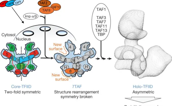
2.2 General transcription factor TFIID ... 47
2.2.1 TFIID structure ... 47
2.2.2 Domain organization of TAFs ... 48
2.2.3 TAF paralogues and their functions ... 50
2.2.4 TFIID assembly ... 51
2.3 Coactivator SAGA complex ... 53
2.4 TRanscription and EXport complex 2 (TREX-2) ... 54
3. Protein complexes ... 56
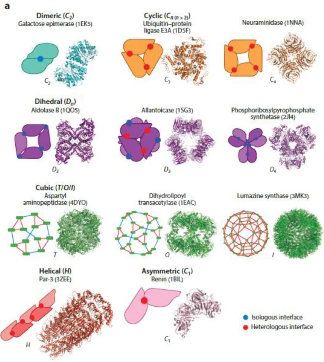
3.1 Homomeric complexes ... 57
3.1.1 TBP-associated factor (TAF) homodimerization ... 60
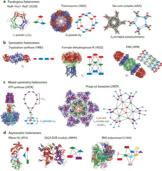
3.2 Heteromeric complexes ... 60
2
3.3.1 Obligate and non-obligate protein complexes ... 63
3.3.2 Transient and stable protein complexes ... 63
3.4 Assembly of protein complexes ... 63
4. Co-translational assembly of protein complexes ... 65
4.1 Co-translational assembly of homomeric protein complexes ... 65
4.1.1 Bacterial beta-galactosidase enzyme ... 65
4.1.2 Myosin heavy chain ... 66
4.1.3 Tenascin intermediate filament ... 66
4.1.4 Reovirus cell attachment protein ... 67
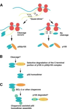
4.1.5 NFKB ... 68
4.1.6 p53 ... 69
4.1.7 Peripherin ... 70
4.2 Co-translational assembly of heteromeric protein complexes ... 71
4.2.1 Immunoglobulin ... 71
4.2.2 Signal recognition particle receptor ... 72
4.2.3 D1 protein of photosystem II ... 73
4.2.4 Membrane ionchannel ... 74
4.2.5 IgE receptor (Fc€RI) ... 75
4.2.6 Co-translation assembly of diverse heteromeric complexes in yeast ... 76
4.2.7 Bacterial Lux Operon ... 79
4.2.8 Co-translational assembly of protein complexes in mammalian cells ... 79
4.3 Ribosomal pause associated with co-translational assembly ... 80
5. How are co-translationally assembling partners brought in close proximity to each other?... 82
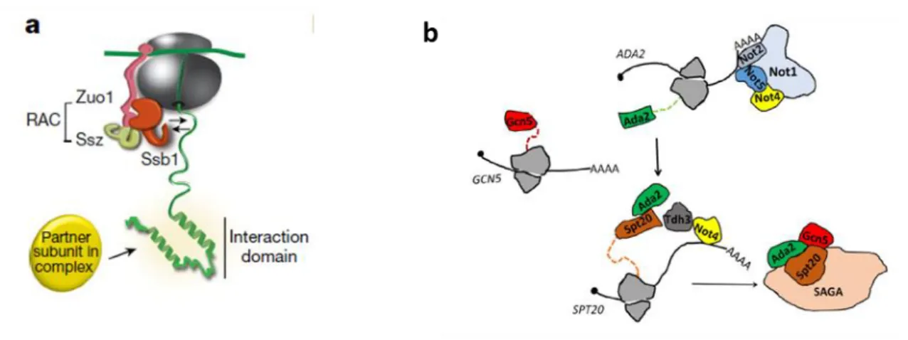
5.1 Chaperones associated with co-translational assembly ... 82
5.2 Cis-acting RNA sequence elements ... 83
5.2.1 AU-rich element (ARE) ... 84
5.2.2 GU-rich element (GRE) ... 84
5.2.3 Polyadenylation sequences ... 85
5.2.4 3’UTRs ... 86
5.2.5 5’UTRs ... 88
5.3 RNA granules ... 89
5.3.1 mRNP granules ... 90
5.3.1.1 Stress granules and Processing bodies (P-bodies) ... 90
3
5.3.1.3 Not1 containing assemblysomes ... 92
6. mRNA and protein surveillance in cells ... 93
6.1 mRNA Quality Control pathways ... 94
6.1.1 mRNA surveillance in the nucleus ... 94
6.1.2 Nonsense- mediated decay (NMD) ... 94
6.1.3 No-go decay (NGD) ... 97
6.1.4 Non-stop decay (NSD) ... 98
6.1.5 Ribosome-associated protein quality control (RAPP) ... 99
6.2 Chaperone, the key player assisting protein folding ... 100
6.2.1 Cytosolic Chaperones ... 101
6.2.2 PAQosome (particle for arrangement of quaternary structure) ... 105
6.2.3 Chaperonins ... 107
6.2.4 Chaperone dysregulation under disease condition ... 108
7. Overview of techniques used to study co-translational assembly ... 110
7.1 Biochemical Approach ... 110 7.1.1 Indirect Approach ... 110 7.1.2 Direct Approach ... 110 7.2 Imaging approaches ... 112 THESIS OBJECTIVES ... 113 RESULTS ... 115
1. Co-translational assembly of mammalian nuclear multisubunit complexes ... 116
2. Unpublished Results ... 153
2.1 Co-translational assembly of TFIID submodules ... 153
2.2 Co-translational assembly of SAGA and ATAC complex ... 157
2.3 Identification of chaperones guiding co-translational assembly of TFIID complex ... 159
DISCUSSION ... 164
1. General transcription factor TFIID assembles co-translationally ... 165
2. Position of dimerization domain drives co-translationally assembly ... 166
3. Co-translational assembly is essential for the cell ... 167
4. Co-translational assembly is a general mechanism of complex assembly ... 168
PERSPECTIVES ... 170
1. Comprehensive study of the assembly of TFIID submodules. ... 171
4
3. Mechanism of subunit distribution between two complexes? ... 173
4. What is the mechanism behind TAF8 mRNA degradation in the absence of its co-translationally assembling partner TAF10? ... 174
CONCLUSIONS ... 175
METHODS ... 178
5
List of Figures
Figure 1: Multiple levels of chromatin folding.. ... 30
Figure 2: Schematic representation of post-translational modifications of histone tails.. ... 32
Figure 3: Schematic representation of key post-translational modifications within globular domains of histones and their functions.. ... 32
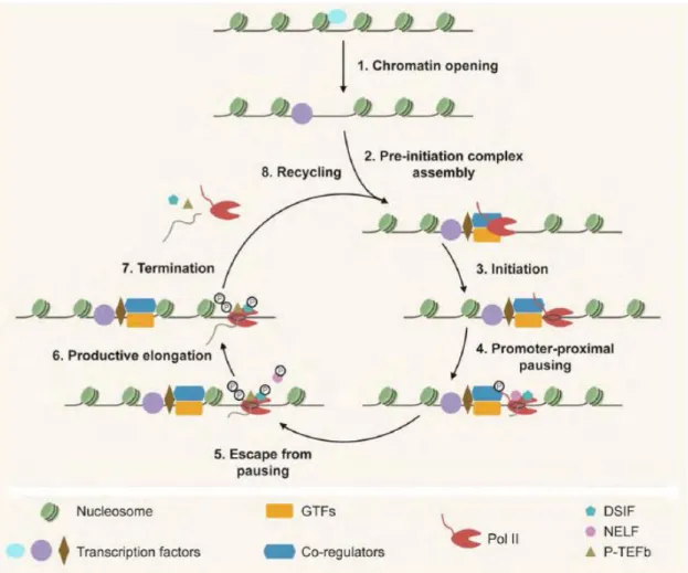
Figure 4: Schematic representation of the different steps of RNA Pol II transcription. ... 36
Figure 5: Transcription factor binding models.. ... 37
Figure 6: RNA Pol II transcription PIC assembly and transcription reinitiation.. ... 41
Figure 7: Subunit sharing between different transcription complexes.. ... 46
Figure 8: Schematic representation of general transcription factor TFIID.. ... 47
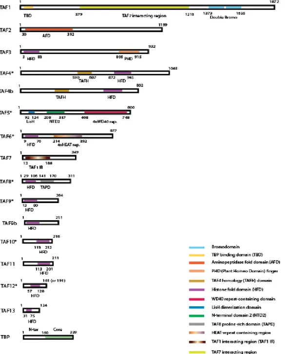
Figure 9: Domain organization of TAFs.. ... 49
Figure 10: Stepwise assembly of TFIID complex.. ... 52
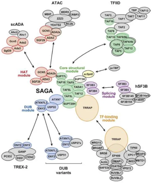
Figure 11: Schematic representation of SAGA complex.. ... 54
Figure 12: Schematic representation of human TREX-2 complex.. ... 55
Figure 13: Different levels of protein organization.. ... 56
Figure 14: Classification of homomeric protein complexes based on shape.. ... 59
Figure 15: Schematic representation of core-TFIID complex.. ... 60
Figure 16: Classification of heteromeric protein complexes based on shape.. ... 62
Figure 17: Models of protein complex assembly... 64
Figure 18: Models depicting β-galactosidase co-translational assembly.. ... 66
Figure 19: Model for the biogenesis of reovirus σ1 trimer.. ... 67
Figure 20: Models of p50 homodimer formation. ... 68
Figure 21: Model for p53 homodimer formation.. ... 69
Figure 22: Model depicting dynamic co-translational assembly of intermediate filaments.. ... 70
Figure 23: Model showing the structure of a typical immunoglobulin molecule (A) and co-translational formation of intrachain disulphide bond of an immunoglobulin molecule (B).. ... 71
6
Figure 25: Model showing stepwise co-translational assembly of D1 protein into PSII
complex and thylakoid membrane.. ... 73
Figure 26: Model for co-translational association of hERG subunit mRNAs.. ... 74
Figure 27: Schematic representation of Fc€RI receptor.. ... 75
Figure 28: Model for co-translational assembly of the SET1C complex.. ... 76
Figure 29: Co-translational assembly of SAGA complex in yeast.. ... 77
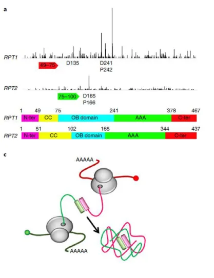
Figure 30: Model for co-translational assembly of Rpt1 and Rpt2 in Not1 containing assemblysomes... 78
Figure 31: Co-translational assembly bacterial Lux operon subunits... 79
Figure 32: An example of ribosome pausing during co-translational assembly from (Panasenko et al. 2019).. ... 81
Figure 33: Possible mechanisms by which co-translationally assembling protein partners are brought near each other.. ... 82
Figure 34: Chaperones assisting co-translational assembly in yeast………83
Figure 35: Schematic representation of the different types of isoforms generated due to the usage of different poly(A) sites in a particular mRNA.. ... 86
Figure 36: Schematic representation of cytoplasmic movement of mRNAs through P-body and stress granules.. ... 91
Figure 37: Schematic representation of TIGER domain cellular compartment.. ... 92
Figure 38: Schematic representation of co-translational assembly of Rpt1 and Rpt2 in NCA.. ... 93
Figure 39: Mechanism of NMD pathway (EJC model).. ... 96
Figure 40: Mechanism of NGD pathway.. ... 98
Figure 41: Mechanism of NSD pathway.. ... 99
Figure 42: Cytoplasmic chaperone pathways in (a) bacteria and (c) eukarya.. ... 101
Figure 43: Composition of ribosome-associated chaperones from bacteria to human.. ... 102
Figure 44: Schematic representation of yeast and human (mRAC) RAC complex.. ... 103
Figure 45: Schematic representation of chaperone interactions with nascent polypeptide of cytosolic proteins (A) and secretory proteins (B) at ribosome exit site.. ... 104
7
Figure 46: Schematic representation of PAQosome structure, Adaptors and
Substrates/Clients.. ... 106
Figure 47: Major steps of selective ribosome profiling (SeRP).. ... 111
Figure 48: Co-translational assembly of TFIID heterotrimer TAF2-TAF8-TAF10.. 154
Figure 49: Co-translational assembly of TFIID heterotrimer. ... 155
Figure 50: Co-translational assembly of TBP and TAF1 in (A) HEK293T and (B) NIH3T3 cell lines. ... 156
Figure 51: Co-translational assembly of (A) SAGA and (B) ATAC complex.. ... 158
Figure 52: Western Blot analysis of HeLa polysome extract.. ... 159
Figure 53: Potential TAF10 and TBP interacting factors from HeLa polysome
containing cell cytoplasmic extracts.. ... 161
Figure 54: Identification of TAF8 mRNA associated factors by TAF8-MS2-TRAP.. ... 163
Figure 55: Schematic representation of polysome RIP assay ... 181
Figure 56: Schematic representation of working principle of smiFISH... 186
List of Tables
Table 1: Different coactivator families and their functions ... 38
Table 2: General transcription factors (GTFs) involved in Pol II transcription. ... 39
Table 3: TAF paralogues and their sequence similarity ... 50
Table 4: Different clusters of AU-rich elements (ARE) and GU-rich elements (GRE).) ... 85
8
Abbreviations
ADA2 : Adenosine deaminase 2
ADA3 : Alteration/Deficiency in Activation 3
ADP-ribosylation : Adenosine diphosphate-ribosylation
ARE : AU-rich element
Arp4 : Actin-related protein 4
Ascl1 : Achaete-Scute Family BHLH Transcription Factor 1
ATAC : Ada2a-containing complex
ATRX : α-thalassaemia/mental retardation syndrome X-linked ATXN7L3 : Ataxin 7 like 3
BAF : BRG1-Associated Factor
BLOC-1 : biogenesis of lysosome-related organelles complex 1
BORC : BLOC-one-related complex
Bre : Brain and Reproductive organ-expressed protein
BRF1 : B-related factor 1
BTAF1 : B-TFIID TATA-Box Binding Protein Associated Factor 1
CAF-1 : Chromatin Assembly Factor-1
CARM1 : Coactivator Associated Arginine Methyltransferase 1
CBP : CREB-binding protein
Ccr4 : C-C Motif Chemokine Receptor 4
CCT : Chaperonin Containing TCP1 or TriC-TCP-1 Ring Complex
CHD : chromodomain helicase DNA-binding
COMPASS : complex proteins associated with Set1p
CPSF : cleavage and polyadenylation specificity factor
cryo-EM : cryo-electron microscopy
CSB : Cockayne syndrome group B
CSTF : cleavage stimulation factor
CTD : C-terminal domain
DOT1L : Disruptor of telomeric silencing 1-like
DP : Aspartic acid-Proline
9
DSS1 : deleted in split hand/split foot
DUB module : Deubiquitination module
Eaf3 : Esa1-associated factor 3
ECDs : extracellular domains
EGFR : epidermal growth factor receptor
EJC : exon junction complex
ENY2 : Enhancer of Yellow 2
ER : Endoplasmic reticulum
FACT : facilitates chromatin transcription
FoxA : Forkhead box A
GABAA : γ-aminobutyric acid
GANP : Germinal-center Associated Nuclear Protein
GAPDH : Glyceraldehyde 3-phosphate dehydrogenase
Gata : (A/T)GATA(A/G)
GCN5 : General Control Nondrepressible 5
GDP : guanosine diphosphate
GFP : green fluorescent protein
GRE : GU-rich element
GTFs : General Transcription Factors
GTP : guanosine triphosphate
HAT : Histone acetyltransferase
HEAT : Huntingtin-elongation factor 3 (EF3)- protein phosphatase 2A (PP2A)- yeast kinase TOR1
hERG : human ether-à-go-go-related Gene
HFD : histone fold domain
Hsc70 : heat shock cognate70
Hsp70 : heat shock protein 70kD
Hsp70L1 : Hsp70-like protein 1
HSPB1 : heat shock protein beta-1
ICDs : intracellular domain
IF : Immunofluorescence
IF : Intermediate filament
10
Ig : immunoglobulin
INO80 : inositol requiring 80
IP : Immunoprecipitation
ISWI : imitation switch
J protein : J domain protein
KDM5 : lysine (K) demethylase 5
Klf4 : Kruppel Like Factor 4
KO : Knockout
Lid : Little imaginal discs
MCP : MS2-coat protein
MERFISH : multiplexed error robust FISH
mESCs : mouse embryonic stem cells
miRNA : microRNA
MLL : mixed lineage leukemia
MPP11 : M phase phosphoprotein 11
mRAC : mammalian RAC
mRNA : messenger RNA
mRNP : mRNA-ribonucleoprotein
MS2-TRAP : MS2-tagged RNA affinity purification
MYB : myeloblastosis
NAC : nascent-chain-associated complex
NC2beta : Negative co-factor 2
NCA : Not1 containing assemblysomes
NEFs : nucleotide exchange factors
NELF : Negative elongation factor
NGD : no-go decay
NLS : Nuclear Localisation Signal
NMD : nonsense-mediated decay
Not : negative on TATA
NPCs : Nuclear pore complexes
NSD : non-stop decay
NTD : N-terminal domain
NTP : Nucleoside triphosphate
11
NuRD : nucleosome remodelling and deacetylase
NURF : Nucleosome Remodeling Factor
Oct3/4 : octamer-binding transcription factor 3/4
ORF : Open reading frame
P bodies : Processing bodies
PAF1C : Polymerase associated factor-1 complex
PAQosome : particle for arrangement of quaternary structure
PAS : polyadenylation signal
Pax7 : Paired box 7
PCID2 : PCI domain containing 2
PCNA : Proliferating Cell Nuclear Antigen
Pfd : Prefoldin
PHD : plant homeodomain
PIC : pre-initiation complex
PPIs : protein-protein interactions
PRMT1 : Protein arginine N-methyltransferase 1
PS1 : Presenilin-1
PSII : Photosystem II
PTC : premature stop codon
P-TEFb : positive transcription elongation factor b
R2TP : Rvb1–Rvb2–Tah1–Pih1 RAC : ribosome-associated complex
RAP30/74 : RNA polymerase II-associating protein 30/74
RAPP : Ribosome-associated protein quality control
RIP : RNA Immunoprecipitation
RNA FISH : RNA Fluorescent in situ hybridization
RNA-BP : RNA-binding protein
RPBs : ribosome-bound protein biogenesis factors
Rpd3 : Reduced potassium dependency-3
Rpt : Proteasome Regulatory particle base subunit
rRNA : ribosomal RNA
SAGA : Spt-Ada-Gcn5 acetyltransferase
SeRP : selective ribosome profiling
12
SETD2 : SET Domain Containing 2
SL1 : Selective factor 1
SMAT : small TAF complex
smFISH : single molecule fluoresencent in situ hybridization
SMG1 : Serine-threonine protein kinase
smiFISH : single molecule inexpensive fluorescent in situ hybridization
snRNA : small nuclear RNA
Sox2 : SRY-Box 2
SPT : Suppressor of Ty
SRα : signal recognition particle receptor α
SUMOylation : Small Ubiquitin-like Modifier (SUMO) -ylation
SURF : SMG1-UPF1-eRF
SWI/SNF : switch/ sucrose non-fermentable
SWR1 : SWI2/SNF2-Related 1
TADs : topologically associating domains
TAF : TBP associated factor
TAFH : TAF4 homology domain
TAND : TAF1 N-terminal domain
TBP : TATA-box binding protein
TCP-1 : tailless complex polypeptide-1
TCR : T-cell receptor
TF : Trigger Factor
TFII : Transcription factor II
TIGER : TIS Granules-ER
Tip60 : Tat interactive protein 60-kDa
TIS : TPA-induced
TLF : TBP-like factor
TLP : TBP-like protein
TP53 : Tumor protein p53
TRRAP : Transformation-transactivation domain-associated protein
TREX-2 : TRanscription and EXport complex 2
TRF : TBP-related factor
13
TRP : TBP related protein
UPF : Up-frameshift
UPF1 : Regulator of nonsense transcripts 1
USP22 : Ubiquitin Specific Peptidase 22
UTR : Untranslated region
WD-40 : Trp-Asp (W-D)-40
XRN2 : 5′-3′ exoribonuclease 2
YEATS2 : YEATS Domain Containing 2
14
Abstract
Biological processes in the cell are mainly are carried out by multisubunit protein complexes and a significant amount of energy is required by the cells to build these huge complexes. Unlike bacteria, genes encoding proteins are dispersed in the genome of eukaryotes and this makes the assembly of protein complexes more complicated. For many years, it was thought that protein complexes are formed by random collisions of their subunits diffusing freely in the cell cytoplasm. However, random collisions could also lead to non-specific interactions and aggregations in the extremely crowded cellular environment. In this respect, co-translational assembly of nuclear and cytoplasmic protein complexes has been put forward in bacteria and yeast that involves the association of nascent ribosome-associated proteins subunits with each other, thereby preventing unwanted interactions. It was shown earlier that operon organisation in bacteria facilitates co-translational assembly of their protein complexes due to proximity of the encoding genes. Additionally, several well-characterised protein complexes in yeast were also reported to assemble co-translationally. In this study, we show that the mammalian multisubunit transcription complexes assemble co-translationally by using several alternate approaches like RNA immunoprecipitation followed by genome-wide detection of mRNAs by microarray analysis, single molecule RNA FISH, immunofluoresence, knock-out mouse embryonic stem cells and domain swapping approaches. We also demonstrate that the dimerization domains and their positions in the interacting subunits determine the co-translational assembly pathway (simultaneous or sequential). Furthermore, cytoplasmic IF-smFISH and two-colour smFISH experiments indicate that the described co-translational assembly is clearly occurring in the cytoplasm of human cells. Identical results in yeast, mouse and human cells suggest that co-translational assembly is a general mechanism of protein complex assembly in eukaryotes.
15
Thesis summary in English
Introduction
Majority of the biological processes are carried out by multisubunit protein complexes in mammalian cells. Transcription is the process by which the information in the DNA is copied into RNA molecules. Eukaryotic RNA Polymerase II transcription is driven by six general transcription factors namely TFIID, TFIIA, TFIIB, TFIIF, TFIIE, TFIIH and the multisubunit mediator complex. In mammalian cells TFIID nucleates the assembly of other transcription factors in most of the expressed protein coding gene promoters and loss of TFIID was shown to be embryonic lethal. TFIID is composed of TBP (TATA-box binding protein) and 13 TAFs (TBP associated factors). Majority of the TAFs contain a common structural motif called the histone fold domain (HFD), which facilitates pairwise interaction between specific TAFs, specifically, TAF10-TAF8, TAF3-TAF10, TAF6-TAF9, TAF4-TAF12, TAF11-TAF13 heterodimers. The histone fold is not conserved at the level of sequence but is conserved structurally; it is composed of three α-helices connected by two loops, which allow heterodimeric interactions between specific TAFs. Moreover, the histone fold containing proteins are not soluble when they are expressed individually. In spite of extensive studies on the structure and function of the multisubunit transcription complexes, very few studies have focused on the assembly mechanism and pathways of the multisubunit protein complexes. It has been reported previously that the assembly of TFIID takes place in a stepwise manner before it embarks on its function in the nucleus. Several submodules of TFIID have been shown to assemble in the cytoplasm before it forms the holo-TFIID complex. Stable heterotrimers of TAF5-TAF6-TAF9, TAF2-TAF8-TAF10 and TAF7-TAF11-TAF13 have been shown to form in the cytoplasm. In this context, the main aim of my project was to study the mechanism of assembly of TFIID submodules in the cytoplasm. A protein dimer can assemble in two possible ways: “posttranslational assembly”, where individual subunits are fully translated and released at random places in the cytoplasm eventually finding their interacting partners, or “co-translational assembly”, where protein-protein interactions form in the cytoplasm during the translation of the interacting protein partners. Co-translational assembly could be beneficial for the cell to prevent non-specific interactions in the very crowded cytoplasmic environment of the cell. One of the earliest evidences of
co-16
translational protein assembly was reported in 2009 where the authors studied 31 proteins lacking RNA-binding domains from Schizosaccharomyces pombe and among
them ∼38% co-purified with mRNAs that encode interacting proteins. This observation
was further supported by a very recent report which showed co-translational assembly
in Saccharomyces cerevisiae by selective ribosome profiling. Taken together, various
evidences point towards the fact that co-translational assembly could be a widespread mechanism for assembly of a large number of complexes in yeast. Here, we study the co-translational assembly mechanism of the general transcription factor TFIID in detail and further extend our observations to other multisubunit complexes in mammalian cells.
Aims:
1. Do TFIID histone-fold domain pairs assemble co-translationally? 2. What drives the co-translational assembly of TFIID HFD pairs? 3. Do non-HFD pairs also assemble co-translationally?
4. Do other multisubunit complexes also assemble co-translationally?
5. Study the co-localisation of co-translationally assembling proteins and RNAs? 6. Study the role of chaperones in co-translational assembly?
Results:
To test whether HFD-containing TAFs assemble co-translationally, we used a monoclonal antibody against the N-terminus of the HFD containing TAF10 to immunoprecipitate (IP) endogenous TAF10 from human HeLa cell cytosolic polysome extracts. Immunoprecipitation of a protein followed by the study of its associated mRNAs is called RNA Immunoprecipitation (RIP) assay. Protein–protein interactions between nascent proteins still associated with translating ribosomes would be revealed by enrichment of mRNAs coding for the interacting partners in the IPs. Global microarray analysis of mRNAs precipitated by the anti-TAF10 RNA IPs (RIPs) revealed enrichment of TAF8 mRNA, suggesting that the well-characterised TAF8-10 HFD dimer forms co-translationally. The microarray results were further supported by anti-TAF10 RIP coupled to RT-qPCR in both HeLa and mouse embryonic stem cells (mESCs). Puromycin treatment resulted in a significant loss of the enrichment of associated mRNAs thereby ruling out the possibility of direct interaction between TAF8
17
mechanism of TAF10-TAF8 co-translational assembly. We generated expression vectors expressing TAF10 and TAF8 with either N-terminal or C-terminal tags. These constructs enabled us to pulldown either nascent (with N-terminal tag) or full-length protein (with C-terminal tag) and thereby determine the order of co-translational assembly of the proteins. In accordance with our endogenous immunoprecipitation (IP) results, nascent TAF10 RIP revealed the presence of its own mRNA, along with
TAF8 mRNA. Interestingly, however, TAF10 mRNA was not co-immunoprecipitated with nascent TAF8 protein. This observation was further supported by pulldown experiments with C-terminal tagged TAF10 and TAF8 constructs. Fully translated mature TAF10 protein revealed the presence of only TAF8 mRNA, but not its own mRNA (as expected) but mature TAF8 protein did not yield TAF10 or TAF8 mRNA. In all cases, TAF8 protein was co-immunoprecipitated with TAF10 protein thereby ruling out the possibility of unsuccessful protein IP experiment. Together these results indicate that TAF10 protein is assembling co-translationally with TAF8 nascent protein unidirectionally by sequential assembly model. We hypothesised that this sequential binding is specific to the localisation of the dimerization domains of TAF10 and TAF8, HFD of TAF10 being towards C-terminus and HFD of TAF8 being towards N-terminus. To test this, we engineered a mutation in the HFD of TAF8 disrupting the interaction between TAF10 and TAF8. This mutation resulted in a nearly complete loss of the co-precipitated TAF8 mRNA, as compared with the wild-type controls, indicating that the dimerization of TAF8 and TAF10 through their HFDs is crucial for co-translational assembly. In addition to that, IF-smiFISH co-localization experiments in fixed HeLa cells showed significant co-localisation between TAF10 protein and TAF8 mRNA in the cytoplasm by confocal microscopy. Importantly, the co-localisation between mutant TAF8 mRNA and TAF10 protein was lost. In addition, TAF8 protein detection by IF and TAF10 mRNA by smiFISH, showed no significant co-localisation, thereby further lending support to the sequential model of TAF10-TAF8 co-translational assembly. Next, we wanted to study if co-translational assembly is essential for the cell. To answer this question, we applied an indirect approach. We hypothesized that if nascent chains of a subunit cannot co-translationally interact with its partner, it may become prone to misfolding and degradation by the proteasome, but the fully translated partner should stay stable. By using these mouse ESCs we observed that the deletion of Taf10 not only ablated Taf10 mRNA and TAF10 protein levels, but significantly reduced both Taf8 mRNA and TAF8 protein expression. These results
18
were also confirmed in Taf10 KO mouse embryos. In contrast, the deletion of Taf8, decreased only its own mRNA and protein levels, without affecting the Taf10 mRNA expression and TAF10 protein levels. In both KO mouse ESCs, other tested TFIID subunits remained unchanged, thereby ruling out the possibility of a primary transcriptional response. Additionally, we also observed that TAF10 re-expression rescued TAF8 from degradation. Thus, the nascent TAF8 HFD, in the absence of its interaction partner TAF10, may serve as a signal for both protein and mRNA degradation, while TAF10 is stable in the absence of TAF8.
Having shown the sequential co-translational assembly of TAF10 and TAF8, we next studied the assembly of a protein pair whose dimerization domains are localised towards the N-terminus. To test this, we studied the co-translational assembly of TAF6-TAF9 HFD pair as they interact through their N-terminal HFDs. Our nascent RIPs revealed that both TAF6 and TAF9 co-IP their partners' mRNA suggesting that they assemble in a bidirectional way, presumably as the neosynthesised interaction domains of both proteins are exposed early during their synthesis on the ribosomes. Further evidence from two colour smiFISH co-localization experiments showed a significantly higher co-localisation of the TAF6 and TAF9 mRNAs in the cytoplasm than several unrelated negative control mRNAs.
In order to rule out the possibility that this form of assembly is specific to only HFD protein pairs, we wanted to study the co-translational assembly of a non-HFD interacting protein pair. In TFIID, the evolutionary conserved core domain of TBP interacts with TAF1 via N-terminal TAND region of TAF1. Genome-wide microarray analysis of TBP-associated RNAs from HeLa cell polysome extracts revealed an enrichment of its own mRNA as well as 19 coding and non-coding RNAs. Among these, we found mRNAs coding for known TBP-interacting proteins: BRF1 coding for a factor important for Pol III transcription, BTAF1 coding for a B-TFIID subunit, as well as TAF1. RIP-qPCR analysis in human HeLa cells and mouse ESCs confirmed the microarray data and revealed a strong enrichment of the TAF1 mRNA. To further investigate the specificity of TBP-TAF1 interaction, we generated a ΔTAF1 expression vector, in which sequences coding for the first 168 residues containing the TAND region were deleted. Anti-TBP RIPs from cells expressing ΔTAF1 resulted in complete loss of TAF1 mRNA enrichment and a reduction of the co-immunoprecipitated protein. These results are consistent with a requirement of the N-terminal TAF1 domain to
19
recruit TBP to the nascent TAF1 polypeptide. As the protein interface is formed by the C-terminal portion of TBP and the very N-terminus of TAF1, we predicted that similar to TAF8-TAF10 assembly, a sequential assembly is also involved in the TBP-TAF1interaction. Indeed, nascent anti-TAF1 RIP from an engineered GFP-TAF1 HeLa cell line resulted in the enrichment of TAF1 mRNA, but not that of TBP, thus supporting the sequential co-translational assembly model of TBP-TAF1 by the sequential pathway.
To extend our findings beyond TFIID, we examined co-translational assembly of the ENY2 subunit with its respective partners. ENY2 is subunit of the TREX-2 mRNA export complex and the DUB module of the SAGA transcription coactivator. In TREX-2, two ENY2 proteins wrap around the central portion of the large GANP helical scaffold. Similarly, human ENY2 wraps around the N-terminal helix of human ATXN7L3 in the highly intertwined SAGA DUB module. To test whether the co-translational model is generally applicable to multisubunit complexes, we analysed ENY2- associated mRNAs from HeLa cells stably expressing ENY2 with an N-terminal GFP-tag. Interestingly, we found that an anti-GFP-ENY2 RIP co-immunoprecipitates predominantly endogenous GANP mRNA and protein (the partner of ENY2 in TREX-2), and also endogenous ATXN7L3 mRNA and protein (the binding partner of ENY2 in the SAGA DUB module).
Conclusions:
Together, our study demonstrates that co-translational assembly is involved in the assembly of mammalian transcription complexes of diverse architecture and function. We also show that the localisation of the interacting domains of a protein dimer determines the co-translational assembly pathway, sequential or simultaneous. We also demonstrate that the nascent protein partner and its mRNA prone are to degradation in the absence of the fully translated co-translationally assembling partner.
20
Publication:
Kamenova I*, Mukherjee P*, Conic S, Mueller F, El- Saafin F, Bardot P, Marie Garnier J, Dembele D, Capponi S, HT Timmers M, Vincent S, Tora L (2019) Co-translational assembly of mammalian nuclear multisubunit complexes. Nature Communications
*equal first authors, names appear in alphabetical order
Communications:
1. Flash talk and poster presentation: Co-translation drives the assembly of mammalian nuclear multisubunit complexes
Pooja Mukherjee, Ivanka Kamenova, Laszlo Tora
SFB 1036 conference "Networks of Cellular Surveillance Mechanisms" October 22-24, 2018, Heidelberg, Germany.
2. Poster presentation: Co-translational assembly of mammalian nuclear multisubunit complexes
Pooja Mukherjee, Ivanka Kamenova, Laszlo Tora
TriRhena Transcription and Chromatin Club, March 26, 2019, Strasbourg, France.
3. Short talk: Co-translational assembly of mammalian nuclear multisubunit complexes
Pooja Mukherjee, Ivanka Kamenova, Laszlo Tora
Cell Symposia: Regulatory RNAs, May 12-14, Berlin, Germany.
21
Thesis summary in French
Introduction
La majorité des processus biologiques sont réalisés par des complexes protéiques contenant plusieurs sous unités. La transcription est le processus par lequel l'information contenue dans l'ADN est copiée dans des molécules d'ARN. La transcription par l’ARN polymérase II chez les eucaryotes est régie par six facteurs généraux de transcription, à savoir TFIID, TFIIA, TFIIB, TFIIF, TFIIE, TFIIH et le complexe multi protéique médiateur. Dans les cellules de mammifère, TFIID génère l'assemblage d'autres facteurs de transcription sur les promoteurs des gènes codants pour des protéines et la perte de TFIID est létale à l’état embryonnaire. TFIID est composé de la protéine TBP (TATA-binding protein) et de 13 TAF (TBP associated
factors). La majorité des TAF partagent un domaine structurel commun appelé Histone
Fold Domain (HFD), qui permet l’hétérodimérisation entre certains TAF, en particulier
TAF10-TAF8, TAF3-TAF10, TAF4-TAF12 et TAF11-TAF13. Le domaine HFD n'est pas conservé au niveau de la séquence mais est conservé d’un point de vue structurelle, il est composé de trois hélices α reliées par deux boucles, qui permettent des interactions entre les TAF spécifiques. De plus, les protéines contenant un HFD ne sont pas solubles lorsqu'elles sont exprimées individuellement. Malgré des études approfondies sur la structure et la fonction de complexes transcriptionnels multi-protéiques, peu d’études se sont intéressées aux mécanismes d’assemblage de ces complexes. Il a déjà été montré que l’assemblage de TFIID se fait par étapes avant qu’il ne soit fonctionnel dans le noyau. Plusieurs sous-modules de TFIID ont été mis en évidence dans le cytoplasme avant la formation du complexe holo-TFIID. La présence d’hétérotrimères stables TAF5-TAF6-TAF9, TAF2-TAF8-TAF10 et TAF7-TAF11-TAF13 a été démontrée dans le cytoplasme. Dans ce contexte, le principal objectif de mon projet était d’étudier le mécanisme d’assemblage des sous-modules TFIID dans le cytoplasme. Un dimère protéique peut s'assembler de deux manières : un assemblage post-traductionnel où des sous-unités individuelles sont entièrement traduites et distribuées de manière aléatoire dans le cytoplasme, ou un assemblage co-traductionnel, où des interactions protéine-protéine se produisent au cours de la traduction des partenaires protéiques. L'assemblage co-traductionnel pourrait être
22
bénéfique pour la cellule en permettant d'éviter des interactions non spécifiques dans l'environnement cytoplasmique très encombré de la cellule. Une des premières observations d’assemblage co-traductionnel a été obtenue en 2009 où les auteurs ont étudié 31 protéines ne contenant pas de domaine de liaison à l'ARN de
Schizosaccharomyces pombe. Parmis ces protéines, ~ 38% co-purifient avec des
ARNm codant pour des protéines partenaires. Cette observation a été corroborée par une publication très récente qui montre un assemblage co-traductionnel chez
Saccharomyces cerevisiae par profilage sélectif des ribosomes. Pris ensemble, ces
diverses évidences montrent que l’assemblage co-traductionnel pourrait être un mécanisme répandu pour l’assemblage d’un grand nombre de complexes multi-protéiques dans la levure. Ici, nous avons étudié en détail le mécanisme d'assemblage co-traductionnel du facteur général de transcription TFIID et avons étendu nos observations à d'autres complexes multi-protéiques dans des cellules de mammifère.
Objectifs:
1. Est-ce que les hétérodimères TAFs contenant des HFD s'assemblent de manière co-traductionnelle ?
2. Qu'est-ce qui dirige l'assemblage co-traductionnelle de hétérodimères TAFs à HFD ?
3. Est-ce que les hétérodimères TAFs ne contenant pas d’HFD s'assemblent également de manière co-traductionnelle ?
4. Est-ce que d'autres complexes multi-protéiques s'assemblent également de manière co-traductionnelle ?
5. Étude de la localisation des ARN et des protéines assemblées de manière co-traductionnelle.
6. Étude du rôle des chaperons dans l’assemblage co-traductionnel.
Résultats:
Afin de tester si les TAF contenant un HFD s'assemblent de manière co-traductionnelle, nous avons utilisé un anticorps monoclonal dirigé contre l'extrémité N-terminale de TAF10 contenant un HFD, pour immunoprécipiter la protéine TAF10 endogène à partir d'extraits de polysomes cytosoliques de cellules HeLa. L’immunoprécipitation d’une protéine associée à l’étude des ARNm associés est
23
appelée RNA – ImmunoPrecipitation (RIP). Les interactions protéine-protéine entre les protéines naissantes encore associées aux ribosomes sont révélées par l'enrichissement de l'ARNm codant pour les partenaires en interaction dans les IP. L'analyse globale de puces à ADN des ARNms précipités par les IP anti-TAF10 (RIP) a révélé un enrichissement de l'ARNm codant pour TAF8, suggérant que le dimère HFD TAF8-TAF10 bien caractérisé, se forme de manière co-traductionnelle. Les résultats des puces ont également été corroborés par le RIP anti-TAF10 couplé à la RT-qPCR dans les cellules HeLa et les cellules souches embryonnaires de souris (ESCs). Le traitement à la puromycine entraîne une perte significative de l'enrichissement des ARNm associés, excluant ainsi la possibilité d'une interaction directe entre l'ARNm codant pour TAF8 et la protéine TAF10. Ensuite, nous nous sommes intéressés à l’étude du mécanisme sous-jacent de l’assemblage co-traductionel de TAF10-TAF8. Nous avons généré des vecteurs d'expression exprimant TAF10 et TAF8 avec des étiquettes N-terminales ou C-terminales. Ces constructions nous ont permis d’immunoprécipiter la protéine naissante (étiquette N-terminale) et la protéine complète (étiquette C-N-terminale) et de déterminer ainsi l'ordre d'assemblage co-traductionnel des protéines. Conformément à nos résultats d'immunoprécipitation endogène (IP), le RIP de TAF10 naissant a révélé la présence de son propre ARNm, ainsi que l'ARNm de TAF8. Cependant, l'ARNm de TAF10 n’a
pas été co-immunoprécipité avec la protéine naissante de TAF8. Cette observation est corroborée également par des expériences d’immunoprécipitation avec des constructions TAF10 et TAF8 marqué à leurs extrémités C-terminale. La protéine mature complètement traduite de TAF10 a révélé la présence de seulement l'ARNm de TAF8, mais pas de son propre ARNm (comme attendu), mais la protéine mature de TAF8 ne donnait pas l'ARNm de TAF10 ou TAF8. Dans tous les cas, la protéine TAF8 a été co-immunoprécipitée avec la protéine TAF10, ce qui exclut la possibilité d'une expérience IP infructueuse. Dans l’ensemble, ces résultats indiquent que la protéine TAF10 s'assemble de manière co-traductionnelle avec la protéine naissante de TAF8 de manière unidirectionnelle par un modèle d'assemblage séquentiel. Nous avons émis l’hypothèse que cette liaison séquentielle est spécifique à la localisation des domaines de dimérisation de TAF10 et TAF8, le HFD de TAF10 étant vers l’extrémité C-terminale et le HFD de TAF8 étant vers l’extrémité N-terminale. Pour tester cela, nous avons réalisé une mutation dans le HFD de TAF8 perturbant l’interaction entre TAF10 et TAF8. Cette mutation a entraîné une perte presque
24
complète de l'ARNm de TAF8 co-précipitée par rapport à la condition contrôle, ce qui indique que la dimérisation de TAF8 et TAF10 via leurs HFD est cruciale pour l'assemblage co-traductionnel. En outre, des expériences de co-localisation IF-smiFISH dans des cellules HeLa fixées ont montré une co-localisation significative entre la protéine TAF10 et l'ARNm de TAF8 dans le cytoplasme par microscopie confocale. Notablement, la co-localisation entre l'ARNm de TAF8 mutée et la protéine TAF10 a été perdue. En outre, la détection de la protéine TAF8 par IF et l’ARNm de TAF10 par smiFISH n'a montré aucune co-localisation significative, ce qui a permis de soutenir davantage le modèle séquentiel d'assemblage co-traductionnel de TAF10-TAF8. Ensuite, nous voulions étudier si l’assemblage co-traductionnel est essentiel pour la cellule. Pour répondre à cette question, nous avons utilisé une approche indirecte. Nous avons supposé que si les chaînes naissantes d'une sous-unité ne peuvent pas interagir de manière co-traductionnelle avec son partenaire, elles peuvent devenir sujettes au mauvais repliement et à la dégradation par le protéasome, mais le partenaire entièrement traduit devrait rester stable. En utilisant ces cellules souche de souris, nous avons observé que la suppression de Taf10 non seulement supprimait les niveaux d'ARNm de Taf10 et de la protéine de TAF10, mais réduisait de manière significative l'expression de l’ARNm de Taf8 et la protéine de TAF8. Ces résultats ont également été confirmés chez des embryons de souris KO pour Taf10. En revanche, la suppression de Taf8 n'a entraîné que la diminution des niveaux de ses propres ARNm et protéines, sans affecter l'expression de l'ARNm de Taf10 ni les taux de protéines TAF10. Dans les deux lignes KO des cellules souches de souris, les autres sous-unités TFIID testées sont demeurées inchangées, excluant ainsi la possibilité d'une réponse transcriptionnelle primaire. De plus, nous avons également observé que la ré-expression de TAF10 a empêché la dégradation de TAF8. Ainsi, le HFD naissant de TAF8, en l'absence de son partenaire d'interaction TAF10, peut servir de signal pour la dégradation de ses protéines et ses ARNm, tandis que TAF10 est stable dans l'absence de TAF8. Après avoir montré l'assemblage séquentiel co-traductionnel de TAF10 et TAF8, nous avons ensuite étudié l'assemblage d'une paire de protéines dont les domaines de dimérisation sont localisés vers l'extrémité N-terminale. Pour tester cela, nous avons étudié l'assemblage co-traductionnel de la paire de HFD de TAF6-TAF9 lors de leur interaction par leurs HFD N-terminaux. Nos RIP naissants ont révélé que TAF6 et TAF9 co-immunoprécipitent les ARNm de leurs partenaires suggèrent qu'ils s'assemblent de manière bidirectionnelle, vraisemblablement au
25
moment où les domaines d'interaction néosynthétisés des deux protéines sont exposés tôt pendant leur synthèse par les ribosomes. D'autres preuves provenant d'expériences de localisation smiFISH à deux couleurs ont montré une co-localisation significativement plus élevée des ARNm de TAF6 et TAF9 dans le cytoplasme que plusieurs ARNm de contrôle négatif non corrélatifs. Dans le but d’exclure la possibilité que cette forme d’assemblage protéique est spécifique aux sous-unités de TFIID contenant des HFDs, nous voulions étudier l’assemblage
co-traductionnel de pair de protéine ne contenant pas d’HFDs. Au sein de TFIID, l’interaction entre TBP et TAF1 se fait d’une part par un domaine central évolutivement conservé et le côté N-terminal contenant une région TAND respectivement. Une analyse sur micro-puce à l’échelle du génome global des ARNm associés à TBP, obtenus à partir d’extrait de polysome de cellules HeLa a révélé que la protéine TBP était majoritairement associée à ses propres ARNm ainsi qu’à 19 autres ARNs codant et non codant. Parmi eux se trouvent les ARNm codant pour des protéines connues pour interagir avec TBP comme BRF1 qui est important pour la transcription médiée par l’ARN polymérase III, BTAF1 présent dans le complexe B-TFIID ainsi que TAF1. Des analyses par RIP-qPCR à partir d’extrait de cellules HeLa et de cellules ES
murines ont confirmé les résultats obtenus sur micro-puces, dont un fort enrichissement de l’ARNm TAF1. Afin d’étudier la spécificité de l’assemblage de TAF1-TBP, un vecteur exprimant une version mutée de TAF1 (noté ΔTAF1) a été généré dans laquelle 168 acide-aminés présent au sein de la région TAND ont été supprimés. Une expérience de RIPs contre TBP à partir des cellules exprimant ΔTAF1
a montré une complète absence d’enrichissement en ARNm TAF1 et une réduction de la quantité de protéine TAF1 immunoprécipité. Ces résultats sont en accord avec le fait que le domaine en position N-terminal de TAF1 est requis pour recruter TBP au niveau de TAF1 en train d’être synthétisé. Comme l’interaction se fait par l’extrémité C-terminal de TBP et par l’extrémité N-terminal de TAF1, nous prédisons que similairement à l’assemblage de TAF8-TAF10, un assemblage séquentiel a aussi lieu pour l’interaction TBP-TAF1. En effet, une expérience de RIPs contre TAF1 naissant à partir d’une lignée cellulaire HeLa exprimant GFP-TAF1 a montré un enrichissement pour l’ARNm TAF1, mais pas de l’ARNm TBP, ce qui supporte cette hypothèse. Pour élargir ces découvertes au-delà de TFIID, nous avons examiné l’assemblage co-traductionnel de ENY2 avec ses partenaires respectifs. ENY2 est une sous-unité du
26
complexe d’export des ARNm nommé TREX-2 ainsi que du module DUB du co-activateur de la transcription SAGA. Dans le complexe TREX-2, deux protéines ENY2 s’enroulent au niveau du squelette hélicoïdal de la protéine GANP. De façon similaire, dans le module DUB, ENY2 humain s’enroule autour de l’hélice en position N-terminal d’ATXN7L3. Pour tester si le modèle d’assemblage co-traductionnel peut-être généralement appliqué à l’assemblage des complexes protéiques, nous avons analysé les ARNm associés à ENY2 au sein de cellules HeLa exprimant de façon stable ENY2 fusionné en N-terminal à un tag GFP. Nous avons trouvé que GFP-ENY2 co-immunoprécipite principalement les ARNm de la protéine GANP endogène ainsi que GANP lui-même (le partenaire d’ENY2 dans TREX-2), et aussi les protéines et les ARNm de ATXN7L3 endogène (le partenaire d’ENY2 dans le module DUB).
Conclusions :
Notre étude démontre que l’assemblage co-traductionnel est impliqué dans l’assemblage de complexes de transcription de mammifères d’architecture et de fonctions diverses. Nous montrons également que la localisation des domaines en interaction d'un dimère de protéine détermine la voie d'assemblage co-traductionnel, séquentielle ou simultanée. Nous démontrons également que le partenaire protéique naissant et son ARNm sont susceptibles de se dégrader en l'absence du partenaire d'assemblage complètement traduit.
27
INTRODUCTION
28
Introduction
The cell uses a vast array of protein complexes to carry out important biological functions. Such protein complexes are made up of either multiple copies of same subunits (homomeric protein complexes) or different subunits (heteromeric protein complexes). The latter group includes various transcription regulatory and chromatin remodelling complexes. In order to achieve correct assembly of protein complexes, the genes encoding the subunits (dispersed in the genome) must be transcribed in the nucleus, transported to the cytoplasm, translated by the ribosomes and finally find their interacting partners. All these processes must be correctly orchestrated to achieve efficient assembly of protein complexes. Following translation in the cytoplasm the protein subunits can find their interacting partners either post-translationally by random collision in the cytoplasm. This can lead to non-specific interactions and the cell might end up spending more energy to assemble protein complexes by this method. Co-translational association of protein complexes can result in efficient formation of protein complexes preventing unwanted interactions. In fact, this model of protein assembly has been put forward in bacteria and yeast.
The major step linking our genome and its final product, protein that carries out functions in the cell, is transcription (synthesis of RNA from DNA). One of the key regulatory steps is transcription initiation. The protein complexes involved in transcription initiation include general transcription factors (GTFs), activators, co-activators, etc. As mentioned above, majority of these complexes are multisubunit. Though their functional aspects in vivo have been studied extensively, their assembly is not well-studied so far. Certain reports suggest the existence of submodules of transcription complexes including RNA Polymerases and TFIID but there has been no concrete report so far on their order of assembly. Another intriguing aspect that is still unanswered is how the cell allocates the common subunits to different complexes as many of these transcription complexes consist of common subunits.
The first two sections of my introduction will focus on the various steps of transcription and the key protein complexes involved in it. The third section mainly deals with different types of protein complexes in the cell. The fourth, fifth and seventh sections describe the various aspects of co-translational protein assembly. Finally, the sixth section describes the various mRNA and protein quality control mechanisms.
29
1. Eukaryotic gene expression
1.1 Chromatin organisation
Living organisms store genetic information in the form of the nucleic acids DNA and RNA. The message in our genome is transferred to RNA by a process called transcription. These RNAs in turn act as messengers of protein synthesis or translation and it is the protein that carries out various functions in the cell. This sequential flow of information constitutes the central dogma of molecular biology (Crick 1970). Although this is the basis of molecular biology, certain discoveries also describe the backward flow of genetic information from RNA to DNA (Iwre 1970; Baltimore 1970). Our genetic material DNA is organised hierarchically in the form of chromatin in the cell (Figure 1). The first level of organisation is the wrapping of 145-147 bp DNA around a scaffold of core histone octamer (two copies each of histone H2A, H2B, H3, H4) forming a dynamic protein-DNA complex called the nucleosome core particle (NCP) (Arents et al. 1991; Fa 1973; Richmond et al. 2003; Woodcock et al. 1976; Richmond et al. 1997; Arents 1993). H2A-H2B and H3-H4 form heterodimeric protein pairs through a specific domain called the histone fold domain (HFD) and their long N-terminal unstructured regions exist in the form histone tail extensions ( Richmond et al. 1997; Arents et al. 1991). The DNA is further packaged into chromatosome core particle which is a recurring structural unit consisting of linker histone H1 bound to the nucleosome core particle (Simpson 1978). The linker histone further aids in the formation of a chromatin fiber of diameter 30 nm (Finch and Klug 1976). In addition, chromatin can also form loops and topologically associating domains (TADs) (Sexton et al. 2012; Hou et al. 2012; Dixon et al. 2012). Several TADs can associate and form a chromatin superdomain (Pueschel et al. 2016). The chromatin structure finally gets condensed to a chromosome. (Figure 1) (R. Kornberg 1974). The structure of chromatin is essential for the regulation of several DNA-related metabolic processes including transcription, recombination, DNA repair, replication, kinetochore, centromere formation, etc (Fyodorov et al. 2018). Over ten decades ago, it was established that some fractions of the chromosomes were stained very intensely with nuclear dyes, while other areas were only weakly stained (Passarge 1979). This finding formed the basis for defining “euchromatin” and “heterochromatin” respectively. Euchromatin is gene-rich and has open chromatin conformation whereas
30
heterochromatin is condensed and contains mainly repetitive elements (Huisinga et al. 2006; Grewal and Jia 2007).
Figure 1: Multiple levels of chromatin folding. DNA is wrapped around the nucleosome and undergoes compaction to accommodate the DNA in the limited volume of the nucleus. Image is from (Zhou and Bai 2019).
31
1.2 Nucleosome remodelling
Chromatin structure imposes significant obstacles upon the transcription machinery. The multiple interactions between histone and DNA makes the nucleosome one of the most stable protein-DNA complexes under physiological conditions (Richmond et al. 1997). At the same time, chromatin is dynamic as well and is tightly regulated by various protein complexes involved in histone modification, chromatin remodelling, histone variant incorporation, and histone eviction. Both histone-modifying enzymes
and chromatin remodelling complexes bind and make DNA accessible to transcription machinery.
1.2.1 Histone-modifying enzymes
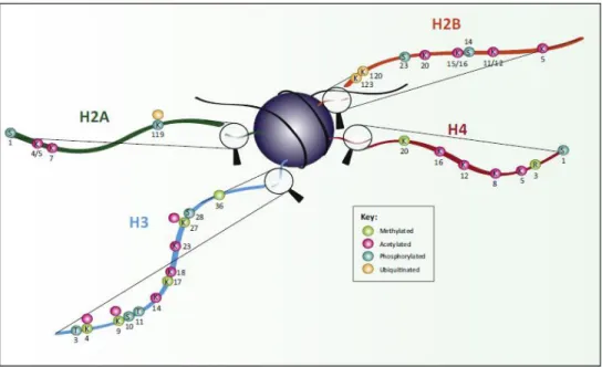
Histone-modifying enzymes target both histone tails and globular domains for posttranslational modifications. Some of the main modifications and their functions are shown in Figure 2 and Figure 3. Modifications associated with active transcription include acetylation of H3 and H4, di- or trimethylation of H3K4 and are commonly referred to as euchromatin modifications. Modifications can also occur at specific amino acid residues, for example, methylation of arginine residues, phosphorylation of serine and threonine, lysine residues can undergo a wide range of modifications including methylation, acetylation, ubiquitination, ADP-ribosylation and SUMOylation. Other modifications like propionylation and butyrylation have also been described very recently (Kebede et al. 2015). A detailed description of all histone modifications and their functions are reviewed in (Lawrence et al .2016).
32
Figure 2: Schematic representation of post-translational modifications of histone tails.
The amino acid modified is shown (K = lysine, R = arginine, S = serine, T = threonine) and the position of each modification is shown in black. Colours depict the nature of modification of each residue (green = methylated, pink = acetylated, turquoise = phosphorylated, beige = ubiquitinated). Image is from (Lawrence et al. 2016).
Figure 3: Schematic representation of key post-translational modifications within globular domains of histones and their functions. Methyl marks are shown in light green, acetyl marks in pink, and phosphorylated residues in light blue. Image is from (Lawrence et al. 2016).
33 1.2.2 Chromatin remodelling complexes
Chromatin remodelling complexes (also called chromatin remodellers, (Clapier et al. 2017) are protein complexes that alter nucleosome structure in an ATP-dependent manner. Various subfamilies of chromatin remodelling complexes carry out diverse functions including transient unwrapping of DNA-histone octamers, forming DNA loops, and nucleosome sliding. Based on the subunit composition and their catalytic ATPases, chromatin remodellers can be classified into four subfamilies: 1) imitation switch (ISWI), 2) chromodomain helicase DNA-binding (CHD), 3) switch/ sucrose non-fermentable (SWI/SNF) and 4) inositol requiring 80 (INO80). Higher eukaryotes contain multiple remodeller subtypes within each family that are specific to certain cell types or developmental stages (Lessard and Crabtree 2010; Bao and Shen 2007). In addition to this, other remodellers, which do not belong to the previously mentioned subfamilies (for example, α-thalassaemia/mental retardation syndrome X-linked (ATRX (Law et al. 2010; Lewis et al. 2010) and Cockayne syndrome group B (CSB) (Citterio et al. 2000)) also exist but are less well-characterized mechanistically.
ISWI family of remodellers include ISWI complexes in yeast and NURF (Nucleosome Remodeling Factor) complex in humans. They are made up of 2 to 4 subunits and their functions include organising nucleosome spacing to facilitate chromatin assembly and therefore transcription repression. However, certain complexes in this family can also assist in RNA Polymerase II (RNA Pol II) activation, thereby suggesting that the functional diversity is mainly imparted by the presence of distinct subunits in each complex (Becker and Workman 2013). The CHD family of remodellers consists of complexes that can promote transcription by sliding or ejecting nucleosomes or they may also have transcription repressive roles. For example, the vertebrate NuRD (nucleosome remodelling and deacetylase) complex plays a role in chromatin compaction by its histone deacetylase activity (Fei et al. 2015). SWI/SNF family of chromatin remodellers include SWI/SNF complex in yeast and BAF (BRG1-Associated Factor complex) complex in human, are composed of 8 to 14 subunits. This family of remodellers has a role in sliding and ejection of nucleosomes at many loci (Narlikar et al. 2013). INO80 group of remodellers include SWR1-related (SWI2/SNF2-Related 1) complexes, (which was first purified from yeast) and human Tip60/TRAPP (Tat interactive protein 60-kDa/ transformation-transactivation domain-associated protein) complex. They are made up of more than 10 subunits and play
34
roles in diverse functions including transcriptional activation and DNA repair. Although belonging to INO80 group, SWR1 has the unique ability to replace H2A of canonical H2A-H2B dimer with histone variant H2AZ to form H2A.Z-H2B dimer. Due to functional differences among the various families of remodelling complexes, earlier models suggested the use of distinct enzymatic mechanisms to achieve their diverse functions. In contrast, a more unified model was proposed recently which puts forward the fact that all histone remodellers use a common ATP-dependent DNA translocation mechanism to move DNA along the histone surface (Clapier et al. 2017). These complex series of events finally result in the accessibility of nucleosome wrapped DNA to transcription factors. Following this, transcription is carried out on “naked” DNA by the transcription machinery. The basic mechanism of transcription is conserved between prokaryotes and eukaryotes even though the transcription machinery is more complex in eukaryotes (Hahn 2004).
1.3 RNA Polymerases
Dedicated transcription machinery exists in the cell that carries out transcription. The enzyme responsible for transcription is RNA Polymerase. The enzymatic activity of RNA Polymerase was first discovered decades ago in rat liver nuclei (Weiss and Gladstone 1959). It was later discovered in E.coli as well (Hurwitz et al. 1961; A. Kornberg 1961) . Five different RNA polymerases (named I to V) have been discovered so far in eukaryotes whereas only one RNA Polymerase has been identified so far in prokaryotes and Archaea. The archaeal RNA Polymerase and eukaryotic RNA Polymerase II are structurally and mechanistically closely related (Korkhin et al. 2009). In eukaryotes, different polymerases transcribe different classes of cellular RNAs (Kedinger et al. 1970; Weinmann et al. 1974; Warfel 1970; Zylber and Penman 1971). Pol I synthesizes 18S and 28S ribosomal RNAs (rRNA), Pol II synthesizes messenger RNAs (mRNAs), small nuclear RNAs (snRNAs) and microRNAs (miRNAs), Pol III synthesizes transfer RNAs (tRNAs) and 5S rRNAs and certain viral RNAs (Zylber and Penman 1971; Weil and Blatti 1976; Roeder and Rutter 1970; Reinberg et al. 2004). The recently identified RNA Polymerase IV and V are required for the production of siRNAs (small interfering RNAs) in plants, mediating RNA-directed DNA methylation, transcriptional silencing and heterochromatin formation (Wierzbicki et al. 2009; Onodera et al. 2005; Kanno et al. 2005). Nevertheless, despite differences in functionality, all RNA Polymerases contain a