HAL Id: inserm-01855905
https://www.hal.inserm.fr/inserm-01855905
Submitted on 8 Aug 2018
HAL is a multi-disciplinary open access
archive for the deposit and dissemination of
sci-entific research documents, whether they are
pub-lished or not. The documents may come from
teaching and research institutions in France or
abroad, or from public or private research centers.
L’archive ouverte pluridisciplinaire HAL, est
destinée au dépôt et à la diffusion de documents
scientifiques de niveau recherche, publiés ou non,
émanant des établissements d’enseignement et de
recherche français ou étrangers, des laboratoires
publics ou privés.
Constance Alabert, L Rogers, L Kahn, S Niellez, Patrick Fafet, S Cerulis,
Jean-Marie Blanchard, Robert Hipskind, Marie-Luce Vignais, R Rogers
To cite this version:
Constance Alabert, L Rogers, L Kahn, S Niellez, Patrick Fafet, et al.. Cell type-dependent control
of NF-Y activity by TGF-β. Oncogene, Nature Publishing Group, 2006, 25 (24), pp.3387 - 3396.
�10.1038/sj.onc.1209385�. �inserm-01855905�
ORIGINAL ARTICLE
Cell type-dependent control of NF-Y activity by TGF-b
C Alabert, L Rogers, L Kahn, S Niellez, P Fafet, S Cerulis, JM Blanchard, RA Hipskind
and M-L Vignais
Institut de Ge´ne´tique Mole´culaire de Montpellier, CNRS-UMR5535-IFR122, Montpellier, France
Transforming growth factor b (TGF-b) is a pluripotent cytokine that regulates cell growth and differentiation in a cell type-dependent fashion. TGF-b exerts its effects through the activation of several signaling pathways. One involves membrane proximal events that lead to nuclear translocation of members of the Smad family of transcriptional regulators. TGF-b can also activate MAPK cascades. Here, we show that TGF-b induces nuclear translocation of the NF-YA subunit of the transcription factor NF-Y by a process that requires activation of the ERK cascade. This results in increased binding of endogenous NF-Y to chromatin and TGF-b-dependent transcriptional regulation of the NF-Y target gene cyclin A2. Interestingly, the kinetics of NF-YA relocalization differs between epithelial cells and fibro-blasts. NIH3T3 fibroblasts show an elevated basal level of phosphorylated p38 and delayed nuclear accumulation of NF-YA after TGF-b treatment. In contrast, MDCK cells show low basal p38 activation, higher basal ERK phosphorylation and more rapid localization of NF-YA after induction. Thus, NF-Y activation by TGF-b1 involves ERK1/2 and potentially an interplay between MAPK pathways, thereby opening the possibility for finely tuned transcriptional regulation.
Oncogene (2006) 25, 3387–3396. doi:10.1038/sj.onc.1209385; published online 23 January 2006
Keywords: TGF-b; NF-Y; MAPK
Introduction
Transforming growth factor b (TGF-b) signaling is implicated in biological processes ranging from growth control to cell differentiation and is disrupted in many cancers (Siegel and Massague, 2003; Waite and Eng, 2003; Yingling et al., 2004). The biological outcome of TGF-b exposure depends on both the cellular context and environmental cues. The effects of TGF-b are triggered by binding of the ligand to its cell surface receptor, which leads to the formation of the active
receptor kinase and to phosphorylation of downstream targets (de Caestecker, 2004). Several signaling path-ways have been identified downstream of TGF-b receptors, among which that involving the Smad family of transcription factors is the best characterized. TGF-b can also activate the ERK, p38 and SAPK/JNK mitogen activated protein kinase (MAPK) cascades to different extents, depending on cell type (Attisano and Wrana, 2000; Derynck and Zhang, 2003). One potential intermediate is TGF-b-activated kinase 1 (TAK1) (Yamaguchi et al., 1995), a MAPKKK that can activate the JNK (Wang et al., 1997) or p38 pathway (Moriguchi et al., 1996).
Outside of Smads, few transcription factors are known to be targeted by TGF-b. We focused our study on the ubiquitous and evolutionarily conserved tran-scription factor NF-Y, also known as CCAAT-binding factor (CBF). Y is composed of three subunits: NF-YA, NF-YB and NF-YC (Maity et al., 1992). NF-YB and NF-YC contain histone-fold motives that are required for dimerization, as well as association with NF-YA, and all three subunits are necessary for specific-DNA binding (Sinha et al., 1995). While the large glutamine-rich domains of the NF-YA and NF-YC subunits can activate transcription (Coustry et al., 1996), NF-Y can also mediate repression of target genes (Manni et al., 2001). NF-Y helps control the expression of many genes, including the cell cycle regulatory genes cyclin A2, cyclin B2 and E2F1 (Hu and Maity, 2000; Manni et al., 2001). Although NF-Y was originally considered a constitutive transcription factor, recent data indicate that its activity is regulated, in particular through NF-YA (Mantovani, 1999; Marziali et al., 1999). The knockout of NF-YA leads to early embryo-nic lethality and accordingly NF-YA!/!mouse
embryo-nic fibroblasts show a block in cell growth, confirming a role for NF-Y in regulating cell proliferation (Bhatta-charya et al., 2003).
In this study, we show that NF-Y is a target of TGF-b signaling. TGF-b treatment leads to localization of NF-YA to the nucleus. This nuclear accumulation correlates with increased binding of endogenous NF-Y to chro-matin and transcriptional regulation as shown for the cyclin A2 gene. NF-YA localization is dependent upon ERK activation in both fibroblasts and epithelial cells. However, the kinetics of NF-YA nuclear accumulation differs in these two cell types, a difference that may be linked to the basal level of activation of both the ERK
Received 16 May 2005; revised 5 December 2005; accepted 9 December 2005; published online 23 January 2006
Correspondence: Dr M-L Vignais, Institut de Ge´ne´tique Mole´culaire de Montpellier, CNRS, 1919 Route de Mende, Montpellier, Cedex 5, 34293, France.
E-mail: [email protected]
Oncogene (2006) 25, 3387–3396
&2006 Nature Publishing Group All rights reserved 0950-9232/06 $30.00 www.nature.com/onc
and p38 cascades. An interplay between MAPK cas-cades would allow a cell type-dependent fine tuning of the expression of key cell cycle genes, such as cyclin A2, thereby providing specificity for the TGF-b response.
Results
NF-YA localizes to the nucleus in response to TGF-b Results from our laboratory have previously implicated the trimeric transcription factor NF-Y in the transcrip-tional regulation of the cyclin A gene (Plet et al., 1997) and preliminary data suggested its likely involvement in the transcriptional response to TGF-b (data not shown). Therefore, we investigated whether this involved a change in the subcellular localization of NF-Y similar to the TGF-b-dependent nuclear translocation of the Smad proteins (Derynck and Zhang, 2003; Shi and Massague, 2003; ten Dijke and Hill, 2004). Our study focused on the NF-YA subunit, since it provides the DNA binding specificity to the trimeric transcription factor. Moreover, the A subunit is regulated in numerous situations (Mantovani, 1999). NIH3T3 fibro-blasts were treated with TGF-b and then processed for indirect immunofluorescence at various times in order to visualize changes in the cellular localization of endo-genous NF-YA and Smad3 (Figure 1). As expected, Smad3 accumulated in the nucleus within 15 min of TGF-b treatment (Figure 1a). NF-YA localized within the cytoplasm and nucleus in cycling cells and then became nuclear in response to TGF-b. Interestingly, NF-YA nuclear localization peaked at around 90 min of TGF-b treatment with kinetics that clearly differed from Smads, for which the peak occurred at 15 min. In contrast, NF-YB localization was restrained to the nucleus in NIH3T3 cycling cells and remained so during TGF-b treatment (Supplementary data). To confirm the nuclear relocalization of NF-YA and Smad3 biochemi-cally, western blots of nuclear extracts from TGF- b-treated NIH3T3 cells were incubated with antibodies against these proteins (Figure 1b). Consistent with the results above, NF-YA levels progressively increased in the nucleus after TGF-b treatment but with delayed kinetics relative to Smad3. Quantification of the nuclear accumulation of Smad3 and NF-YA was performed from blots obtained from three independent sets of protein extracts. It showed increases in nuclear concen-trations of respectively 5.1-fold for Smad3 (s.d.: 0.7) and 2.3-fold for NF-YA (s.d.: 0.2). This nuclear accumula-tion reflected translocaaccumula-tion of the cytoplasmic pool of NF-YA, since TGF-b induction did not alter the total amount of the protein (Figure 1b).
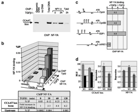
TGF-b-induced NF-YA nuclear accumulation correlates with increased binding to chromatin of CCAAT box-containing promoters and with transcriptional regulation To demonstrate a functional role for the nuclear relocalization of NF-YA, we first investigated whether this correlated with increased binding to its target promoters. As the transcription factor NF-Y can
modify chromatin structure and associate with both histones and other transcription factors, we tested this using chromatin immunoprecipitation (ChIP). Chroma-tin was prepared from TGF-b-treated NIH3T3 fibro-blasts and immunoprecipitated with antibodies specific for NF-YA. To follow NF-YA binding, we used both semiquantitative and real-time PCR to detect the
Figure 1 TGF-b treatment leads to nuclear accumulation of NF-YA in NIH3T3 cells. (a) NIH3T3 fibroblasts were treated for the indicated times (in min) with 80 pMTGF-b1. Endogenous NF-YA and Smad3 were visualized by immunofluorescence. Nuclei were stained with Hoechst. (b) Lysates were prepared from NIH3T3 cells incubated with TGF-b for the indicated times. Equal amounts of nuclear extracts (upper panel) and total extracts (lower panel) were analysed by immunoblotting with antibodies specific for NF-YA, Smad3, TBP and GAPDH.
inverted CCAAT box of the cyclin A2 promoter that binds NF-Y in vitro and in vivo (Plet et al., 1997) (Figure 2a and b). TGF-b stimulation led to increased recruit-ment of NF-YA to the CCAAT box but not to an upstream region of the cyclin A promoter (see table). Likewise, only background levels of cyclin A CCAAT box chromatin were precipitated by an irrelevant anti-body against the tyrosine kinase Tyk2. These data indicate that TGF-b induces increased NF-YA binding to the cyclin A CCAAT box in vivo. In addition, TGF-b treatment also resulted in increased DNA binding of the NF-YB subunit, an effect that was abolished when, instead of the wild-type (wt) NF-YA, a dominant negative mutant of NF-YA was overexpressed (data not shown). Similarly NF-YA showed increased bind-ing, in response to TGF-b, to the CCAAT boxes present in the promoters of the cyclin B2 and E2F1 genes (Bolognese et al., 1999) (Figure 2c). There was only background binding to the promoter of the house-keeping gene hypoxanthine phosphoribosyltransferase (HPRT) that contains no CCAAT box. These data
confirm that TGF-b signaling results in increased DNA recruitment of NF-YA on CCAAT boxes in vivo. Besides, we also tested NF-Y binding by gel shift assay using nuclear extracts from mock or TGFb-treated cells. It also showed increased NF-Y DNA binding activity in response to TGFb (data not shown).
We used the cyclin A2 promoter to study the activity of NF-Y in response to TGF-b since this gene is repressed by TGF-b in CCL39 cells (Barlat et al., 1993, 1995). Moreover, NF-Y binds to the inverted CCAAT box found near the cyclin A2 transcription initiation sites (Plet et al., 1997) and here, we have shown that TGF-b induces NF-Y binding to this site in chromatin. We compared the activities of cyclin A2 promoters bearing either a wt or a mutated (mut) CCAAT box, using luciferase reporter genes in transient transfection assays. Notably, our cyclin A2 promoter constructs are mutated at the CCRE-CHR site to bypass cell cycle-dependent regulation of the cyclin A2 gene conferred by this site (Huet et al., 1996; Philips et al., 1999; Fajas et al., 2001). We observed a twofold transcriptional
Figure 2 Effect of TGF-b treatment on NF-Y DNA binding and transcriptional activity. Chromatin immunoprecipitation (ChIP) was performed on NIH3T3 fibroblasts after TGF-b1 (80 pM) treatment. The chromatin samples were immunoprecipitated with antibodies specific for either NF-YA or Tyk2. The immunoprecipitated DNAs were analysed by both semiquantitative (a) and real-time (b) PCR. (a) PCR amplification of the CCAAT box region of the endogenous cyclin A promoter. The products of 32 cycles of amplification were detected using [a32P]dCTP and analysed by autoradiography of DNA-PAGE. ‘Genomic DNA’ is a control of native
murine DNA and ‘total DNA’ corresponds to the chromatin input for the immunoprecipitations. (b) Percentage of immunoprecipitated DNA (ratio of immunoprecipitated versus input DNA) for each time point (in min) of TGF-b treatment. PCRs were performed using primer pairs amplifying the CCAAT box region or a region 700 bp upstream of the endogenous cyclin A2 promoter. Tyk2 indicates amplification of ChIPs performed at the 120 min time point with the Tyk2 antibody. The table indicates the increase in YA binding to the endogenous cyclin A2 CCAAT box observed in response to TGF-b treatment. The increase in NF-YA binding upon TGF-b treatment was observed in several ChIP experiments. The diagram shows the data from two independent experiments. (c) ChIP analysis of TGF-b-induced binding of NF-YA to CCAAT boxes in the indicated promoters. The experiments were performed as described above. (d) The CCAAT box and NF-Y are required for TGF-b-dependent repression of cyclin A2 transcription. NIH3T3 cells were transfected with luciferase reporter genes driven by the cyclin A2 promoter bearing either the wt or a mut CCAAT box. Cells were then left untreated or induced with 80 pMTGF-b1. The left panel represents the relative luciferase units
(RLU) measured in control (grey) or TGF-b-treated (black) cells. The right panel presents the degree of cyclin A2 trancriptional repression, measured as the ratio of luciferase activities in untreated versus TGF-b-treated cells.
TGF-b regulates NF-YA nuclear localization via MAPKs
C Alabert et al
3389
repression by TGF-b of the wt cyclin A2 CCAAT box that dropped to 1.3 upon mutation of the CCAAT box (Figure 2d). These differences, obtained from four independent series of experiments, were significant (P-valueo0.0001). Likewise, overexpression of the wt and of a dominant negative mutant of NF-YA had opposing effects on reporter activity (Po0.0005), as the NF-YA dominant negative mutant diminished while the wt protein increased repression of the cyclin A2 by TGF-b (Po0.05). These data indicate that the CCAAT box and NF-Y participate in transcriptional regulation of the cyclin A2 gene in response to TGF-b, therefore supporting a functional role for nuclear localization of the NF-YA subunit in response to TGF-b.
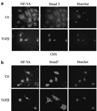
TGF-b-induced nuclear accumulation of NF-YA does not require protein neosynthesis or the Smad2/Smad3 signaling pathway
NF-YA levels did not change in whole cell extracts in response to TGF-b (Figure 1b). However, its delayed accumulation in the nucleus relative to Smads could be dependent on prior activation of the Smads and/or de novo synthesis of other signaling components. To test the latter, we pretreated NIH3T3 cells with cyclo-heximide prior to incubation with TGF-b. Inhibition of protein synthesis did not hinder TGF-b-driven nuclear accumulation of NF-YA (Figure 3a). To test the role of Smad activation, we transfected an expres-sion vector encoding Smad7, which inhibits Smad2/3 signaling (data not shown and (Derynck and Zhang, 2003)). It slightly enhanced TGFb-dependent phosphory-lation of ERK (Supplementary Figure 2). TGF-b still induced nuclear accumulation of NF-YA in Smad7 overexpressing cells while, as previously documented, the recombinant Smad7 protein redistributed to the cytoplasm (Figure 3b). These data indicate that NF-YA nuclear accumulation in response to TGF-b is indepen-dent of Smad activation and protein neosynthesis.
NF-YA nuclear accumulation depends on ERK activation In numerous cultured cell lines, MAPK cascades mediate some TGF-b intracellular signaling events. Therefore, we tested for MAPK activation by TGF-b in our NIH3T3 fibroblasts and whether MAPKs played a role in NF-YA nuclear localization. TGF-b stimulated activating phosphorylation of ERK2 by 15 min and again by 60 min after treatment but did not alter the level of ERKs 1 and 2 (Figure 4a). Activating phosphorylation of p38 in response to TGF-b treatment clearly differed from that of ERK, with a progressive decrease up to 30 min after TGF-b treatment and a sharp transient peak at the 60 min time point. As with ERK1/2, p38 levels remained unaffected by TGF-b addition (Figure 4a). Notably, TGF-b did not activate SAPKs in NIH3T3 cells, while they, as well as ERK1/2 and p38, were readily induced by subinhibitory levels of anisomycin (Figure 4a).
To evaluate the role of these MAPKs in NF-YA nuclear accumulation, we used pharmacological inhibi-tors specific for the ERK and p38 cascades. PD98059
blocks activation of MEK1 and thereby ERK1/2 (Alessi et al., 1995), while SB203580 inhibits p38a and b activity but not their activating phosphorylation (Young et al., 1997). As expected, PD98059 diminished TGF-b-driven phosphorylation of ERK; SB203580 inhibited phos-phorylation of ATF1, a well-characterized target of the p38 cascade (Tan et al., 1996) (Figure 4b) and thus an indicator of p38 kinase activity. SB203580 slightly increased SAPK phosphorylation after TGF-b treat-ment, suggesting that, like in other cell systems, p38 inhibition enhances TAK1 induction and thereby that of SAPK (Cheung et al., 2003).
We next examined the effects of these inhibitors on TGF-b-dependent accumulation of NF-YA in the nucleus. This was blocked by pretreatment of cells with PD98059 but not by the SAPK inhibitor SP600125 (Figure 4c). These data were quantified from two independent experiments, using either FITC- or cy3-conjugated antibodies. While the proportion of cells harboring a high nuclear concentration of NF-YA significantly increased from 16 to 78% (s.d. 4.2) upon TGFb treatment, pretreatment of the NIH3T3 cells with PD98059 prevented this increase and NF-YA remained spread between the nucleus and the cytoplasm in 90% (s.d. 5.7) of the cells. Similar results were obtained in the CCL39 fibroblasts (data not shown).
Figure 3 NF-YA nuclear accumulation in response to TGF-b requires neither protein neosynthesis nor Smad2/Smad3 signaling. NIH3T3 cells were seeded on coverslips and treated with 80 pM
TGF-b for 90 min. The endogenous NF-YA and Smad3 were detected by immunofluorescence. (a) Cycloheximide (CHX, 10mg/ml) was added to NIH3T3 cells 30 min prior to TGF-b treatment. (b) NIH3T3 cells were transfected with a vector encoding Flag-tagged Smad7 protein 24 h prior to TGF-b treatment. Smad7 overexpression was monitored by immunofluorescence using a polyclonal Flag antibody.
In the presence of SB, NF-YA was found around the nuclear periphery after TGF-b induction. This could reflect a failure of NF-YA to localize to the nucleus after TGF-b treatment or an accelerated nuclear export. Consistent with the second possibility are preliminary observations that NF-YA appears more rapidly in the nucleus upon TGF-b induction in cells pretreated with SB alone or together with SP, which blocks SB-induced SAPK activation. We also tested the effect of okadaic acid, a specific inhibitor of type 1 and 2A serine/ threonine phosphatases, as PP2A was both proposed to be involved in TGF-b signaling and to regulate the activities of ERK and p38 (Janssens and Goris, 2001). Okadaic acid activated all three MAPK pathways and led to NF-YA nuclear accumulation independently of TGF-b stimulation, thus emphasizing the important role of serine/threonine phosphorylation in NF-YA nuclear
localization (Figure 4b and c). Interestingly, Smads remained cytoplasmic upon okadaic acid treatment (Figure 4c), providing further evidence that NF-YA nuclear accumulation in response to TGF-b does not require prior Smad3 activation. Taken together, these data show that (1) TGF-b induces NF-YA nuclear accumulation in NIH3T3 fibroblasts with kinetics delayed compared to those of the Smads, and (2) this is dependent upon activation of the ERK1/2 cascade but not that of Smads.
The kinetics of TGF-b-dependent NF-YA nuclear accumulation differ between epithelial cells and fibroblasts As TGF-b has distinct effects depending on cell type, we investigated whether it could also induce nuclear accumulation of NF-YA in epithelial cells. Interestingly,
Figure 4 ERK1/2 activation is required for TGF-b-induced NF-YA nuclear accumulation. (a) Lysates were prepared from NIH3T3 cells treated with 100 pMTGF-b for the indicated times. Equal amounts of total extracts were analysed by western blotting with
antibodies recognizing ERK, p38 and SAPK in both their unmodified and phosphorylated states. For a positive control, cells were treated for 15 min with 50 ng/ml anisomycin (ani). (b, c) NIH3T3 cells were pretreated with 10mMPD98059, 10mMSB203580, 10mM
SP600125 or 1mMokadaic acid 30 min prior to a 60 min (b) or 90 min (c) induction with 100 pMTGF-b. (b) Whole cell extracts were
analysed as in (a). (c) Endogenous NF-YA and Smad3 were visualized by immunofluorescence.
TGF-b regulates NF-YA nuclear localization via MAPKs
C Alabert et al
3391
TGF-b also induced nuclear localization of NF-YA in MDCK epithelial cells that was faster and more transient than that observed in NIH3T3 fibroblasts. The diffuse NF-YA cellular signal observed in cycling MDCK cells concentrated in the nucleus within 15 min of TGF-b treatment and then redistributed within the cell (Figure 5a). Although the basal level of ERK phosphorylation was higher in these cells, it still showed a relative increase at 15 and 60 min after TGF-b addition (Figure 5b). Similar to NIH3T3 cells, inhibition of the ERK cascade with PD98059 blocked nuclear accumulation of NF-YA but not that of Smad3 (Figure 5c). Quantification of these data was done from two independent experiments, using either FITC- or cy3-conjugated antibodies, on a total of 200 cells for each condition. TGFb treatment led to an increase from 7 to 74% (s.d. 3.5) of the proportion of cells harboring a high NF-YA nuclear concentration that was reduced to 13% in the presence of PD98059. Importantly, we obtained the same data in the human epithelial cell line HEK treated with TGF-b (data not shown). Curiously, in PD-treated MDCK cells, NF-YA localized at the membrane, an effect that remains yet to be explained.
To confirm the inhibitory effect of PD98059 on the nuclear relocalization of NF-YA biochemically, nuclear extracts were prepared from TGF-b-treated MDCK cells in the presence or absence of PD98059, separated by SDS-PAGE and the Western blots were incubated with antibodies against this protein (Figure 6a). Consis-tent with the results above, NF-YA levels rapidly increased in the nucleus after TGF-b treatment with kinetics similar to those of Smad3. This increase was blocked by pretreatment with PD98059, an inhibitory effect that was not observed for Smad3. Quantification of the nuclear accumulation of NF-YA (performed from two independent western blots) showed a 2.5-fold (s.d. 0.6) increase in nuclear NF-YA that returned to 1.0 (s.d. 0.1) in the presence of PD98059. This nuclear accumula-tion was followed by an increase in NF-YA associaaccumula-tion with chromatin that peaked at 30 min (Figure 6b). Chromatin solubilization by brief micrococcal nuclease (MNase) treatment showed that NF-YA was indeed associated with chromatin, as opposed to some other insoluble structure (Figure 6c). Pretreatment of the MDCK cells with PD98059 prevented this increase while Smad3 recruitment to chromatin in response to
Figure 5 MDCK epithelial cells and NIH3T3 fibroblasts show different kinetics of TGF-b-induced nuclear accumulation of NF-YA. (a) MDCKs were treated with 80 pMTGF-b1 for the indicated time in minutes. Endogenous NF-YA and Smad3 were visualized by immunofluorescence. (b) Lysates were prepared from MDCKs induced with TGF-b for the indicated times. Equal amounts of whole cell extracts were analysed by Western blotting with the indicated antibodies. (c) MDCKs were pretreated for 30 min with 10mM
PD98059 prior to a 15 min induction with TGF-b. Cells were treated as in (a).
TGFb was unaffected by the pharmacological inhibitor. Association of NF-YA with chromatin was quantified from two independent chromatin preparations. It showed a 2.0-fold (s.d. 0.2) increase in nuclear NF-YA, while PD98059 blocked chromatin association (1.070.1). Together these data show that both TGFb-induced nuclear relocalization and chromatin recruit-ment of NF-YA depend on ERK activation.
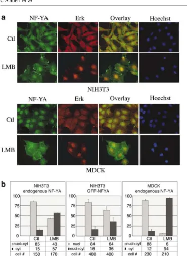
Smads, like numerous other proteins, have been shown to constantly shuttle between the cytoplasm and the nucleus. To test whether this was the case for NF-YA, we treated both NIH3T3 and MDCK cells with leptomycin B (LMB), an inhibitor of CRM1-driven nuclear export. As expected, LMB treatment led to the sequestering of ERK in the nucleus (Adachi et al., 2000) (Figure 7a). In contrast, the endogenous NF-YA protein, which was found in both the nuclear and cytoplasmic compartments in the absence of LMB, appeared to accumulate in the cytoplasm in a number of NIH3T3 cells in the presence of LMB, an effect that was even more pronounced in MDCK cells (Figure 7a, top
and bottom panels). These data were quantified from two independent experiments for each cell line, using either FITC- or cy3-conjugated antibodies to detect NF-YA (Figure 7b, left and right panels for respectively NIH3T3 and MDCK cells). To avoid any possible bias due to cell permeabilization conditions, NIH3T3 cells were also transfected with GFP-NFYA expressing vectors, in which case cell nuclei were Hoechst labeled directly following LMB treatment and fixation. Trans-fection led to a nuclear localization of the recombinant NF-YA fusion protein in a high proportion of the cells (84%), while it was evenly spread between the nucleus and the cytoplasm in the remaining 16% of cells. Even under these conditions, LMB treatment led to a cytoplasmic accumulation of NF-YA as the correspond-ing population increased from 16 to 36% (Figure 7b,
Figure 6 The TGF-b-dependent association of NF-YA with chromatin requires ERK activation. (a, b) MDCK cells were incubated with TGF-b for the indicated times after a 30 min pretreatment with PD98059 (PD) or with carrier only (Ctl). Nuclear extracts (a) and chromatin-enriched fractions (b) were prepared from these cells and analysed by immunoblotting with antibodies specific for Smad2/3, NF-YA and TBP. (c) The chromatin fraction of TGFb-treated cells (30 min time point) was treated with Mnase for the times indicated. The digested suspension was then centrifuged and the supernatant and pellet analysed by immunoblotting with anti NF-YA antibodies.
Figure 7 Leptomycin B (LMB) treatment differentially affects ERK and NF-YA intracellular localization. NIH3T3 and MDCK cells were treated with 20 nM leptomycin B for 16 h. (a) Endogenous NF-YA and ERK were visualized by immunofluor-escence. (b) Statistical analysis was carried out on two independent experiments for each cell line with either FITC- or cy3-conjugated antibodies for the detection of NF-YA. The left and right panels show the data for the endogenous NF-YA for respectively the NIH3T3 and the MDCK cells. The intracellular localization of an overexpressed GFP-tagged NF-YA protein was also investigated in the NIH3T3 cells. The data reflect the results of three independent transfection experiments (middle panel). The total number of cells counted for each condition is indicated (#). The histograms and the tables below show the percentage of cells harboring the different NF-YA intracellular localizations, in the control and LMB culture conditions.
TGF-b regulates NF-YA nuclear localization via MAPKs
C Alabert et al
3393
middle panel). These data were obtained from three independent transfections, with a total cell count of 400 for each condition. Altogether, these data indicate that NF-YA is not retained in the nucleus by leptomycin treatment and accordingly that its nuclear export is independent of CRM1. In fact, LMB has the unexpected effect of increasing cytoplasmic accumulation of NF-YA. This observation confirms that the intracellular distribution of NF-YA is regulated and suggests furthermore that it depends on factors that are themselves controlled by CRM1.
Discussion
We show in this study that the NF-YA subunit of the transcription factor NF-Y, which is found in the nuclear and cytoplasmic subcellular compartments, accumulates in the nucleus upon TGF-b1 induction. Accordingly, it binds to chromatin regions containing CCAAT regula-tory sequences in the promoters of several cell cycle regulatory genes. This TGF-b-dependent nuclear local-ization of NF-YA is observed in epithelial cells and fibroblasts, albeit with different kinetics, and is depen-dent upon activation of the ERK1/2 cascade. Notably, increased basal activation of the p38 cascade and low basal phospho-ERK1/2 levels seem to correlate with slower NF-YA nuclear accumulation in fibroblasts. Thus, the balance of activity of these two MAPK cascades might help control the dynamics of NF-YA nuclear localization, and thus its activity, induced by TGF-b1.
The nuclear accumulation of NF-YA in response to TGF-b is reminiscent of the nuclear shuttling of R-Smads. Translocation of phosphorylated Smad3 protein involves an interaction between its nuclear localizing signal (NLS) and importin-b1 (Xiao et al., 2000; Kurisaki et al., 2001). Although Smad2 contains a NLS region similar to that of Smad3 (Xiao et al., 2000), it appears to use a distinct translocation mechanism that is independent of the NLS and of importin-b, and instead involves direct interactions between Smad2 and the nucleoporins Nup214 and Nup153 (Xu et al., 2002). All three Smad proteins have been reported to constantly shuttle between the cytoplasm and the nucleus (Inman et al., 2002; Nicolas et al., 2004). The export of Smad4 was shown to be dependent of CRM1 while the subcellular distribution of Smad2 and Smad3 is unaffected by leptomycin B (LMB) treatment (Pierreux et al., 2000). After this manuscript was submitted, NF-YA was reported to contain a nonclassi-cal nuclear lononclassi-calization signal (ncNLS) at its C-terminus and to be translocated to the nucleus by an importin b-mediated pathway (Kahle et al., 2005). It remains to be seen whether NF-YA also contains a nuclear export signal and whether it shuttles in and out of the nucleus like Smads. Thus, it is difficult at present to determine whether its nuclear accumulation actually arises from a change in shuttling dynamics. Curiously, leptomycin B treatment drove endogenous NF-YA to the cytoplasm,
while it led to nuclear retention of ERK. A similar effect of LMB, leading to increased cytoplasmic localization, was observed for an ectopically expressed GFP-NFYA fusion protein. This provides further evidence that the subcellular localization of NF-YA is regulated and that the factors controlling this are themselves affected by inhibiting CRM-1-mediated nuclear export. In light of our data, it seems fair to speculate that ERK and p38 are among these regulatory factors. NF-YA was recently shown to be phosphorylated in vivo (Yun et al., 2003). This involved two serine-proline motifs that could be phosphorylated by cdk2 kinase. Notably, these two serine residues lie just C-terminal to the above-men-tioned ncNLS (Kahle et al., 2005). Moreover, mutation of the two serine residues to alanine allowed formation of the NF-Y trimer but inhibited DNA binding. However, the intracellular localization of the NF-YA mutant was not addressed. Based on our data, we can speculate that these two serine residues might also be substrates for ERK and therefore their mut-ation would decrease the nuclear concentrmut-ation of the NF-YA mutant. In addition, the observation that p38 can phosphorylate serine residues that are not directly followed by prolines (Bulavin et al., 2001; Cheung et al., 2003) opens the possibility for other p38 target sites on NF-YA. This remains to be investigated.
We show that, in contrast to the B subunit of NF-Y that is constitutively nuclear, NF-YA is found in both the nuclear and cytoplasmic compartments and accu-mulates in the nucleus in response to TGF-b1 treatment. Thus, our data (Figure 1 and Supplementary Figure 1) demonstrate that TGF-b drives colocalization of two components of NF-Y. NF-YC was also recently described to undergo nuclear relocalization during the cell cycle (Frontini et al., 2004). Unlike NF-YA, NF-YC accumulated in the nucleus following leptomycin B treatment, suggesting that the subcellular distributions of these two NF-Y subunits are controlled by distinct mechanisms. Surprisingly, these investigators found NF-YA to be solely nuclear in their experimental system. This likely reflects different experimental condi-tions, particularly those used for cell permeabilization, as in our hands too their conditions led to a loss of NF-YA cytoplasmic staining.
We observed no variation in the total level of NF-YA in TGF-b-treated cells. In agreement with this, serum stimulation of NIH3T3 cells blocked in G0 was recently shown to result in phosphorylation of NF-YA, while the total level of NF-YA remained constant (Yun et al., 2003). However, the actual level of NF-YA protein can change in certain situations, for instance increasing during monocyte to macrophage differentiation (Marziali et al., 1999) or decreasing in terminally differentiating myoblast C2C12 cells (Farina et al., 1999). We used cyclin A2 as a model gene to test the effect of the activation of NF-YA by TGF-b on transcription. Indeed, NF-Y associates with the pro-moters of several genes whose products help control the cell cycle. In addition, knockout experiments clearly showed the role of NF-Y in cell proliferation (Bhatta-charya et al., 2003). However, recent studies suggest that
NF-Y could regulate a wider spectrum of genes (Testa et al., 2005), opening the question of which subset of these genes might be targeted by TGF-b. One example might be procollagen A1 (COL1A1), as it was recently shown to be activated by TGF-b in cardiac fibroblasts through increased NF-Y binding (Lindahl et al., 2002). As expression of the TGF-b type II receptor is under the transcriptional control of a CCAAT box (Park et al., 2002; Huang et al., 2005), it is tempting to speculate that regulation of NF-Y activity by TGF-b could provide a feedback loop for TGF-b signaling.
We show that TGF-b1-dependent nuclear accumula-tion of NF-YA occurs in fibroblasts and epithelial cells. In both cell types, this requires activation of the ERK1/2 cascade. Although ERK1/2 are activated with similar kinetics by TGF-b1 in fibroblasts and in epithelial cells, the kinetics of NF-YA nuclear localization clearly differs between these two cell types. This difference correlates with the basal level of activation of the p38 cascade. Interestingly, exponentially growing epithelial MDCK or HEK cells (data not shown) contain barely detectable levels of basal p38 phosphorylation and show a much more rapid nuclear accumulation of NF-YA in response to TGF-b1. Taken together, these observations suggest that the activity levels of ERK and p38 help control the dynamics of NF-YA activation in response to TGF-b.
We do not know whether the effect of MAPK cascade activation on the dynamics of NF-YA nuclear reloca-tion might be due to a phosphorylareloca-tion of the NF-YA protein or of NF-YA regulating factors. Negative crosstalk between the ERK and p38 cascades has previously been described in keratinocytes (Efimova et al., 2003) and in the murine myeloid M1 cell lines, where p38 inhibition was shown to upregulate ERK activity (Hall and Davis, 2002). Regardless of the precise mechanism of regulation involved, our findings on NF-YA activation by TGF-b1 suggest that a single activation mechanism can sustain distinct dynamics of activation, depending on the cellular context. Thus, they provide novel insights on how TGF-b cytokines can generate distinct signaling patterns in different cell types.
Materials and methods
Cell lines, cell culture, transfections and reagents
NIH3T3, MDCK and CCL39 cells were grown in DMEM supplemented with 10% fetal bovine serum (FBS). TGF-b1 was from RD Systems. For luciferase experiments, 105CCL39
cells per 3.5 cm diameter Petri dish were transfected for 6 h with calcium phosphate in FBS 0.5% and further grown for 16 h with TGF-b1 (80 pM) in FBS 5%, prior to luciferase activity measurement (Promega). Luciferase experiments were performed in triplicate and data shown represent the average of three such experiments. Other transfections were achieved with Fugene (Roche) and TGF-b treatment was performed 24 h later. When mentioned, cycloheximide (10mg/ml), PD98059, SB203580 (10mM, Alexis Biochemicals), SP600125 (10mM, Calbiochem) and okadaic acid (1mM, Alexis Biochem-icals) were added to the cell culture 30 min prior to TGF-b treatment and left thereafter.
Plasmids and oligonucleotides
Details of all constructs and oligonucleotides used are available upon request. mCCRE-CycA2-Luc is a pGL2-basic vector containing murine cyclin A2 promoter sequences (!177 to þ 100 relative to the most 30 transcription initiation site,
mutated at the CCRE site). The mutation at the CCRE site prevents indirect regulating effects of TGF-b. The CCAAT box sequence was mut to ggccT by splice overlap extension PCR and constructs were sequenced.
Chromatin immunoprecipitation and fractionation
Chromatin immunoprecipitations (ChIP) were performed on NIH3T3 cells essentially as described (Fajas et al., 2001). Cells were incubated for various times with TGF-b1 (80 pM). Approximately 5# 106cells were used per ChIP. An aliquot
of each chromatin sample (‘input’) was taken prior to immunoprecipitation and purified in parallel to the immuno-precipitated DNA. Immunoprecipitations was carried out overnight with antibodies against NF-YA (sc-10779, Santa Cruz) or Tyk2 (#06-375, UBI). Immunoprecipitated and ‘input’ DNAs were assayed by both semiquantitative and real-time PCRs. For the semiquantitative PCR reactions,a32
P-dCTP was incorporated during the amplification and aliquots of the PCR reactions were analysed in parallel with Maxam and Gilbert DNA sequence and analysed by PAGE and autoradiography. Quantitative PCR analysis was carried out with a light cycler apparatus (Roche) and for each locus analysed, both the immunoprecipitated and the input DNAs were quantified. PCR DNA fragments were circa 200–500 bp in length. Small scale chromatin fractionation was carried out as previously described (Wysocka et al., 2001).
Immunofluorescence
NIH3T3 cells were grown on coverslips in six-well dishes (40 000 cells/well) in FCS 10%. After 48 h, they were treated with TGF-b1 (80 pM) for various times. Cells were fixed with PFA (4%) for 30 min and ethanol permeabilized (EtOH 70%, overnight at 41C). Cells were incubated with the antibodies for 1 h at room temperature. Immunofluorescence was monitored by incubation with FITC and Cy-3 secondary antibodies. Coverslips were mounted with Permafluor (Immunon). The following primary antibodies were used: rabbit anti-NF-YB, monoclonal anti-NF-YA (Mab-7) (gifts from R Mantovani), monoclonal anti NF-YA (#556359, BD Pharmingen), Smad3 (sc-8332, Santa-Cruz), rabbit anti-Flag (#7425, Sigma), p44/42 and p38 MAPKs (#9102 and #9212, Cell Signaling). All immunofluorescence data are representative of at least three independent experiments. Protein extracts and immunoblots
Protein extracts were prepared from actively growing subcon-fluent NIH3T3 fibroblasts treated with TGF-b1 (80 pM) for the indicated times. Cells were scraped in PBS containing NaF (50 mM) and vanadate (2 mM, Sigma). Nuclear extracts were prepared as described previously (Vignais et al., 1996). Buffers contained protease inhibitors (Roche) as well as microcystin (0.2mM, Sigma-M2912) and vanadate (0.5 mM). The lysis buffer for whole cell extracts contained: 50 mMTris (pH 8.0),
100 mMNaCl, 1 mMEDTA, 1 mMEGTA, 50 mMNaF, 25 mM
NaPP, 1% NP-40, 1 mM DTT, 1 mM PMSF and 1 mM
vanadate. Proteins were separated by SDS-PAGE (15%) and transferred to nitrocellulose membranes. After incubating with antibodies, blots were finally subjected to enhanced chemi-luminescence substrate (Lumi-Light, Roche). The following primary antibodies were used: NF-YA monoclonal antibody (#556359, BD Pharmingen and gift from R Mantovani), Smad3 (sc-8332, Santa-Cruz), phospho-p44/42 MAPK (E10 and #9101S, Cell Signaling), p44/42 MAPK (#9102, Cell
TGF-b regulates NF-YA nuclear localization via MAPKs
C Alabert et al
3395
Signaling), phospho-p38 and p38 MAPKs (#9211 and #9212, Cell Signaling), phospho-SAPK (#9251, Cell Signaling), SAPK (#9252, NewEngland Biolabs), monoclonal anti-TBP (clone 3G3). All blots shown are representative of at least 2 different experiments performed from different protein extracts. Statistical analysis
Statistical analysis of the results was performed with the statistical package SAS system and with the ANOVA test. A P-valueo0.05 was considered to be statistically significant.
Acknowledgements
We thank R Mantovani (University of Modene, Italy) for discussions and for providing antibodies and plasmids, K Matsumoto for providing plasmids. We also thank Annick Vie´ for her excellent technical support. This work was funded by grants from l’Association pour la Recherche sur le Cancer (contracts 3304 to RAH, 4209 and 3446 to MLV) and La Ligue Contre le Cancer. SC was supported by the Ch Oberling Foundation.
References
Adachi M, Fukuda M, Nishida E. (2000). J Cell Biol 148: 849–856.
Alessi DR, Cuenda A, Cohen P, Dudley DT, Saltiel AR. (1995). J Biol Chem 270: 27489–27494.
Attisano L, Wrana JL. (2000). Curr Opin Cell Biol 12: 235–243.
Barlat I, Fesquet D, Brechot C, Henglein B, Dupuy d’Angeac A, Vie A et al. (1993). Cell Growth Differ 4: 105–113. Barlat I, Henglein B, Plet A, Lamb N, Fernandez A,
McKenzie F et al. (1995). Oncogene 11: 1309–1318. Bhattacharya A, Deng JM, Zhang Z, Behringer R, de
Crombrugghe B, Maity SN. (2003). Cancer Res 63: 8167–8172.
Bolognese F, Wasner M, Dohna CL, Gurtner A, Ronchi A, Muller H et al. (1999). Oncogene 18: 1845–1853.
Bulavin DV, Higashimoto Y, Popoff IJ, Gaarde WA, Basrur V, Potapova O et al. (2001). Nature 411: 102–107.
Cheung PC, Campbell DG, Nebreda AR, Cohen P. (2003). EMBO J 22: 5793–5805.
Coustry F, Maity SN, Sinha S, de Crombrugghe B. (1996). J Biol Chem 271: 14485–14491.
de Caestecker M. (2004). Cytokine Growth Factor Rev 15: 1–11. Derynck R, Zhang YE. (2003). Nature 425: 577–584. Efimova T, Broome AM, Eckert RL. (2003). J Biol Chem 278:
34277–34285.
Fajas L, Paul C, Vie A, Estrach S, Medema R, Blanchard JM et al. (2001). Mol Cell Biol 21: 2956–2966.
Farina A, Manni I, Fontemaggi G, Tiainen M, Cenciarelli C, Bellorini M et al. (1999). Oncogene 18: 2818–2827.
Frontini M, Imbriano C, Manni I, Mantovani R. (2004). Cell Cycle 3: 217–222.
Hall JP, Davis RJ. (2002). J Cell Biochem 86: 1–11. Hu Q, Maity SN. (2000). J Biol Chem 275: 4435–4444. Huang W, Zhao S, Ammanamanchi S, Brattain M,
Venkatasubbarao K, Freeman JW. (2005). J Biol Chem 280: 10047–10054.
Huet X, Rech J, Plet A, Vie A, Blanchard JM. (1996). Mol Cell Biol 16: 3789–3798.
Inman GJ, Nicolas FJ, Hill CS. (2002). Mol Cell 10: 283–294. Janssens V, Goris J. (2001). Biochem J 353: 417–439. Kahle J, Baake M, Doenecke D, Albig W. (2005). Mol Cell
Biol 25: 5339–5354.
Kurisaki A, Kose S, Yoneda Y, Heldin CH, Moustakas A. (2001). Mol Biol Cell 12: 1079–1091.
Lindahl GE, Chambers RC, Papakrivopoulou J, Dawson SJ, Jacobsen MC, Bishop JE et al. (2002). J Biol Chem 277: 6153–6161.
Maity SN, Sinha S, Ruteshouser EC, de Crombrugghe B. (1992). J Biol Chem 267: 16574–16580.
Manni I, Mazzaro G, Gurtner A, Mantovani R, Haugwitz U, Krause K et al. (2001). J Biol Chem 276: 5570–5576. Mantovani R. (1999). Gene 239: 15–27.
Marziali G, Perrotti E, Ilari R, Coccia EM, Mantovani R, Testa U et al. (1999). Blood 93: 519–526.
Moriguchi T, Kuroyanagi N, Yamaguchi K, Gotoh Y, Irie K, Kano T et al. (1996). J Biol Chem 271: 13675–13679. Nicolas FJ, De Bosscher K, Schmierer B, Hill CS. (2004).
J Cell Sci 117: 4113–4125.
Park SH, Lee SR, Kim BC, Cho EA, Patel SP, Kang HB et al. (2002). J Biol Chem 277: 5168–5174.
Philips A, Chambeyron S, Lamb N, Vie A, Blanchard JM. (1999). Oncogene 18: 6222–6232.
Pierreux CE, Nicolas FJ, Hill CS. (2000). Mol Cell Biol 20: 9041–9054.
Plet A, Huet X, Algarte M, Rech J, Imbert J, Philips A et al. (1997). Oncogene 14: 2575–2583.
Shi Y, Massague J. (2003). Cell 113: 685–700.
Siegel PM, Massague J. (2003). Nat Rev Cancer 3: 807–821. Sinha S, Maity SN, Lu J, de Crombrugghe B. (1995). Proc Natl
Acad Sci USA 92: 1624–1628.
Tan Y, Rouse J, Zhang A, Cariati S, Cohen P, Comb MJ. (1996). EMBO J 15: 4629–4642.
ten Dijke P, Hill CS. (2004). Trends Biochem Sci 29: 265–273. Testa A, Donati G, Yan P, Romani F, Huang TH, Vigano
MA et al. (2005). J Biol Chem 280: 13606–13615.
Vignais ML, Sadowski HB, Watling D, Rogers NC, Gilman M. (1996). Mol Cell Biol 16: 1759–1769.
Waite KA, Eng C. (2003). Nat Rev Genet 4: 763–773. Wang W, Zhou G, Hu MC, Yao Z, Tan TH. (1997). J Biol
Chem 272: 22771–22775.
Wysocka J, Reilly PT, Herr W. (2001). Mol Cell Biol 21: 3820–3829.
Xiao Z, Liu X, Henis YI, Lodish HF. (2000). Proc Natl Acad Sci USA 97: 7853–7858.
Xu L, Kang Y, Col S, Massague J. (2002). Mol Cell 10: 271–282.
Yamaguchi K, Shirakabe K, Shibuya H, Irie K, Oishi I, Ueno N et al. (1995). Science 270: 2008–2011.
Yingling JM, Blanchard KL, Sawyer JS. (2004). Nat Rev Drug Discovery 3: 1011–1022.
Young PR, McLaughlin MM, Kumar S, Kassis S, Doyle ML, McNulty D et al. (1997). J Biol Chem 272: 12116–12121. Yun J, Chae HD, Choi TS, Kim EH, Bang YJ, Chung J et al.
(2003). J Biol Chem 278: 36966–36972.
Supplementary Information accompanies the paper on the Oncogene website (http://www.nature.com/onc).