HAL Id: hal-00999541
https://hal.archives-ouvertes.fr/hal-00999541
Submitted on 3 Jun 2014
HAL is a multi-disciplinary open access
archive for the deposit and dissemination of
sci-entific research documents, whether they are
pub-lished or not. The documents may come from
teaching and research institutions in France or
abroad, or from public or private research centers.
L’archive ouverte pluridisciplinaire HAL, est
destinée au dépôt et à la diffusion de documents
scientifiques de niveau recherche, publiés ou non,
émanant des établissements d’enseignement et de
recherche français ou étrangers, des laboratoires
publics ou privés.
Exciton dynamics of a single quantum dot embedded in
a nanowire
Gregory Sallen, Adrien Tribu, Thomas Aichele, Régis André, Lucien
Besombes, Catherine Bougerol, Serge Tatarenko, Kuntheak Kheng,
Jean-Philippe Poizat
To cite this version:
Gregory Sallen, Adrien Tribu, Thomas Aichele, Régis André, Lucien Besombes, et al.. Exciton
dy-namics of a single quantum dot embedded in a nanowire. Physical Review B: Condensed Matter
and Materials Physics (1998-2015), American Physical Society, 2009, 80, pp.085310.
�10.1103/Phys-RevB.80.085310�. �hal-00999541�
Exciton dynamics of a single quantum dot embedded in a nanowire
G. Sallen,1A. Tribu,2T. Aichele,1,
*
R. André,1L. Besombes,1C. Bougerol,1 S. Tatarenko,1K. Kheng,2and J. Ph. Poizat1,†1Institut Néel, CNRS–Université Joseph Fourier, 38042 Grenoble, France 2CEA/INAC/SP2M, 38054 Grenoble, France
共Received 17 March 2009; revised manuscript received 27 July 2009; published 18 August 2009兲
We have carried out a detailed optical characterization of a single CdSe quantum dot embedded in a ZnSe nanowire. Exciton, biexciton, and charged exciton lines have been identified unambiguously using photon correlation spectroscopy. This technique has provided a detailed picture of the dynamics of this system. It has been found that the dark exciton has a strong influence on the optical properties. The most visible influence is the strongly reduced excitonic emission compared to the biexcitonic one. Temperature-dependent lifetime measurements have allowed us to measure a large splitting of ⌬E = 共6.0⫾ 0.2兲 meV between the dark and the bright exciton as well as the spin-flip rates between these two states. This type of semiconducting quantum dot turns out to be a very efficient single photon source in the visible. Its particular growth technique opens additional possibilities as compared to the usual self-assembled quantum dots.
DOI:10.1103/PhysRevB.80.085310 PACS number共s兲: 78.67.Lt, 78.55.Et
I. INTRODUCTION
Semiconductor nanowires 共NWs兲 appear as promising building blocks for nanoscale devices and circuits with im-pressive potential applications including nanoelectronics,1–3
optoelectronics 共light-emitting diodes4,5 and nanolasers6兲,
thermoelectrical energy conversion,7 and biological or
chemical sensors.8 Moreover, high quality defect free NWs can be grown on low-cost routinely used substrates such as silicon, which means that they could easily be used for fab-ricating commercial devices and could possibly be integrated with mainstream Si microelectronics devices.
NW growth methods allow for the variation in the chemi-cal composition9,10or doping11along the longitudinal or
ra-dial directions. This enables the fabrication of well con-trolled one-dimensional nanoscale heterostructures.10 For
example, as shown in this work, it is possible to insert a slice of a low band-gap semiconductor within a high band-gap NW and thus realize a light-emitting quantum dot 共QD兲 op-erating as a single photon source.12,13 So far, work on the
light-emitting properties of single QDs has mainly concerned self-assembled QDs formed by surface forces induced by lattice mismatch between different materials. Such QDs have been widely used in the past decade as single photon sources14 and for their potential application in quantum
in-formation processing 共see, for example, Refs. 15 and 16兲. QDs in NWs appear to be an interesting alternative to self-assembled QDs. The absence of a wetting layer offers a bet-ter confinement which could enable room-temperature pro-duction of single photons.13Radial growth techniques enable
engineering of optical guides allowing more efficient light extraction than in bulk materials.17,18 Furthermore,
NW-based heterostructures, being much less limited by lattice mismatches, greatly widen the possible materials combina-tions and enable well controlled stacking of several QDs in a single NW, offering interesting possibilities for quantum in-formation processing.19
In this paper we present a detailed optical characterization of excitonic emission in a single CdSe QD embedded in a ZnSe NW. We have already shown that this system is an
efficient single photon source operating at temperatures as high as 220 K.13Single photon emission from NWs has oth-erwise only been demonstrated at 4 K in InAs QDs embed-ded in InP NWs.12 Our system emits light around 550 nm
共2.2 eV兲 where silicon avalanche photodiodes 共APDs兲 are very efficient. This has allowed us to perform a thorough spectroscopic analysis of a QD embedded in a NW by using photon correlation spectroscopy.20We have identified
unam-biguously the exciton, biexciton, and charged exciton lines and obtained information on the charging dynamics of this QD 共Sec. III兲. We have fitted the various correlation func-tions with a model including the dark exciton and its param-eters defined by lifetime measurements. The most apparent manifestation of the dark exciton is the large predominance of the biexciton line with respect to the exciton line above saturation. The dark exciton is studied in Sec.IV.
II. SAMPLE AND EXPERIMENTAL SETUP A. Sample fabrication and preparation
The wires are grown by molecular beam epitaxy in the vapor-liquid-solid growth mode catalyzed by gold particles on a Si substrate. In order to make QDs, a small region of CdSe is inserted in the ZnSe NW. This is done by interrupt-ing the ZnSe growth, changinterrupt-ing to CdSe growth for a short time, and then growing ZnSe again.13 Details about the
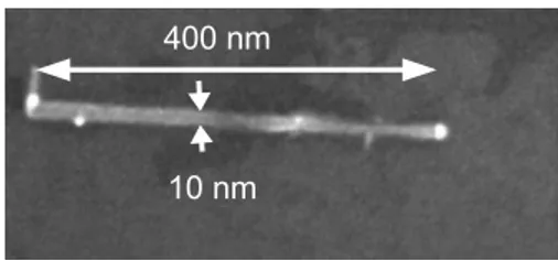
growth of the ZnSe NWs can be found in Ref. 21. The wire diameter 共around 10 nm兲 is on the order of the bulk exciton Bohr diameter for CdSe 共11 nm兲. This means that the carriers in the CdSe QD are in the strong quantum confinement re-gime. For the study of single NWs, the sample is sonicated in methanol causing NWs to break off the substrate into the solution. Droplets of this solution are then deposited on a Si substrate and a low density of individual NWs is obtained after evaporation. As shown in Fig.1, individual NWs can be isolated allowing single QD optical spectroscopy.
B. Experimental setup
The experimental apparatus is a standard microphotolumi-nescence 共PL兲 setup. The samples are mounted on a XYZ
piezomotor system in a He flow cryostat at a temperature of 4 K. The optical excitation is provided by a 405 nm continuous-wave diode laser illuminating the sample via a microscope objective of numerical aperture NA= 0.65 lo-cated in the cryostat. Time-resolved measurements are per-formed by exciting the sample at a wavelength of = 440 nm with a frequency doubled Ti:sapphire laser oper-ating at = 880 nm with pulse duration of 1 ps and a repeti-tion rate of 80 MHz. The NW emission is collected by the same objective and sent to a 50/50 beam splitter for correla-tion measurements. In each arm of the beam splitter, the light is dispersed by a monochromator 共1200 grooves/mm grating, 30 cm and 50 cm focal length, respectively兲. Each mono-chromator has a switchable mirror inside, which can direct the luminescence either onto a charge coupled device camera for the measurement of the PL spectrum or through the exit slit toward a low jitter 共40 ps兲, high quantum efficiency APD. The detectors send electrical pulses into a time-correlated single photon module that builds a histogram of the time delays between successive photons. This histogram is pro-portional to the second-order correlation function g共2兲共t兲.14
The overall temporal resolution of our setup is essentially limited by the jitter of the APDs and the dispersion of the monochromator gratings. This time resolution was measured by recording the autocorrelation function of 1 ps pulses from a frequency-doubled Ti:sapphire laser. A full width at half maximum of 90 ps was obtained for the autocorrelation func-tion peak. For lifetime measurement, the timing resolufunc-tion was measured by sending the 1 ps laser pulses in a mono-chromator and a full width at half maximum of 70 ps was obtained.
C. General optical properties
A typical low-temperaturePL spectrum is shown in the inset of Fig.2 where three lines can be seen. A comparison with relative energy positions of known emission lines in spectra of self-assembled CdSe/ZnSe QDs 共Refs.22and23兲 suggests that these lines correspond to the exciton 共X兲, the biexciton 共XX兲, and the charged exciton 共CX兲. The X-CX 共resp. X-XX兲 energy splitting is found around 10 meV 共resp. 20 meV兲 as compared to 15–22 meV 共resp. 19–26 meV兲 for self-assembled QDs. The mean excitonic energy, 共2.25⫾ 0.08兲 eV, is also similar as compared to 共2.45⫾ 0.2兲 eV for self-assembled CdSe/ZnSe QDs.24
Un-ambiguous proof for the assignment of these lines will be given below using photon correlation spectroscopy. The line-width broadening can be attributed to spectral diffusion.25
This effect might arise from the close proximity of the QD to
the NW surface, where fluctuating charges can be trapped on surface states even at low temperature.12,26
One of the characteristic features of such NW QD struc-tures is their polarization properties. As shown in Ref.13the excitation efficiency and the luminescence are both strongly polarization dependent.27–29 A 90% contrast is obtained for
the excitation efficiency depending on the direction of the linear polarization of the pumping laser. The light emission is also 90% linearly polarized in the same direction as the best pumping polarization, independently of the excitation polar-ization.
It can be seen in Fig.2that the saturation level of the XX line is more than three times larger than that of the X line. As it will be shown in Sec. IV, this is due to a strong storage effect of the dark exciton state.
III. PHOTON CORRELATION SPECTROSCOPY
The results presented in this section all come from the same QD whose spectrum is shown in Fig. 2. The level scheme used to model our system is shown in Fig. 3. It is based on models used in Refs. 30–32where the carriers can enter the QD either individually 共power-dependent rates␥C1
and␥C2兲 or already bound as excitons 共power-dependent rate FIG. 1. Scanning electron microscope image of a single CdSe/
ZnSe nanowire deposited on a silicon substrate.
1 0 2 1 0 3 1 0 4 1 0 5 µP L in te ns ity (c ou nt s/ s) 1 1 0 P o w e r ( µ W ) : X : C X : X X 3 0 2 0 1 0 0 x1 0 3 2 . 2 4 2 . 2 2 2 . 2 0 E n e r g y ( e V ) X X X C X
FIG. 2. 共Color online兲 Inset: PL spectrum of a nanowire quan-tum dot at an excitation power of P = 15 W. The electronic back-ground noise has been subtracted. Main plot: line intensities as a function of excitation power. The spectra were obtained at 4 K.
FIG. 3. 共Color online兲 Level scheme including the empty dot
共E0兲, the dark exciton 共EX
D兲, the bright exciton 共EXB兲, the biexciton
共EXX兲, the charged dot 共EC兲, and the charged exciton 共ECX兲. The
various rates between the different levels are indicated in the figure.
SALLEN et al. PHYSICAL REVIEW B 80, 085310 共2009兲
r兲. We have added the dark exciton, which plays a key role in our system. It includes the bright and dark exciton, the charged exciton, and the biexciton. A triexciton level and a charged biexciton level are also included to avoid artificial saturation of the biexciton and of the charged exciton but they are not represented in Fig. 3.
All of the power-independent parameters of the model can be evaluated independently prior to photon correlation ex-periments by performing lifetime measurements 共see follow-ing Sec.IV兲. Their values are listed in TableI. These values are compatible to observations in self-assembled CdSe/ZnSe QDs.23
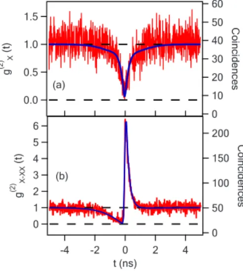
We come now to the results of this work on photon cor-relation experiments. We present first the data concerning the neutral QD in Fig.4. The autocorrelation of the X line emis-sion is shown in Fig. 4共a兲 exhibiting a clear antibunching which is characteristic of the statistics of a single photon emitter.14
Figure4共b兲 shows the cross-correlation measurement be-tween the X and the XX line. It displays the typical asym-metric shape with bunching and antibunching features that is the signature for the cascaded emission of a XX photon fol-lowed by a X photon.33This allows us to identify unambigu-ously these two lines as exciton and biexciton of the same QD. Note that the narrow bunching peak can only be fitted if the dark exciton is included in the model.
For all the correlation graphs 共Figs. 4 and 5兲 the right
vertical axes are the raw number of coincidences. The left axes represent the normalized correlation function according to a Poisson statistics where the coincidences involving background photons have been subtracted. The corrected correlation function g共2兲is related to the uncorrected one gu
共2兲
by g共2兲− 1 = 共gu
共2兲
− 1兲 /2
, where = S / 共S + B兲 with S and B, respectively, the number of signal and background photons as measured in the spectrum of Fig.2.
The autocorrelation of the CX line is shown in Fig. 5共a兲. As for the X line, it exhibits a clear antibunching. Also, on a larger time scale, a bunching effect can be observed. This is due to the hopping from the charged state to the neutral state of the QD as confirmed by the X-CX cross correlation dis-played in Fig.5共b兲. This proves that the CX line comes from the same QD as the X line. The situation is depicted sche-matically in Fig. 5共c兲, which shows that the photons are emitted either from the charged or from the neutral state of the QD. The average time spent by the QD in the charged state is given by the characteristic time of an exponential fitting of the bunching peak in Fig. 5共a兲 which is 5 ns. It should be noticed that the antibunching dip in Fig.5共b兲is not symmetrical. Negative 共positive兲 time corresponds to the probability of detecting a CX 共X兲 photon after having de-tected a X 共CX兲 photon. Formation of a CX in an empty QD 共t ⬍ 0兲 requires the loading of three charges, whereas the formation of an X in a QD with a single charge 共level Ecin Fig.3兲 is faster since it requires only the loading of a single charge 共t ⬎ 0兲.30–32 Although we do not know whether this
charge is positive or negative, comparison with spectra of self-assembled CdSe/ZnSe QDs 共Refs.22and23兲 leads us to the assumption that the charge state is negative.
We also show in Fig.6 the autocorrelation of the biexci-tonic line and the cross correlation between the biexciton and the charged exciton. The biexciton autocorrelation exhibits a narrow antibunching and the XX-CX cross-correlation fea-tures a broad antibunching caused by charge hopping as in the X-CX cross correlation of Fig.5共b兲.
TABLE I. Table of power-independent transition rates in ns−1at
a temperature T = 4 K. ␥X ␥XX ␥CX ␥d ␥u ␥NR 1.4 2.5 1.7 1.4 0 0.2 1 . 5 1 . 0 0 . 5 0 . 0 g (2 ) (t)X 6 0 5 0 4 0 3 0 2 0 1 0 0 C oin cid en ce s ( a ) 6 5 4 3 2 1 0 g (2 ) X-X X (t) - 4 - 2 0 2 4 t ( n s ) 2 0 0 1 5 0 1 0 0 5 0 0 C oin cid en ce s ( b )
FIG. 4. 共Color online兲 共a兲 Exciton emission autocorrelation, 共b兲 exciton-biexciton cross correlation for an excitation power P = 15 W. The left axes are the correlation functions corrected from the background and the right axes are the raw coincidence rates 共see
text兲. The fit is performed using the model based on Fig. 3. The
power-dependent parameters used for the fit are r = 0.6 ns−1, ␥
C1
= ␥C2= 1 ns−1. The other parameters are given in TableI.
1 . 5 1 . 0 0 . 5 0 . 0 g (2 ) CX (t) 1 0 0 8 0 6 0 4 0 2 0 0 C oin cid en ce s 1 . 5 1 . 0 0 . 5 0 . 0 g (2 ) X-C X (t) - 8 - 4 0 4 8 t ( n s ) 6 0 4 0 2 0 0 C oin cid en ce s (c) (b) (a)
FIG. 5. 共Color online兲 共a兲 Charged exciton autocorrelation for an excitation power P = 8 W; 共b兲 exciton-charged exciton cross
cor-relation for an excitation power P = 10 W. The fits are performed
using the model based on Fig.3. The power-dependent parameters
used for the fits are, respectively, r = 0.31 ns−1, ␥
C1= 0.09 ns−1, and
␥C2= 0.058 ns−1 for the CX autocorrelation 共a兲 and r = 0.37 ns−1,
␥C1= 0.25 ns−1, and ␥C2= 0.28 ns−1for the X-CX cross correlation
共b兲. The other parameters are given in TableI. 共c兲 Representation of
the stream of photons coming alternatively from the neutral and the charged QD.
As can be seen in Figs.4–6, the experimental results are fitted very well by the model shown in Fig. 3 taking into account the temporal resolution of our experimental setup 共90 ps兲. Inclusion of the dark exciton turned out to be essen-tial for the modeling of the photon correlation data. Allowing for the coexistence of two excitation mechanisms, namely, charge by charge 共described by␥C1and␥C2兲 or directly
feed-ing the QD with an already bound exciton 共described by r兲 has also turned out to be necessary for the fitting. The QD charge hopping time depends on the value of these param-eters. The model gives also the correct intensities of the spectral lines within 10%.25
IV. DARK EXCITON CHARACTERIZATION
In a QD, lowest energy excitons are the combination of an electron 共spin ⫾1 / 2兲 and a heavy hole 共spin ⫾3 / 2兲. This results in two different energy levels of spin ⫾1 and spin ⫾2. The spin-⫾1 states are optically connected to the zero spin empty dot state and called the bright exciton. The low energy spin ⫾2 states are called the dark exciton because they are not optically active. Indeed a photon is a spin-1 particle that cannot carry away 2 quanta of angular momen-tum.
In this section we have performed temperature-dependent lifetime measurements on this CdSe/ZnSe NW. By fitting these data with a model involving an acoustic phonon bath34
we are able to extract the value of the dark and bright exciton energy splitting ⌬E and the spin-flip rates between these two states. The measurements performed in this section have been carried out on a QD with a similar spectrum but differ-ent than for the photon correlation results.
The ratio between charged and neutral QD luminescence is varying from dot to dot. We have also observed that in-creasing the temperature tends to neutralize the QD. The
large linewidths have been attributed to spectral diffusion.25
The most conspicuous feature is that thePL intensity of the XX line at saturation is always a lot larger than that of the X line as it is shown in the power dependence of the different lines 共Fig.2兲. This effect is the signature of a strong storage effect on the dark exciton 共DX兲 state. The DX state reduces the luminescence of the X line owing to the leakage from the bright to the dark exciton but the DX state remains an effi-cient intermediate state for populating the XX state.35 The
QD photoluminescence properties are well described by the set of rate equations including the bright and dark exciton, and the biexciton as represented in Fig. 3.
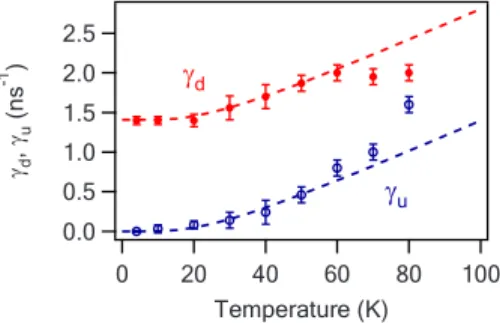
The population transfer between the bright and the dark exciton states is governed by the two temperature-dependent rates␥dand␥u共see Fig.3兲. We assume that these transitions are assisted by acoustic phonons whose energy matches the X-DX energy splitting ⌬E. At a temperature T the number N of acoustic phonons per quantum state of energy ⌬E is given by the Bose-Einstein statistics and reads
N= 1
exp共⌬E/kBT兲− 1
. 共1兲
The downward transition rate ␥dfrom the X to the DX state corresponds to the spontaneous and stimulated emission of a phonon, whereas the upward rate ␥u corresponds to the ab-sorption of a phonon.34They are given by
␥d= 共N + 1兲␥0,
␥u= N␥0, 共2兲
where␥0is the zero-temperature downward rate 共N = 0兲.
We have performed time-resolved photoluminescence of the X state at different temperatures. The results are pre-sented in Fig.7. The decay time of the X level depends not only on the radiative decay rate ␥X but also on the
temperature-dependent ␥d and ␥u rates between the bright and dark excitons. At low temperature 共T = 4 K兲, the lumi-nescence exhibits a fast monoexponential decay with a time scale of the order of 1 / 共␥X+␥d兲 corresponding to the radia-tive decay and the leakage toward the DX state. For interme-diate temperatures 共T = 20 K and T = 40 K兲, the fast decay is still present and there is the apparition of a slow time scale
8 0 6 0 4 0 2 0 0 C oin cid en ce s 2 . 0 1 . 5 1 . 0 0 . 5 0 . 0 g (2 ) (t)XX 8 0 6 0 4 0 2 0 0 C oin cid en ce s - 8 - 4 0 4 8 t ( n s ) 1 . 5 1 . 0 0 . 5 0 . 0 - 0 . 5 g (2 ) XX-C X (t) ( a ) ( b )
FIG. 6. 共Color online兲 共a兲 Autocorrelation of the biexcitonic line 共XX兲 for an excitation power of P = 15 W, 共b兲 biexciton-charged
exciton cross correlation, both for an excitation power P = 15 W.
The fits are performed using the model based on Fig.3. The
power-dependent parameters used for the fits are r = 0.6 ns−1, ␥
C1
= 1 ns−1, and ␥
C2= 2 ns−1. The other parameters are given in Table
I.
Intensity
(arb.
units)
FIG. 7. 共Color online兲 Decay of the X line emission of QD1 for different temperatures. The pumping power is well below saturation
共r Ⰶ ␥X兲 so that the XX state is almost not populated.
SALLEN et al. PHYSICAL REVIEW B 80, 085310 共2009兲
corresponding to the thermally activated reloading of the X state from the long lived DX state. For higher temperature 共T = 80 K兲, the reloading from the dark to the bright exciton becomes even more efficient and the decay appears as mo-noexponential with a time scale intermediate between the previous slow and fast time scales.
By fitting the lifetimes using the model of Fig.3 we can extract the values for ␥X, ␥NR, ␥d, and ␥u. Each of these parameters has a specific influence on the shape of the lumi-nescence decay and can be evaluated with a good precision. The radiative decay rate of the exciton is found to be ␥X
= 1.4 ns−1and is assumed to be temperature independent up to T = 60 K as it was observed for CdSe/ZnSe self-assembled QDs.36This assumption is justified a posteriori by the
qual-ity of the fits up to this temperature. The values for␥dand␥u
are plotted in Fig.8 as a function of temperature. This tem-perature dependence is well fitted using Eqs. 共1兲 and 共2兲. This enables us to obtain the DX-X energy splitting ⌬E = 共6.0⫾ 0.2兲 meV and the zero-temperature downward rate
␥0= 共1.41⫾ 0.02兲 ns−1.
The rather large value for the DX-X energy splitting ⌬E is an indication of the strong exciton confinement within the QD, owing to its relatively small size and the absence of a wetting layer.37The bulk DX-X energy splitting for CdSe is
0.12 meV. According to the calculation performed by Kling-shirn et al.,38confinement-induced enhancement of this split-ting reaches a factor of 50 共that is ⌬E = 6 meV for CdSe兲 for infinite barriers cylindrical dots of radius corresponding to the Bohr radius aB and of height corresponding to aB/4. These dimensions are compatible with the measured diam-eter 共10 nm兲 of the NW and with the height expected from the CdSe growth duration. The DX-X energy splitting has been measured at 1.9 meV for self-assembled CdSe/ZnSe QD.37The larger value that we have observed is an
indica-tion of the larger confinement in NWs owing to the absence of a wetting layer. This ⌬E = 6 meV splitting corresponds to the value for very small 共about 2 nm diameter兲 colloidal spherical CdSe nanocrystal as reported in Ref.39. The value for zero-temperature downward transition rate ␥0= 1.4 ns−1
is comparable to what has been obtained for colloidal CdSe
nanocrystals34 or some self-assembled InP/GaInP QDs.35 Slower rates 共␥0= 0.01 ns−1兲 have also been observed in
InGaAs/GaAs self-assembled QDs.40
A good fitting of the experimental data requires the inclu-sion of an effective nonradiative decay rate ␥NRof the DX
state. This rate slightly increases with temperature from
␥NR= 0.2 ns−1 at 4 K up to ␥NR= 0.5 ns−1 at 80 K. These
values are of similar order of magnitude that what was re-ported in InGaAs/GaAs self-assembled QDs.40Nonradiative
phenomena in nanocrystals are generally slower ranging from hundreds of nanoseconds to a few microseconds in col-loidal nanocrystals.34,41
V. CONCLUSION
In conclusion, we have used photon correlation spectros-copy to characterize a light-emitting QD embedded in a NW. We obtained a very good fit to the experimental data with a model based on a standard excitonic level scheme. This al-lowed us to extract quite complete dynamics of the neutral and charged excitons including charge hopping between these two states of the QD. We have performed temperature-dependent lifetime measurements of a CdSe QD embedded in a ZnSe NW. A careful quantitative analysis of these data has allowed us to confirm the strong influence of the dark exciton and to extract the dark-bright exciton splitting to-gether with the transition rates between these two levels. The rather large dark-bright exciton splitting that we have mea-sured is a signature of the strong confinement of the exciton within the QD in this NW geometry. This value is three times larger than for self-assembled CdSe QDs although the exci-tonic energy and the X-XX and X-CX energy splittings are of same order of magnitude.
CdSe/ZnSe QDs in NWs are nano-objects situated be-tween self-assembled QDs and CdSe-based colloidal nanocrystals.42,43The latter operate at room temperature but
have a blinking problem and a lifetime above 20 ns. On the other hand, self-assembled QDs are nonblinking and feature a subnanosecond lifetime allowing GHz repetition rates. QDs embedded in NWs have the potential to combine the
best of both worlds by offering nonblinking
room-temperature13single photon sources with a high
repeti-tion rate. Furthermore, the versatility of this particular nano-structure growth technique offers interesting perspectives for engineering semiconducting QDs such as coupled QDs or waveguide coupled QDs.
ACKNOWLEDGMENTS
We thank M. Richard for many fruitful discussions, F. Donatini for very efficient technical support, and R. Cox for a careful reading of the paper. T.A. acknowledges support by Deutscher Akademischer Austauschdienst 共DAAD兲. Part of this work was supported by European project QAP 共Contract No. 15848兲. 2 . 5 2 . 0 1 . 5 1 . 0 0 . 5 0 . 0 gd ,gu (n s -1 ) 1 0 0 8 0 6 0 4 0 2 0 0 T e m p e r a t u r e ( K ) gd gu
FIG. 8. 共Color online兲 Downward and upward spin-flip rates between X and DX as a function of temperature. The dotted lines
*Present address: Physics Institute, Humboldt University, Berlin, Germany; aichele@physik.hu-berlin.de
†jean-philippe.poizat@grenoble.cnrs.fr
1X. Duan, Y. Huang, Y. Cui, J. Wang, and C. M. Lieber, Nature
共London兲 409, 66 共2001兲.
2W. Lu and C. M. Lieber, Nature Mater. 6, 841 共2007兲.
3C. Thelander, T. Martensson, M. T. Björk, B. J. Ohlsson, M. W.
Larsson, L. R. Wallenberg, and L. Samuelson, Appl. Phys. Lett.
83, 2052 共2003兲.
4R. Könenkamp, Robert C. Word, and C. Schlegel, Appl. Phys.
Lett. 85, 6004 共2004兲.
5H. M. Kim, Y. H. Cho, H. Lee, S. I. Kim, S. R. Ryu, D. Y. Kim,
T. W. Kang, and K. S. Chung, Nano Lett. 4, 1059 共2004兲.
6X. Duan, Y. Huang, R. Agarwal, and C. M. Lieber, Nature
共Lon-don兲 421, 241 共2003兲.
7A. I. Hochbaum, R. Chen, R. D. Delgado, W. Liang, E. C.
Gar-nett, M. Najarian, A. Majumdar, and P. Yang, Nature 共London兲
451, 163 共2008兲.
8Y. Cui, Q. Wei, H. Park, and C. M. Lieber, Science 293, 1289
共2001兲.
9M. S. Gudiksen, L. Lauhon, J. Wang, D. C. Smith, and C. M.
Lieber, Nature 共London兲 415, 617 共2002兲.
10M. T. Björk, B. J. Ohlsson, T. Sass, A. I. Persson, C. Thelander,
M. H. Magnusson, K. Deppert, L. R. Wallenberg, and L. Sam-uelson, Nano Lett. 2, 87 共2002兲; Appl. Phys. Lett. 80, 1058 共2002兲.
11C. Yang, Z. Zhong, and C. M. Lieber, Science 310, 1304 共2005兲.
12M. T. Borgström, V. Zwiller, E. Müller, and A. Imamoglu, Nano
Lett. 5, 1439 共2005兲.
13A. Tribu, G. Sallen, T. Aichele, R. André, J.-Ph. Poizat, C.
Boug-erol, S. Tatarenko, and K. Kheng, Nano Lett. 8, 4326 共2008兲.
14P. Michler, A. Kiraz, C. Becher, W. V. Schoenfeld, P. M. Petroff,
L. Zhang, E. Hu, and A. Imamoglu, Science 290, 2282 共2000兲.
15I. Fushman, D. Englund, A. Faraon, N. Stoltz, P. Petroff, and J.
Vuckovic, Science 320, 769 共2008兲.
16R. Hanson and D. D. Awschalom, Nature 共London兲 453, 1043
共2008兲.
17P. J. Pauzauskie and P. Yang, Mater. Today 9, 36 共2006兲.
18N. Gregersen, T. R. Nielsen, J. Claudon, J. M. Gérard, and J.
Mørk, Opt. Lett. 33, 1693 共2008兲.
19C. Simon, Y. M. Niquet, X. Caillet, J. Eymery, J. P. Poizat, and
J. M. Gérard, Phys. Rev. B 75, 081302共R兲 共2007兲.
20A. Kiraz, S. Fälth, C. Becher, B. Gayral, W. V. Schoenfeld, P. M.
Petroff, Lidong Zhang, E. Hu, and A. Imamoğlu, Phys. Rev. B
65, 161303共R兲 共2002兲.
21T. Aichele, A. Tribu, C. Bougerol, K. Kheng, R. André, and S.
Tatarenko, Appl. Phys. Lett. 93, 143106 共2008兲.
22V. Türck, S. Rodt, R. Heitz, O. Stier, M. Strassburg, U. W. Pohl,
and D. Bimberg, Phys. Status Solidi B 224, 217 共2001兲.
23B. Patton, W. Langbein, and U. Woggon, Phys. Rev. B 68,
125316 共2003兲.
24G. Bacher, R. Weigand, J. Seufert, V. D. Kulakovskii, N. A.
Gippius, A. Forchel, K. Leonardi, and D. Hommel, Phys. Rev. Lett. 83, 4417 共1999兲; F. Gindele, K. Hild, W. Langbein, and U. Woggon, Phys. Rev. B 60, R2157 共1999兲; F. Gindele, U.
Wog-gon, W. Langbein, J. M. Hvam, K. Leonardi, D. Hommel, and H. Selke, ibid. 60, 8773 共1999兲; J. C. Kim, H. Rho, L. M. Smith, Howard E. Jackson, S. Lee, M. Dobrowolska, and J. K. Furdyna, Appl. Phys. Lett. 75, 214 共1999兲; T. Kümmell, R. Weigand, G. Bacher, A. Forchel, K. Leonardi, D. Hommel, and H. Selke,
ibid. 73, 3105 共1998兲; S. M. Ulrich, S. Strauf, P. Michler, G. Bacher and A. Forchel, ibid. 83, 1848 共2003兲; V. D. Kulak-ovskii, G. Bacher, R. Weigand, T. Kümmell, A. Forchel, E. Borovitskaya, K. Leonardi, and D. Hommel, Phys. Rev. Lett.
82, 1780 共1999兲.
25G. Sallen, Ph.D. thesis, Université Joseph Fourier, Grenoble,
2009 共http://tel.archives-ouvertes.fr/tel-00362497/fr/兲.
26M. Bayer and A. Forchel, Phys. Rev. B 65, 041308共R兲 共2002兲.
27J. Wang, M. S. Gudiksen, X. Duan, Y. Cui, and C. M. Lieber,
Science 293, 1455 共2001兲.
28Y. M. Niquet and D. C. Mojica, Phys. Rev. B 77, 115316 共2008兲.
29M. H. M. van Weert, N. Akopian, F. Kelkensberg, U. Perinetti,
M. P. van Kouwen, J. Gómez Rivas, M. T. Borgström, R. E. Algra, M. A. Verheijen, E. P. A. M. Bakkers, L. P. Kouwen-hoven, and V. Zwiller, arXiv:0808.2908, Small 共to be pub-lished兲.
30C. Santori, D. Fattal, J. Vučković, G. S. Solomon, E. Waks, and
Y. Yamamoto, Phys. Rev. B 69, 205324 共2004兲.
31M. H. Baier, A. Malko, E. Pelucchi, D. Y. Oberli, and E. Kapon,
Phys. Rev. B 73, 205321 共2006兲.
32J. Suffczyński, T. Kazimierczuk, M. Goryca, B. Piechal, A.
Tra-jnerowicz, K. Kowalik, P. Kossacki, A. Golnik, K. P. Korona, M. Nawrocki, J. A. Gaj, and G. Karczewski, Phys. Rev. B 74, 085319 共2006兲.
33E. Moreau, I. Robert, L. Manin, V. Thierry-Mieg, J. M. Gérard,
and I. Abram, Phys. Rev. Lett. 87, 183601 共2001兲.
34O. Labeau, P. Tamarat, and B. Lounis, Phys. Rev. Lett. 90,
257404 共2003兲.
35M. Reischle, G. J. Beirne, R. Roßbach, M. Jetter, and P. Michler,
Phys. Rev. Lett. 101, 146402 共2008兲.
36I. C. Robin, R. André, Le Si Dang, H. Mariette, S. Tatarenko, J.
M. Gérard, K. Kheng, F. Tinjod, M. Bartels, K. Lischka, and D. Schikora, Phys. Status Solidi B 241, 542 共2004兲; I. C. Robin, R. André, and J. M. Gérard, Phys. Rev. B 74, 155318 共2006兲.
37J. Puls, M. Rabe, H.-J. Wünsche, and F. Henneberger, Phys. Rev.
B 60, R16303 共1999兲.
38C. Klingshirn, M. Hetterich, J. M. Hvam, W. Langbein, U.
Wog-gon, and F. Gindele, Solid State Commun. 106, 653 共1998兲.
39Al. L. Efros, M. Rosen, M. Kuno, M. Nirmal, D. J. Norris, and
M. Bawendi, Phys. Rev. B 54, 4843 共1996兲.
40J. M. Smith, P. A. Dalgarno, R. J. Warburton, A. O. Govorov, K.
Karrai, B. D. Gerardot, and P. M. Petroff, Phys. Rev. Lett. 94, 197402 共2005兲.
41M. Nirmal, D. J. Norris, M. Kuno, M. G. Bawendi, A. L. Efros,
and M. Rosen, Phys. Rev. Lett. 75, 3728 共1995兲.
42P. Michler, A. Imamoğlu, M. D. Mason, P. J. Carson, G. F.
Strouse, and S. K. Buratto, Nature 共London兲 406, 968 共2000兲.
43B. Mahler, P. Spinicelli, S. Buil, X. Quelin, J. P. Hermier, and B.
Dubertet, Nature Mater. 7, 659 共2008兲.
SALLEN et al. PHYSICAL REVIEW B 80, 085310 共2009兲