HAL Id: hal-02345719
https://hal.archives-ouvertes.fr/hal-02345719
Submitted on 18 Mar 2021HAL is a multi-disciplinary open access
archive for the deposit and dissemination of sci-entific research documents, whether they are pub-lished or not. The documents may come from teaching and research institutions in France or abroad, or from public or private research centers.
L’archive ouverte pluridisciplinaire HAL, est destinée au dépôt et à la diffusion de documents scientifiques de niveau recherche, publiés ou non, émanant des établissements d’enseignement et de recherche français ou étrangers, des laboratoires publics ou privés.
Bottom-up effects on biomechanical properties of the
skeletal plates of the sea urchin Paracentrotus lividus
(Lamarck, 1816) in an acidified ocean scenario
Valentina Asnaghi, Marie Collard, Luisa Mangialajo, Jean-Pierre Gattuso,
Philippe Dubois
To cite this version:
Valentina Asnaghi, Marie Collard, Luisa Mangialajo, Jean-Pierre Gattuso, Philippe Dubois. Bottom-up effects on biomechanical properties of the skeletal plates of the sea urchin Paracentrotus lividus (Lamarck, 1816) in an acidified ocean scenario. Marine Environmental Research, Elsevier, 2019, 144, pp.56-61. �10.1016/j.marenvres.2018.12.002�. �hal-02345719�
Title: Bottom-up effects on biomechanical properties of the skeletal plates of the sea
1
urchin Paracentrotus lividus (Lamarck, 1816) in an acidified ocean scenario
2
Valentina Asnaghi 1*, Marie Collard 2, Luisa Mangialajo 3, Jean-Pierre Gattuso4,5, Philippe Dubois2
3 4
1 Department of Earth, Environment and Life Science, DiSTAV, University of Genoa, Italy 5
2 Laboratoire de Biologie marine CP160/15, Université Libre de Bruxelles, av F.D. Roosevelt, 50, B-1050 Bruxelles, 6
Belgium 7
3 Université de Nice-Sophia Antipolis, EA 4228 ECOMERS, Nice, France, 8
4 Sorbonne Université, CNRS, Laboratoire d'Océanographie de Villefranche, 181 chemin du Lazaret, F 06230, 9
Villefranche-sur-mer, France 10
5 Institute for Sustainable Development and International Relations, Sciences Po, 27 rue Saint Guillaume, F75007, Paris, 11
France 12
*corresponding author: valentina.asnaghi@unige.it 13 14 15 16 17 18 19 Keywords 20
Ocean Acidification; Temperate Reefs; Sea Urchins; Macroalgae; Paracentrotus lividus; Trophic Cascade
21 22 23 24 25 Authors contribution 26
VA, LM, JPG, PD conceived of or designed study; VA, MC performed research; VA analyzed data, VA, MC,
27
PD wrote the paper.
Abstract
29
Sea urchins, ecologically important herbivores of shallow subtidal temperate reefs, are considered particularly
30
threatened in a future ocean acidification scenario, since their carbonate structures (skeleton and grazing
31
apparatus) are made up of the very soluble high-magnesium calcite, particularly sensitive to a decrease in pH.
32
The biomechanical properties of their skeletal structures are of great importance for their individual fitness,
33
because the skeleton provides the means for locomotion, grazing and protection from predators. Sea urchin
34
skeleton is composed of discrete calcite plates attached to each other at sutures by organic ligaments. The
35
present study addressed the fate of the sea urchin Paracentrotus lividus (Lamarck, 1816) skeleton in acidified
36
oceans, taking into account the combined effect of reduced pH and macroalgal diet, with potential cascading
37
consequences at the ecosystem level. A breaking test on individual plates of juvenile specimens fed different
38
macroalgal diets has been performed, teasing apart plate strength and stiffness from general robustness,.
39
Results showed no direct short-term effect of a decrease in seawater pH nor of the macroalgal diet on single
40
plate mechanical properties. Nevertheless, results from apical plates, the ones presumably formed during the
41
experimental period, provided an indication of a possible diet-mediated response, with sea urchins fed the
42
more calcified macroalga sustaining higher forces before breakage than the one fed the non-calcified algae.
43
This supports the need of longer term experiments to observe substantial differences on skeletal plate structure.
44 45 46 47
Introduction
48
Sea urchins are important calcifiers in shallow subtidal areas of temperate regions and play a key 49
ecological role in these ecosystems being generally the most effective benthic herbivores and 50
controlling, through their grazing activity, the dynamic, structure and composition of macroalgal 51
assemblages (Jangoux and Lawrence 1982; Ruitton et al., 2000; Bulleri et al., 2002; Privitera et al., 52
2008; Bonaviri et al., 2011). Their skeleton, spines and grazing apparatus are made of high-53
magnesium calcite, a form of calcium carbonate that is particularly vulnerable to dissolution under 54
low pH conditions (Andersson et al., 2008; Hermans et al., 2010). For this reason, sea urchins have 55
long been regarded as particularly threatened by the ongoing decrease of pH and calcium carbonate 56
saturation states of the oceans, referred to as ocean acidification (Kurihara and Shirayama 2004; 57
Dupont et al., 2010; Byrne et al., 2011). 58
Echinoid skeleton is made up of discrete ossicles located in the dermis. Each ossicle consists of a 59
three-dimensional network of mineralized trabeculae, the stereom, delimiting an internal and 60
complementary network filled by connective tissue, the stroma (Dubois and Chen 1989). The 61
perforated calcite plates are attached to each other at sutures by ligaments that wrap around calcite 62
rods, thus sewing together adjacent plates. Trabeculae project from one plate into holes in the adjacent 63
plates, thus interlocking the plates (Moss and Meehan 1967). These processes ensure a relative 64
rigidity of the test. The stereom consists of high-magnesium calcite and of 0.1% (w/w) organic 65
material (the intrastereomic organic matrix; e.g. Weiner 1985). Sutural ligaments among plates 66
strengthen sea urchin skeleton (Kidwell and Baumiller 1990) and, on the basis of histological and 67
morphological evidence, these ligaments may be interpreted as “stress-breakers” that evenly 68
distribute stresses and thus contribute to the structural integrity of echinoid skeletons (Moss and 69
Meehan 1967). The strengthening role of sutural ligaments is different according to size, age, diet 70
and taxa (Ellers et al., 1998). Sutural ligaments are known to reinforce urchin skeletons under natural 71
loads such as the action of crab claws, apical or lateral forces from waves and forces generated when 72
an urchin wedges itself in a crack (Ellers et al., 1998). 73
The biomechanical properties of skeletal structures have a great importance for individual fitness 74
(Currey 1989; Meyers et al., 2008) because skeletons provide the means for locomotion, grazing and 75
protection from predators. Few studies have investigated the skeletal biomechanical properties of 76
echinoderms with respect to ocean acidification (reviewed in Dubois 2014; Collard et al., 2016; Dery 77
et al., 2017). Sea urchin spines and test plates are differently affected by seawater acidification. 78
Spines, more vulnerable, showed reduced fracture forces at reduced pH also in short term studies 79
(Holtmann et al., 2013; Dery et al., 2017; Emerson et al., 2017) while plates were not impacted by 80
acidified conditions in short and long term experiments (Holtmann et al., 2013; Moulin et al., 2015; 81
Collard et al., 2016). 82
Only few studies focused on the mechanical resistance of the whole test under low pH conditions. 83
Byrne et al. (2014) reported a reduced crushing force in live Tripneustes gratilla juveniles grown 84
from metamorphosis at pHNBS 7.6 (0.5 pH units below control) but this was attributed to differences
85
in urchin size. A decrease in test robustness, also related to test size, was observed on Paracentrotus 86
lividus and Diadema africanum juveniles kept under reduced pH (7.6 pHNBS) for 100 days, compared
87
to control conditions (8.0 pHNBS; Rodriguez et al., 2017). Similarly, in juvenile Paracentrotus lividus
88
maintained for one month at pHT 7.7 (0.4 pHT units below control), the test was less robust (in terms
89
of resistance to an increasing crushing load, tested on dried skeletons) than at higher pHT (7.84, 7.89,
90
8.09; Asnaghi et al., 2013). These differences in test robustness were mirrored by diet-related 91
differences (calcified vs. non-calcified macroalgae) in skeletal composition (particularly Mg/Ca ratio; 92
Asnaghi et al., 2013, 2014), suggesting that diet is another potentially relevant source for bicarbonate 93
uptake (in addition to possible uptake from the seawater). In Asnaghi et al. (2013), the crushing test 94
was performed on entire dry preserved juvenile sea urchins, where organic material and ligaments 95
were still present. This dried organic material is flexurally stiff relative to the calcite plates and might 96
provide tensile reinforcement to the skeleton (Ellers et al., 1998), even if performing the test on dried 97
ligaments could have resulted in biased resistance compared to fresh organisms (Ellers et al., 1998). 98
In the present study, the fate of sea urchin skeletons in acidified oceans has been addressed, teasing 99
apart plate strength and stiffness from general robustness of the test, due to calcium carbonate plates 100
and organic material associated. We performed a breaking test, using a motorized load frame (Instron 101
5543 tensile tester), on individual plates detached from the skeleton and cleaned of organic material 102
in juvenile sea urchins fed different diets. The individuals were part of the experiment presented in 103
Asnaghi et al. (2013, 2014). The individuals tested in this study were not previously tested for whole 104
test strength to avoid any confounding factors such as cracks or breaks. 105
Juvenile sea urchins were treated for one month under four pH levels and fed one of three species of 106
macroalgae with variable carbonate content (i.e. calcified vs. not-calcified). Since sea urchin 107
skeletons grow both by the accretion of calcite at the edges and faces of the plates and by the addition 108
of new plates at the apex resulting in gradual migration of initially apical plates adorally during 109
growth (Deutler 1926; Märkel 1975), we can assume that in our juvenile sea urchins the largest part 110
of the most apical plates were formed during the experimental period, while ambital ones were 111
already formed before the experiment (as proposed by Collard et al., 2015). Consequently, we 112
expected different responses to lowered pH, in terms of individual plate strength and stiffness, for 113
plates under formation (apical) and for already formed ones (ambital), and according to the different 114
macroalgal diets. 115
116
Materials and methods
117
Experimental set-up
118
The experimental set-up was thoroughly described in Asnaghi et al. (2013). Briefly, a total of 144 4-119
month old juveniles of Paracentrotus lividus (Lamarck, 1816), provided by a sea urchin hatchery in 120
Camogli (NW Mediterranean Sea, Italy), with an average test diameter of 5.8 mm (± 0.1 standard 121
error, SE), were randomly selected and transferred to the Laboratoire d’Océanographie de 122
Villefranche (NW Mediterranean Sea, France) where the experiment was performed in July 2011. 123
Four pHT levels, corresponding to future pCO2 conditions chosen according to best practices (Barry
124
et al., 2010) and three scenarios were used: (1) present day, pCO2 = 390 µatm (pHT ≈ 8.1, control);
125
(2) optimistic scenario (SRES scenario B1), pCO2 = 550 µatm (pHT ≈ 8.0); (3) realistic scenario
126
(midway between SRES scenario AB1 and A2), pCO2 = 750 µatm (pHT ≈ 7.8) and (4) pessimistic
127
scenario (A1F1), pCO2 = 1000 µatm (pHT ≈ 7.7). pCO2 was controlled by bubbling pure-CO2,
128
according to best practices for ocean acidification experiments (Riebesell et al., 2010). 129
Unfiltered seawater, pumped from a depth of 10 m, was continuously supplied to four 200 l header 130
tanks and bubbled with air. pH was continuously monitored by a pH-stat system (IKS, Karlsbad, 131
Aquastar) and small amounts of pure CO2 were added to keep pH at the desired level. Manipulated
132
seawater from the four header tanks was delivered to experimental units at a rate of about 6 l h-1 in
133
an open system. 134
The experiment was performed within a thermostatic chamber kept at 22°C. Irradiance values in the 135
aquaria were maintained at about 215 µmol photons m-2s-1, with a 12:12 h L:D photoperiod. Juvenile
136
sea urchins were fed three different macroalgal species: a calcified species, Ellisolandia elongata 137
(J.Ellis & Solander) K.R.Hind & G.W.Saunders, 2013 (previously known as Corallina elongata) and 138
two non-calcified species Dictyota dichotoma (Hudson) J.V.Lamouroux, 1809 and Cystoseira 139
amentacea (C.Agardh) Bory de Saint-Vincent, 1832. Macroalgae were collected prior to the start of
140
the experiment and kept for at least one week in distinct aquaria at the same pH levels as the sea 141
urchins. Feeding was ad libitum with macroalgae from the corresponding pH level. 142
Six juvenile sea urchins were placed in each of the 24 experimental units (2 independent replicates 143
for each combination of pH and diet - see details of experimental set-up and sea water parameters in 144
Asnaghi et al., 2013). The experiment lasted one month. At the end of the experiment, all specimens 145
were measured, air-dried and stored pending future analyses. 146
Mechanical test
147
For the mechanical test, a subset of 24 sea urchins (one from each experimental unit, representing an 148
independent replicate) was selected. First of all, a cleaning protocol was set up in order to remove the 149
soft tissues surrounding the skeleton without damaging the stereom and allow separation of the plates. 150
Different exposure times were tested in order to select the best cleaning protocol and effectiveness of 151
the different methods was checked under a scanning electron microscope (SEM). The best procedure 152
for cleaning plates without damaging their structure consisted in placing entire dried urchins in 2.5% 153
(v:v) sodium hypochlorite for 1 h to detach the spines and Aristotle’s lantern, rinsing 3 times in MilliQ 154
water and air drying for 1 h. Tests devoid of spines and lantern were further cleaned for 30 min in 155
1% (v:v) sodium hypochlorite, rinsed 3 times in MilliQ water and air-dried overnight. 156
Single interambulacral plates were separated under a stereo-microscope; 3 apical (presumably formed 157
during the experimental period) and 3 ambital plates (already formed before the beginning of the 158
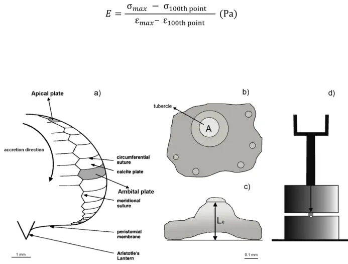
experiment) for each specimen were used for biomechanical measurements (Figure 1a). 159
Top and lateral view photographs of each sea urchin plate were obtained with a Nikon Coolpix 995 160
3Mpi under a binocular lens. Photographs were analyzed, using the software ImageJ, to measure 161
tubercle areas (A), which is the area over which the force was actually applied (Figure 1b) and 162
effective length (Le), which is the height to the edge of the tubercles (Figure 1c). These parameters
163
are necessary for Strain and Stress calculations (see equations below). 164
Due to the small size and flatness of both apical and ambital plates of juvenile sea urchins, an ad hoc 165
system, halfway between compression test and three-point bending test, was created to measure the 166
fracture force. Individual cleaned and detached sea urchin plates were placed on a metal stand with a 167
groove in the middle and the mechanical test was carried out using a second metal block fixed on the 168
load frame of the force stand, Instron 5543 tensile tester, at a speed of 0.1 mm min-1, applying constant
169
compression till breakage (Figure 1d). Displacement and force were recorded at a frequency of 10 170
Hz. For each plate, the Bluehill software (Instron) provided information about force at fracture (Fmax)
171
and displacement (DL). 172
Stiffness was measured through Young's modulus (E), a quantity used to characterize materials and 173
defined as the ratio of the stress along an axis over the strain along that same axis. 174
Strain and Stress were calculated for each plate, using the following equations: 176 Strain:
σ
= F/A 177 Stress:ε
= DL/Le 178where F: force at fracture, A: tubercle area, DL: displacement, Le: effective length.
179
The Young's modulus (E) was calculated as the slope between two points of the final linear part of 180
the curve, in this case the maximum force and the 100th point before that, using the following 181 formula: 182 𝐸 = σ%&' − σ)**+, -./0+ ε%&'– ε)**+, -./0+ (Pa) 183 184 185 186
Figure 1: Schematic representation of a) interambulacral plates, apical and ambital, and suture geometry with 187
corresponding scale bar at the bottom (modified from Ellers et al., 1998; b) upper and c) lateral view of one plate, in order 188
to perform tubercle area (A) and effective length (Le) measurements, with corresponding scale bar at the bottom; d) simple 189
ad hoc compression device composed by two metal blocks, the lower, where the plate should be placed, with a groove
190
and the upper fixed on the load frame (modified from Collard et al., 2016) 191
192 193
Data analyses
194
In order to assess the effect of pH level, algal diet and their interaction on apical and ambital plates 195
strain (Fmax/A) and stiffness (Young’s modulus, square root transformed data), a crossed ANOVA
196
design was applied, using ‘pH’ and ‘diet’ as fixed crossed factors. Since multiple observations were 197
performed on each specimen, a random effect specimen nested in the interaction (pH * diet) was 198
added in order to account for the dependency structure in the data. 199
Normality and homogeneity of variance were verified for both the considered response variables 200
(Fmax/A and ÖE) using Shapiro-Wilk test and Bartlett tests, respectively. All statistical analyses were
201
performed using the software R (R Core Team 2014). 202
203
Results
204
No significant effect of the interaction between the two fixed factors, pH and diet, was observed in 205
strain (fracture force/surface on which the force is applied) or in stiffness neither for ambital nor for 206
apical plates. Moreover, no significant difference according to factors seawater pH or diet singularly 207
were detected in ambital plates (Tables 1, 2). Similarly, no effect of seawater pH or diet was 208
evidenced in apical plates strain and stiffness (Tables 1, 2). 209
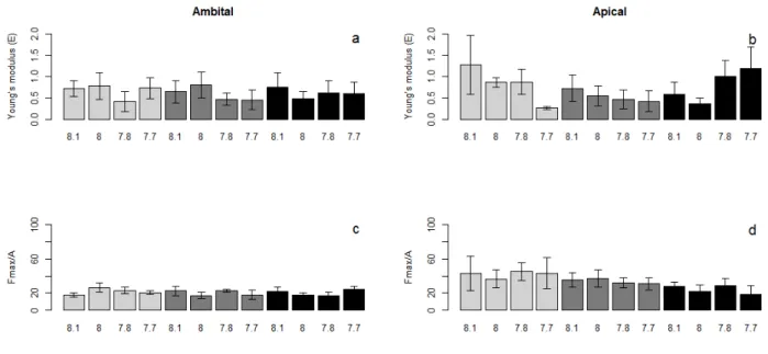
Both strain and stiffness of ambital plates (Fig. 2 a, c) showed more homogeneous values among 210
treatments compared to apical ones (Fig. 2 b, d). Apical plates stiffness slightly decreased according 211
to the pH decrease for E. elongata and C. amentacea, but not in D. dichotoma, where the trend seems 212
to be the opposite (Fig. 2b), even if differences are not statistically significant. A weak gradient of 213
apical plates strain values according to the algal diet can be observed, i.e. gradual decrease of the 214
strain value for sea urchins fed E. elongata, C. amentacea and D. dichotoma, respectively (Figure 215
2d). 216
Table 1: ANOVA results table for strain data (Fmax/A) of ambital and apical plates under factors pH and Diet and their 218
interaction. The random effect specimen nested in the interaction (pH x* diet) is considered in order to account for the 219
dependency structure in the data 220
AMBITAL PLATES APICAL PLATES
Df SS MS F signif. Df SS MS F signif. pH 3 2. 8 0.95 0.01 0.998 3 271 90.2 0.16 0.918 Diet 2 51.1 25.55 0.38 0.694 2 3798 1898.9 3.47 0.065 pH * diet 6 641 106.84 1.58 0.236 6 529 88.1 0.16 0.983 specimen(pH * diet) 12 776.5 67.71 2.91 0.004 12 6567 547.3 3.32 0.001 Residuals 48 1116 23.24 48 7906 164.7 221 222
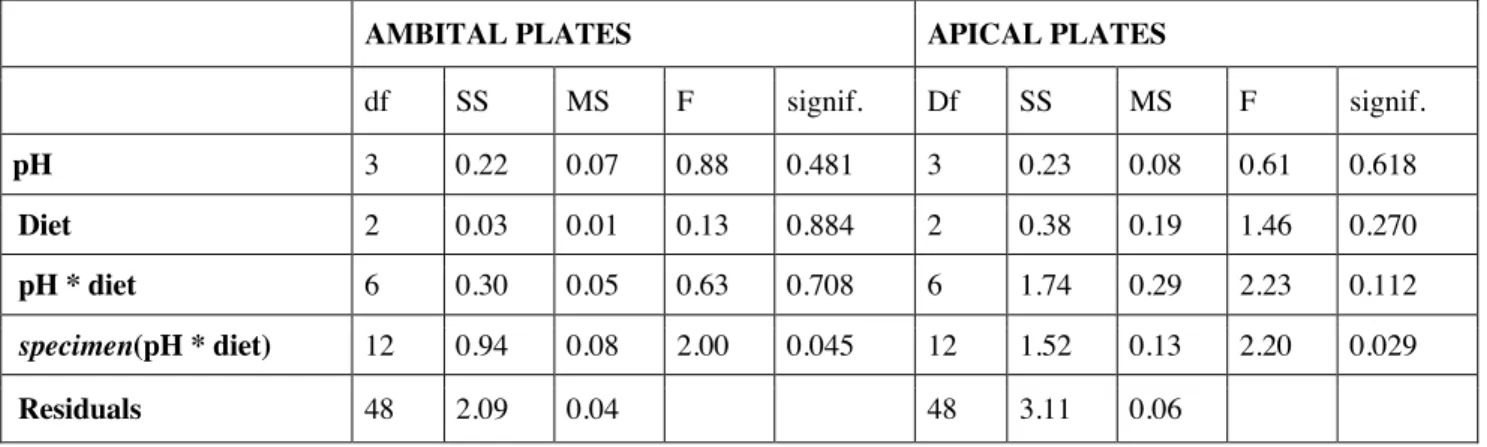
Table 2: ANOVA results table on square root transformed Young's modulus data (ÖE) of ambital and apical plates under 223
factors pH and Diet and their interaction. The random effect specimen nested in the interaction (pH x* diet) is considered 224
in order to account for the dependency structure in the data 225
AMBITAL PLATES APICAL PLATES
df SS MS F signif. Df SS MS F signif. pH 3 0.22 0.07 0.88 0.481 3 0.23 0.08 0.61 0.618 Diet 2 0.03 0.01 0.13 0.884 2 0.38 0.19 1.46 0.270 pH * diet 6 0.30 0.05 0.63 0.708 6 1.74 0.29 2.23 0.112 specimen(pH * diet) 12 0.94 0.08 2.00 0.045 12 1.52 0.13 2.20 0.029 Residuals 48 2.09 0.04 48 3.11 0.06 226 227
228
Figure 2: Barplots of Young's modulus (E), upper panels, and strain data (Fmax/A), lower panels, measured for ambital (a, 229
c) and apical (b, d) plates under different sea water pH (x axis) and different macroalgal diets (light gray= E. elongata, 230
gray= C. amentacea, dark gray= D. dichotoma). Error bars: standard error. 231
232
Discussion
233
Ocean acidification caused by anthropogenic carbon dioxide emissions is known to pose major threats 234
for marine organisms, particularly calcifying ones (e.g. Kroeker et al., 2010; Riebesell et al., 2010), 235
since their calcium carbonate structures are potentially susceptible to dissolution in acidic waters (Orr 236
et al. 2005). Sea urchins are important marine calcifiers, playing a relevant ecological role in 237
temperate ecosystems (Jangoux and Lawrence 1982; Ruitton et al., 2000; Bulleri et al., 2002; Privitera 238
et al., 2008; Bonaviri et al., 2011). Among calcifying organisms, highly calcified sea urchins are 239
expected to be the echinoderms more affected by reduced pH, with differences at species, population 240
and stage level (reviewed in Dupon et al., 2010). 241
Early life stages are acknowledged to be the most sensitive to ocean acidification (Dupont et al., 242
2010; Moulin et al., 2011; Byrne et al., 2013; Stumpp et al., 2013), but also post-metamorphic phases 243
(juveniles and adults) showed to be affected, mainly in terms of survival and growth, both from 244
laboratory experiments (e.g. Shirayama and Thornton 2005; Byrne et al., 2011; Albright et al., 2012; 245
Asnaghi et al., 2013, 2014; Moulin et al., 2015; Collard et al., 2016), and from in situ records in 246
naturally acidified areas (only on adults, e.g. Hall-Spencer et al., 2008; Bray et al., 2014; Collard et 247
al., 2016). Moreover, different studies investigated coelomic fluid regulatory capacities in adult sea 248
urchins exposed to hypercapnic conditions, reporting contrasting results about their ability to partially 249
or fully compensate extracellular pH (Stumpp et al., 2012; Collard et al., 2013, 2014; Calosi et al., 250
2013; Kurihara et al., 2013; Moulin et al., 2014, 2015) and prevent skeletal dissolution at low pH 251
(reviewed by Dery et al., 2017). 252
Only few studies addressed the combined effect of reduced pH and macroalgal diet on sea urchins, 253
highlighting potential cascading consequences at the ecosystem level (Johnson and Carpenter, 2012; 254
Asnaghi et al., 2013; 2014). Macroalgae exhibit a broad range of responses to ocean acidification 255
(Hall-Spencer et al., 2008; Nelson, 2009; Martin and Gattuso, 2009; Connell and Russell, 2010; 256
Cornwall et al. 2011, 2017; Poore et al., 2013), mainly linked to their calcium carbonate content 257
(Cornwall et al., 2014; James et al, 2014) and inorganic carbon physiology (Cornwall et al., 2017). 258
The loss of calcium carbonate and tissue modification in macroalgal thalli caused by reduced pH may 259
enhance their palatability to grazers (Duarte et al., 2016; Rich et al., 2018, Rodriguez et al., 2018), 260
leading to potential shifts in the ecosystem equilibrium under an acidified scenario. 261
Different algal feedings, quantity and species of macroalgae available as food are known to directly 262
affect somatic and gonadal production in sea urchins, both from field and laboratory studies (e.g. 263
Ebert 1968; Lawrence 1975; Lilly 1975; Vadas 1977; Larson et al., 1980; Lawrence and Lane 1982; 264
Thompson 1983, 1984; Privitera et al., 2008). Food shortage (i.e. the lack of macroalgal biomass for 265
feeding) have been reported to cause modifications in plastic resource allocation (Ebert 1980; Haag 266
et al., 2016), differences in mechanical properties of sea urchin spines (Moureaux and Dubois 2012) 267
and, in an acidified scenario, reduction of the buffer capacity of the coelomic fluid to compensate the 268
decrease of external pH (Collard et al., 2013). 269
In the present study, the combined effect of pH level and macroalgal diet was investigated in terms 270
of sea urchin plates biomechanical properties, showing no direct short-term effect of seawater pH nor 271
diet on single plate strain and stiffness, in agreement with previous laboratory studies (Holtmann et 272
al., 2013; Moulin et al., 2015; Collard et al., 2016). 273
A field study, instead, highlighted a role of the diet in mediating sea urchins biomechanical properties, 274
showing that test plates from P. lividus living in tide pools mainly covered by encrusting calcareous 275
algae exhibit a higher fracture force than test plates of sea urchins living in pools containing erected 276
non-calcifying algae (Collard et al., 2016). Similar role of calcifying macroalgae in strengthen P. 277
lividus juvenile tests has been shown by Asnaghi et al. (2013) on specimens exposed to the same
278
experimental conditions of the present study, but it is not easy to disentangle the role of plates, 279
ligaments and calcified locking structures in providing test robustness. 280
Results from the present study showed more homogeneous responses of the ambital plates (the ones 281
already formed before the start of the experiment) in terms of strain and stiffness (Fig. 2 a, c), while 282
apical plates, the one presumably formed during the one-month experimental period, showed more 283
variable values, with strain data suggesting a possible diet-mediated response, maybe visible on a 284
longer term (Fig. 2 b, d): sea urchins fed the more calcified macroalga (E. elongata) sustained higher 285
fracture force than the one fed the non-calcified algae (C. amentacea and D. dichotoma). The high 286
variability at the level of the specimen (Table 1 and 2) may have masked patterns on the short term. 287
Mechanical properties can be affected by changes in the growth rate (e.g. Moureaux et al., 2010), 288
which may affect the three-dimensional morphology or density of the stereom (Smith 1980). 289
Alternatively, the structural properties of the material itself may be affected by the formation of 290
imperfections during the CaCO3 precipitation (e.g. Moureaux et al., 2011).
291
Sea urchins from the present experiment showed different growth rates according to their diet. 292
Ellisolandia elongata allowed for a faster growth rate of the sea urchins compared to that of the ones
293
fed Cystoseira amentacea and Dictyota dichotoma. (Asnaghi et al., 2014). This could be linked to the 294
supply of calcium by calcified algae (Powell et al., 2010) rather than to the carbonate ions, which are 295
not transported through cell membranes, or bicarbonate ions which are available in high concentration 296
in seawater from which they are readily absorbed (from this source; Collard et al., 2014). 297
In the present study, the choice to use juveniles specimens, characterized by higher growth rates 298
compared to adults, was driven by the possibility to observe potential modifications linked to pH and 299
diet treatments on a short time scales (one month). Indeed, a diet-medieted decrease in growth and 300
test robustness in these P. lividus juveniles under acidified conditions was proven (Asnaghi et al., 301
2013), even if not mirrored by substantial modification of test thickness and single plates 302
biomechanical properties. Rodriguez et al. (2017) reported significantly thinner test plates in juvenile 303
Paracentrotus lividus and Diadema africanum kept for 100 days in one tank at low pH conditions
304
compared to the ones in the control tank, that may have led to less robust test, even if several other 305
sources of variability were present in the experimental design. 306
Those considerations suggest that, in order to observe substantial differences on skeletal plate 307
structure, it is necessary to perform longer term experiments (Dupont et al., 2010; Hendricks et al., 308
2010), taking into particular account feeding conditions, that are frequently neglected in ocean 309
acidification studies on post-metamorphic individuals (Dubois et al., 2014). 310
The impact of algal diet on sea urchin test resistance to breakage is particularly relevant in the context 311
of global change. Coralline algae are renowned impacted by ocean acidification, while non-calcified 312
algae will be most likely favored in future oceans (Hall-Spencer et al., 2008; Porzio et al., 2011; Koch 313
et al., 2013; Sunday et al., 2017). As a consequence, some sea urchin species might be affected by 314
the expected change in macroalgal diet availability, growing slower and producing a less robust 315
skeleton, with potential relevant cascading consequences on their ability to graze, move, search for 316
shelters and defend from predation and hydrodynamics. 317 318 319 320 321 322 323
Acknowledgements 324
Philippe Dubois is a Research Director of the National Fund for Scientific Research (NFSR, Belgium), Marie
325
Collard was holder of a FRIA grant (Belgium). Valentina Asnaghi scientific stay in France was funded by the
326
“Bando VINCI 2010” of the Italian-French University and in Belgium by a grant of the National Fund for
327
Scientific Research (NFSR, Belgium). Work supported by NFSR project 26112046 and J.0219.16 SOFTECHI.
328
This work is a contribution to the “European Project on Ocean Acidification” (EPOCA) funded by European
329
Community’s Seventh Framework Programme (FP7/2007-2013; grant agreement n 211384). We thank Saloua
330
M’Zoudi for technical help, Marco Asnaghi for helping in plate measurements and Mariachiara Chiantore for
331
the scientific support. We thank Andy Davis and the other two anonymous reviewers for their precious
332 comments. 333 334 335 References 336
1. Albright R, Bland C, Gillette P, Serafy JE, Langdon C, Capo TR 2012. Juvenile growth of the 337
tropical sea urchin Lytechinus variegatus exposed to near-future ocean acidification scenarios. 338
Journal of Experimental Marine Biology and Ecology 426–427: 12–17. 339
2. Andersson AJ, Mackenzie FT, Bates NB, 2008. Life on the margin: implications of ocean 340
acidification on Mg-calcite, high latitude and cold-water marine calcifiers. Marine Ecology 341
Progress Series 373: 265-273. 342
3. Asnaghi V, Chiantore M, Mangialajo L, Gazeau F, Francour P, Alliouane S, Gattuso J-P 2013. 343
Cascading effects of ocean acidification in a rocky subtidal community. PLOS ONE 8 (4): 344
e61978. 345
4. Asnaghi V, Mangialajo L, Gattuso JP, Francour P, Privitera D, Chiantore M 2014. Effects of
346
ocean acidification and diet on thickness and carbonate elemental composition of the test of 347
juvenile sea urchins. Marine Environmental Research 93: 78-84. 348
5. Barry JP, Tyrrell T, Hansson L, Gattuso J-P 2010. CO2 targets for ocean acidification
349
perturbation experiments. Riebesell U, Fabry VJ, Hansson L, Gattuso J-P, editors. Guide to 350
best practices for ocean acidification research and data reporting. Luxembourg: Publications 351
Office of the European Union, p53-66. 352
6. Bonaviri C, Vega Fernández T, Fanelli G, Badalamenti F, Gianguzza P 2011. Leading role of 353
the sea urchin Arbacia lixula in maintaining the barren state in southwestern Mediterranean. 354
Marine Biology 158: 2505–2513. 355
7. Bray L, Pancucci-Papadopulou MA, Hall-Spencer JM 2014. Sea urchin response to rising 356
pCO2 shows ocean acidification may fundamentally alter the chemistry of marine skeletons.
357
Mediterranean Marine Science 15(3): 510–519. 358
8. Bulleri F, Bertocci I, Micheli F 2002. Interplay of encrusting coralline algae and sea urchins 359
in maintaining alternative habitats. Marine Ecology Progress Series 243:101–109. 360
9. Byrne M, Ho M, Wong E, Soars NA, Selvakumaraswamy P, Shepard-Brennand H, 361
Dworjanyn SA, Davis AR 2011. Unshelled abalone and corrupted urchins: Development of 362
marine calcifiers in a Changing Ocean. Proceedings of the Royal Society B-BiolSci 363
278(1716): 2376-2383. 364
10. Byrne M, Lamare M, Winter D, Dworjanyn SA, Uthicke S 2013. The stunting effect of a high 365
CO2 ocean on calcification and development in sea urchin larvae, a synthesis from the tropics
366
to the poles. Philosophical Transaction of the Royal Society B 368: 20120439. 367
11. Byrne M, Smith AM, West S, Collard M, Dubois P, Graba-landry A, Dworjanyn SA 2014. 368
Warming influences Mg2+ content, while warming and acidification influence calcification
369
and test strength of a sea urchin. Environmental science & technology 48(21): 12620-12627. 370
12. Calosi P, Rastrick SPS, Graziano M, Thomas SC, Baggini C, Carter HA, Hall-Spencer JM, 371
Milazzo M, Spicer JI 2013. Distribution of sea urchins living near shallow water CO2 vents
372
is dependent upon species acid–base and ion-regulatory abilities. Marine Pollution Bulletin 373
73: 470–484. 374
13. Collard M, Laitat K, Moulin L, Catarino A, Grosjean P, Dubois P 2013. Buffer capacity of 375
the coelomic fluid in echinoderms. Comparative Biochemistry and Physiology Part A, 166: 376
199 – 206. 377
14. Collard M, Dery A, Dehairs F, Dubois P. 2014. Euechinoidea and Cidaroidea respond 378
differently to ocean acidification. Comparative Biochemistry and Physiology Part A, 174: 45– 379
55. 380
15. Collard M, Rastrick SPS, Calosi P, Demolder Y, Dille J, Findlay HS, Hall-Spencer JM, 381
Milazzo M, Moulin L, Widdicombe S, Dehairs F, Dubois P 2016. The impact of ocean 382
acidification and warming on the skeletal mechanical properties of the sea urchin 383
Paracentrotus lividus from laboratory and field observations. ICES Journal of Marine Science
384
73(3): 727-738. 385
16. Connell SD, Russell BD, 2010. The direct effects of increasing CO2 and temperature on
non-386
calcifying organisms: increasing the potential for phase shifts in kelp forests. Proceedings of 387
the Royal Society of London B: Biological Sciences, rspb20092069. 388
17. Cornwall CE, Hepburn CD, Pritchard D, Currie KI, McGraw CM, Hunter KA, Hurd CL, 389
2011. Carbon-use strategies in macroalgae: differential responses to lowered pH and 390
implications for ocean acidification. J. Phycol. 48: 137–144 391
18. Cornwall CE, Boyd PW, McGraw CM, Hepburn CD, Pilditch CA, Morris JN, ... Hurd CL, 392
2014. Diffusion boundary layers ameliorate the negative effects of ocean acidification on the 393
temperate coralline macroalga Arthrocardia corymbosa. PLoS. One 9, e97235. 394
19. Cornwall CE, Revill AT, Hall-Spencer JM, Milazzo M, Raven JA, Hurd CL, 2017. 395
Inorganic carbon physiology underpins macroalgal responses to elevated CO2.
396
Scientific reports, 7, 46297 397
20. Currey JD 1989. Biomechanics of mineralized skeletons. Short Courses in Geology 5: 11-25 398
21. Dery A, Collard M, Dubois P 2017. Ocean acidification reduces spine mechanical strength in 399
euechinoid but not in cidaroid sea urchins. Environmental Science & Technology 51: 3640-400
3648. 401
22. Deutler F 1926. Uber die Wachstum des Seeigelskeletts. Zoologische Jahrbücher. Abteilung 402
für Anatomie und Ontogenie der Tiere Abteilung für Anatomie und Ontogenie der Tiere 48: 403
119-200. 404
23. Duarte C, López J, Benítez S, Manríquez PH, Navarro JM, Bonta CC, Torres R, Quijón P, 405
2016. Ocean acidification induces changes in algal palatability and herbivore feeding behavior 406
and performance. Oecologia 180: 453-462. 407
24. Dubois P, Chen CP 1989. Calcification in echinoderms. Jangoux M, Lawrence JM, editors. 408
Balkema, Rotterdam, The Netherlands. Echinoderm Studies 3, p55–136. 409
25. Dubois P 2014. The skeleton of postmetamorphic echinoderms in a changing world. 410
Biological Bulletin 226: 223–236. 411
26. Dupont S, Ortega-Martínez O, Thorndyke MC 2010. Impact of near-future ocean acidification 412
on echinoderms. Ecotoxicology 19: 449–462. 413
27. Ebert TA 1968. Growth rates of the sea urchin Strongylocentrotus purpuratus related to food 414
availability and spine abrasion. Ecology 49: 1075:1091. 415
28. Ebert TA 1980. Relative growth of sea urchin jaws: an example of plastic resource allocation. 416
Bulletin of Marine Science 30: 467–474. 417
29. Ellers O, Johnson AS, Moberg PE 1998. Structural strengthening of urchin skeletons by 418
collagenous sutural ligaments. Biological Bulletin 195: 136- 144. 419
30. Emerson CE, Reinardy HC, Bates NR, Bodnar AG 2017. Ocean acidification impacts spine 420
integrity but not regenerative capacity of spines and tube feet in adult sea urchins. Royal 421
Society open science 4(5): 170140. 422
31. Haag N, Russell MP, Hernandez JC 2016. Effects of spine damage and microhabitat on 423
resource allocation of the purple sea urchin Strongylocentrotus purpuratus (Stimpson 1857). 424
Journal of Experimental Marine Biology and Ecology 482: 106-117. 425
32. Hall-Spencer JM, Rodolfo-Metalpa R, Martin S, Ransome E, Fine M, Turner SM, Rowley SJ, 426
Tedesco D, Buia M-C 2008. Volcanic carbon dioxide vents show ecosystem effects of ocean 427
acidification. Nature 454(7200) : 96-99. 428
33. Hermans J, Borremans C, Willenz P, André L, Dubois P 2010. Temperature, salinity and 429
growth rate dependences of Mg/Ca and Sr/Ca ratios of the skeleton of the sea urchin 430
Paracentrotus lividus (Lamarck): an experimental approach. Marine Biology 157: 1293–
431
1300. 432
34. Holtmann WC, Stumpp M, Gutowska MA, Syre S, Himmerkus N, Melzner F, Bleich M 2013. 433
Maintenance of coelomic fluid pH in sea urchins exposed to elevated CO2: the role of body
434
cavity epithelia and stereo dissolution. Marine Biology 160(10):2631-2645. 435
35. James RK, Hepburn CD, Cornwall CE, McGraw CM, Hurd CL, 2014. Growth response of an 436
early successional assemblage of coralline algae and benthic diatoms to ocean acidification. 437
Marine Biology 161: 1687–1696. 438
36. Jangoux M, Lawrence JM 1982. Echinoderm Nutrition. Balkema, Rotterdam. 439
37. Johnson MD, Carpenter RC, 2012. Ocean acidification and warming decrease calcification in 440
the crustose coralline alga Hydrolithon onkodes and increase susceptibility to grazing. Journal 441
of Experimental Marine Biology and Ecology, 434 : 94-101. 442
38. Kidwell SM, Baumiller T 1990. Experimental disintegration of regular echinoids: roles of 443
temperature, oxygen, and decay thresholds. Paleobiology 16: 247-271. 444
39. Koch M, Bowes G, Ross C, Zhang XH 2013. Climate change and ocean acidification effects 445
on seagrasses and marine macroalgae. Global change biology 19(1): 103-132. 446
40. Kroeker KJ, Kordas RL, Crim RN, Singh GG, 2010. Meta‐analysis reveals negative yet 447
variable effects of ocean acidification on marine organisms. Ecology letters, 13(11): 1419-448
1434. 449
41. Kurihara H, Shirayama Y 2004. Effects of increased atmospheric CO2 on sea urchin early
450
development. Marine Ecology Progress Series 274: 161–169. 451
42. Kurihara H, Yin R, Nishihara GN, Soyano K, Ishimatsu A 2013. Effect of ocean acidification 452
on growth, gonad development and physiology of the sea urchin Hemicentrotus pulcherrimus. 453
Aquatic Biology 18: 281–292. 454
43. Larson BR, Vadas RL, Keser M 1980. Feeding and nutritional ecology of the sea urchin 455
Strongylocentrotus droebachiensis. Marine Biology 59: 49-62.
456
44. Lawrence JM 1975. On the relationship between marine plants and sea urchins. Oceanography 457
and Marine Biology, An Annual Review 13: 213-286. 458
45. Lawrence JM, Lane JM 1982. The utilization of nutrients by post-metamorphic echinoderms. 459
Echinoderm nutrition: 331-371 460
46. Lilly GR 1975. The influence of diet on the growth and bioenergetics of the tropical sea 461
urchin, Tripneustes ventricosus (Lamark). Thesis. Vancouver: Univ. of British Columbia. 462
47. Martin S, Gattuso JP, 2009. Response of Mediterranean coralline algae to ocean acidification 463
and elevated temperature. Global Change Biol. 15: 2089–2100. 464
48. Martin S, Richier S, Pedrotti ML, Dupont S, Castejon C, Gerakis Y, Kerros ME, Oberhänsli 465
F, Teyssié, J.L., Jeffree, R., Gattuso, J.P., 2011. Early development and molecular plasticity 466
in the Mediterranean sea urchin Paracentrotus lividus exposed to CO2-driven acidification.
467
Journal of Experimental Biology 214(8): 1357-1368. 468
49. Märkel K 1975. Wachstum des Coronarskeletes von Paracentrotus lividus 469
Lmk.(Echinodermata, Echinoidea). Zoomorphologie, 82(2-3): 259-280. 470
50. Meyers MA, Chen P-Y, Lin AY-M, Seki Y 2008. Biological materials: structure and 471
mechanical properties. Progress in Material Science 53: 1–206. 472
51. Moss ML, Meehan MM 1967. Sutural connective tissues in the test of an echinoid Arbacia 473
punctulata. Acta Anatomica 66: 279-304.
474
52. Moulin L, Catarino AI, Claessens T, Dubois P 2011. Effects of seawater acidification on early 475
development of the intertidal sea urchin Paracentrotus lividus (Lamarck 1816). Marine 476
Pollution Bulletin 62(1): 48-54. 477
53. Moulin L, Grosjean P, Leblud J, Batigny A, Dubois P 2014. Impact of elevated pCO2 on
acid-478
base regulation of the sea urchin Echinometra mathaei and its relation to resistance to ocean 479
acidification: a study in mesocosms. Journal of Experimental Marine Biology and Ecology 480
457: 97–104. 481
54. Moulin L, Grosjean P, Leblud J, Batigny A, Collard M, Dubois P 2015. Long-term 482
mesocosms study of the effects of ocean acidification on growth and physiology of the sea 483
urchin Echinometra mathaei. Marine environmental research 103: 103-114. 484
55. Moureaux C, Pérez-Huerta A, Compère P, Zhu W, Leloup T, Cusack M, Dubois P 2010. 485
Structure, composition and mechanical relations to function in sea urchin spine. Journal of 486
Structural Biology 170: 41– 49. 487
56. Moureaux C, Simon J, Mannaerts G, Catarino AI, Pernet P, Dubois P 2011. Effects of field 488
contamination by metals (Cd, Cu, Pb, Zn) on biometry and mechanics of echinoderm ossicles. 489
Aquatic Toxicology 105: 698–707. 490
57. Moureaux C, Dubois P 2012. Plasticity of biometrical and mechanical properties of 491
Echinocardium cordatum spines according to environment. Marine Biology 159: 471-492
479. 493
58. Nelson WA (2009) Calcified macroalgae critical to coastal ecosystems and vulnerable 494
to change: A review. Mar Freshw Res 60(8):787–801. 495
59. O'Donnell MJ, Todgham AE, Sewell MA, Hammond LM, Ruggiero K, Fangue NA, Zippay 496
ML, Hofmann GE 2010. Ocean acidification alters skeletogenesis and gene expression in 497
larval sea urchins. Marine Ecology Progress Series 398: 157-171. 498
60. Orr JC, Fabry VJ, …, Yool A, 2005. Anthropogenic ocean acidification over the twenty-first 499
century and its impact on calcifying organisms. Nature, 437(7059): 681. 500
61. Poore AGB, Graba-Landry A, Favret M, Sheppard Brennand H, Byrne M, Dworjanyn SA, 501
2013. Direct and indirect effects of ocean acidification and warming on a marine plant– 502
herbivore interaction. Oecologia 173: 1113–1124. 503
62. Porzio L, Buia MC, Hall-Spencer JM, 2011. Effects of ocean acidification on macro-algal 504
communities. Journal of Experimental Marine Biology and Ecology 400: 278–287. 505
63. Privitera D, Chiantore M, Mangialajo L, Glavic N, Kozul W, Cattaneo-Vietti R 2008. Inter-506
and intra-specific competition between Paracentrotus lividus and Arbacia lixula in resource-507
limited barren areas. Journal of Sea Research 60(3): 184-192. 508
64. Rich WA, Schubert N, Schläpfer N, Carvalho VF, Horta AC, Horta PA, 2018. Physiological 509
and biochemical responses of a coralline alga and a sea urchin to climate change: Implications 510
for herbivory. Marine environmental research, 142, 100-107. 511
65. Riebesell U, Fabry VJ, Hansson L, Gattuso J-P (Eds.), 2010. Guide to best practices for ocean 512
acidification research and data reporting. Luxembourg: Publications Office of the European 513
Union, pp. 53–66. 514
66. Rodríguez A, Hernández JC, Brito A, Clemente S, 2017. Effects of ocean acidification on 515
juvenile sea urchins: predator-prey interactions. J. Exp. Mar. Biol. Ecol. 493: 31–40. 516
67. Rodríguez A, Clemente S, Brito A, Hernández JC, 2018. Effects of ocean acidification on 517
algae growth and feeding rates of juvenile sea urchins. Marine environmental research, 140, 518
382-389. 519
68. Ruitton S, Francour P, Boudouresque C 2000. Relationships between algae, benthic 520
herbivorous invertebrates and fishes in rocky sublittoral communities of a temperate sea 521
(Mediterranean). Estuarine, Coastal and Shelf Science 50(2), 217-230. 522
69. Shirayama Y, Thornton H 2005. Effect of increased atmospheric CO2 on shallow water
523
marine benthos. Journal of Geophysical Research 110(C9): C09S08. 524
70. Smith AB 1980. Stereom microstructure of the echinoid test. Special Papers in Palaeontology 525
25: 1-81. 526
71. Stumpp M, Dupont S, Thorndyke MC, Melzner F 2011. CO2 induced seawater acidification
527
impacts sea urchin larval development II: Gene expression patterns in pluteus larvae. 528
Comparative Biochemistry and Physiology - A Molecular and Integrative Physiology 160(3): 529
320-330. 530
72. Stumpp M, Trubenbach K, Brennecke D, Hu MY, Melzner F 2012. Resource allocation and 531
extracellular acid-base status in the sea urchin Strongylocentrotus droebachiensis. Aquatic 532
Toxicology 110 – 111: 194–207. 533
73. Stumpp M, Hu M, Casties I, Saborowski R, Bleich M, Melzner F, Dupont S 2013. Digestion 534
in sea urchin larvae impaired under ocean acidification. Nature climate change 3(12): 1044. 535
74. Sunday JM, Fabricius KE, Kroeker KJ, Anderson KM, Brown NE, Barry JP, Connell SD, 536
Dupont S, Gaylord B, Hall-Spencer JM, Klinger T, Milazzo M, Munday PL, Russell BD, 537
Sanford E, Thiyagarajan V, Vaughan MLH, Widdicombe S, Harley CDG 2017. Ocean 538
acidification can mediate biodiversity shifts by changing biogenic habitat. Nature Climate 539
Change 7(1): 81. 540
75. Thompson RJ 1983. The relationship between food ration and reproductive e ort in the green 541
sea urchin, Strongylocentrotus droebachiensis. Oecologia 56: 50:57. 542
76. Thompson RJ 1984. Partitioning of energy between growth and reproduction in three 543
populations of the sea urchin Strongylocentrotus droebachiensis. In: Engels W, editor. 544
Advances in invertebrate reproduction. 3. Elsevier Science Publishers BV, Amsterdam, p425-545
431. 546
77. Todgham AE, Hofmann GE 2009. Transcriptomic response of sea urchin larvae 547
Strongylocentrotus purpuratus to CO2-driven seawater acidification. Journal of Experimental
548
Biology 212: 2579-2594. 549
78. Vadas RL 1977. Preferential feeding: an optimization strategy in sea urchins. Ecological 550
Monographs 47: 337-371. 551
79. Weiner S.1985. Organic matrix-like macromolecules associated with the mineral phase of sea 552
urchin skeletal plates and teeth. Journal of Experimental Zoology 234: 7–15. 553