ORIGINAL PAPER
Risk factors for dislocation arthropathy after Latarjet
procedure: a long-term study
Alexandre Lädermann&Anne Lubbeke&
Richard Stern&Grégory Cunningham&
Vittorio Bellotti&Dominique F. Gazielly
Received: 12 February 2013 / Accepted: 19 February 2013 / Published online: 13 March 2013 # Springer-Verlag Berlin Heidelberg 2013
Abstract
Purpose The purpose of this study was to analyse the long-term incidence of dislocation arthropathy after a modified Latarjet procedure for glenohumeral instability.
Methods Long-term follow-up information was obtained from a consecutive series of patients who had under-gone a modified Latarjet procedure by one surgeon between 1986 and 1999. Multivariable regression anal-ysis was performed to examine the relation between the development of a dislocation arthropathy and patients and surgery-related factors.
Results There were 117 patients (117 shoulders) for evalu-ation, (35 women and 82 men) with a mean age 28.4±8.5
(range, 16–55). The mean follow-up was 16.2 years (range, ten to 22.2 years). Signs of dislocation arthropathy were found in 36 % of patients, graded as Samilson 1 in 30 %, Samilson 2 in 3 %, and 3 % Samilson 3 in 3 % of patients. Risk factors for dislocation arthropathy included surgery in patients older than 40 years of age (64.3 vs. 34.4 %; adjust-ed RR 2.2, 95 % CI 1.7–2.9) and lateral positioning of the transferred coracoid process in relation to the glenoid rim (82.4 vs. 30.4 %; adjusted RR 2.3, 95 % CI 1.7–3.2). Patients with hyperlaxity developed less dislocation arthrop-athy (15 vs. 42.5 %; adjusted RR 0.4, 95 % CI 0.1–0.95). Conclusion The development of dislocation arthropathy after the Latarjet procedure remains a source of concern in the long term. It correlates with surgery after the age of 40 and lateral coracoid transfer in relation to the glenoid rim. On the other hand, hyperlaxity seems to have a protective effect on the development of disloca-tion arthropathy.
Introduction
The Latarjet procedure [1] and its subsequent modifications [2,3] are becoming increasingly popular and are currently considered as an efficient method to stabilise the shoulder primarily or after recurrent dislocation [4,5]. The procedure can be performed open [6,7] or arthroscopically [8,9]. Only a few studies with a large sample size have reported long-term outcomes [10–13], and only one has focused on dislo-cation arthropathy as defined by Samilson, which can occur after either shoulder dislocation or surgical repair [14].
Several risks factors such as patient age at the time of surgery or the details of the procedure itself have been described and might be responsible for the development of dislocation arthropathy following anterior shoulder stabilisation [11,13–15].
Level of evidence Level III, therapeutic case series. A. Lädermann
:
A. Lubbeke:
R. Stern:
G. Cunningham:
D. F. GaziellyDivision of Orthopaedics and Trauma Surgery, Department of Surgery, Geneva University Hospitals, Rue Gabrielle-Perret-Gentil 4,
1211 Geneva, Switzerland A. Lädermann
Faculty of Medicine, University of Geneva, Rue Michel-Servet 1, 1211 Geneva, Switzerland
A. Lädermann (*)
Division of Orthopaedics and Trauma Surgery, La Tour Hospital, Av. J.-D. Maillard 3,
1217 Meyrin, Switzerland
e-mail: [email protected] V. Bellotti
ICATME, Instituto Universitario Dexeus, Carrer Sabino Arana 5-19, 2a Planta, 08028 Barcelona, Spain
D. F. Gazielly
Shoulder Unit, Clinique Genolier, Rue du Muids 3, 1272 Genolier, Switzerland
The objective of this study was first to evaluate the long-term clinical results of a modified Latarjet procedure and second to establish the incidence of dislocation arthropathy, its risk factors, and its relation to clinical results.
Materials and methods Patient selection
We performed a retrospective review of 324 patients who had undergone an open modified Latarjet procedure by the senior author (DFG) from January 1986 to November 1999. The inclusion criteria included a primary bone Latarjet procedure, a minimum follow-up of ten years, and complete preoperative medical records. Exclusion criteria included a previous bone block procedure such as Eden-Hybinette stabilisation [16,17], lack of follow-up data, and incomplete radiographic examination.
Surgical technique
The surgical technique for the open Latarjet procedure has been previously described [1, 4]. The senior author who performed all the operations in this study used a modified operative technique. The approach was delto-pectoral with an L-shaped incision in the superior two-thirds of the subscapularis muscle. Drilling was done through the hori-zontal part of the coracoid, the graft was attached, flush to the glenoid neck below the equator, in a supine position with a 4.5-mm cortical screw and washer (Fig.1). All patients took part in a standardised rehabilitation protocol [18]. Study variables
The outcomes of interest were (1) the long-term clinical results and (2) the long-term incidence of dislocation ar-thropathy. Additionally, the incidences of other radiological complications such as osteolysis, migration, fracture, or pseudarthrosis were recorded.
Long-term clinical results were assessed using a patient-administered questionnaire including (1) the Walch-Duplay score [19] (maximum of 100 points) which is a functional score consisting of objective as well as subjective parame-ters, and has been used to classify sports activities and pain; (2) the Walch-Duplay pain score is graded from 0 points (pain during activities of daily living) to 25 points (no pain); (3) hyperlaxity as defined by external rotation greater than 90° with the elbow at the side [4], whereby the method of self-determining forward elevation has been previously val-idated [20]; (4) patient satisfaction (very satisfied, satisfied or dissatisfied); (5) return to sports or activities (yes/no); and (6) need for repeat surgery. In addition, the following
baseline characteristics were assessed: age, sex, and side of surgery.
Radiological evaluation
At latest follow-up patients underwent radiological evalua-tion including anteroposterior views in internal and external rotation, one subcoracoid view [10] and one Bernageau view [21]. All radiographs were independently assessed by two orthopaedic surgeons who had not been involved in the surgical procedures. The diagnosis of dislocation arthropa-thy was defined according to the Samilson criteria [14], whi ch tak es in to ac cou nt th e a pp ea ran ce o f t he glenohumeral joint and the size of inferior humeral and/or glenoid osteophytes. Mild arthritis was diagnosed when osteophytes were less than three millimetres. When the osteophytes measured between three and seven millimetres with mild gleno-humeral irregularity, arthropathy was clas-sified as moderate. Severe arthropathy was recorded if there was severe sclerosis of the glenohumeral joint or if the osteophytes were greater than seven millimetres in height.
The position of the graft was considered too far lateral if any part of the screw, washer or graft itself was overhanging lateral to the joint line on the Bernageau view. The position was too far medial if the lateral aspect of the coracoid was greater than four millimetres medial to the joint line. Fig. 1 The coracoid and glenoid have been drilled using 4.5- and 3.2-mm drill bits, respectively. The coracoid graft is secured to the anterior glenoid by means of a 4.5-mm screw and washer
Osteolysis, migration, fracture, or pseudarthrosis of the graft was recorded using the criteria of Hovelius et al. [22]. The graft was considered as united when there was no visible radiolucent zone between the graft and the scapular neck on all radiographs. Pseudarthrosis was diagnosed when the graft showed separation from the scapular neck by a radiolucent zone not wider than five millimetres. If greater than five millimetres the graft was classified as migrated.
Statistical analysis
To determine risk factors for dislocation arthropathy defined as presence of mild to severe arthritis (Samilson grade 1–3) we calculated crude and adjusted risk ratios (RR) and their 95 % confidence intervals (CI). Adjusted risk ratios were obtained with the use of the general linear model (GLM) for the binomial family (STATA version 11.1).
Sex, dominant side and sports activity were not included in the multivariable model, because no substantial effect on the risk of developing dislocation arthropathy was seen in the univariate analyses.
To evaluate whether the presence or absence of arthrop-athy influenced clinical results, the unpaired Student’s t-test for continuous variables and the chi-square test for categor-ical variables was used to obtain p-values.
Ethics
Patients gave their permission to be included in this consec-utive series. Ethical Committee approval was not required.
Results
Demographic data
All eligible patients were contacted for study recruit-ment. Of the 324 patients who had undergone the pro-cedure during the study interval, 207 did not respond. Thus there were 117 patients (117 procedures, 35 wom-en and 82 mwom-en) with a mean age of 28.4 ± 8.5 years (range, 16–55 years) for final analysis. Of those 110
patients could be graded for the presence or absence of dislocation arthropathy. Mean follow-up was 16.2 years (range, ten to 22.2 years).
Complications
Postoperative complications included one postoperative in-fection, one transient lesion of the musculocutaneus nerve, and one superficial vein thrombosis.
Long-term clinical outcomes
Thirty-two patients (17 %) had hyperlaxity. Four patients reported persistent apprehension. However, none of them showed hyperlaxity. Two patients (1.7 %) sustained a re-dislocation or re-subluxation, but none required further op-eration. The mean Walch-Duplay score was 92.8 ± 10.3 (range, 25–100), and results were good or excellent in 97.4 % of cases. Sixty percent of patients were pain free, 37 % described occasional pain, and 3 % of patients suffered from pain during activities of daily living. Seventy-eight percent of patients were very satisfied, 18 % were satisfied and 3 % were dissatisfied with their outcome. Return to sports activities was possible for 83 % of the patients. Radiological results
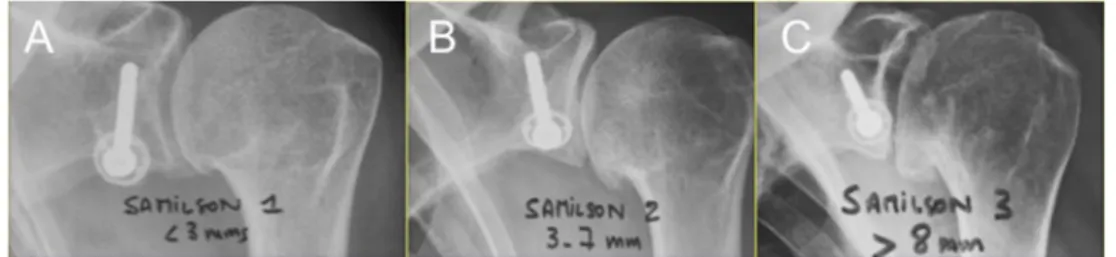
Radiological complications included two patients with a pseudarthrosis (1.7 %), four cases of osteolysis (3.4 %), one fracture (0.9 %), and one migration (0.9 %). The pres-ence of dislocation arthropathy was found in 42 of 110 patients (36 %). Of those 30 % were graded Samilson 1, 3 % Samilson 2 and 3 % were graded Samilson 3 (Fig.2). The coracoid was positioned laterally to the glenoid in 19 patients (14.5 %). Risk factors for dislocation arthropathy (Table 1) in the multivariable analysis were surgery in patients older than 40 years of age compared to those younger than 40 (64.3 % vs. 34.4 %; adjusted RR 2.2, 95 % CI 1.7–2.9) and lateral positioning of the transferred coracoid process in relation to the glenoid rim (82.4 % vs. 30.4 %; adjusted RR 2.3, 95 % CI 1.7–3.2). Presence of hyperlaxity was protective (15 % vs. 42.5 %; adjusted RR 0.4, 95 % CI 0.1–0.95). No difference in risk was seen regarding sex, sports activity and dominant side.
Fig. 2 Samilson 1 (a), 2 (b) and 3 (c) dislocation
arthropathy was found in 30 %, 3 % and 3 % of cases, respectively
None of the clinical outcome parameters except for pa-tient satisfaction differed substantially between papa-tients with dislocation arthropathy and those without (Table2). Patients with radiological signs of arthropathy chose more often the rating“satisfied” instead of “very satisfied” as compared to those without arthropathy (p=0.012).
Discussion
The results of this study indicate that the long-term clinical results of the modified Latarjet procedure are good in terms of stability, pain, return to sports and patient satisfaction. Similar clinical results have previously been described
following this procedure and its subsequent modifications [23–25].
A rate of re-dislocation or re-subluxation has been reported by several author as ranging between 0 and 10 % [5,12, 23–25]. In our study there were only two patients (1.7 %) who sustained a re-dislocation or re-subluxation, but there were four other patients (3.4 %) who felt that their shoulder was not stable. There was no correlation between this finding and the presence of arthropathy. A possible explanation for this discrepancy may be that some patients still suffer from apprehension despite a stable shoulder.
The aetiology of dislocation arthropathy is controversial. Lateral protrusion of the graft or osteosynthesis material is known to lead to secondary arthritis [10, 26]. However, Table 1 Association between
potential risk factors and pres-ence of radiographic osteoarthri-tis (Samilson grades 1–3)
aInformation on dominant side
was missing for three patients
b
Hyperlaxity was defined as an external rotation of more than 85° elbow at the side; informa-tion was missing for three patients
c We used the general linear
model (GLM) for the binomial family (STATA version 11.1) to obtain adjusted risk ratios and their 95 % confidence intervals
Potential risk factors Samilson grade 1–3 Total (n) Crude risk ratio (95 % CI) Adjusted risk ratio (95 % CI)c Men (%) 30 (39.5) 76 Ref. Women (%) 12 (35.3) 34 0.9 (0.5–1.5) Age at operation (%) <40 years 33 (34.4) 96 Ref. ≥40 years 9 (64.3) 14 1.9 (1.2–3.0) 2.2 (1.7–2.9) Dominant side (%)a No 20 (39.2) 51 Ref. Yes 19 (33.9) 56 0.9 (0.5–1.4) Sports activity (%) No 7 (41.2) 17 Ref. Yes 35 (37.6) 93 0.9 (0.5–1.7)
Lateral overhanging of the graft relative to the glenoid (%)
No 28 (30.4) 92 Ref.
Yes 14 (82.4) 17 2.7 (1.9–4.0) 2.3 (1.7–3.2)
Hyperlaxity (%)b
No 37 (42.5) 87 Ref.
Yes 3 (15.0) 20 0.4 (0.1–1.0) 0.4 (0.1–0.95)
Table 2 Long-term clinical outcomes according to presence or absence of dislocation arthropathy
Overall, 110 patients had com-plete information on Samilson grades and clinical outcomes
a
p-values were obtained with use of the unpaired Student’s t-test for continuous variables and chi-square test for categorical variables
Outcome measure Presence of arthropathy (Samilson grade 1–3) (n=42) Absence of arthropathy (n=68) p-valuea
Walch-Duplay score, mean (SD) 91.7 (±9.8) 93.7 (±10.3) 0.313
Walch-Duplay pain score (%) 0.927
None 25 (59.5) 42 (61.8)
Occasional 16 (38.1) 25 (36.8)
Pain during daily activities 1 (2.4) 1 (1.5)
Sports activity yes (%) 35 (83.3) 58 (85.3) 0.782
Satisfaction (%) 0.012
Very satisfied 26 (61.9) 59 (86.8)
Satisfied 15 (35.7) 7 (10.3)
while 14.5 % of patients had such an overhang dislocation arthropathy was found in 36 % of patients as a whole. This difference is probably related to the natural history of this disorder and not the surgery itself, a point previously noted by Hovelius et al. [10]. The 30 % incidence of dislocation arthropathy found in our study is similar (19–30 %) to other series [12,13,27]. The radiological findings did not corre-late with functional outcome but with patient satisfaction.
Dislocation of the shoulder prior to 22 years of age is a risk factor for recurrent dislocation [28]. Undergo-ing shoulder stabilization after the age of 40 is a risk factor for the development of dislocation arthropathy. Indeed, a prolonged delay between the initial dislocation and surgery contributes to a greater likelihood of devel-oping dislocation arthropathy. Perhaps this finding might be explained by a greater number of shoulder disloca-tions or subluxadisloca-tions prior to stabilisation. An additional factor may be less favourable biology secondary to aging which correlates with poorer cartilage properties and less capacity for self-repair, leading to extended cartilage damage at the time of stabilisation. Surprising-ly, Hovelius et al. did not find a similar relationship [10]. Hyperlaxity, on the other hand, had a protective effect on dislocation arthropathy in our study. Some authors [13] have postulated that subluxation of the shoulder remains possible after a Latarjet procedure and the repeated sliding of the humeral head leads to arthropathy. Conversely, we believe that hyperlaxity may decrease postoperative contact pressure of the hu-meral head on the glenoid and thus prevent develop-ment of secondary arthritis.
Strengths and limitations
A major strength of our study is the long follow-up after the Latarjet procedure, which is one of the longest to date. As an increasing number of Latarjet procedures are being performed, a better understanding of factors that contribute to dislocation arthropathy is crucial. An additional strength is that all operations were performed by a single surgeon who used the same techniques for repair according to the type of lesion. Limitations in-clude the retrospective design that led to a high loss to follow-up rate, and the inability to determine the num-ber of previous dislocations or subluxations prior to surgery. This information might have correlated with the development of dislocation arthropathy, as such a link probably exists. Additionally, since only postopera-tive imaging for the ipsilateral side was available it was impossible to compare the incidence of postoperative dislocation arthropathy to preoperative or contralateral side arthritis.
Conclusion
The development of dislocation arthropathy after the Latarjet procedure remains a source of concern in the long term. It correlates with surgery after the age of 40 and lateral coracoid transfer in relation to the glenoid rim. On the other hand, hyperlaxity seems to have a protective effect on the development of dislocation arthropathy.
Acknowledgments The authors state that this study or any part of this work has never been submitted or published elsewhere. This work has been read and approved by all authors. Each author believes that the manuscript represents honest work.
Conflict of interest and funding The authors, their immediate fam-ilies, and any research foundation with which they are affiliated did not receive any financial payments or other benefits from any commercial entity related to the subject of this article. No funding was received for this study.
References
1. Latarjet M (1954) Treatment of recurrent dislocation of the shoul-der. Lyon Chir 49:994–997
2. Helfet AJ (1958) Coracoid transplantation for recurring dislocation of the shoulder. J Bone Joint Surg Br 40-B(2):198–202
3. May VR Jr (1970) A modified Bristow operation for anterior recurrent dislocation of the shoulder. J Bone Joint Surg Am 52(5):1010–1016
4. Young AA, Maia R, Berhouet J, Walch G (2011) Open Latarjet procedure for management of bone loss in anterior instability of the glenohumeral joint. J Shoulder Elbow Surg 20(2 Suppl):S61– S69. doi:10.1016/j.jse.2010.07.022
5. Burkhart SS, De Beer JF, Barth JR, Cresswell T, Roberts C, Richards DP (2007) Results of modified Latarjet reconstruction in patients with anteroinferior instability and significant bone loss. Arthroscopy 23(10):1033–1041. doi:10.1016/j.arthro.2007.08.009
6. Doursounian L, Debet-Mejean A, Chetboun A, Nourissat G (2009) Bristow-Latarjet procedure with specific instrumentation: study of 34 cases. Int Orthop 33(4):1031–1036. doi:10.1007/s00264-008-0606-z
7. Walch G, Charret P, Pietro-Paoli H, Dejour H (1986) Anterior recurrent luxation of the shoulder. Postoperative recurrences. Rev Chir Orthop Reparatrice Appar Mot 72(8):541–555
8. Boileau P, Mercier N, Roussanne Y, Thelu CE, Old J (2010) Arthroscopic Bankart-Bristow-Latarjet procedure: the develop-ment and early results of a safe and reproducible technique. Arthroscopy 26(11):1434–1450. doi:10.1016/j.arthro.2010.07.011
9. Lafosse L, Lejeune E, Bouchard A, Kakuda C, Gobezie R, Kochhar T (2007) The arthroscopic Latarjet procedure for the treatment of anterior shoulder instability. Arthroscopy 23(11):1242 e1241–1245. doi:10.1016/j.arthro.2007.06.008
10. Hovelius L, Sandstrom B, Saebo M (2006) One hundred eighteen Bristow-Latarjet repairs for recurrent anterior dislocation of the shoulder prospectively followed for fifteen years: study II-the evolution of dislocation arthropathy. J Shoulder Elbow Surg 15(3):279–289. doi:10.1016/j.jse.2005.09.014
11. Hovelius LK, Sandstrom BC, Rosmark DL, Saebo M, Sundgren KH, Malmqvist BG (2001) Long-term results with the Bankart and Bristow-Latarjet procedures: recurrent shoulder instability and ar-thropathy. J Shoulder Elbow Surg 10(5):445–452. doi:10.1067/ mse.2001.117128
12. Allain J, Goutallier D, Glorion C (1998) Long-term results of the Latarjet procedure for the treatment of anterior instability of the shoulder. J Bone Joint Surg Am 80(6):841–852
13. Singer GC, Kirkland PM, Emery RJ (1995) Coracoid transposition for recurrent anterior instability of the shoul-der. A 20-year follow-up study. J Bone Joint Surg Br 77(1): 73–76
14. Samilson R, Prieto V (1983) Dislocation arthropathy of the shoul-der. J Bone Joint Surg Am 65:456–460
15. Lehmann L, Magosch P, Mauermann E, Lichtenberg S, Habermeyer P (2010) Total shoulder arthroplasty in dislocation arthropathy. Int Orthop 34(8):1219–1225. doi: 10.1007/s00264-009-0928-5
16. Eden R (1918) Zur Operation der habituellen Schulterluxation unter Mitteilung eines neuen Verfahrens bei Abriss am inneren Pfannenrande. Dsch Z Chir 144:269
17. Hybinette S (1932) De la transplantation d’un fragment osseux pour remédier aux luxations récidivantes de l’épaule. Acta Chir Scand 71:411–445
18. Gazielly D (2006) Rééducation et chirurgie de l'épaule au quotidien, vol 1, 2nd edn. Sauramps Médical, Montpellier 19. Walch G (1987) The Walch-Duplay rating sheet for anterior
insta-bility of the shoulder. SECEC/ESSSE, Paris, pp 51–55
20. Carter CW, Levine WN, Kleweno CP, Bigliani LU, Ahmad CS (2008) Assessment of shoulder range of motion: introduction of a novel patient self-assessment tool. Arthroscopy 24(6):712–717. doi:10.1016/j.arthro.2008.01.020
21. Bernageau J, Patte D, Debeyre J, Ferrane J (1976) Value of the glenoid profile in recurrent luxations of the shoulder. Rev Chir Orthop Reparatrice Appar Mot 62(Suppl 2):142–147
22. Hovelius L, Vikerfors O, Olofsson A, Svensson O, Rahme H (2011) Bristow-Latarjet and Bankart: a comparative study of shoulder stabilization in 185 shoulders during a seventeen-year f o l l ow - u p. J S ho u l d e r E l bo w Su rg 2 0( 7 ) : 10 95–1101. doi:10.1016/j.jse.2011.02.005
23. Hovelius L, Sandstrom B, Sundgren K, Saebo M (2004) One hundred eighteen Bristow-Latarjet repairs for recurrent anterior dislocation of the shoulder prospectively followed for fifteen years: study I—clinical results. J Shoulder Elbow Surg 13(5):509–516. doi:10.1016/S1058274604000916
24. Huguet D, Pietu G, Bresson C, Potaux F, Letenneur J (1996) Anterior instability of the shoulder in athletes: apropos of 51 cases of stabilization using the Latarjet-Patte intervention. Acta Orthop Belg 62(4):200–206
25. Matton D, Van Looy F, Geens S (1992) Recurrent anterior dislo-cations of the shoulder joint treated by the Bristow-Latarjet proce-dure. Historical review, operative technique and results. Acta Orthop Belg 58(1):16–22
26. Vander Maren C, Geulette B, Lewalle J, Mullier J, Autrique JC, Thiery J, Deneufbourg J (1993) Coracoid process abutment according to Latarjet versus the Bankart operation. A comparative study of the results in 50 cases. Acta Orthop Belg 59(2):147–155 27. Rahme H, Wikblad L, Nowak J, Larsson S (2003) Long-term clinical and radiologic results after Eden-Hybbinette operation for anterior instability of the shoulder. J Shoulder Elbow Surg 12(1):15–19. doi:10.1067/mse.2002.128138
28. Porcellini G, Campi F, Pegreffi F, Castagna A, Paladini P (2009) Predisposing factors for recurrent shoulder dislocation after arthro-scopic treatment. J Bone Joint Surg Am 91(11):2537–2542. doi:10.2106/JBJS.H.01126