Roughness of enamel surfaces after different bonding and
debonding procedures. An in vitro study
Einfluss von Konditionierung und Politur auf die
Schmelzoberflächenrauheit. Eine In-vitro-Studie
Abstract
Background and aim. Maintaining an intact enamel surface is an
essential aspect of orthodontic therapy; however, various therapeutic measures can affect this surface. The aim of our study was to evaluate roughness of the enamel surface after different conditioning and pol-ishing procedures.
Materials and methods. 42 bovine incisors were submitted to
conventional abrasion (using 37% phosphoric acid), to air abrasion, and a combination of the two. Brackets were put in place and then debonded, and the remaining adhesive removed with a carbide bur or via air abrasion. The enamel surface's roughness was assessed us-ing a confocal laser scannus-ing microscope (CLSM).
Results. Mean roughness (Ra) was 33.1. There were no statistically
significant differences among the six groups, or in Rq values. Under
CLSM, the roughness after polishing via air abrasion appeared even. Although it was macroscopically smoother after polishing with a car-bide bur, the surface showed a wave-like pattern.
Conclusion. The method of enamel conditioning revealed no
sig-nificant effect on the enamel surface after debonding. Neither polish-ing via air abrasion nor carbide bur resulted in differences in superfi-cial roughness. However, the carbide bur left a wave-like pattern on the enamel surface.
Keywords
Air abrasion · Enamel · Surface roughness · Etching · Debonding
Zusammenfassung
Hintergrund und Ziel. Der Erhalt einer intakten
Schmelzoberflä-che sollte während der kieferorthopädisSchmelzoberflä-chen Therapie eine Selbstver-ständlichkeit darstellen. Dabei können unterschiedliche Therapie-phasen einen Einfluss auf die Schmelzoberfläche nehmen. Ziel dieser Studie war die Evaluation der Schmelzoberflächenrauheit nach unterschiedlichen Konditionierungs- und Politurverfahren.
Material und Methodik. Zweiundvierzig bovine Inzisivi wurden
mittels konventioneller Ätzung (37%ige Phosphorsäure), Air Abra-sion oder einer Kombination (Air AbraAbra-sion und Ätzung) konditioniert. Nach dem Aufbringen und Entfernen von Brackets wurde der verblei-bende Kunststoff mit einem Karbid-Finierer oder Air Abrasion ent-fernt. Die Oberflächenrauheiten wurden mittels CLSM (konfokalem Laser-Scanning-Mikroskop)-Aufnahmen evaluiert.
Ergebnisse. Der arithmetische Mittelwert der Rauheit (Ra) betrug
33,1. Es gab keinen statistisch signifikanten Unterschied zwischen den sechs Gruppen. Auch für die Rq (quadratischer Mittelwert der
Rauheit)-Werte wurden keine statistisch signifikanten Unterschiede gefunden. Die visuelle Beurteilung der CLSM-Bilder zeigte eine gleichmäßige Rauheit bei Politur mittels Air Abrasion. Nach Politur mit dem Karbid-Finierer zeigte sich die Oberflächenstruktur zwar ma-kroskopisch glatter, allerdings resultierten leichte Rillen und Riefen.
Schlussfolgerung. Die Schmelzkonditionierung hatte keinen
sig-nifikanten Einfluss auf die Oberflächenrauheit nach Debonding. Auch die Politur mittels Air Abrasion oder Karbid-Finierer führte nicht zu signifikant unterschiedlichen Oberflächenrauheiten. Allerdings hin-terließ der Karbid-Finierer ein Wellenmuster auf der Schmelzoberflä-che.
Schlüsselwörter
Air Abrasion · Schmelz · Oberflächenrauheit · Ätzung · Debonding
J Orofac Orthop 2011; 72:61-67 DOI 10.1007/s00056-010-0002-3
1 Clinic of Orthodontics and Pedodontics, Dental School, University of
Basel, Switzerland
2 Department of Orthodontics, Ludwig Maximilian University, Munich,
Germany
Received: August 18, 2010; accepted: December 1, 2010
Introduction
Adhesive systems for the bonding of brackets have come to play a key role in fixed orthodontic therapy, and many research teams have evaluated bond strengths of the adhesive interface [5, 18, 26, 35, 42], with special attention paid to various adhesives [3, 18, 42, 46], enamel pre-conditioning methods [26-28, 35, 40, 42] and bracket types [7]. Relevant factors in bracket debonding [34, 46] and subsequent polishing of the enamel surface have also been in-vestigated [3, 7, 11, 12, 29, 30, 44, 46].
Incorrect removal of brackets and adhesive can lead to perma-nent damage of the enamel as well as an extended period of debond-ing [15, 30, 43]. Various methods for removdebond-ing the adhesive resin from the enamel surface safely and efficiently have been described. The most common method is resin removal with a carbide bur [4] [12, 25, 36, 44], but other systems such as scalers and band-remov-ing pliers [38], diamond-polishband-remov-ing discs [14], zirconia slurry and ultrasonic instruments [3] have also been suggested. Each debond-ing procedure can result in variable surface quality accorddebond-ing to the instruments used [7, 8, 12, 46] and different degrees of enamel loss [7, 44].
Another interesting influence on surface roughness is the polish-ing effect through mastication. Roughness could be reduced signifi-cantly and spontaneously over a 12-week period following interprox-imal stripping [33]. This smoothing process probably also occurs on labial enamel surfaces. The effect of surface roughness on deminer-alization is unclear. Studies on interproximal stripping [6, 16, 28, 45] have demonstrated no elevated caries risk after interproximal enamel reduction, even though the roughening occurs at a site vulnerable to caries, which is not the case with labial enamel surfaces. However, the orthodontist's ultimate goal should be to restore the enamel surface to its original state after debonding a patient.
Surface roughness after debonding can be measured in many ways. Linear contact measuring tools can be used, yet they have the disadvantage of only evaluating a limited surface area. There are optical 3D scanners which allow the digitization of large surface ar-eas, thus minimizing error in a restricted area of analysis [1, 23, 31, 39, 41]. Finally, there is visual analysis via electron microscopy [7, 8, 12, 44, 46].
The aim of this investigation was to evaluate enamel-surface roughness using different pre-conditioning and polishing methods.
Material and methods
A total of 42 bovine incisors were extracted 1 week prior to assess-ment in this study. Three groups comprising 14 teeth each were assigned according to the pre-conditioning method employed. Group 1 was conditioned with 37% ortho-phosphoric acid (Etch-ing Gel 712–039; 3M Unitek, Monrovia, CA, USA) for 15 s. Group 2 was conditioned with air abrasion using a Rondoflex handpiece (Rondoflex 2013; KAVO Dental AG, Brugg, Switzerland) with 50 μm Al2O3 particles: operational pressure was 3.2 bar and the
parti-cles were accelerated to a speed of 20 m/s. The teeth were submit-ted to air abrasion for 2 s from a distance of 5 mm. In group 3, both conditioning methods were combined, with etching follow-ing air abrasion.
Einleitung
Adhäsivsysteme sind heute ein wichtiger Bestandteil der festsit-zenden orthodontischen Therapie. Zahlreiche Untersuchungen beschäftigten sich mit dem adhäsiven Verbund [5, 18, 26, 35, 42]. Untersucht wurden dabei insbesondere verschiedene Adhäsivsys-teme [3, 18, 42, 46], Schmelzkonditionierungen [26-28, 35, 40, 42], Bracket-Typen [7]. Ebenso wurden die Bracket-Entfernung [46, 34] und die nachfolgende Schmelzoberflächenpolitur [3, 7, 11, 12, 29, 30, 44, 46] betrachtet.
Fehler in der Bracket-Entfernung können zu dauerhaften Schä-den im Schmelz und zu verlängerten Debonding-Zeiten führen [15, 30, 43]. Unterschiedliche Methoden zur sicheren und effizien-ten Entfernung der Adhäsivreste von der Schmelzoberfläche sind beschrieben worden. Dabei wurde die Entfernung mittels Karbid-Finierer am häufigsten genannt [4, 12, 25, 36, 44]. Des Weiteren wurden Scaler oder Bandentfernungszangen [38], diamantierte Polierscheiben [14], Zirkoniumpaste und Ultraschallinstrumente [3] vorgeschlagen. Diese Politurverfahren führten nicht nur zu ver-schiedenen Schmelzoberflächenqualitäten [7, 8, 12, 46], sondern auch zu unterschiedlichen Schmelzverlusten [7, 44].
Bei der Betrachtung der Schmelzoberflächenrauheit muss der Politureffekt durch Mastikation beachtet werden. Im Rahmen einer Untersuchung zur interproximalen Schmelzreduktion [33] konnte eine signifikante spontane Verringerung der Oberflächenrauheit über zwölf Wochen beobachtet werden. Dieser Politureffekt wird wahrscheinlich auch auf den labialen Schmelzoberflächen eintre-ten. Des Weiteren ist die Auswirkung der Schmelzrauhigkeit auf die Demineralisierungstendenz unklar. Studien zur interproximalen Schmelzreduktion [6, 16, 28, 45] zeigten, dass nach Schmelzreduk-tion kein erhöhtes Kariesrisiko besteht, obwohl die Aufrauhung des Schmelzes dabei – anders als bei der Politur nach Debonding – an einer Kariesprädilektionsstelle stattfand. Dennoch sollte es Ziel beim Debonding sein, eine möglichst intakte Schmelzoberfläche zu hinterlassen.
Zur Messung der Oberflächenrauheit sind diverse Methoden be-kannt. Vielfach wurden lineare Abtastmessgeräte verwendet. Diese haben jedoch den Nachteil, dass sie nur ein sehr limitiertes Ober-flächenareal vermessen. Optische 3D-Scanner, welche die Digitali-sierung größerer Oberflächen erlauben, führten zu einer Verringe-rung des Fehlers, der durch die Messung zu kleiner Areale bedingt war [1, 23, 31, 39, 41]. Schließlich wurden auch visuelle Analysen über elektronenmikroskopische Untersuchungen beschrieben [7, 8, 12, 44, 46].
Ziel der vorgestellten Untersuchung war die Evaluation der Schmelzoberflächenrauheit nach unterschiedlichen Konditionie-rungsverfahren und Poliermethoden.
Material und Methodik
Eine Woche vor Studienbeginn wurden 42 bovine Inzisivi extra-hiert und je nach Konditionierung in drei Gruppen mit jeweils 14 Zähnen eingeteilt. Gruppe 1 wurde mit 37%iger Orthophosphor-säure (Etching-Gel 712–039; 3M Unitek, Monrovia, CA, USA) für 17 s behandelt. Eine zweite Gruppe wurde mit Air Abrasion kon-ditioniert; dafür wurde ein Rondoflex-Handstück (Rondoflex
All roughened enamel surfaces were then primed with Trans-bond™ MIP (3M Unitek, Monrovia, CA, USA). The bracket was bonded with Transbond™ XT (3M Unitek, Monrovia, CA, USA) and an Ortholux diode lamp (ORTHOLUX™ LED Curing Light; 3M Unitek, Monrovia, CA, USA) was used to cure the adhesive for 20 s for each bracket. Thereafter the samples were stored for 1 week in Ringer's solution.
The brackets were then debonded with Weingart pliers, and the surfaces evaluated according to the ARI score [2]: ARI 0 no sive on enamel, ARI 1 <50% adhesive on enamel, ARI 2 >50% adhe-sive on enamel, ARI 3 all adheadhe-sive remaining on enamel.
Each group was split into two subgroups according to the tech-nique used for adhesive removal (carbide bur or air abrasion). Dia-mond discs, slurry or rubber cups were used to polish, and the sam-ples were evaluated via confocal laser scanning microscopy (CLSM Leica TCS SP2 AOBS; Leica Microsystems, Heidelberg, Germany). Five 0.9 mm2 areas in the debonded section of each tooth were
measured. Mean values and standard deviations were calculated from a total of 35 measurements for each subgroup, 70 measure-ments for the three conditioning methods, and 105 measuremeasure-ments for both methods of adhesive removal (means and standard devia-tions of Ra and Rq). Whereas Ra describes the arithmetic mean of the
variance in the profile, Rq represents the root mean-square average
which is more sensitive to peaks in the profile.
In addition, 3D images were visually inspected for surface struc-ture and adhesive remnants. InStat (GraphPad Software, San Diego, CA, USA) was used for the descriptive statistical analysis. All sub-groups were tested for normal distribution (Shapiro-Wilk). The descriptive analysis was calculated with Bonferroni multiple com-parison tests for Ra and Rq, comparing all subgroups, as well as the
three pre-conditioning methods. The two procedures for enamel removal were compared using an unpaired t-test.
Results
ARI scores, mean Ra and Rq as well as standard deviations are
pro-vided in Table 1.
2013; KAVO Dental, Brugg, Schweiz) mit 50 μm Al2O3-Pulver
verwendet. Der verwendete Luftdruck betrug 3,2 bar, das Pulver wurde auf eine Geschwindigkeit von 20 m/sec beschleunigt. Die Zähne wurden der Air Abrasion über 2 s aus einer Distanz von 5 mm ausgesetzt. In der dritten Gruppe wurden beide Methoden kombiniert, dabei folgte die Ätzung der Air Abrasion.
Alle konditionierten Schmelzoberflächen wurden mit Trans-bond™ MIP (3M Unitek, Monrovia, CA, USA) behandelt. Die Bra-ckets wurden mit Transbond™ XT (3M Unitek, Monrovia, CA, USA) geklebt und der Kunststoff mit einer Ortholux-Diodenlampe (ORTHOLUX™ LED Curing Light; 3M Unitek, Monrovia, CA, USA) für jeweils 20 s ausgehärtet. Die Proben wurden eine Woche in Ringer-Lösung gelagert.
Danach wurden die Brackets mit einer Weingart-Zange entfernt und die Oberflächen nach einem „adhesive remnant index“ (ARI; [2]) beurteilt. ARI 0 = kein Adhäsiv auf dem Schmelz, ARI 1 = <50% auf dem Schmelz, ARI 2 = >50% auf dem Schmelz, ARI 3 = sämtliches Adhäsiv auf dem Schmelz.
Die drei Gruppen wurden entsprechend des Polierverfahrens – Karbid-Finierer oder Air Abrasion – jeweils in zwei Untergruppen unterteilt. Eine Feinpolitur mit diamantierten Scheiben, Politurpas-te oder Gummipolierern wurde nicht durchgeführt. Die Proben wurden mit einem konfokalen Laser-Scanning-Mikroskop (CLSM; Cosmo Leica TCS SP2 AOBS; Leica Microsystems, Heidelberg, Deutschland) untersucht. Auf jedem Zahn wurden fünf Areale von jeweils 0,9 mm2 betrachtet. Die Mittelwerte wurden anhand von
insgesamt 35 Messungen pro Untergruppe, 70 Messungen für die drei Konditionierungsmethoden und 105 Messungen für die bei-den Poliermethobei-den berechnet (Mittelwert und Standardabwei-chung der Oberflächenrauheit Ra und Rq). Ra beschreibt dabei den
arithmetischen Mittelwert der Profilabweichung, während Rq den
quadratischen Mittelrauheitswert bezeichnet und empfindlicher auf einzelne Spitzen und Riefen im Messbereich reagiert.
Zusätzlich wurden 3D-Bilder zur visuellen Inspektion von Ober-flächenstruktur und Adhäsivresten aufgenommen. Eine deskripti-ve statistische Analyse wurde mit InStat (GraphPad Software, San
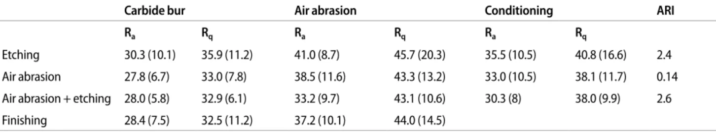
Table 1. Roughness (Ra, Rq) and standard deviation (in parenthesis) for all groups. The column ”Conditioning” provides the combined values
ac-cording to the conditioning technique and the row ”Finishing” to the method chosen for adhesive removal. ARI scores are shown for all three con-ditioning methods. With the exception of the ARI scores for air abrasion alone compared to the other concon-ditioning methods, none of the differenc-es were significant
Tabelle 1. Rauhigkeit (Ra, Rq) und Standardabweichung (in Klammern) für alle Gruppen. In der Spalte „Conditioning” (Schmelzkonditionierung)
sind die kombinierten Werte je nach Vorbehandlung angegeben, in der Spalte „Finishing“ je nach der zur Adhäsiventfernung verwendeten Meth-ode. Für alle drei Konditionierungsmethoden sind ARI („adhesive remnant index“)-Scores angegeben. Mit Ausnahme des ARI-Scores für die allein-ige Air Abrasion im Vergleich mit den anderen Methoden ergaben sich keinerlei signifikante Unterschiede
Carbide bur Air abrasion Conditioning ARI
Ra Rq Ra Rq Ra Rq
Etching 30.3 (10.1) 35.9 (11.2) 41.0 (8.7) 45.7 (20.3) 35.5 (10.5) 40.8 (16.6) 2.4 Air abrasion 27.8 (6.7) 33.0 (7.8) 38.5 (11.6) 43.3 (13.2) 33.0 (10.5) 38.1 (11.7) 0.14 Air abrasion + etching 28.0 (5.8) 32.9 (6.1) 33.2 (9.7) 43.1 (10.6) 30.3 (8) 38.0 (9.9) 2.6 Finishing 28.4 (7.5) 32.5 (11.2) 37.2 (10.1) 44.0 (14.5)
After debonding the brackets, the ARI scores revealed significant differences between the samples pre-conditioned with air abrasion and those with etching. Of the 14 samples in the air abrasion group 13 had an ARI score of 0 (no adhesive left on the enamel) and only one a score of 2 (>50% adhesive left on the enamel). The ARI scores of the two remaining groups were similarly distributed and no sig-nificant differences were observed. Most etched samples showed various amounts of residual adhesive (which usually constituted more than 50% of the resin); only two samples revealed less than that amount.
Mean surface roughness (Ra) and root mean-square roughness
(Rq) were calculated for all subgroups. All subgroups revealed a
normal distribution and no statistically significant differences. The mean Ra for all subgroups was 33.1 (SD 5.5) and Rq 38.9 (SD 5.7).
For the three pre-conditioning methods, the differences between the mean Ra/Rq values for air abrasion (33/38.1), etching (35.5/40.8)
and the combination of air abrasion and etching (30.3/38) did not statistically differ. The two enamel-removal methods did not reveal a statistical significance either. Ra and Rq values were 28.4/37.2 for
the carbide bur and 32.5/44 for air abrasion (Table 1).
Visual inspection of the CLSM images yielded obvious differ-ences according to the method of enamel removal. Air abrasion leaves a roughened but relatively homogenous surface (Figure 1a), while the carbide bur leaves a typical wave-like pattern (Figure 1b).
Discussion
It has always been the aim of orthodontists to remove fixed appli-ances without leaving any residual adhesive or damaging the enamel. Most adhesive nowadays is removed with a carbide bur followed by various polishing steps. Many authors have examined air abrasion for enamel conditioning prior to bracket bonding [5, 10, 18, 19, 26, 35, 42]. However, few studies have addressed the ef-fect of air abrasion on adhesive removal [20, 42]. It was this study's aim to evaluate air abrasion and subsequent surface rough-ness as an alternative technique for adhesive removal.
Diego, CA, USA) durchgeführt. Alle Subgruppen wurden auf Nor-malverteilung getestet (Shapiro-Wilk-Test). Die vergleichende Sta-tistik bezüglich Ra und Rq wurde über multiple Bonferroni-Tests für
alle Subgruppen und Konditionierungsmethoden durchgeführt. Die beiden Vorgehensweisen der Schmelzpolitur wurden mit einem ungepaarten t-Test verglichen.
Resultate
Die ARI-Werte, Mittelwerte für Ra und Rq sowie die
entsprechen-den Standardabweichungen sind in Tabelle 1 wiedergegeben. Die ARI-Werte nach Bracketentfernung zeigten signifikante Unterschiede zwischen den Gruppen, die mit Air Abrasion und mit Ätzung vorbehandelt wurden. In der Gruppe mit Air Abrasion zeigten 13 von 14 Proben einen ARI-Wert von 0 (kein Adhäsivrest auf dem Schmelz) und nur eine Probe einen Wert von 2 (mehr als 50% Adhäsivreste auf dem Schmelz). Die ARI-Werte für die ande-ren zwei Gruppen waande-ren ähnlich verteilt, und es bestanden keine signifikanten Unterschiede. Die meisten geätzten Proben wiesen unterschiedliche Anteile an verbliebenem Adhäsiv auf dem Schmelz auf und nur zwei Proben zeigten weniger als 50% auf dem Schmelz verbliebene Adhäsivreste.
In allen Untergruppen zeigte sich eine Normalverteilung für die Oberflächenrauheiten Ra und Rq. Statistisch signifikante
Unter-schiede konnten nicht gefunden werden. Der mittlere Ra-Wert für
alle Untergruppen betrug 33,1 (Standardabweichung 5,5), der mitt-lere Rq-Wert 38,9 (Standardabweichung 5,7). Es bestand kein
statis-tisch signifikanter Unterschied zwischen den Ra/Rq-Werten der
drei Konditionierungsmethoden Air Abrasion (33/38,1), Ätzung (35,5/40,8) und der Kombination aus Air Abrasion und Ätzung (30,3/38). Auch der Vergleich der beiden Methoden zur Adhäsiv-entfernung zeigte keine signifikanten Unterschiede; die Ra/Rq
-Wer-te betrugen 28,4/37,2 für den Karbid-Finierer und 32,5/44 für die Air Abrasion (Tabelle 1).
Die visuelle Überprüfung der CLSM-Bilder ließ deutliche Untschiede zwischen den beiden Methoden der Adhäsiventfernung
er-Figure 1. Surface structure of
enamel (40x magnification) after adhesive removal with air abrasion (a) and carbide bur (b). A uniform rough surface is visible following air abrasion, whereas the carbide bur leaves a typical wavy pattern
Abbildung 1.
Schmelzober-flächenstruktur (Vergr. 40:1) nach Adhäsiventfernung mit Air Abrasion (a) und Karbid-Fi-nierer (b). Eine homogene Oberfläche wurde für die Air Abrasion gefunden, während der Karbid-Finierer ein Wellen-muster hinterließ
Freshly-extracted bovine incisors were used as investigative sam-ples. Unlike bovine dentin, which is not recommended as a substi-tute for human dentin [37], bovine enamel very closely resembles human enamel and is frequently used in research [17, 18, 22, 24, 42], as suggested by the ISO 11405/TS recommendation.
The ARI scores correlated closely to the pre-conditioning meth-od. A low ARI score (as associated with air abrasion) would be ben-eficial at the debonding stage from a clinical point of view, as very little adhesive remains on the tooth surface to remove. However, the advantage of a low ARI score is compromised by lower bond strengths [5, 42].
The confocal laser scanning microscope (CLSM) used to measure and visualize the enamel surfaces had a reproducible accuracy of 5 μm in the z-axis, which describes the depth of the surface roughness. Accuracy was considered sufficient regarding the values of Ra and Rq
between 28 and 45 and standard deviation of 5.5 for Ra and 5.7 for Rq.
In addition, CLSM allowed to compute three-dimensional (3D) im-ages, which could be viewed in 3D using special glasses. This was very useful for visually inspecting the areas scanned, although a two-dimensional representation was chosen for this publication (Figure 1), as the fundamental difference between enamel removal with a carbide bur and air abrasion is still apparent in two dimensions. The surface of teeth treated with air abrasion was very homogenous, whereas the surface after use of a carbide bur showed a regular wave pattern previously described in the literature [7, 12, 32, 44, 46]. This is most likely due to unequal pressure during adhesive removal or a slightly excentric rotation of the carbide burs.
The Ra and Rq values of the two adhesive removal methods were
similar and revealed no statistical differences. This finding was sur-prising, as we expected the opaque surface after air abrasion to show increased roughness. Previous publications have supported our finding of equal roughness values in association with both tech-niques [8, 20]. The roughness values found in this study are higher than those previously described [8, 20]. Thus the conditioning method did not play a decisive role in the degree of roughness after debonding. Although surface roughness did not play an additive role, enamel loss certainly must have. Thus, careful conditioning is mandatory to maintain an intact enamel surface.
When enamel loss via the debonding procedure is considered, the study's roughness values lie within the range of enamel loss de-scribed in the literature [1, 9, 13, 21, 30, 42]. One could argue that the final enamel-surface polish leads to a reduction in roughness equaling the arithmetic mean of all the individual enamel surface roughnesses.
The results of this study support both techniques of adhesive re-moval in relation to surface roughness, as no differences were de-tected. However, there is the potential risk of inhaling increased amounts of ambient dust from air abrasion. Conditioning with air abrasion alone led to low ARI scores and may be associated with a higher rate of bracket failure.
Conclusion
– The confocal laser scanning microscope is a useful tool for measuring 3D surface structures.
kennen: Die Air Abrasion hinterließ eine raue, aber homogene Oberfläche (Abbildung 1 a), der Karbid-Finierer dagegen führte zu einer Wellenstruktur (Abbildung 1 b).
Diskussion
Die Adhäsiventfernung nach kieferorthopädischer Therapie sollte möglichst ohne Verletzung der Schmelzoberfläche erfolgen. Am häufigsten wird heute zur Adhäsiventfernung ein Karbid-Finierer angewandt, gefolgt von diversen Politurschritten. Air Abrasion wurde zwar mehrfach als Methode zur Konditionierung der Schmelzoberfläche untersucht [5, 10, 18, 19, 26, 35, 42], allerdings befassten sich nur wenige Studien mit dem Effekt der Air Abra-sion auf die Adhäsiventfernung [20, 42]. Ziel der hier vorgestellten Studie war die Evaluation der Oberflächenrauheit verschiedener Schmelzkonditionierungen in Kombination mit unterschiedlichen Methoden der Adhäsiventfernung.
Als Probenmaterial wurden frisch extrahierte bovine Inzisivi verwendet. Während bovines Dentin nicht als Ersatz für Human-dentin verwendet werden sollte [37], entspricht boviner Schmelz weitgehend dem humanen und wurde häufig für Forschungszwe-cke eingesetzt [17, 18, 22, 24, 42]. Dies entspricht der Empfehlung ISO 11405/TS.
Die ARI-Werte zeigten eine enge Korrelation mit den Konditio-nierungsmethoden. Klinisch betrachtet ist der bei der Air Abrasion gefundene tiefe ARI-Wert beim Debonding vorteilhaft, weil wenig Adhäsiv auf dem Schmelz verbleibt. Der Vorteil beruht allerdings auf einer Verminderung der Adhäsion zum Schmelz, was zu ver-mehrten Bracket-Verlusten führen kann [5, 42].
Das CLSM, das für die Messungen und Visualisierungen der Schmelzoberflächen verwendet wurde, zeigte eine reproduzierbare Genauigkeit von 5 μm in der Z-Achse, welche die Tiefe der Ober-flächenrauheit beschreibt. In Relation zu den gemessenen Ra/Rq
-Werten zwischen 28 und 45 und den Standardabweichungen von 5,5 und 5,7 wurde die Genauigkeit als hinreichend eingestuft. Zu-dem erlaubte die Verwendung des CLSM die Erstellung dreidimen-sionaler Bilder, die mit 3D-Gläsern betrachtet werden konnten. Für die visuelle Interpretation der gescannten Bereiche war dies vorteil-haft, obwohl für diese Publikation eine zweidimensionale Darstel-lung gewählt wurde (Abbildung 1), da der grundlegende Unter-schied zwischen der Adhäsionsentfernung mit dem Karbid-Finie-rer und der Air Abrasion auch zweidimensional noch offensichtlich ist. Während die Zahnoberflächen nach Air Abrasion sehr homo-gen wirkten, zeigten sie nach Politur mit dem Karbid-Finierer ein regelmäßiges Wellenmuster, wie es schon früher beschrieben wur-de [7, 12, 32, 44, 46]. Es wird vermutet, dass dies durch ungleich-mäßigen Druck und leicht exzentrische Rotation der Karbid-Finie-rer zustande kam.
Die Ra- und Rq-Werte für beide Methoden der
Adhäsiventfer-nung zeigten ähnliche Werte ohne signifikante Unterschiede. Dies war erstaunlich, da die opake Oberfläche nach Air Abrasion mak-roskopisch deutlich rauer schien. Die Ähnlichkeit beider Methoden in Hinblick auf die Schmelzoberflächenrauheit bestätigt die Anga-ben in früheren Studien [8, 20], wobei die Rauheitswerte in der hier vorgestellten Studie insgesamt höher lagen. Die
Konditionierungs-– with low scores in association with air abrasion and high scores for both methods involving etching.
– A surface roughness of 30 can be expected with both tech-niques, and none of our three groups revealed any statistical dif-ferences.
– Visual inspection of the debonded areas under 40x magnifica-tion showed a wave pattern of enamel removal with the carbide bur and a more homogeneous surface following air abrasion. – Due to its efficiency and easy manageability, we recommend
re-moval with the carbide bur.
Conflict of interest
The corresponding author states that there are no conflicts of in-terest.
technik stellte sich als unbedeutend für die Schmelzoberflächen-rauheit nach Debonding heraus. Wenngleich die Rauheit nicht ad-ditiv schien, so muss für den Schmelzverlust davon ausgegangen werden. Insofern ist auch die schonende Konditionierung für die Intaktheit der Schmelzoberfläche von Bedeutung.
Betrachtet man den Schmelzverlust während des Debonding, so kann festgehalten werden, dass die durchschnittlichen Rauheitstie-fen in der vorgestellten Studie mit dem in der Literatur beschriebe-nen Schmelzverlust bei Debonding übereinstimmten [1, 9, 13, 21, 30, 42]. Demzufolge reduziert die abschließende Politur der Schmelzoberfläche die verbleibenden Rauheiten ungefähr um das arithmetische Mittel der einzelnen Schmelzoberflächenrauheiten.
Die Ergebnisse der hier vorgestellten Studie erlauben den klini-schen Einsatz der geschilderten Methoden gleichermaßen, da sich keine Unterschiede in der Rauheit ergaben. Allerdings muss bei Kunststoffentfernung mittels Air Abrasion auf ein potenzielles Risi-ko durch Einatmung erhöhter intraoraler Mengen an Strahlguts-taub hingewiesen werden. Die Konditionierung mit alleiniger Air Abrasion hob sich aufgrund der niedrigen ARI-Werte von den an-deren Verfahren ab und könnte zu erhöhten Bracket-Verlustraten führen.
Schlussfolgerung
– Das CLSM erwies sich als sinnvolles Messgerät für die 3D-Oberflächendarstellung.
– Die ARI-Werte korrelierten gut mit den Konditionierungsme-thoden. Es ergaben sich niedrige Werte für die Air Abrasion und hohe Werte für die Methoden, die einen Ätzvorgang be-inhalteten.
– Beide Techniken der Adhäsiventfernung wiesen eine Oberflä-chenrauheit von etwa 30 auf; es bestanden keine statistisch sig-nifikanten Unterschiede.
– Die visuelle Evaluation der Bereiche nach Schmelzentfernung mit 40-facher Vergrößerung zeigten ein wellenförmiges Muster für den Karbid-Finierer und eine homogene Oberfläche für die Air Abrasion.
– Aufgrund der höheren Effizienz wird der Karbid-Finierer als Methode zur Entfernung von Adhäsivresten empfohlen.
Interessenkonflikt
Der korrespondierende Autor gibt an, dass kein Interessenkonflikt besteht.
References
1. Al Shamsi AH, Cunningham JL, Lamey PJ, Lynch E (2007) Three-dimensional measurement of residual adhäsive and enamel loss on teeth after debonding of orthodontic brackets: An in-vitro study. Am J Orthod Dentofacial Orthop 131:301.e9–301.e15
2. Artun J, Bergland S (1984) Clinical trials with crystal growth as an alternative to acid-etch enamel pretreatment. Am J Orthod 85:333–340
3. Burapavong V, Marshall GW, Apfel DA, Perry HT (1978) Enamel surface charac-teristics on removal of bonded orthodontic brackets Am J Orthod 74:176–187 4. Campbell PM (1995) Enamel surfaces after orthodontic bracket debonding.
Angle Orthod 65:103–110
5. Canay S, Kocadereli I, Akça E (2000) The effect of enamel air abrasion on the retention of bonded metallic orthodontic brackets. Am J Orthod Dentofacial Orthop 117:15–19
6. Crain G, Sheridan JJ (1990) Susceptibility to caries and periodontal disease after posterior air-rotor stripping. J Clin Orthod 24:84–85
7. Diedrich P (1981) Enamel alterations from bracket bonding and debonding: A study with the scanning electron microscope. Am J Orthod 79:500–522 8. Eliades T, Gioka C, Eliades G, Makou M (2004) Enamel surface roughness
follo-wing debonding using two resin grinding methods. Eur J Orthod 26:333–338 9. Fitzpatrick DA, Way DC (1977) The effects of wear and etching and bond
remo-val on human enamel. Am J Orthod 72:671–681
10. Goldstein RE, Parkins FM (1995) Using air-abrasive technology to diagnose and restore pit and fissure caries. J Am Dent Assoc 126:761–766
11. Gwinnett AJ, Gorelick L (1977) Microscopic evaluation of enamel after debon-ding: Clinical application. Am J Orthod 71:651–665
12. Hong YH, Lew KK (1995) Quantitative and qualitative assessment of enamel surface following five composite removal methods after bracket debonding. Eur J Orthod 17:121–128
13. Hosein I, Sherriff M, Ireland AJ (2004) Enamel loss during bonding, debon-ding, and cleanup with use of a self-etching primer. Am J Orthod Dentofacial Orthop 126:717–724
14. Howell S, Weekes WT (1990) An electron microscopic evaluation of the ena-mel surface subsequent to various debonding procedures. Aust Dent J 35:245–252
15. Ireland AJ, Hosein I, Sheriff M (2005) Enamel loss at bond-up, debond and clean-up following the use of conventional light cured composite and resin modified glass polyalkenoate cement. Eur J Orthod 27:413–419
16. Jarjoura K, Gagnon G, Nieberg L (2006) Caries risk after interproximal enamel reduction. Am J Orthod Dentofacial Orthop 130:26–30
17. Jost-Brinkmann PG (1998) The influence of air polishers on tooth enamel. J Orofac Orthop 59:1–16
18. Jost-Brinkmann PG, Drost C, Can S (1996) In vitro study of the adhesive strengths of brackets on metal, ceramic and composite. J Orofac Orthop 57:76–87
19. Katora ME, Jubach T, Polimus MM (1981) Airabrasive etching of the enamel surface. Quintessence 9:967–968
20. Kim SS, Park WK, Son WS et al (2007) Enamel surface evaluation after removal of orthodontic composite remnants by intraoral sandblasting: A 3-dimensio-nal surface profilometry study. Am J Orthod Dentofacial Orthop 132:71–76 21. Krell KV, Courey JM, Bishara SE (1993) Orthodontic bracket removal using
con-ventional and ultrasonic debonding techniques, enamel loss, and time requi-rements. Am J Orthod Dentofacial Orthop 103:258–266
22. Levinkind M, Vandernoot TJ, Elliott JC (1983) Electrochemical impedance cha-racterization of human and bovine enamel. J Dent Res 69:1806–1811 23. McDowell GC, Bloem TJ, Lang BR, Asgar K (1988) In vivo wear. Part I: The
Michi-gan computer-graphic measuring system. J Prosthet Dent 60:112–120 24. Nakamichi I, Iwaku M, Fusayama T (1990) Bovine teeth as possible substitutes
25. Oliver RG, Griffiths J (1992) Different techniques of residual composite remo-val following debonding – time taken and surface enamel appearance. Br J Orthod 19:131–137
26. Olsen ME, Bishara SE, Damon P, Jakobson JR (1997) Comparison of shear bond strength and surface structure between conventional acid etching and air-abrasion of human enamel. Am J Orthod Dentofacial Orthop 112:502–506 27. Olsen ME, Bishara SE, Damon P, Jakobson JR (1997) Evaluation of scotchbond
multipurpose and maleic acid as alternative methods of bonding orthodontic brackets. Am J Orthod Dentofacial Orthop 111:498–501
28. Osorio R, Toledano M, Garcia-Godoy F (1999) Bracket bonding with 15- or 60 second etching and adhesive remaining on enamel after debonding. Angle Orthod 69:45–48
29. Osorio R, Toledano M, Garcia-Godoy F (1998) Enamel surface morphology af-ter bracket debonding. ASDC J Dent Child 65:313–317
30. Pus MD, Way DC (1980) Enamel loss due to orthodontic bonding with filled and unfilled resins using various clean-up techniques. Am J Orthod 77:269– 283
31. Quick DC, Holtan JR, Ross GK (1992) Use of a scanning laser three-dimensional digitizer to evaluate dimensional accuracy of dental impression materials. J Prosthet Dent 68:229–235
32. Radlanski RJ (2001) A new carbide bur for bracket debonding. J Orofac Orthop 62:296–304
33. Radlanski RJ, Jäger A, Zimmer B, Bertzbach F (1990) Scanning electron micro-scopic studies on the clinical use of interdental stripping. J Orofac Orthop 51:117–122
34. Reicheneder C (1992) Bracketentfernung mit Hilfe verschiedener Methoden und daraus resultierende Schmelzläsionen: eine rasterelektronenmikroskopi-sche Studie. Med Dissertation, Ludwig Maximilian University, Munich 35. Reisner KR, Levitt HL, Mante FK (1997) Enamel preparation for orthodontic
bonding: a comparison between the use of a sandblaster and current techni-ques. Am J Orthod Dentofacial Orthop 111:366–373
36. Retief DH, Denys FR (1979) Finishing of enamel surfaces after debonding of orthodontic attachments. Angle Orthod 49:1–10
37. Retief DH, Mandras RS, Russel CM, Denys FR (1990) Extracted human versus bovine teeth in laboratory studies. Am J Dent 3:253–258
38. Rouleau BD, Marshall GW Jr, Cooley RO (1982) Enamel surface evaluations af-ter clinical treatment and removal of orthodontic brackets. Am J Orthod 81:423–426
39. Roulet JF, Mettler P, Friedrich U (1980) The abrasion of composites in the regi-on of the lateral teeth – results after 3 years. Dtsch Zahnarztl Z 35:493–497
40. Sargison AE, McCabe JF, Millet DT (1999) A laboratory investigation to compa-re enamel pcompa-reparation by sandblasting or acid etching prior to bracket bon-ding. Br J Orthod 26:141–146
41. VanWaes H, Matter T, Krejci I (1997) Three dimensional measurement of ena-mel loss caused by bonding and debonding of orthodontic brackets. Am J Orthod Dentofacial Orthop 112:666–669
42. Van Waveren Hogervorst WL, Feilzer AJ, Prahl-Andersen B (2000) The air-ab-rasion technique versus the conventional acid-etching technique: A quantifi-cation of surface enamel loss and a comparison of shear bond strength. Am J Orthod Dentofacial Orthop 117:20–26
43. Yamada R, Hayakawa T, Kasai K (2002) Effect of using self-etching primer for bonding orthodontic brackets. Angle Orthod 72:558–564
44. Zachrissson BU, Arthun J (1979) Enamel surface appearance after various de-bonding techniques. Am J Orthod 75:121–137
45. Zachrisson BU, Nyøygaard L, Mobarak K (2007) Dental health assessed more than 10 years after interproximal enamel reduction of mandibular anterior teeth. Am J Orthod Dentofacial Orthop 131:162–169
46. Zarrinia K, Eid NM, Kehoe MJ (1995) The effect of different debonding techni-ques on the enamel surface: An in vitro qualitative study. Am J Orthod Dento-facial Orthop 108:284–293
Correspondence address
Dr. Dr. Lorenz M. Brauchli
Klinik für Kieferorthopädie und Kinderzahnmedizin Hebelstr. 3
4056 Basel Schweiz
Phone: (+41/61) 26726-41, Fax: -57 e-mail: [email protected]