CASE REPORT
Inflammatory myofibroblastic tumor of the lung: a rare cause
of atelectasis in children
Amira Dhouib&Constance Barrazzone&
Alexandra Reverdin&Mehrak Anooshiravani&
Sylviane Hanquinet
Received: 8 March 2012 / Revised: 12 June 2012 / Accepted: 19 July 2012 / Published online: 13 November 2012 # Springer-Verlag 2012
Abstract Although rare, inflammatory myofibroblastic tu-mor is the most common primary lung mass in children. We report the case of an 11-year-old boy investigated for persis-tent cough and dyspnea with complete left lung atelectasis mimicking pneumonia. CT and MRI showed an endobron-chial mass of the left main bronchus. The boy underwent endoscopic resection of the tumor and histology was in favor of an inflammatory myofibroblastic tumor of the lung. This diagnosis should be suspected in children with recurrent pneumonia. The prognosis is good after complete resection.
Keywords Inflammatory myofibroblastic tumor . Atelectasis . Children
Introduction
Inflammatory myofibroblastic tumor (IMT) of the lung is a rare benign tumor first described by Brunn in 1939, as cited by
Jeba et al. [1]. It belongs to the group of soft-tissue tumors and is also called inflammatory pseudotumor, fibrous histiocy-toma, plasma cell granuloma, plasmocyhistiocy-toma, solitary mast cell tumor, pseudoneoplastic pneumonia or fibroxanthoma.
IMT can involve any anatomical location, but the lung is the most commonly affected site in children as well as in adults. The other anatomical locations are head and neck, mesentery, liver and bladder. In children, IMT is the most common primary lung mass that can behave like malignant tumors. Clinical presenta-tion and radiologic findings are variable and not specific. Diag-nostic confirmation is based on histological examination.
Case report
An 11-year-old otherwise healthy boy had a 2-month history of dyspnea and coughs and was treated for asthma with no clinical improvement. One month after the onset of respira-tory symptoms, he developed fever and the cough became persistent. Chest radiograph showed left lower lobe atelec-tasis. The diagnosis of pneumonia was established and anti-biotherapy prescribed. One month later, he was referred to our emergency department because of unsatisfactory clinical course with complete left lung radiologic atelectasis (Fig.1). White blood cell count, blood chemistry and CRP level were unremarkable. Tuberculin skin test was negative. Chest US showed pulmonary consolidation without signif-icant pleural effusion. Chest CT was performed on a 16-slice machine (GE LightSpeed, Waukesha, WI, USA) after intra-venous 2 ml/kg injection of a 300 mgI/ml contrast medium (CTDI of 6.44 mGy). Besides total atelectasis of the left lung with mediastinal shift to the left and compensatory overinflation of the right lung, CT showed a well-delineated homogeneous mass of 1.8× 1.2× 1.7 cm in the left main bronchus (Fig. 2). This lesion completely obstructed the
A. Dhouib
:
M. Anooshiravani:
S. HanquinetDepartment of Pediatric Radiology, University Children’s Hospital
Geneva,
Geneva, Switzerland C. Barrazzone
Department of Pediatric Pneumology, University Children’s Hospital Geneva,
Geneva, Switzerland A. Reverdin
Department of Pneumology, Clinique La Tour, Geneva, Switzerland
A. Dhouib (*)
Unité de Radiopédiatrie, Hôpital Universitaire des Enfants, 6 rue Willy Donzé,
1211, Genève 14, Suisse e-mail: amira.dhouib@hcuge.ch Pediatr Radiol (2013) 43:381–384 DOI 10.1007/s00247-012-2508-x
main bronchus and was not enhanced by contrast material. Chest MRI was performed on a 1.5-T (Magnetom Avanto, Siemens, Erlangen, Germany) and imaging protocol
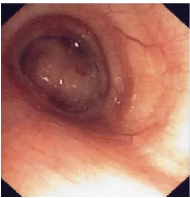
included Balanced Steady-State Free Precession (bSSFP), coronal T1-weighted turbo spin-echo sequences (TR/TE 641/22 ms), axial and coronal T2-weighted turbo spin echo (TR/TE 1,000/80 ms) and fat-suppressed T1-weighted spin-echo sequences after intravenous administration of gadobutro-lum (Gadovist, Bayer, Zurich, Switzerland) at a dose of 0.1 mmol/kg body weight. MRI confirmed the presence of a homogeneous mass in the left main bronchus with high signal intensity on T2- and T1-weighted images with no significant contrast enhancement (Fig. 3). Whole-body MRI was also performed and the protocol included 3-D inversion recovery (inversion time0160 ms) T2-weighted turbo spin-echo (SPACE, TR/TE 2,000/151 ms, isotropic voxel dimensions, 1.4 mm), coronal T1-weighted spin-echo sequences (TR/TE 550/11) and diffusion. No other lesion was found. On [F-18]-fluoro-2-deoxyglucose (FDG) PET/CT (Biograph 16; Siemens, Erlangen, Germany), this mass showed increased metabolism (SUV max 21.8) (Fig.4). No other hypermetabolic lesions were found on PET/CT. Lung volumes were markedly decreased with a restrictive syndrome (total lung capacity 55% of pre-dicted) on spirometric studies. Bronchoscopy visualized the endobronchial tumor, which was obstructing incompletely the distal end of the left main bronchus (Fig.5). The polypoid shape
Fig. 1 The chest radiograph shows complete left lung atelectasis with compensatory overinflation of the right lung
Fig. 2 Contrast-enhanced and reformatted coronal (a) and axial (b) CT images show an obstructive homogeneous, well-circumscribed and
non-enhanced mass in the left main bronchus (arrows)
Fig. 3 Axial T2-weighted MR image shows a well-delineated mass
(arrow) with high signal intensity in the left main bronchus and
atelectasis of the left lung
Fig. 4 Fused coronal PET/CT image demonstrates intense 18F-FDG
uptake in the pulmonary mass (arrow)
of the tumor allowed total endoscopic resection with a diather-mic snare combined with argon plasma coagulation and no complications. Histological analysis revealed a mesenchymal tumor more commonly called inflammatory myofibroblastic tumor. Cytogenetic analysis confirmed this diagnosis with pos-itive characteristic rearrangement at 2p23.9 of the gene coding for the anaplastic lymphoma kinase receptor tyrosine kinase (found in 50% of IMT cases). Chest radiographs after resection showed complete resolution of the atelectasis. Spirometry showed normal total lung capacity, and repeated bronchoscopies at 1-month and 3-month follow-ups were normal.
Discussion
Most of the causes of atelectasis in childhood are related to airway obstruction. Intrabronchial obstruction may be exog-enous due to foreign body, or to mucous plugs. Endobron-chial tumors are a rare cause of atelectasis.
IMT is a rare lesion; it belongs to the group of soft-tissue tumors and has also been called inflammatory pseudotumor, fibrous histiocytoma, plasma cell granuloma, plasmocytoma, solitary mast cell tumor, pseudoneoplastic pneumonia, and fibroxanthoma. This is probably due to its predominant cellular component. It can involve any anatomical location; however, the lung is the most commonly affected site [2]. Other frequent locations include the bladder, mesentery, omentum, retroperito-neum, pelvis, gastrointestinal tract, liver, spleen, kidney and pancreas. Some tumors can be multifocal and involve several organs. The true incidence of IMT of the lung is difficult to establish because of various nomenclatures used. However, it is relatively rare among all primary lung neoplasms, accounting for only 0.04%–0.7% of them [3]. According to many series,
IMT is the most common primary childhood lung tumor, with an incidence ranging from 12% to 57% [2]. There is a near equal male/female occurrence [2,4].
Pathophysiologically, an unregulated proliferation of in-flammatory cells seems to be present [3]. The cause of this inflammatory response remains unknown. It has been hypoth-esized that an initial infection leads to IMT, and about 30% of patients have had a history of respiratory infection. In a case report Fiocchi et al. [5] described a direct progression from infectious disease to an IMT. Inflammatory lesions are thought to occur locally as a result of excessive response to tissue damage [6]. Others believe it to be a real neoplasm induced by genic reorganizations on the chromosome 23 [7].
Most frequent symptoms are cough, hemoptysis, chest pain, dyspnea and fever. It may also be asymptomatic [7].
Radiologic findings are variable and unspecific. The most common radiologic presentation is a solitary peripheral well-circumscribed lung mass, with variable size between 1.5 cm and 14 cm according to a series of 28 cases reported by Kim et al. [8]. The endobronchial polypoid forms arising in the tra-chea or the bronchus are the rarest but have also been reported, as in our case. They can show atelectasis and obstructive pneumonia and can spread into the mediastinum.
Contrast-enhanced CT or MRI is necessary to view the endobronchial mass that is not visible on the chest radiograph. CT and MRI allow a better characterization and delineation of the tumor. Contrast-enhanced CT shows a homogeneous or heterogeneous attenuated mass. Intralesional calcifications are possible but rare, and are shown more frequently in pediatric cases [4,7]. IMTs are also known to rarely form a cavity. In such cases, MRI shows intermediate signal intensity on T1-weighted images and high signal intensity on T2-T1-weighted images. Absent or delayed enhancement with contrast injec-tion on CT or MRI is described in the literature. Takayama et al. [4] reported two cases of IMT that were intra-pulmonary well circumscribed masses. Dynamic contrast enhancement CT in both cases showed delayed enhancement. They specu-late that the delayed enhancement could be attributed to abundant fibrous tissues such as myofibroblasts [4]. High fixation of 18F-FDG of IMT on PET has been described [7] and may be used for assessment of multifocal forms.
Concerning transbronchial biopsy and transthoracic needle biopsy, Cerfolio et al. [2] believe that they often lead to confusion between IMT and fibrohistiocytic neoplasms, nodular sclerosing Hodgkin lymphoma, primary lung cancer and mediastinal fibro-sis. Indeed, histological findings of a fragment cannot exclude a neoplasm with surrounding inflammation. Complete resection for both diagnosis and treatment is mandatory.
While inflammatory myofibroblastic tumor is a rare endo-bronchial tumor, it should be known as a possible cause of atelectasis in children and distinguished from a malignancy. The treatment is surgical, and a favorable prognosis relies on complete resection.
Fig. 5 Endoscopic view of the left main bronchus shows an obstruc-tive endobronchial mass
References
1. Jeba J, John S, Backiyanathan S et al (2008) Inflammatory pseudo-tumour of the lung with sarcomatous brain metastasis. Eur J Cancer Care (Engl) 17:412–414
2. Cerfolio RJ, Allen MS, Nascimento AG et al (1999) Inflammatory pseudotumors of the lung. Ann Thorac Surg 67:933–936
3. Rasalkar DD, Chu WC, To KF et al (2010) Radiological appearance of inflammatory myofibroblastic tumour. Pediatr Blood Cancer
54:1029–1031
4. Takayama Y, Yabuuchi H, Matsuo Y et al (2008) Computed tomographic and magnetic resonance features of inflammatory
myofibroblastic tumor of the lung in children. Radiat Med
26:613–617
5. Fiocchi A, Mirri GP, Santini I et al (1999) Progression from bron-chopneumonia to inflammatory pseudotumor in a seven-year-old girl. Pediatr Pulmonol 27:138–140
6. Ochs K, Hoksch B, Frey U et al (2010) Inflammatory myofibro-blastic tumour of the lung in a five-year-old girl. Interact Cardiovasc Thorac Surg 10:805–806
7. Hantous-Zannad S, Esseghaier S, Ridene I et al (2009) Inflamma-tory myofibroblastic tumors of the lung: imaging features. J Radiol
90:1851–1855
8. Kim JH, Cho JH, Park MS et al (2002) Pulmonary inflammatory
pseudotumor—a report of 28 cases. Korean J Intern Med 17:252–258