Review Article
Skin bioavailability of dietary vitamin E, carotenoids, polyphenols, vitamin C,
zinc and selenium
Myriam Richelle*, Magalie Sabatier, Heike Steiling and Gary Williamson
Nestle´ Research Center, Nestec Ltd, PO Box 44, CH-1000 Lausanne 26, Switzerland (Received 30 August 2005 – Revised 1 March 2006 – Accepted 29 March 2006)
Dietary bioactive compounds (vitamin E, carotenoids, polyphenols, vitamin C, Se and Zn) have beneficial effects on skin health. The classical route of administration of active compounds is by topical application direct to the skin, and manufacturers have substantial experience of formu-lating ingredients in this field. However, the use of functional foods and oral supplements for improving skin condition is increasing. For oral consumption, some dietary components could have an indirect effect on the skin via, for example, secondary messengers. However, in the case of the dietary bioactive compounds considered here, we assume that they must pass down the gastrointestinal tract, cross the intestinal barrier, reach the blood circulation, and then be distributed to the different tissues of the body including the skin. The advantages of this route of admin-istration are that the dietary bioactive compounds are metabolized and then presented to the entire tissue, potentially in an active form. Also, the blood continuously replenishes the skin with these bioactive compounds, which can then be distributed to all skin compartments (i.e. epidermis, dermis, subcutaneous fat and also to sebum). Where known, the distribution and mechanisms of transport of dietary bioactive compounds in skin are presented. Even for compounds that have been studied well in other organs, information on skin is relatively sparse. Gaps in knowledge are identified and suggestions made for future research.
Bioavailability: Skin: Vitamin E: Tocopherol: Vitamin C: Ascorbate: Carotenoid: Polyphenol: Zinc: Selenium
The skin is the largest organ in man, consisting of different layers of epidermis and dermis. Its primary physiological function is that of a barrier between the body and the external environment, protecting against mechanical damage, radiation, toxic com-pounds and micro-organisms. Skin plays a role in the regulation of body temperature and is involved in body water homeostasis. Skin is constantly exposed to pro-oxidant environmental stresses from an array of sources, such as air pollutants, solar UV light, chemical oxidants, micro-organisms, cigarette smoke and ozone (Thiele et al. 1997; Cross et al. 1998). Reac-tive oxygen species have been implicated in the aetiology of several skin disorders including skin cancer and photoageing (Perchellet & Perchellet, 1989; Dalle & Pathak, 1992; Emerit, 1992; Guyton & Kensler, 1993). These reactive oxygen species are capable of oxidizing lipids, proteins or DNA leading to the formation of oxidized products such as lipid hydroperoxides, protein carbonyls or 8-hydroxyguano-sine, respectively (Beehler et al. 1992; Hu & Tappel, 1992; Podda et al. 1998). Reactive oxygen species are generated constantly in skin, and are rapidly neutralized by non-enzy-matic and enzynon-enzy-matic antioxidant substances, which prevent their harmful effects and maintain a pro-oxidant – antioxidant
balance, resulting in cell and tissue stabilization. If the antiox-idant defence is exhausted, cell damage can occur. Known non-enzymatic scavengers of free radicals in human skin are b-carotene, vitamin C and vitamin E, and enzymatic scaven-gers are Se-dependent gluthathione peroxidases, Cu/Zn-super-oxide dismutase, Mn-superCu/Zn-super-oxide dismutase and catalase (Steenvoorden & van Henegouwen, 1997; Thiele et al. 2000). In recent years, particular antioxidants have gained considerable attention as a means to neutralize reactive oxygen species (Mukhtar & Ahmad, 1999). Green tea poly-phenols (Katiyar & Mukhtar, 1997), resveratrol (Jang et al. 1997), curcumin (Stoner & Mukhtar, 1995), ginger (Katiyar et al. 1996) and diallyl sulfide (Sadhana et al. 1988; Perchellet et al. 1990) afford protection against the development of skin cancer, both in vitro (in culture systems) as well as in vivo (in animal models). Additionally, diets rich in bioactive com-pounds such as vitamins C and E, b-carotene, lycopene, Zn and Se have also demonstrated a photoprotective effect against solar irradiation in human subjects (Gollnick et al. 1996; Fuchs, 1998; Fuchs & Kern, 1998; McKenzie, 2000; Stahl et al. 2000, 2001; Greul et al. 2002; Rostan et al. 2002; Stahl & Sies, 2002; Cesarini et al. 2003).
* Corresponding author: Dr M. Richelle, fax þ 41 21 785 85 44, email myriam.richelle@rdls.nestle.com
Abbreviations: DCT1, Hþ-coupled divalent cation transporter; hZIP4, human zrt-,irt-like protein; hZTL1, human zinc T-like transporter; MMP, matrix metalloproteinase; OATP, organic anion transporting polypeptide.
An increase in cellular antioxidants in skin is observed after the exogenous administration of antioxidant compounds. In the case of the skin, the classical route of antioxidant adminis-tration is topical application. However, this topical route of administration can be achieved efficiently only if the particular antioxidant is stable in the preparation as well as on skin, is able to penetrate the skin and is present in its active form (i.e. possible metabolites). In addition, penetration of antioxidants into the skin is influenced by environmental factors, such as tem-perature, hydration and the presence of other chemicals. Another means to deliver antioxidants to the skin is through oral admin-istration via the diet and dietary supplements. In this case, anti-oxidants cross the intestinal barrier and reach the blood circulation from where they are distributed to different tissues; specifically for skin to subcutaneous adipose tissue, dermis, epi-dermis and sebum. The advantages of this oral administration are that antioxidants are metabolized and then presented to the entire skin potentially in their active forms. In addition, the blood continuously replenishes the skin with these antioxidants, which are distributed to all skin compartments in which they could exert a biological activity.
In order to be active in skin, dietary bioactive compounds must be able to cross the intestinal barrier and reach the blood circulation. This step could be a limiting factor of the efficacy of these dietary bioactive compounds in skin. The present paper reviews current knowledge on the journey of dietary bioactive compounds from the mouth to skin with a special focus on antioxidants such as vitamin C, vitamin E, carotenoids, polyphenols, Zn and Se.
For the purposes of the present review, we have considered dietary compounds that have some ‘antioxidant’ properties, either direct or indirect. Because this group includes antioxi-dant vitamins (C and E), phytochemicals (carotenoids and polyphenols) and minerals (Zn and Se), we have grouped these compounds under the term ‘dietary bioactives’. Definition of bioavailability
Bioavailability is defined here by the relative amount of a diet-ary bioactive consumed that crosses the intestinal barrier,
reaches the blood circulation and is available for metabolic processes or storage in the body; in this context, the skin. Bioavailability comprises various steps summarized by the acronym LADME. L means liberation of the molecule from the dietary matrix (food or supplement); A means absorption, i.e. transfer of the molecule from the gut lumen into the blood circulation; D refers to distribution of the molecule from the blood circulation into all body tissues (in this case the skin); M takes account of metabolism, consisting of the further pro-cessing of the molecule in the body either in the gastrointesti-nal tract or in various tissues; and E refers to elimination from the body in urine, stools, sweat, tears or expired air.
A brief comment on mechanisms of absorption in the gut Dietary bioactives such as vitamin C, vitamin E, carotenoids (a- and b-carotene, lycopene, lutein, zeaxanthin and b-cryp-toxanthin, astaxanthin, canthaxanthin), polyphenols (hesperi-din, quercetin, rutin, genistein, daidzein, procyanidins, catechins), Zn and Se belong to two main groups: lipid-soluble and water-soluble dietary bioactives (Table 1). The solubility of a dietary bioactive markedly affects its mechanisms of
bioavailability, which are reported in the following
paragraphs.
Lipid-soluble or lipophilic dietary bioactives, e.g. vitamin E and carotenoids, are absorbed by the same pathway. In the stomach, these dietary bioactives are released from the food matrix and transferred into oil droplets, which are transformed in the small intestine into mixed micelles. The micellar solu-bilization of these lipophilic dietary bioactives is mandatory for their absorption; if lipophilic nutrients escape this solubil-ization, they continue their journey in the gastrointestinal tract and are subjected to either microflora metabolism in the colon or elimination. The uptake of lipid-soluble dietary bioactives by enterocytes was long considered a passive mechanism (Hollander et al. 1975; Hollander & Ruble, 1978; Fig. 1), but there is increasing evidence for facilitated, protein-car-rier-mediated transport involving lipid transporters (During et al. 2002, 2005; Reboul et al. 2004; Fig. 2). Once inside
Table 1. Characteristics of dietary bioactive compounds (‘dietary bioactives’)
Solubility
Octanol – water partition coefficient
at pH 7, 378C (Cooper et al. 1997) Class Dietary bioactive Example of food source
Lipid-soluble dietary bioactives 17·62 Carotenoid a- and b-Carotene Carrots, green leaves
17·64 Lycopene Tomato
14·82 Lutein Green leaves
14·95 Zeaxanthin Green leaves, corn
16·08 b-Cryptoxanthin Apples, apricots
13·27 Asthaxanthin Salmon, trout
14·1 Canthaxanthin Salmon, trout, egg yolk
12·2 Vitamin Vitamin E Vegetable oil
Water-soluble dietary bioactives 23·98 Vitamin Vitamin C Citrus, orange
2·44 Polyphenol Hesperidin Orange
1·48 Quercetin Onions 21·11 Rutin Tea 2·53 Genistein Soya 2·55 Daidzein Soya 2·26 Procycanidin Cocoa 2·6 (þ )-Catechin Tea
Trace element Se Cereals, seafood
the enterocyte lining the small intestine, these lipid-soluble dietary bioactives are packed into new oil-droplet structures called prechylomicrons, which are expulsed by exocytosis into the extracellular space, entering the lymphatic system and then reaching the general blood circulation.
In the particular case of water-soluble dietary bioactives, such as polyphenols and vitamin C, they remain in the bulk of the meal and arrive in the small intestine, since most polyphenols are stable in the stomach (Rios et al. 2002; Fig. 3). Many polyphenols are glycosylated in food, i.e. they are linked usually through a b-linkage to one or more glucose, arabinose, galactose or rhamnose residue(s). The glucose, and to a certain extent the arabinose and galac-tose residues, are hydrolysed by endogenous enzymes such as lactase present in the small intestine (Day et al. 2000; Nemeth et al. 2003) and the polyphenol molecule (aglycone) is then absorbed by the intestine. On the other hand, when polyphenols are bound to a rhamnose sugar such as in quer-cetin rhamnoglucoside (rutin), the brush border enzymes are not efficient at hydrolysing the polyphenol – sugar bond and
therefore these molecules continue their journey in the gas-trointestinal tract and reach the colon, where microflora pos-sess the enzymes necessary to remove the rhamnose moiety from the polyphenol. At this stage, the polyphenol is either absorbed as such or metabolized further by bacteria into lower-molecular-weight metabolites, which are then absorbed by the colon. Some polyphenols, especially catechins and procyanidins, are not glycosylated, and so do not require deglycosylation before absorption. Within the enterocyte, most polyphenols are again conjugated but with glucuronide and sulfate moieties, although a proportion of some polyphe-nols, i.e. galloylated catechins and isoflavones such as genis-tein and daidzein, escapes this conjugation and is found partially in the unconjugated form in plasma (Manach et al. 2005). In consequence, blood contains a mixture of unconju-gated and conjuunconju-gated polyphenols resulting from intestinal metabolism and subsequent hepatic metabolism. Note that the exact nature of the polyphenol conjugates in blood is
known only for a limited number of compounds
(Kroon et al. 2004). Liver RRR-α Intestine Blood Skin RRR-α All-rac-α RRR-α All-rac-α RRR-α All-rac-α ? ? All-rac-α TTP ? RRR-α
Fig. 1. A highly simplified representation of the mechanism of vitamin E absorption. RRR-a, RRR-a-tocopherol or natural vitamin E; all-rac-a, all-racemic-a-toco-pherol or synthetic form of vitamin E containing eight different stereoisomers; TTP, tocoall-racemic-a-toco-pherol transfer protein.
Carot Carot ester Carot ester Intestine Blood Skin Carot Carot Liver Carot ? ? ? Carot ester Lipid transporters ? ? Carot ester
Fig. 2. A highly simplified representation of the mechanism of carotenoid absorption. Carot, carotenoids; Carot ester, carotenoid ester is carotenoid linked to a fatty acid moiety.
Vitamin C and dehydroascorbic acid are readily, rapidly and efficiently absorbed from the upper part of the small intestine into the blood circulation (Moser & Bendich, 1991) via Naþ -dependent active transport processes or by facilitated-diffusion glucose transporters (Liang et al. 2001; Fig. 4). Above 400 mg daily intake, plasma vitamin C remains constant due to higher renal and faecal excretion.
Trace elements occur in food in different forms, either inor-ganic forms such as oxyanions or orinor-ganic forms such as linked to amino acids; these forms determine their pathway of absorption, i.e. passive or transporter-mediated. In the case of inorganic Se, selenate is absorbed by a sulfate transporter while selenite is absorbed by passive diffusion (Wolffram, 1995). Organic Se such as selenomethionine uses the same transporter as methionine (Fig. 5).
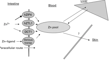
Zn is absorbed mainly by facilitated processes involving several transporters (Fig. 6). Ionized Zn uses different trans-porters such as hZIP4 (human zrt-,irt-like protein), which is the most predominant (Wang et al. 2002), while a cation dif-fusion facilitator (hZTL1, human zinc T-like transporter; Ford, 2004) and a Hþ-coupled divalent cation transporter (DCT1) also play a role in Zn absorption. Complexed Zn, e.g. with
amino acids, enters enterocytes via an Hþ/peptide co-transpor-ter (Evans, 1980; Hambidge et al. 1986; Tapiero & Tew, 2003). Moreover, a small proportion of Zn is absorbed by the paracellular route by either passive diffusion or solvent drag. Transporter-mediated or facilitated uptake is limited due to saturation of the transport systems or ion channels employed, and passive diffusion may be limited owing to numerous factors, e.g. physico-chemical properties (chemical form, degree of ionization and size). The concentration of trace elements in plasma is well controlled. Indeed, when intake is greater than the immediate tissue requirements, the excess is stored in some specific tissues (Fe in liver), excreted in urine (Se) or excreted back into the gastrointestinal lumen via gastrointestinal secretions or intestinal mucosal cell shed-ding (Zn).
Distribution and delivery to the skin
When dietary bioactives arrive in the blood circulation, they are ready to be distributed to all body tissues where they can exhibit a biological activity. Although certain dietary bioactives have been reported to exert a biological activity
Polyphenol conjugate OATP ? Intestine Blood Skin Polyphenol glucoside Polyphenol Polyphenol Polyphenol conjugate Liver ? Polyphenol LPH ? Polyphenol conjugate
Fig. 3. A highly simplified representation of the mechanism of polyphenol absorption. LPH, lactase phloridzin hydrolase; OATP, organic anion transporting poly-peptide (a transporter). AA Intestine Blood Skin AA DHA AA DHA Liver DHA SVCT GLUT GLUT? SVCT? DHA reductase
Fig. 4. A highly simplified representation of the mechanism of vitamin C absorption. AA, ascorbic acid; DHA, dehydroascorbic acid; SVCT, a Naþ
-dependent trans-porter; GLUT, a glucose transporter.
in skin such as photoprotection, collagen synthesis and cancer prevention, information on their delivery mechanisms to skin is quite scarce.
Vitamin E
Vitamin E consists of a mixture of different molecules, i.e. a-, b-, g- and d-tocopherols and a-, b-, g- and d-tocotrienols. Due to the presence of three chiral atoms, these molecules exhibit different stereoisomers ranging from RRR, RSR, etc. to SSS. In response to supplementation, the concentration of vitamin E increases immediately in plasma while a rise in concentration is observed only after several days (7 d) in sebum (Vaule et al. 2004). The basis of this delay may be related to the process of sebum production, because it has been reported (Downing et al. 1981) to take approximately 8 d for newly synthesized lipids to be secreted in sebum. The delivery of vitamin E to skin seems to be very specific for certain isomeric forms (Fig. 1). Indeed, following supplementation, both the natural form (RRR-a-tocopherol) and the synthetic form (all-rac-a-tocopherol) appear in the blood circulation, whereas only
RRR-a-tocopherol appears in sebum (Vaule et al. 2004). This indicates that a specific protein could selectively transport this form of the vitamin into the sebum. A similar specificity has already been described in the liver (Hosomi et al. 1997). g-Toco-pherol is absorbed into plasma but it is still uncertain whether g-tocopherol exerts a biological activity in man since this form is mostly eliminated by the liver. However, g-tocopherol is present in sebum and skin (Thiele et al. 1999; Vaule et al. 2004) and so it might exert an activity in human skin, although this needs further investigation. Skin vitamin E exhibits a gradi-ent of concgradi-entration with a higher level in the dermis and a lower level in the stratum corneum (Shindo et al. 1994). In addition, there are regional variations of skin vitamin E; facial skin con-tains a several-fold higher level of vitamin E than unexposed skin sites such as skin from the upper arm (Thiele et al. 1998). This regional variability of vitamin E is supported by the fact that vitamin E is continuously delivered to sebum via the sebaceous glands (Thiele et al. 1999; Vaule et al. 2004). As a consequence, skin sites with high sebum production such as
forehead skin exhibit higher vitamin E concentration
(Lang et al. 1986). Liver Intestine Blood Skin Selenite Se pool Selenoprotein Selenoprotein P ? AA trans Diffusion Mediated Organic forms Selenate
Fig. 5. A highly simplified representation of the mechanism of selenium absorption. AA trans, amino acid transporter.
Paracellular route Intestine Blood Skin Zn–ligand Liver ? Various transp Zn2+ DCT1 hZTL1 hZIP4 Zn pool
Fig. 6. A highly simplified representation of the mechanism of zinc absorption. hZIP4 (human zrt-,irt-like protein), hZTL1 (human zinc T-like transporter) and DCT1 (Hþ
Carotenoids
Because carotenoids are lipophilic molecules, they are well placed to act as chain-breaking antioxidants in the skin pro-tecting epidermal PUFA from peroxidation by oxygen radicals (Krinsky and Denecke, 1982). Carotenoid determination in skin is classically assessed by HPLC; however, this method is tedious and also quite invasive for the subject since it requires skin biopsy collection. Due to the polyene molecular structure of carotenoids, new non-invasive methods for the measurement of skin total carotenoid concentration have been developed such as reflection spectrometry (Jungmann et al. 1996) and Raman spectroscopy (Hata et al. 2000; Hammond and Wooten, 2005). Although skin total carotenoid levels measured with these two non-invasive techniques corre-late with those obtained with HPLC, these methods provide the total carotenoid concentration rather than the concentration of individual carotenoids. In addition, these techniques measure carotenoids in the upper part of the skin, whereas punch biopsies provide information on the epidermal plus dermal content. It is difficult to quantify individual caroten-oids because, for example, b-carotene exhibits a fourfold higher Raman scattering cross-section than lycopene with laser excitation at 488 nm (Hata et al. 2000). However, more recently, Raman spectrometry methodology has been adapted to quantify not only total carotenoids but also b-carotene and lycopene individually (Darvin et al. 2005) by taking into con-sideration the difference in absorbance between both sub-stances and using an algorithm for the calculation of the absolute concentration of both carotenoids.
Carotenoid deposition in skin is not equal throughout the body (Hata et al. 2000; Stahl & Sies, 1996; Darvin et al. 2005) with the concentration decreasing in the following order: forehead . palm of hand . dorsal . inside arm ¼ back of hand. Similarly to vitamin E, carotenoids might also be secreted in sebum and in consequence might explain the non-uniform level of skin carotenoids, but this needs to be further investigated. Carotenoids exhibit a concentration gra-dient with a higher amount in the dermis and lower levels in the stratum corneum. In addition, subcutaneous adipose tissue is rich in carotenoids, which could be a storage site of carotenoids for skin (Ribaya-Mercado et al. 1995). Skin b-car-otene and lycopene are lower in smokers than in non-smokers and higher in vegetarians (Darvin et al. 2005).
Following supplementation, carotenoid concentration
increases in skin (Stahl & Sies, 1996; Stahl et al. 1998, 2001; Postaire et al. 1997; Alaluf et al. 2002; Heinrich et al. 2003) but again to different extents at various skin sites: the increase is 2·4-fold in forehead, 0·7-fold in dorsal skin, 2·2-fold in the palm of the hand, 17-2·2-fold in the back of the hand and 1·7-fold for the inside of the arm. Most of the increase in skin carotenoid levels occurs within the first four weeks of supplementation but no plateau is reached during 12 weeks of supplementation. Cessation of supplementation induces a prompt drop of carotenoid level in all skin sites, decreasing by 56 % in forehead, 14 % in dorsal, 31 % for palm of the hand, 35 % for back of the hand and 47 % inside the arm (Stahl et al. 1998).
Besides the amount of total carotenoids delivered to skin, some attention needs to be given to how carotenoids reach the skin. For example, the carotenoid lycopene is a highly
unsaturated molecule, containing thirteen double bonds among which eleven are conjugated. Of the theoretical 211 geometrical isomer forms, only seventy-two are thermodyna-mically possible. All-trans-lycopene is the predominant lyco-pene isomer in nature, representing about 80 – 97 % in tomatoes and related products (Boileau et al. 2002). However, more than 50 % total lycopene is found in various cis forms in body fluids such as plasma (Krinsky et al. 1990) and breast milk, and in the prostate, testis and skin (Stahl & Sies, 1992; Clinton et al. 1996; Schierle et al. 1997; Wu et al. 2003). Investigators propose several explanations for this high proportion of cis isomers of lycopene in human, such as the preferential absorption of cis-lycopene isomers within the intestine (Stahl & Sies, 1992) or the isomerization of all-trans-lycopene in the stomach or intestine (Re et al. 2001); however, further investigation is required to identify the role of these cis isomers in human. Xanthophylls such as lutein and zeaxanthin are carotenoids containing hydroxyl groups and in consequence they could be either free or esterified with fatty acids. In fruits and vegetables, most xanthophylls are esterified. It is generally believed that these xanthophyll esters are hydrolysed during intestinal absorption. However, xanthophyll esters are present in skin, suggesting that either some xanthophyll esters have been absorbed as such or xanthophylls have been re-esterified in skin (Wingerath et al. 1998). Another aspect is the accumulation of specific caroten-oids in specific tissues, such as lutein and zeaxanthin in the eye (Bone et al. 1988, 1997; Handelman et al. 1988) and lyco-pene and b-carotene in prostate (Clinton et al. 1996). This specific tissue accumulation suggests that the delivery of caro-tenoids to tissues involves facilitated processes. Indeed, evi-dence points to the implication of lipid transporters in carotenoid absorption by intestinal cells in vitro (Reboul et al. 2004; During et al. 2005). This facilitated transport of carotenoids could be also relevant in skin, but this needs to be investigated further (Fig. 2).
Polyphenols
Polyphenols are mostly present in plasma bound to albumin in conjugated forms, i.e. with glucuronide or sulfate, or meth-ylated (Day et al. 2001; Kroon et al. 2004). Glucuronide con-jugates of polyphenols would need to be transported actively into peripheral tissues because they are relatively hydrophilic and therefore diffuse through membranes only very slowly (Fig. 3). In certain tissues, conjugated polyphenols are hydro-lysed by cellular b-glucuronidase activity, found both in the lysosomal fraction and in the lumen of the endoplasmic reti-culum; in liver cells, this enzyme is active on quercetin glu-curonides (O’Leary et al. 2003). Sulfatase activity is also present (Pasqualini & Nguyen, 1991) and acts on steroids and other sulfates inside the cell, thereby producing intra-cellular aglycone forms of polyphenols. The entry of poly-phenol conjugates into hepatic cells is at least in part due to the organic anion transporting polypeptide (OATP) trans-porter class (O’Leary et al. 2003), and keratinocytes express some OATP transporters (Schiffer et al. 2003). While poly-phenols are associated with a beneficial effect on skin (Kami-mura & Takahashi, 2002; Ni et al. 2002; Singh & Agarwal, 2002; Mittal et al. 2003; Wie et al. 2003; Kenny et al. 2004; Widyarini et al. 2005), the exact mechanism of action is still
unknown, i.e. whether this action takes place directly via an increase of skin polyphenol content or indirectly through a systemic effect on the vascular system. Indeed, in vitro, the aglycone form of hesperidin is efficiently taken up by skin fibroblasts but does not protect them against UVA-induced
damage, while hesperetin-7-glucuronide could not be
detected in skin fibroblasts as an aglycone or a conjugated form, but is protective against UVA radiation (Proteggente et al. 2003). Silibinin, a polyphenol from milk thistle, appeared in mouse skin rapidly after absorption, but 90 % was metabolized or excreted within 4 h. The maximum amount of conjugated silibinin in skin was similar to that found in lung, liver and prostate, although the amount of the free form was lower: free silibinin, 1·4 mg/g (v. 8·8 mg/g in liver, 4·3 mg/g in lung, 2·5 mg/g in prostate and 5·8 mg/g in pancreas), and conjugated silibinin, 4·3 mg/g (v. 5·7 mg/g in liver, 2·8 mg/g in lung, 6·1 mg/g in prostate and 10·6 mg/g in pancreas) from a dose of 50 mg/kg. The liver exhibited the fastest elimination half-life, but the other organs were similar (half-life of about 100 min). There was also an increase in skin of the phase II enzymes, quinone reductase and glutathione S-transferase, which required 15 d administration to be maximal (Zhao and Agarwal, 1999). ( – )-Epigallocatechin gallate, a polyphenol present in green tea, has been administered to the mouse. After consumption of 3H-labelled compound, there was some radioactivity in skin (equivalent to 0·11 mg per 100 mg skin for a 1 mg dose). However, this is likely to be metabolites or breakdown products, protein-bound forms or even exchange with body pools of water, since the plasma kinetics using3H measure-ment did not match the pharmacokinetics using HPLC (Suga-numa et al. 1998).
Vitamin C
Vitamin C is an effective antioxidant and an essential cofactor in numerous enzymatic reactions. It comprises two major forms:L-ascorbic acid, the reduced form, andL
-dehydroascor-bic acid, the oxidized form. Man and other primates have lost the ability to synthesize vitamin C as a result of a mutation in the gene encoding forL-gulono-g-lactone oxidase, an enzyme
required for vitamin C biosynthesis. In man, plasma ascorbic acid concentrations are maintained between 10 and 160 mM
(1 – 15 mg/ml) and any excess of the vitamin is excreted by the kidney (Fuchs and Podda, 1997). Analysis of tissue ascor-bate levels in human subjects revealed highest amounts in adrenal glands (550 mg/kg), brain (140 mg/kg) and liver (125 mg/kg), followed by lungs (70 mg/kg), kidneys (55 mg/kg), heart (55 mg/kg), skeletal muscle (35 mg/kg) and skin (30 mg/kg), and low levels in adipose tissue (10 mg/kg) and blood (9 mg/kg; Brown & Jones, 1996; Fuchs and Podda, 1997). Considering the different size and weight of the organs it is evident that ascorbic acid is concentrated in specific tissues, with the highest concentrations in adrenal glands, brain and liver, and the highest total amount present in skeletal muscle. In most of these tissues high ascorbate levels are probably important for maintaining structural integ-rity through collagen fibres, as well as for more specific
func-tions, e.g. hormone synthesis, immune response and
antioxidant protection. This concentration difference between different tissues clearly indicates that ascorbic acid uptake
and distribution into tissues is mediated by an active transport mechanism. Indeed, two different ascorbic acid transporters, SVCT1 and SVCT2, have been identified by screening a rat kidney cDNA library (Tsukaguchi et al. 1999). SVCT1 is lar-gely confined to epithelial surfaces involved in bulk transport, such as those of the intestine or kidney. In contrast, SVCT2 appears to account for tissue-specific uptake of vitamin C and is widely expressed, occurring in neurons, the endocrine system and other tissues. Recently, SVCT2-null mice have been generated. Surprisingly, the knockout is lethal perina-tally, owing to respiratory failure and intraparenchymal brain haemorrhage in the animals. Most likely the observed haemorrhages are not a result of scurvy because the knockout mice showed no haemorrhages in other tissues and their skin had normal 4-hydroxyproline levels despite low ascorbate concentration. However, uptake of vitamin C by fibroblasts cultured from these mice was virtually abolished (Sotiriou et al. 2002). Interestingly, the organs with the most severe phenotypes were those that possessed the highest ascorbate concentration in man. In skin, in vitro studies in HaCaT ker-atinocytes demonstrated the presence and functional activity of both transporters, as well as an efficient ascorbate recycling system (Savini et al. 1999, 2000, 2002; Liang et al. 2001; Fig. 4). The latter might be an explanation for the mild skin phenotype of the SVCT2 knockout mice (Sotiriou et al. 2002). However, more work needs to be done to elucidate ascorbate transport in human skin.
Vitamin C distribution within the skin has also been deter-mined in the murine model. Ascorbic acid concentration in the epidermis and dermis was 1·3 mmol/g (229 mg/g) and 1·0 mmol/g (176 mg/g), respectively; for dehydroascorbate,
the concentration was 1·3 mmol/g in epidermis and
0·9 mmol/g in dermis (Shindo et al. 1994). Within the murine stratum corneum, the outermost layer of the epider-mis, ascorbic acid exhibits a gradient of concentration with low levels in the outer layers and a steep increase in the deeper parts (Weber et al. 1999). Although skin was always thought to be the most sensitive organ during deficiency status (e.g. scurvy), recent results indicate that skin can cope with marginal amounts of vitamin C whereas other organs like brain and lungs suffer much more. How-ever, under certain conditions the skin vitamin C pool can be depleted selectively, e.g. upon UV irradiation or in atopic dermatitic lesions (Shindo et al. 1994; Podda et al. 1998; Leveque et al. 2003), but whether that is due to a higher vitamin C turnover or a less efficient transport or recy-cling is not known at present. In contrast to ascorbate, the intestinal uptake of dehydroascorbate is mediated by facili-tated-diffusion glucose transporters GLUT1, GLUT3 and GLUT4 (Liang et al. 2001). Under physiological conditions however, the reduced form of vitamin C will predominate (95 % in human plasma), and, thus, it is unlikely that GLUT-mediated dehydroascorbic acid uptake will be suffi-cient for the cellular demand of most cells. Furthermore,
cir-culating levels of glucose are 1000-fold higher than
dehydroascorbic acid levels (2 – 5 mM) and marked compe-tition by glucose of dehydroascorbic acid influx is most likely (Liang et al. 2001). Furthermore, dehydroascorbic acid is nearly undetectable in most tissues (Rumsey and Levine, 1998). However, higher concentrations may occur transiently during oxidative stress.
Selenium
Most ingested Se, whether in organic or inorganic form, is converted by the liver into selenocysteine, which is used in the biosynthesis of selenoproteins including glutathione per-oxidase and thioredoxin reductase. Evidence supports that selenoprotein P is a major transporter of Se from blood to other tissues, especially to brain and testis (Burk & Hill, 2005; Richardson, 2005). However, it is not clear how Se is delivered into cells. It would require specific membrane receptors to transport the selenoproteins; but evidence suggests the presence of such receptors has been found only in animal models (Wilson & Tappel, 1993; Burk & Hill, 1994). Se is present in skin cells (Fig. 5) as part of thioredoxin reductase and glutathione peroxidase, which exhibit major roles in the cellular defence against oxidative stress. Studies have showed that thioredoxin reductase is located on the cell membrane in keratinocytes and that this plays a vital role in protection from UV-induced free radical damage. Membrane-associated thioredoxin reductase corre-lates with different skin phototypes I – VI (Fitzpatrick classi-fication), where darker skin has significantly higher enzyme activity than very fair skin. Higher levels of thioredoxin reductase have been found in black v. Caucasian skin (Schall-reuter et al. 1987). Moreover, it is possible that the
compo-sition of selenoproteins in different skin cell types
contributes to their well-known different susceptibility to UV-induced damage, and that genetic and racial differences in susceptibility to solar damage may reflect different geneti-cally determined levels of expression of selenoproteins (McKenzie, 2000). Qualitative and quantitative differences have been shown in selenoprotein expression between kerati-nocytes, melanocytes and fibroblasts in culture (Rafferty et al. 1998). Keratinocytes have twice the specific activity of glutathione peroxidase of fibroblasts, and keratinocytes
are more resistant to UV damage than fibroblasts
(Leccia et al. 1998).
Total-body Se is 13 to 20 mg. Liver and kidney have the highest Se concentration (per weight of tissue), but these organs contain only a relatively small amount of total-body Se (4 % for kidney and 8 % for liver). About 40 to 50 % total-body Se is contained in skeletal muscles. It is important to note that some Se in muscles is incorporated unspecifically as selenomethionine instead of its methionine analogue. Brain, nervous and lung tissue have relatively low Se concentrations (Oster et al. 1988). Data related to Se concentration or distri-bution in skin are sparse and may depend on racial differences. One study showed that the epidermal:dermal ratio of Se is much lower than for Zn, Cu or Mn and varies between 0·8 and 1·5 in abdominal skin. As for other nutrients with antiox-idant properties, this ratio depends on skin site and is much lower in plantar skin (Molokhia et al. 1979). Such a distri-bution is likely to be influenced by the type of environmental insult faced by skin on different parts of the body. There is some evidence of a hierarchy of tissue retention when Se deficiency occurs, with preferential accumulation of Se in brain, gonad, thyroid, pituitary and adrenal tissue over liver, erythrocytes, heart and muscle (Behne et al. 1988; Richardson, 2005). This could imply that a less-sensitive organ like skin could be depleted of its store in the case of inadequate Se intake or dietary restriction.
Zinc
In blood, 69 % Zn is transported with albumin, 30 % with a2
-macroglobulin and 1 % with the amino acids, cysteine and his-tidine (Hallman et al. 1971). The mechanism of delivery of Zn to skin is still unknown but might involve carrier proteins (Ackland et al. 1988; Guiraud et al. 1992; Fig. 6). The adult human body contains between 1·2 and 2·3 g Zn, of which 57 % is found in muscles (51 mg/g wet weight), 29 % in bone (100 mg/g wet weight) and 6 % in skin (32 mg/g wet weight; Jackson, 1989; King et al. 2000). The remaining Zn is found in all other tissues with 5 %, 1·5 %, 0·7 %, 0·5 %, 0·4 % and 0·1 % in liver (58 mg/g wet weight), brain (11 mg/g wet weight), kidneys (55 mg/g wet weight), blood (1 mg/g wet weight), heart (23 mg/g wet weight) and hair (150 mg/g wet weight), respectively. In skin, Zn exhibits a gradient of con-centration, with five- to sixfold higher concentration in the epidermis (17 mg/g dry weight) than in the dermis (Molokhia & Portnoy, 1969). Zn plays an important role in the three skin functions, i.e. morphogenesis, repair and maintenance, and in protection and defence, since Zn is essential for catalytic, structural and/or regulatory functions of proteins and/or enzymes involved in these processes. The best described and with relevant activity in skin are the matrix metalloproteinases (MMP), superoxide dismutase, metallothionenein, alkaline phosphatase and those involved in regulation of gene expression, such as DNA and RNA polymerases. MMP, including collagenase (MMP-1), elastase (MMP-12) and gela-tinase (MMP-2), are involved in the formation of extracellular matrix. Superoxide dismutase is important for its antioxidant properties, and metallothioneins store Zn and also have antioxidant properties. Alkaline phosphatase is involved in AMP metabolism, which plays a role in suppressing the inflammatory process. No tissue acts as a Zn store. Conse-quently when adaptation to low intake fails, deficiency can occur rapidly (Miller et al. 1994). During experimental Zn depletion in human volunteers, skin lesions were the most prevalent clinical sign (Prasad, 1982; Baer & King, 1984; Baer et al. 1985). Among other clinical signs and after bio-chemical modifications, moderate Zn deficiency is manifest by rough skin and delayed wound healing. In severe Zn deficiency such as acrodermatitis enteropathica, a gene mutation coding for hZIP4 transporter is associated with a defective absorption of Zn inducing dermatological manifes-tations such as bullous pustular dermatitis, erythema, areas of eczema and alopecia.
Conclusions
Different data support the fact that dietary bioactives such as vitamins, carotenoids, polyphenols and trace elements contrib-ute to maintenance and improvement of skin integrity and physiology, as well as preventing deleterious effects induced by ageing and environmental stress. Beneficial effects have been demonstrated in various experimental systems including topical application of some of these ingredients. More recently, oral supplements containing various dietary bioac-tives have also been reported to be beneficial for skin. How-ever, oral consumption does not guarantee obtaining a beneficial effect on human skin. Although some components of the diet could act by secondary messengers from the gut
or other organs to the skin, we assume that the dietary bioac-tives described here must cross the intestinal barrier and be metabolized and distributed to the skin in order to be effective. On the basis of present understanding of the key parameters involved in the absorption process, it is likely that the absorption of dietary bioactives in the gut, but also in skin, could be modulated by e.g. transfer proteins, the physical and chemical properties of the dietary bioactive, and compe-tition and/or interaction with other dietary bioactives.
There are large gaps in knowledge in the area of skin bioa-vailability of dietary bioactives, despite some good evidence for beneficial effects of some of these components on skin in several in vitro and in vivo models. For example, the distri-bution and activity in skin of transporters for these compounds are almost completely unknown. The distribution between different compartments and body areas of skin is only known for some compounds, and turnover and export of diet-ary bioactives in skin are poorly studied.
The administration of dietary bioactives by the oral route offers several advantages over their topical application: intes-tinal absorption of bioactives, which are sometimes compro-mised in topical application owing to their low stability or low skin penetration; bioactives reach the entire skin of the body; and bioactives are distributed to all skin compartments, e.g. epidermis, dermis, hypodermis, blood vessels and sebum, allowing bioefficacy in all these compartments. The oral administration of these bioactives could also be complemen-tary to the topical application.
The effects of ageing on skin health will always remain an important issue for the population, and educating them about what has or has not been established scientifically is an important role for health-care professionals. Additionally, further education on these ingredients, products and oral skin health supplements in general may ultimately drive future research that will distinguish truly efficacious nutrients from those with misleading claims.
Acknowledgement
The authors thank Dr Birgit Holst for her critical comments during the preparation of the manuscript.
References
Ackland ML, Danks DM & McArdle HJ (1988) Studies on the mech-anism of zinc uptake by human fibroblasts. J Cell Physiol 135, 521 – 526.
Alaluf S, Heinrich U, Stahl W, Tronnier H & Wiseman S (2002) Diet-ary carotenoids contribute to normal human skin color and UV photosensitivity. J Nutr 132, 399 – 403.
Baer MT & King JC (1984) Tissue zinc levels and zinc excretion during experimental zinc depletion in young men. Am J Clin Nutr 39, 556 – 570.
Baer MT, King JC, Tamura T, Margen S, Bradfield RB, Weston WL & Daugherty NA (1985) Nitrogen utilization, enzyme activity, glu-cose intolerance and leukocyte chemotaxis in human experimental zinc depletion. Am J Clin Nutr 41, 1220 – 1235.
Beehler BC, Przybyszewski J, Box HB & Kulesz-Martin MF (1992) Formation of 8-hydroxydeoxyguanosine within DNA of mouse keratinocytes exposed in culture to UVB and H2O2. Carcinogen-esis 13, 2003 – 2007.
Behne D, Hilmert H, Scheid S, Gessner H & Elger W (1988) Evidence for specific selenium target tissues and new biologically important selenoproteins. Biochim Biophys Acta 966, 12 – 21. Boileau TW, Boileau AC & Erdman JW Jr (2002) Bioavailability of
all-trans and cis-isomers of lycopene. Exp Biol Med (Maywood) 227, 914 – 919.
Bone RA, Landrum JT, Fernandez L & Tarsis SL (1988) Analysis of the macular pigment by HPLC: retinal distribution and age study. Invest Ophthalmol Vis Sci 29, 843 – 849.
Bone RA, Landrum JT, Friedes LM, Gomez CM, Kilburn MD, Menendez E, Vidal I & Wang W (1997) Distribution of lutein and zeaxanthin stereoisomers in the human retina. Exp Eye Res 64, 211 – 218.
Brown LAS & Jones DP (1996) The biology of ascorbic acid. In Handbook of Antioxidants, pp. 117 – 154 [E Cadenas and L Packer, editors]. New York: Marcel Dekker Inc.
Burk RF & Hill KE (1994) Selenoprotein P. A selenium-rich extra-cellular glycoprotein. J Nutr 124, 1891 – 1897.
Burk RF & Hill KE (2005) Selenoprotein P: an extracellular protein with unique physical characteristics and a role in selenium homeo-stasis. Annu Rev Nutr 25, 215 – 235.
Cesarini JP, Michel L, Maurette JM, Adhoute H & Bejot M (2003) Immediate effects of UV radiation on the skin: modification by an antioxidant complex containing carotenoids. Photodermatol Photoimmunol Photomed 19, 182 – 189.
Clinton SK, Emenhiser C, Schwartz SJ, Bostwick DG, Williams AW, Moore BJ & Erdman JW (1996) Cis-trans lycopene isomers, caro-tenoids and retinol in the human prostate. Cancer Epidemiol Biomarkers Prev 5, 823 – 833.
Cooper DA, Webb DR & Peters JC (1997) Evaluation of the potential for olestra to affect the availability of dietary phytochemicals. J Nutr 127, Suppl., 1699S – 1709S.
Cross CE, van der Vliet A, Louie S, Thiele JJ & Halliwell B (1998) Oxidative stress and antioxidants at biosurfaces: plants, skin, and respiratory tract surfaces. Environ Health Perspect 106, 1241 – 1251.
Dalle CM & Pathak MA (1992) Skin photosensitizing agents and the role of reactive oxygen species in photoaging. J Photochem Photo-biol B 14, 105 – 124.
Darvin ME, Gersonde I, Meinke M, Sterry W & Landemann J (2005) Non-invasive in vivo determination of the carotenoids b-carotene and lycopene concentrations in the human skin using the Raman spectroscopic method. J Phys D Appl Phys 38, 2696 – 2700. Day AJ, Canada FJ, Diaz JC, Kroon PA, McLauchlan WR, Faulds
CB, Plumb GW, Morgan MRA & Williamson G (2000) Dietary flavonoid and isoflavone glycosides are hydrolysed by the lactase site of lactase phlorizin hydrolase. FEBS Lett 468, 166 – 170. Day AJ, Mellon FA, Barron D, Sarrrazin G, Morgan MRA &
Williamson G (2001) Human metabolism of dietary flavonoids: identification of plasma metabolites of quercetin. Free Radic Res 212, 941 – 952.
Downing DT, Stewart ME & Strauss JS (1981) Estimation of sebum production rates in man by measurement of the squalene content of skin biopsies. J Invest Dermatol 77, 358 – 360.
During A, Dawson HD & Harrison EH (2005) Carotenoid transport is decreased and expression of the lipid transporters SR-BI, NPC1L1, and ABCA1 is downregulated in Caco-2 cells treated with ezeti-mibe. J Nutr 135, 2305 – 2312.
During A, Hussain MM, Morel DW & Harrison EH (2002) Carotenoid uptake and secretion by CaCo-2 cells: b-carotene isomer selectivity and carotenoid interactions. J Lipid Res 43, 1086–1095. Emerit I (1992) Free radicals and aging of the skin. EXS 62,
328 – 341.
Evans GW (1980) Normal and abnormal zinc absorption in man and animals: the tryptophan connection. Nutr Rev 38, 137 – 141. Ford D (2004) Intestinal and placental zinc transport pathways. Proc
Fuchs J (1998) Potential and limitation of the natural antioxidants RRR a tocopherol,L-ascorbic acid and b-carotene in cutaneaous photoprotection. Free Radic Biol Med 25, 848 – 873.
Fuchs J & Kern H (1998) Modulation of UV-light-induced skin inflam-mation byD-a-tocopherol andL-ascorbic acid: a clinical study using solar simulated radiation. Free Radic Biol Med 25, 1006 – 1012. Fuchs J & Podda M (1997) Vitamin C in cutaneous biology. In
Vita-min C in Health and Disease, pp. 333 – 340 [L Packer and J Fuchs, editors]. New York: Marcel Dekker Inc.
Gollnick HPM, Hopfenmuller W, Hemmes C, Chun SC, Schmid C, Sundermeier K & Biesalski HC (1996) Systemic b carotene plus topical UV-sunscreen are an optimal protection against harmful effects of natural UV-sunlight: results of the Berlin-Eilath study. Eur J Dermatol 6, 200 – 205.
Greul AK, Grundmann JU, Heinrich F, Pfitzincer I, Bernhardt J, Ambach A, Biesalski HK & Gollnick H (2002) Photoprotection of UV-irradiated human skin: an antioxidative combination of vita-mins E and C, carotenoids, selenium and proanthocyanidins. Skin Pharmacol Appl Skin Physiol 15, 307 – 315.
Guiraud P, Lepee M, Monjo AM, Richard MJ & Favier A (1992) Cul-tured human skin fibroblasts absorb 65Zn. Optimization of the method and study of the mechanisms involved. Biol Trace Elem Res 32, 213 – 225.
Guyton KZ & Kensler TW (1993) Oxidative mechanisms in carcino-genesis. Br Med Bull 49, 523 – 544.
Hallman PS, Perrin DD & Watt AE (1971) The computed distribution of Cu (II) and zinc (II) ions among seventeen amino acids present in human blood plasma. Biochem J 121, 549 – 555.
Hambidge K, Casey C & Krebs N (1986) Zinc. In Trace Elements in Human and Animal Nutrition, pp. 1 – 137 [W Mertz, editor]. New York: Academic Press.
Hammond BR & Wooten BR (2005) Resonance Raman spectroscopic measurement of carotenoids in the skin and retina. J Biomed Opt 10(5), 054002 (12 pages).
Handelman GJ, Dratz EA, Reay CC & van Kuijk FJGM (1988) Caro-tenoids in the human macula and whole retina. Invest Ophthalmol Vis Sci 29, 850 – 855.
Hata TR, Scholz TA, Ermakov IV, McClane RW, Khachik F, Geller-mann W & Pershing LK (2000) Non-invasive Raman spectroscopic detection of carotenoids in human skin. J Invest Dermatol 115, 441 – 448.
Heinrich U, Gartner C, Wiebusch M, Eichler O, Sies H, Tronnier H & Stahl W (2003) Supplementation with b-carotene or a similar amount of mixed carotenoids protects humans from UV-induced erythema. J Nutr 133, 98 – 101.
Hollander D, Rim E & Muralidhara KS (1975) Mechanism and site of small intestinal absorption of a-tocopherol in the rat. Gastroenter-ology 68, 1492 – 1499.
Hollander D & Ruble PE (1978) b-Carotene intestinal absorption: bile, fatty acid, pH, and flow rate effects on transport. Am J Physiol 235, E686 – E691.
Hosomi A, Arita M, Sato Y, Kiyose C, Ueda T, Igarashi O, Arai H & Inoue K (1997) Affinity for a-tocopherol transfer protein as a determinant of the biological activities of vitamin E analogs. FEBS Lett 409, 105 – 108.
Hu ML & Tappel AL (1992) Potentiation of oxidative damage to pro-teins by ultraviolet-A and protection by antioxidants. Photochem Photobiol 56, 357 – 363.
Jackson MJ (1989) Physiology of zinc: general aspects. In Zinc in Human Biology, pp. 1 – 14 [CF Mills, editor]. London: Springer-Verlag.
Jang M, Cai L, Udeani GO, et al. (1997) Cancer chemopreventive activity of resveratrol, a natural product derived from grapes. Science 275, 218 – 220.
Jungmann H, Heinrich U, Wiebusch M & Tronnier H (1996) Der ein-satz der reflektionsspektroskopie in der dermatologie am beispiel der b-carotins. Kosmet Med 1, 50 – 57.
Kamimura A & Takahashi T (2002) Procyanidin B-2, extracted from apples, promotes hair growth: a laboratory study. Br J Dermatol 146, 41 – 51.
Katiyar SK, Agarwal R & Mukhtar H (1996) Inhibition of tumor pro-motion in SENCAR mouse skin by ethanol extract of Zingiber offi-cinale rhizome. Cancer Res 56, 1023 – 1030.
Katiyar SK & Mukhtar H (1997) Tea antioxidants in cancer chemo-prevention. J Cell Biochem 27, 59 – 67.
Kenny TP, Keen CL, Jones P, Kung HJ, Schmitz HH & Gershwin ME (2004) Cocoa procyanidins inhibit proliferation and angiogenic sig-nals in human dermal microvascular endothelial cells following stimulation by low-level H2O2. Exp Biol Med (Maywood) 229, 765 – 771.
King JC, Shames DM & Woodhouse LR (2000) Zinc homeostasis in humans. J Nutr 130, Suppl., 1360S – 1366S.
Krinsky NI & Deneke SM (1982) Interaction of oxygen and oxy-rad-icals with carotenoids. J Natl Cancer Inst 69, 205 – 210.
Krinsky NI, Russett MD, Handelman GJ & Snodderly DM (1990) Structural and geometrical isomers of carotenoids in human plasma. J Nutr 120, 1654 – 1662.
Kroon PA, Clifford MN, Crozier A, Day AJ, Donovan JL, Manach C & Williamson G (2004) How should we assess the effects of exposure to dietary polyphenols in vitro? Am J Clin Nutr 80, 15 – 21.
Lang JK, Gohil K & Packer L (1986) Simultaneous determination of tocopherols, ubiquinols, and ubiquinones in blood, plasma, tissue homogenates, and subcellular fractions. Anal Biochem 157, 106 – 116.
Leccia MT, Richard MJ, Joanny-Crisci F & Beani JC (1998) UV-A1 cytotoxicity and antioxidant defence in keratinocytes and fibro-blasts. Eur J Dermatol 8, 478 – 482.
Leveque N, Robin S, Muret P, Mac-Mary S, Makki S & Humbert P (2003) High iron and low ascorbic acid concentrations in the dermis of atopic dermatitis patients. Dermatology 207, 261 – 264.
Liang WJ, Johnson D & Jarvis SM (2001) Vitamin C transport sys-tems of mammalian cells. Mol Membr Biol 18, 87 – 95.
McKenzie RC (2000) Selenium, ultraviolet radiation and the skin. Clin Exp Dermatol 25, 631 – 636.
Manach C, Williamson G, Morand C, Scalbert A & Remesy C (2005) Bioavailability and bioefficacy of polyphenols in humans. I. Review of 97 bioavailability studies. Am J Clin Nutr 81, Suppl., 230S – 242S.
Miller LV, Hambidge KM, Naake VL, Hong Z, Westcott JL & Fen-nessey PV (1994) Size of the zinc pools that exchange rapidly with plasma zinc in humans: alternative techniques for measuring and relation to dietary zinc intake. J Nutr 124, 268 – 276.
Mittal A, Elmets CA & Katiyar SK (2003) Dietary feeding of proanthocyanidins from grape seeds prevents photocarcinogenesis in SKH-1 hairless mice: relationship to decreased fat and lipid per-oxidation. Carcinogenesis 24, 1379 – 1388.
Molokhia MM & Portnoy B (1969) Neutron activation analysis of trace elements in skin. 3. Zinc in normal skin. Br J Dermatol 81, 759 – 762.
Molokhia A, Portnoy B & Dyer A (1979) Neutron activation analysis of trace elements in skin. VIII. Selenium in normal skin. Br J Der-matol 101, 567 – 572.
Moser U & Bendich A (1991) Vitamin C. In Handbook of Vitamins, pp. 195 – 232 [LJ Machlin, editor]. New York: Marcel Dekker Inc. Mukhtar H & Ahmad N (1999) Cancer chemoprevention: future holds
in multiple agents. Toxicol Appl Pharmacol 158, 207 – 210. Nemeth K, Plumb GW, Berrin JG, Juge N, Jacob R, Naim HY,
Williamson G, Swallow DM & Kroon PA (2003) Deglycosylation by small intestinal epithelial cell b-glucosidases is a critical step in the absorption and metabolism of dietary flavonoid glycosides in humans. Eur J Nutr 42, 29 – 42.
Ni Z, Mu Y & Gulati O (2002) Treatment of melasma with Pycno-genol. Phytother Res 16, 567 – 571.
O’Leary KA, Day AJ, Needs PW, Mellon FA, O’Brien NM & Williamson G (2003) Metabolism of quercetin-7-and quercetin-3-glucuronides by an in vitro hepatic model: the role of human b-glucuronidase, sulfotransferase, catechol-O-methyl-transferase and multi-resistant protein 2 (MRP2) in flavonoid metabolism. Biochem Pharmacol 65, 479 – 491.
Oster O, Schmiedel G & Prellwitz W (1988) The organ distribution of selenium in German adults. Biol Trace Elem Res 15, 23 – 45. Pasqualini JR & Nguyen BL (1991) Estrone sulfatase activity and
effect of antiestrogens on transformation of estrone sulfate in hor-mone-dependent vs independent human breast-cancer cell-lines. Breast Cancer Res Treat 18, 93 – 98.
Perchellet JP & Perchellet EM (1989) Antioxidants and multistage carcinogenesis in mouse skin. Free Radic Biol Med 7, 377 – 408. Perchellet JP, Perchellet EM & Belman S (1990) Inhibition of
DMBA-induced mouse skin tumorigenesis by garlic oil and inhi-bition of two tumor-promotion stages by garlic and onion oils. Nutr Cancer 14, 183 – 193.
Podda M, Traber MG, Weber C, Yan LJ & Packer L (1998) UV-irradiation depletes antioxidants and causes oxidative damage in a model of human skin. Free Radic Biol Med 24, 55 – 65. Postaire E, Jungmann H, Bejot M, Heinrich U & Tronnier H (1997)
Evidence for antioxidant nutrients-induced pigmentation in skin: results of a clinical trial. Biochem Mol Biol Intern 42, 1023 – 1033. Prasad A (1982) Clinical and biochemical spectrum of zinc deficiency in human subjects. In Clinical, Biochemical, and Nutritional Aspects of Trace Elements, pp. 3 – 62 [AS Prasad, editor]. New York: Alan R. Liss.
Proteggente AR, Basu-Modak S, Kuhnle G, Gordon MJ, Youdim K, Tyrrell R & Rice-Evans CA (2003) Hesperetin glucuronide, a photoprotective agent arising from flavonoid metabolism in human skin fibroblasts. Photochem Photobiol 78, 256 – 261. Rafferty TS, McKenzie RC, Hunter JA, Howie AF, Arthur JR, Nicol F
& Beckett GJ (1998) Differential expression of selenoproteins by human skin cells and protection by selenium from UVB-radi-ation-induced cell death. Biochem J 332, 231 – 236.
Re R, Fraser PD, Long M, Bramley PM & Rice-Evans C (2001) Iso-merization of lycopene in the gastric milieu. Biochem Biophys Res Commun 281, 576 – 581.
Reboul E, Abou L, Mikail C, Ghiringhelli O, Andre M, Portugal H, Jourdheuil-Rahmani D, Amiot MJ, Lairon D & Borel P (2004) Lutein transport by Caco-2 TC-7 cells occurs partly by a facilitated process involving the scavenger receptor class B type I (SR-BI). Biochem J 387, 455 – 461.
Ribaya-Mercado JD, Garmyn M, Gilchrest BA & Russell RM (1995) Skin lycopene is destroyed preferentially over b-carotene during ultraviolet irradiation in humans. J Nutr 125, 1854 – 1858. Richardson DR (2005) More roles for selenoprotein P: local selenium
storage and recycling protein in the brain. Biochem J 386, e5 – e7. Rios L, Bennett RN, Lazarus SA, Remesy C, Scalbert A & William-son G (2002) Cocoa proanthocyanidins are stable during gastric transit in humans. Am J Clin Nutr 76, 1106 – 1110.
Rostan EF, DeBuys HV, Madey DL & Pinnell SR (2002) Evidence supporting zinc as an important antioxidant for skin. Int J Dermatol 41, 606 – 611.
Rumsey SC & Levine M (1998) Absorption, transport, and disposi-tion of ascorbic acid in humans. J Nutr Biochem 9, 116 – 160. Sadhana AS, Rao AR, Kucheria K & Bijani V (1988) Inhibitory
action of garlic oil on the initiation of benzo[a ]pyrene-induced skin carcinogenesis in mice. Cancer Lett 40, 193 – 197.
Savini I, Catani MV, Rossi A, Duranti G, Melino G & Avigliano L (2002) Characterization of keratinocyte differentiation induced by ascorbic acid: protein kinase C involvement and vitamin C homeo-stasis. J Invest Dermatol 118, 372 – 379.
Savini I, D’Angelo I, Ranalli M, Melino G & Avigliano L (1999) Ascorbic acid maintenance in HaCaT cells prevents radical for-mation and apoptosis by UV-B. Free Radic Biol Med 26, 1172 – 1180.
Savini I, Duflot S & Avigliano L (2000) Dehydroascorbic acid uptake in a human keratinocyte cell line (HaCaT) is glutathione-indepen-dent. Biochem J 345, 665 – 672.
Schallreuter KU, Hordinsky MK & Wood JM (1987) Thioredoxin reductase. Role in free radical reduction in different hypopigmen-tation disorders. Arch Dermatol 123, 615 – 619.
Schierle J, Bretzel W, Bu¨hler I, Faccin N, Hess D, Steiner K & Schu¨ep W (1997) Content and isomeric ratio of lycopene in food and human plasma. Food Chem 59, 459 – 465.
Schiffer R, Neis M, Holler D, et al. (2003) Active influx transport is mediated by members of the organic anion transporting polypep-tide family in human epidermal keratinocytes. J Invest Dermatol 120, 285 – 291.
Shindo Y, Witt E, Han D, Epstein W & Packer L (1994) Enzymic and non-enzymic antioxidants in epidermis and dermis of human skin. J Invest Dermatol 102, 122 – 124.
Singh RP & Agarwal R (2002) Flavonoid antioxidant silymarin and skin cancer. Antioxid Redox Signal 4, 655 – 663.
Sotiriou S, Gispert S, Cheng J, Wang Y, Chen A, Hoogstraten-Miller S, Miller GF, Kwon O, Levine M, Guttentag SH & Nussbaum RL (2002) Ascorbic-acid transporter Slc23a1 is essential for vitamin C transport into the brain and for perinatal survival. Nat Med 8, 514 – 517.
Stahl W, Heinrich U, Jungmann H, Sies H & Tronnier H (2000) Caro-tenoids and caroCaro-tenoids plus vitamin E protect against ultraviolet light-induced erythema in humans. Am J Clin Nutr 71, 795 – 798. Stahl W, Heinrich U, Jungmann H, von Laar J, Schietzel M, Sies H &
Tronnier H (1998) Increased dermal carotenoid levels assessed by noninvasive reflection spectrometry correlate with serum levels in women ingesting betatene. J Nutr 128, 903 – 907.
Stahl W, Heinrich U, Wiseman S, Eichler O, Sies H & Tronnier H (2001) Dietary tomato paste protects against ultraviolet light-induced erythema in humans. J Nutr 131, 1449 – 1451.
Stahl W, Schwarz W, Sundquist AR & Sies H (1992) Cis-trans iso-mers of lycopene and b-carotene in human serum and tissues. Arch Biochem Biophys 294, 173 – 177.
Stahl W & Sies H (1992) Uptake of lycopene and its geometrical iso-mers is greater from heat processed than from unprocessed tomato juice in humans. J Nutr 122(11), 2161 – 2166.
Stahl W & Sies H (1996) Lycopene: a biologically important caroten-oid for humans? Arch Biochem Biophys 336, 1 – 9.
Stahl W & Sies H (2002) Carotenoids and protection against solar UV radiation. Skin Pharmacol Appl Skin Physiol 15, 291 – 296. Steenvoorden DP & van Henegouwen GM (1997) The use of
endogenous antioxidants to improve photoprotection. J Photochem Photobiol B 41, 1 – 10.
Stoner GD & Mukhtar H (1995) Polyphenols as cancer chemopreven-tive agents. J Cell Biochem Suppl 22, 169 – 180.
Suganuma M, Okabe S, Oniyama M, Tada Y, Ito H & Fujiki H (1998) Wide distribution of [3H]( – )-epigallocatechin gallate, a cancer pre-ventive tea polyphenol, in mouse tissue. Carcinogenesis 19, 1771 – 1776.
Tapiero H & Tew KD (2003) Trace elements in human physiology and pathology: zinc and metallothioneins. Biomed Pharmacother 57, 399 – 411.
Thiele JJ, Dreher F & Packer L (2000) Cosmeceuticals. In Drugs vs Cosmetics, pp. 145 – 188 [P Elsner and H Maibach, editors]. New York: Dekker.
Thiele JJ, Podda M & Packer L (1997) Tropospheric ozone: an emer-ging environmental stress to skin. Biol Chem 378, 1299 – 1305. Thiele JJ, Traber MG & Packer L (1998) Depletion of human
stratum corneum vitamin E: an early and sensitive in vivo marker of UV induced photo-oxidation. J Invest Dermatol 110, 756 – 761.
Thiele JJ, Weber SU & Packer L (1999) Sebaceous gland secretion is a major physiologic route of vitamin E delivery to skin. J Invest Dermatol 113, 1006 – 1010.
Tsukaguchi H, Tokui T, Mackenzie B, Berger UV, Chen XZ, Wang Y, Brubaker RF & Hediger MA (1999) A family of mamma-lian Naþ-dependent L-ascorbic acid transporters. Nature 399, 70 – 75.
Vaule H, Leonard SW & Traber MG (2004) Vitamin E delivery to human skin: studies using deuterated a-tocopherol measured by APCI LC-MS. Free Radic Biol Med 36, 456 – 463.
Wang K, Zhou B, Kuo YM, Zemansky J & Gitschier J (2002) A novel member of a zinc transporter family is defective in acroder-matitis enteropathica. Am J Hum Genet 71, 66 – 73.
Weber SU, Thiele JJ, Cross CE & Packer L (1999) Vitamin C, uric acid, and glutathione gradients in murine stratum corneum and their susceptibility to ozone exposure. J Invest Dermatol 113, 1128 – 1132.
Widyarini S, Husband AJ & Reeve VE (2005) Protective effect of the isoflavonoid equol against hairless mouse skin carcinogenesis induced by UV radiation alone or with a chemical cocarcinogen. Photochem Photobiol 81, 32 – 37.
Wie H, Saladi R, Lu Y, Wang Y, Palep SR, Moore J, Phelps R, Shyong E & Lebwohl MG (2003) Isoflavone genistein: photoprotection and clinical implications in dermatology. J Nutr 133, Suppl., 3811S – 3819S.
Wilson DS & Tappel AL (1993) Binding of plasma selenoprotein P to cell membranes. J Inorg Biochem 51, 707 – 714.
Wingerath T, Sies H & Stahl W (1998) Xanthophyll esters in human skin. Arch Biochem Biophys 355, 271 – 274.
Wolffram S (1995) Mechanisms of intestinal absorption of selenium. Med Klein (Munich) 90, Suppl., 1 – 5.
Wu K, Schwartz SJ, Platz EA, Clinton SK, Erdman JW Jr, Ferruzzi MG, Willett WC & Giovannucci EL (2003) Variations in plasma lycopene and specific isomers over time in a cohort of US men. J Nutr 133, 1930 – 1936.
Zhao J & Agarwal R (1999) Tissue distribution of silibinin, the major active constituent of silymarin, in mice and its association with enhancement of phase II enzymes: implications in cancer chemo-prevention. Carcinogenesis 20, 2101 – 2108.