C L I N I C A L R E S E A R C H
Physeal Distraction for Joint Preservation in Malignant
Metaphyseal Bone Tumors in Children
Michael Betz MD, Charles E. Dumont MD, Bruno Fuchs MD, G. Ulrich Exner MD
Received: 23 June 2011 / Accepted: 12 December 2011 / Published online: 28 December 2011 Ó The Association of Bone and Joint Surgeons1 2011
Abstract
Background Physeal distraction facilitates metaphyseal bone tumor resection in children and preserves the adjacent joint. The technique was first described by Can˜adell. Tumor resection procedures allowing limb-sparing recon-struction have been used increasingly in recent years without compromising oncologic principles.
Questions/purposes We report our results with Can˜a-dell’s technique by assessing tumor control, functional outcome, and complications.
Methods Six consecutive children with primary malig-nant metaphyseal bone tumors underwent physeal
distraction as a part of tumor resection. Tumor location was the distal femur in four patients, the proximal humerus in one patient, and the proximal tibia in one patient. The functional outcome was evaluated after a minimum of 18 months (median, 62 months; range, 18–136 months) using the Musculoskeletal Tumor Society (MSTS) score and the Toronto Extremity Salvage Score (TESS). Results At latest followup, five patients were alive and disease-free and one had died from metastatic disease. All tumor resections resulted in local control; there were no local recurrencies. The mean MSTS score was 79% (range, 53%–97%) and corresponding mean TESS was 83% (range, 71%–92%). In one case, postoperative infection required amputation of the proximal lower leg. All physeal distractions were successful except for one patient in whom distraction resulted in rupturing into the tumor. This situ-ation was salvaged by transepiphyseal resection.
Conclusions We consider Can˜adell’s technique a useful tool in the armamentarium to treat children with malignant tumors that are in close proximity to an open physis. Level of Evidence Level IV, therapeutic study. See Guidelines for Authors for a complete description of levels of evidence.
Introduction
Seventy-five percent of malignant bone tumors in children and adolescents are located close to the growth plate [16]. In tumor surgery, physeal distraction allows for preserva-tion of the epiphysis in the growing bone and can provide a safe margin of excision [9]. This technique was first reported by Can˜adell et al. [8] in 1994.
In Can˜adell’s technique, physeal distraction is not used for bone lengthening, as is also described by Can˜adell and others [7,11,12]. It is the first part of tumor resection that
Each author certifies that he or she, or a member of their immediate family, has no commercial associations (eg, consultancies, stock ownership, equity interest, patent/licensing arrangements, etc) that might pose a conflict of interest in connection with the submitted article.
All ICMJE Conflict of Interest Forms for authors and Clinical Orthopaedics and Related Research editors and board members are on file with the publication and can be viewed on request. Clinical Orthopaedics and Related Research neither advocates nor endorses the use of any treatment, drug, or device. Readers are encouraged to always seek additional information, including FDA approval status, of any drug or device before clinical use. Each author certifies that his or her institution approved the human protocol for this investigation, that all investigations were conducted in conformity with ethical principles of research, and that informed consent for participation in the study was obtained.
This work was performed at University of Zurich, Balgrist, Zurich, Switzerland.
M. Betz (&), B. Fuchs
University of Zurich, Balgrist, Forchstrasse 340, 8008 Zurich, Switzerland
e-mail: michael.betz@balgrist.ch C. E. Dumont, G. U. Exner
Orthopa¨die Zentrum Zu¨rich, Zurich, Switzerland Clin Orthop Relat Res (2012) 470:1749–1754
DOI 10.1007/s11999-011-2224-0
and Related Research
®
allows separation of the epiphysis from the tumor-bearing metaphysis.
Can˜adell’s technique is indicated for pediatric bone sarcomas located in the metaphysis. The physis has to be open and the tumor must not have transgressed the physis [9]. MRI is the imaging method of choice in evaluating physeal tumor involvement [20].
If the tumor is in contact with part of the physis, physeal distraction can be tried. Nevertheless, it is possible that tumor cells have already crossed the physis. Consequently, Can˜adell’s group recommends intraoperative histology [9]. If tumor cells are found in the physeal margin of the resection, surgical treatment is completed by transepiphy-seal or epiphytransepiphy-seal resection. When the tumor has crossed the physis or if the tumor is in contact with all of the physis, Canadell’s technique is contraindicated [9].
Alternatives to Can˜adell’s technique are transepiphyseal resection, joint resection, or amputation [1,6,15,17,18]. We are not aware of reports on this technique other than Can˜adell’s. We therefore analyzed and report our results with this technique by assessing tumor control, functional outcome, and complications in all our patients treated with Can˜adell’s technique.
Patients and Methods
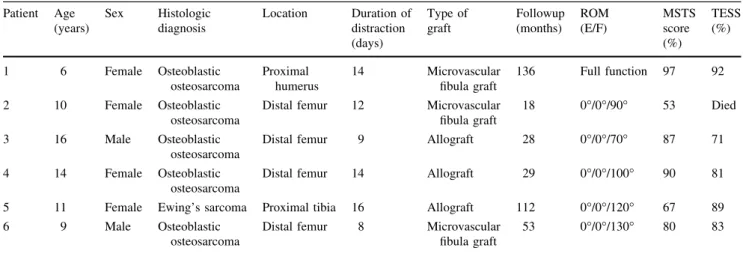
From 1998 to 2007, six patients (two boys, 9 and 16 years old, and four girls between 6 and 14 years old) with a malignant metaphyseal bone tumor underwent physeal distraction and subsequent joint-preserving tumor resection (Table1). Tumor location was the distal femur in four patients, the proximal humerus in one, and the proximal tibia in one. The histologic diagnosis was osteosarcoma in five patients and Ewing’s sarcoma in one. Preoperative staging revealed met-astatic disease in one patient with osteosarcoma. The
minimum followup was 18 months (median, 62 months; range, 18–136 months). Local tumor control was based on clinical and radiographic (plain radiographs, CT scans) information. Approval for collecting these data was obtained from the responsible ethics committee.
All patients received neoadjuvant chemotherapy. Patients with osteosarcoma were treated either according to the COSS-96 [5] or EURAMOS-1 [21] protocol. The EURO-EWING [14] protocol was used for the patient with Ewing’s sarcoma.
There was no delay in chemotherapy related to the placement of the external fixator and subsequent physeal distraction. Chemotherapy was commenced 2 to 5 months preoperatively and physeal distraction was begun 12 days (range, 8–16 days) before tumor resection.
All patients were operated on under the responsibility of the senior author (GUE). The surgical technique consisted of three parts: physeal distraction, resection of the tumor, and reconstruction of the defect [8]. The initial stage was application of an external fixator at an adequate distance from the tumor. The pins were stiff to allow direct trans-mission of mechanical forces to the physis with minimal risk of gradual malalignement. Distraction was commenced in the operating room and continued at the rate of 1 mm/ day. Separation of the epiphysis from the tumor-bearing metaphysis was monitored radiographically. Rupture of the physis occurred abruptly after 7 to 15 days and usually was accompanied by some discomfort. There were no pin tract infections in our series. Resection of the tumor and reconstruction were performed as soon as rupture of the physis had occurred. Reconstruction of the defect was performed with massive bone allograft or autograft or a combination thereof (Table1).
Postoperative results were evalutated at final followup by one individual (MB) using the Musculoskeletal Tumor Society (MSTS) score [13] and the Toronto Extremity
Table 1. Demographic and clinical data Patient Age (years) Sex Histologic diagnosis Location Duration of distraction (days) Type of graft Followup (months) ROM (E/F) MSTS score (%) TESS (%) 1 6 Female Osteoblastic osteosarcoma Proximal humerus 14 Microvascular fibula graft 136 Full function 97 92 2 10 Female Osteoblastic osteosarcoma
Distal femur 12 Microvascular fibula graft
18 0°/0°/90° 53 Died
3 16 Male Osteoblastic osteosarcoma
Distal femur 9 Allograft 28 0°/0°/70° 87 71
4 14 Female Osteoblastic osteosarcoma
Distal femur 14 Allograft 29 0°/0°/100° 90 81
5 11 Female Ewing’s sarcoma Proximal tibia 16 Allograft 112 0°/0°/120° 67 89 6 9 Male Osteoblastic
osteosarcoma
Distal femur 8 Microvascular fibula graft
53 0°/0°/130° 80 83
Salvage Score (TESS) [10]. In addition, active ROM of the knee or shoulder was recorded.
Results
At latest followup, five patients were alive and disease-free, and one patient with metastatic disease on first presentation had died from metastatic disease. No postoperative deaths were related to the procedure or local recurrence. All tumor resections resulted in local control until the end of followup. The mean MSTS score was 79% (range, 53%–97%) and corresponding mean TESS was 83% (range, 71%–92%) (Table1).
Two cases are presented in detail, one to illustrate the potential of the technique for functional preservation and the other to draw the attention to a possible complication.
Patient 1
A 6-year-old girl presented with arm pain after minimal trauma. MRI showed a metaphyseal tumor localized in the proximal humerus (Fig.1A). The tumor had no contact with the physis and biopsy revealed an osteoblastic oste-osarcoma. Neoadjuvant chemotherapy was given according to the COSS-96 protocol. The external monolateral fixator was applied (Fig.1B) and rupture of the physis occurred 11 days later (Fig.1C). Tumor resection and subsequent reconstruction of the bone defect with a vascularized fibula autograft were performed (Fig.1D). The resection margins were tumor free. The patient received postoperative che-motherapy according to the COSS-96 protocol. At her 10-year followup, the patient was disease free. She has a short upper arm ( 4 cm) (Fig.1E) but otherwise full elbow and shoulder function (Fig.1F).
Fig. 1A–F (A) A preoperative MR image shows the tumor not reaching the physis. AP radio-graphs show (B) the situation after application of the external fixator, (C) separation of the epiphysis from the metaphysis, (D) the situation 1 day after tumor resection and reconstruc-tion of the defect with a microvascularized fibula graft, and (E) a short upper arm ( 4 cm) at the 8-year followup. (F) Free shoulder function was seen at the 8-year followup.
Patient 2
A 10-year-old girl with osteosarcoma in the left distal femur (Fig.2A) received neoadjuvant chemotherapy according to the COSS-96 protocol. The girl refused amputation, rota-tionplasty, and endoprosthetic replacement proposed at other institutions. She accepted the proposed biologic recon-struction with a free microvascular fibula after physeal separation. Despite documented lung metastases, the resec-tion and reconstrucresec-tion using the proposed technique were performed as curative resection of the lung metastases appeared possible. The monolateral fixator was mounted and distraction began the following day. Twelve days later, 1 day before definitive tumor surgery was planned, radiography showed separation of the physis but possible rupture into the tumor similar to a Salter-Harris II fracture [18] (Fig.2B). This was confirmed by CT (Fig.2C). Surgery was performed with transepiphyseal resection, leaving the physis with the
tumor specimen but preserving the epiphysis. The bone defect was reconstructed with a microvascular fibula graft. Histologic analysis of the resected specimen showed tumor-free margins. Postoperative chemotherapy was performed according to the COSS-96 protocol. Excision of metastasis in both lungs was performed 2 months after tumor resection. Recurrence of metastasis in the left lung required an addi-tional intervention with metastasis removal 8 months after tumor resection. Five months later, mediastinal metastases were discovered. The patient refused further interventions. Active knee ROM (extension/flexion) of 0°/0°/90° was achieved 13 months postoperatively. Radiographs showed fusion of the reconstruction (Fig.2D). The girl died 18 months after the intervention at the age of 12 years. There was no local recurrence of the primary tumor.
Postoperative complications required a total number of 13 reoperations, which corresponds to an average of 2.2 reoperations per patient after tumor resection.
Fig. 2A–D (A) An MR image shows the tumor at diagnosis. (B) An AP radiograph shows separation of the physis 12 days after application of the external fixator. Rupture occurred into the tumor-bearing metaphysis (arrow). (C) A CT scan recon-struction confirms rupture into the tumor (arrow) 15 days after application of the external fixa-tor. (D) An AP radiograph shows fusion of the reconstruction 13 months after the intervention.
Complications included delayed wound healing (five patients), infection (two patients), nonunion of the graft (two patients), and others (four patients). The five patients with delayed wound healing were treated successfully by de´bridement and secondary wound closure. Covering was obtained without additional plastic surgery procedures. Allograft infection occurred in Patient 4, 8 months after tumor resection, and was treated successfully with systemic antibiotics (followup after infection, 21 months). For Patient 5, nonunion of the allograft-host junction and implant breakage were managed by revision osteosynthe-sis. Sixteen months later, allograft infection necessitated allograft removal and resection of the proximal lower leg. The foot was fixed to the remaining stump. A below-knee prosthesis was customized and well tolerated. For Patient 6, nonunion of the autograft-host junction was solved with partial autograft removal and simultaneous allograft reconstruction. The other complications included contrac-ture of the flexor hallucis longus and flexor digitorum longus muscle after fibula removal for autograft recon-struction in Patient 1 treated with two lengthening procedures for the flexor hallucis longus and flexor digi-torum longus tendon; vascular anastomotic leakage (femoral vessels) in Patient 3 requiring surgical revision 1 week after tumor resection; peroneal nerve palsy in Patient 4, 2 days after tumor resection owing to hematoma, requiring surgical exploration and decompression with full peroneal nerve recovery; and leg length discrepancy ( 4 cm) of the surgically treated leg in Patient 6 requiring contralateral definitive epipysiodesis of the distal femoral physis 4 years after tumor resection.
Discussion
Complete tumor resection is the main objective in surgical treatment of bone sarcomas. In tumor surgery, physeal distraction can provide a safe margin of excision [9] and allows for preservation of the epiphysis in the growing bone of children and adolescents. Physeal distraction was first reported by Can˜adell et al. [8] in 1994. We therefore analyzed and reported our results with this technique by assessing tumor control, functional outcome, and compli-cations in all our patients treated with Can˜adell’s technique.
Our study has two major limitations. First, our study group is small and might not be representive of a larger collective. Second, we had no control group with another surgical technique.
In our small series, physeal distraction and subsequent tumor resection allowed for local tumor control until the end of followup. One patient died from preoperatively documented metastatic disease.
Can˜adell’s technique permits limb-sparing reconstruc-tion, which has been used increasingly in recent years without compromising oncologic principles [4]. Limb-sparing surgery is superior to amputation in terms of function [2] . In our series, the functional outcome, with an MSTS score of 79% and a TESS of 83%, is similar to that of other limb-sparing procedures [2].
When compared with other ephiphyseal-sparing proce-dures such as transepiphyseal resection [17] or multiplanar osteotomy [3], physeal distraction delivers the advantage of greater intraoperative safety. The structure of the growth plate is highly complex with irregular surfaces. Consequently, transepiphyseal osteotomy or multiplanar osteotomy is more difficult to perform and may result in incomplete tumor resection [9]. Physeal separation by external fixator distrac-tion is the first part of tumor resecdistrac-tion. Physeal distracdistrac-tion is begun preoperatively and must be understood as a blunt dis-section. With the rupture of the growth plate, the metaphyseal osteotomy is already performed preoperatively and tumor resection can be completed by a diaphyseal osteotomy [9].
A prerequisite for Can˜adell’s technique is a clearly open physis and a physis not invaded by the tumor [20]. San-Julian et al. [20] reported good results even if the tumor was in close contact with the physis.
MRI is currently the most accurate method for evalua-tion of potential physeal involvement in osteosarcoma and Ewing’s sarcoma, with a sensitivity of 100% and the best accuracy compared with other imaging methods [20]. In Patient 2, we recognized physeal separation was not complete but had partially ruptured into the tumor (com-parable to a Salter-Harris II fracture [19]). Close contact of the tumor to the physis increases the risk that physeal distraction may not provide clear margins. We therefore consider transepiphyseal resection leaving the intact physis on the resection specimen in these cases.
Except for incomplete physeal separation in Patient 2, all other complications were related to reconstruction of the defect. Most of these complications occurred early after tumor resection and could be solved without any sequelae. The most severe complication was seen in Patient 5 for whom allograft infection required allograft removal and resection of the proximal lower leg. The knee could be preserved and a below-knee prosthesis provided an excel-lent functional outcome (Table1).
We suggest Can˜adell’s technique should be considered in the technical armamentarium for biologic reconstruction in the treatment of malignant bone tumors in children. The potential of the technique for functional preservation is illustrated in Patient 1; at her 10-year followup, she has unlimited arm function. We believe it is important to draw attention to the complication of incomplete distraction and recommend careful monitoring to ensure the complete distraction of the physis.
Acknowledgments We thank Mikel San-Julian, MD, PhD, Department of Orthopedic Surgery, University of Navarra, Spain for his kind support regarding Patient 2 and his personal contributions for applying this technique.
References
1. Abudu A, Grimer R, Tillman R, Carter S. The use of prostheses in skeletally immature patients. Orthop Clin North Am. 2006; 37:75–84.
2. Aksnes LH, Bauer HC, Jebsen NL, Folleras G, Allert C. Haugen GS, Hall KS. Limb- sparing surgery preserves more function than amputation: a Scandinavian sarcoma group study of 118 patients. J Bone Joint Surg Br. 2008;90:786–794.
3. Avedian RS, Haydon RC, Peabody TD. Multiplanar osteotomy with limited wide margins: a tissue preserving surgical technique for high-grade bone sarcomas. Clin Orthop Relat Res. 2010; 468:2754–2764.
4. Ayerza MA, Farfalli GL, Aponte-Tinao L, Musculo DL. Does increased rate of limb- sparing surgery affect survival in osteo-sarcoma? Clin Orthop Relat Res. 2010;468:2854–2859. 5. Bielack S, Kempf-Bielack B, Schwenzer D, Birkfellner T,
Delling G, Ewerbeck V, Exner GU, Fuchs N, Go¨bel U, Graf N, Heise U, Helmke K, von Hochstetter AR, Ju¨rgens H, Maas R, Mu¨nchow N, Salzer-Kuntschik M, Treuner J, Veltmann U, Werner M, Winkelmann W, Zoubek A, Kotz R. [Neoadjuvant therapy for localized osteosarcoma of extremities: results from the Cooperative osteosarcoma study group COSS of 925 patients] [in German]. Klin Pa¨diatr. 1999;211:260–270.
6. Campanacci L, Manfrini M, Colangeli M, Alı´ N, Mercuri M. Long-term results in children with massive bone osteoarticular allografts of the knee for high-grade osteosarcoma. J Pediatr Orthop. 2010;30:919–927.
7. Can˜adell J, de Pablos J. Breaking bony bridges by physeal dis-traction: a new approach. Int Orthop. 1985;9:223–229.
8. Can˜adell J, Forriol F, Cara JA. Removal of metaphyseal bone tumours with preservation of the epiphysis: physeal distraction before excision. J Bone Joint Surg Br. 1994;76:127–132. 9. Can˜adell J, San-Julian M. Pediatric Bone Sarcomas. Berlin,
Germany: Springer; 2009.
10. Davis AM, Wright JG, Williams JI, Bombardier C, Griffin A, Bell RS. Development of a measure of physical function for patients with bone and soft tissue sarcoma. Qual Life Res. 1996;5:508–516.
11. de Pablos J, Can˜adell J. [Elongation of the lower extremities: experience at the University Clinic of Navarre][in Spanish]. Rev Med Univ Navarra. 1987;31:43–52.
12. de Pablos J, Villas C, Can˜adell J. Bone lengthening by physial distraction: an experimental study. Int Orthop. 1986;10:163–170. 13. Enneking WF, Dunham W, Gebhardt MC, Malawar M, Pritchard DJ. A system for the functional evaluation of reconstructive procedures after surgical treatment of tumors of the muskulo-skeletal system. Clin Orthop Relat Res. 1993;286:241–246. 14. Juergens C, Weston C, Lewis I, Whelan J, Paulussen M, Oberlin
O, Michon J, Zoubek A, Juergens H, Craft A. Safety assessment of intensive induction with vincristine, ifosfamide, doxorubicin, and etoposide (VIDE) in the treatment of Ewing tumors in the EURO-E.W.I.N.G. 99 clinical trial. Pediatr Blood Cancer. 2006;47:22–29.
15. McDonald DJ, Scott SM, Eckardt JJ. Tibial turn-up for long distal femoral bone loss. Clin Orthop Relat Res. 2001;383:214–220. 16. Mercuri M, Capanna R, Manfrini M, Bacci G, Picci P, Ruggieri
P, Ferruzzi A, Ferraro A, Donati D, Biagini R, et al. The man-agement of malignant bone tumors in children and adolescents. Clin Orthop Relat Res. 1991;264:156–168.
17. Muscolo DL, Ayerza M, Aponte-Tinao L, Ranalletta M. Partial epiphyseal preservation and intercalary allograft reconstruction in high-grade metaphyseal osteosarcoma of the knee. J Bone Joint Surg Am. 2005;87(suppl 1):226–236.
18. Muscolo DL, Ayerza MA, Aponte-Tinao LA, Ranalletta M. Use of distal femoral osteoarticular allografts in limb salvage surgery: surgical technique. J Bone Joint Surg Am. 2006;88(suppl 1 pt 2):305–321.
19. Salter RB, Harris WR. Injuries involving the epiphyseal plate. J Bone Joint Surg Am. 1963;45:587–622.
20. San-Julian M, Aquerreta JD, Benito A, Can˜adell J. Indications for epiphyseal preservation in metaphyseal malignant bone tumors of children: relationship between image methods and histological findings. J Pediatr Orthop. 1999;19:543–548.
21. The European and American Osteosarcoma Study Group. EUR-AMOS I trial. Available at:http://www.euramos.org. Accessed November 14, 2010.