Received: 23 January 2013 / Revised: 1 April 2013 / Accepted: 21 April 2013 / Published online: 15 May 2013 # ISS 2013
Abstract
Purpose To prospectively characterize the MR appearance of the carpometacarpal (CMC) joint of the thumb in asymp-tomatic volunteers.
Materials and methods Thirty-four asymptomatic volun-teers (17 women, 17 men, mean age, 33.9 ± 9.2 years) underwent MR imaging of the thumb after approval by the local ethical committee. Two musculoskeletal radiologists independently classified visibility and signal intensity (SI) characteristics of the anterior oblique (AOL/beak ligament), the posterior oblique (POL), the intermetacarpal (IML), and the dorsoradial ligaments (DRL) on a three-point Likert scale. The thickness of all ligaments, cartilage integrity, and presence of joint fluid were assessed. The alignment of the first metacarpal base with the trapezium was quanti-fied on sagittal and coronal planes.
Results The ligaments of the CMC joint were constantly visible in all volunteers for the POL and IML, and in all but one for the AOL and DRL. On intermediate-weighted fat-saturated images the POL (65 %/74 % reader 1/reader 2) and DRL (58 %/64 %) were commonly of increased SI, while the IML had a striated appearance in 91 %/76 % of
subjects. The AOL showed a variable SI (36 %/42 % low, 27 %/27 % increased, 36 %/30 % striated). The IML was the thickest ligament with a mean of 2.9 mm/3.1 mm and the DRL the thinnest (1.2 mm/1.4 mm). There was a mean dorsal subluxation of 1.8 mm/2.0 mm and radial subluxation of 2.8 mm/3.4 mm of the metacarpal base. The AOL was significantly thicker in men (1.7 mm) than in women (1.2 mm; p = 0.02). Radial subluxation was significantly larger in men (3.4 mm) than in women (2.2 mm; p=0.02). No subluxation in palmar or ulnar direction was seen. Conclusions Radial and dorsal subluxation of the CMC joint can be a normal finding in a resting position at MR imaging. The CMC ligaments showed a considerable vari-ability of signal intensity with a typically striated IML; thickness of the AOL is typically less than 2.2 mm, of the POL typically less than 2.9 mm.
Keywords Carpometacarpal joint . Thumb . Magnetic resonance imaging
Introduction
The carpometacarpal (CMC) joint of the thumb is a saddle joint with a biconcave-convex shape. The trape-zium articulates with the first and second metacarpal base distally [1]. This unique morphology leads to a wide range of motion [2–4]. Stability is given by a ligament complex, which consists of five main compo-nents (Fig. 1): the anterior oblique ligament (AOL/beak ligament), the posterior oblique ligament (POL), the dorsoradial ligament (DRL), the intermetacarpal liga-ment (IML) and the ulnar collateral ligaliga-ment (UCL) [2,3,
5,6]. Due to the exceptional anatomy of the joint, it is lax and incongruent in the resting thumb position while it is stable and tightly congruent in the final phase of thumb opposition [4, 6, 7].
A. Hirschmann (*)
:
R. Sutter:
C. W. A. Pfirrmann Department of Radiology, Orthopedic University Hospital Balgrist, University of Zurich, Forchstrasse 340, 8008 Zurich, Switzerland e-mail: anna.hirschmann@balgrist.ch R. Sutter e-mail: reto.sutter@balgrist.ch C. W. A. Pfirrmann e-mail: christian.pfirrmann@balgrist.ch A. SchweizerDepartment of Orthopedic Surgery, Orthopedic University Hospital Balgrist, University of Zurich, Forchstrasse 340, 8008 Zurich, Switzerland
Isolated dislocation of the CMC joint is uncommon and occurs in less than 1 % of all hand injuries [6,8]. It is more often combined with a fracture of the first metacarpal base [7,9]. A pure dislocation of the CMC joint typically leads to a rupture of the DRL and POL [6–8]. Magnetic resonance (MR) imaging can be used to accurately evaluate these ligament injuries [5].
The ligamentous anatomy of the CMC joint has been extensively studied in cadaveric dissections [1, 10–18]. Normal CMC ligament appearance on MR images is often described as low signal intensity (SI) with a thin linear shape in all sequences [3,5], although there are reports of variable SI in asymptomatic healthy volunteers in elbow and ankle ligaments [19,20]. Only a few studies have addressed the complex MR anatomy of the CMC ligaments in detail [3,5]. Also, the spectrum of normal MR morphology of the CMC joint structures in healthy volunteers is not known. The purpose of this study was to prospectively characterize the MR anatomy and variants of the CMC joint in asymptom-atic volunteers.
Materials and methods
Thirty-four asymptomatic healthy volunteers (17 women, mean age 33.5 ± 9.8 years, range, 20–49 years; 17 men, mean age 34.4±8.8 years, range, 23–49 years) were pro-spectively included in this study. For each decade between 20 and 50 years, at least five women and five men were included. All volunteers completed a questionnaire to ensure that they had not undergone prior thumb and hand surgery, had no thumb and hand pain, had never seen a physician for thumb and hand complaints, had never injured the thumb and hand and did not suffer from a systemic inflammatory disease. Daily and labor activities of the thumb were
evaluated as well as the handedness. This study was ap-proved by the local ethics committee and informed consent was obtained from each volunteer.
MR imaging protocol
MR imaging was performed with a 1.5-T extremity scanner (Optima MR 430 s, GE Healthcare, Waukesha, WI, USA). All subjects were examined in the static resting position of the thumb in a dedicated 100-mm coil. The patient was sitting next to the extremity scan-ner, the arm was placed in the neutral position, the thumb rested parallel to the second digit. Intermediate-weighted fat-saturated (FS) images were acquired in the sagittal plane (repetition time ms/echo time ms, 2,000/24; section thickness, 2 mm; field of view, 67 mm; acqui-sition time, 5 min 24 s), in the coronal plane (2,039/25; section thickness, 2 mm; field of view, 135 mm; acqui-sition time, 5 min 29 s) and in the axial plane perpen-dicular to the first metacarpal bone (2,079/22; section thickness, 2.5 mm; field of view, 60 mm; acquisition time, 5 min 48 s). T1-weighted turbo spin echo images were obtained in the sagittal plane (598/14; section thick-ness, 2 mm; field of view, 67 mm; acquisition time, 4 min 55 s) and in the axial plane perpendicular to the first metacarpal bone (557/13; section thickness, 2.5 mm; field of view, 60 mm; acquisition time, 5 min 5 s).
Definition of ligaments
Anterior oblique/beak ligament (AOL)
The AOL has its origin at the palmar tubercle of the trapezium and attaches at the volar beak of the first metacarpal base (Figs. 1 and 2). The AOL runs in a Fig. 1 3D reconstructions show the volar view (a), the dorsal view (b),
and the radial view (c) of the carpometacarpal (CMC) ligament complex of the thumb. The dark (AOL) and white (POL) stripes demonstrate the ligament orientation. AOL = anterior oblique ligament/beak,
POL=posterior oblique ligament, DRL=dorsoradial ligament, IML= intermetacarpal ligament, 1st MC = first metacarpal bone, 2nd MC=second metacarpal bone, T=trapezium
sagittal orientation. The ligament spans volarly around the CMC joint and runs in a diagonal fashion from proximal-radial to distal-ulnar.
The AOL is composed of two layers, a superficial and a deeper intraarticular one. Most authors do not differentiate between these two layers and term the AOL“beak ligament” [4, 7, 10, 17, 21], while a few authors name the deep bundles“beak ligament” [3,16].
Posterior oblique ligament (POL)
The POL runs obliquely from the dorsal tubercle of the trapezium to the dorsoulnar tubercle of the metacarpal base (Figs. 1 and 2). Best visualization is in the sagittal imaging plane.
Dorsoradial ligament (DRL)
The DRL origins at the dorsoradial tubercle of the trapezium, attaches to the adjacent aspect of the metacarpal base and has a coronal orientation (Figs.1and3).
Intermetacarpal ligament (IML)
This extracapsular ligament arises from the radial base of the second metacarpal bone and runs transversally to the ulnar side of the first metacarpal base (Figs. 1 and 3). It is best visualized in coronal images.
Qualitative analysis
MR images were analyzed independently by two fellowship-trained musculoskeletal radiologists. They were blinded to the results of each other’s readings and to any information regard-ing the volunteers.
Visibility (no/yes) and SI (low as the normal low signal of ligaments and tendons/increased compared to the normal low signal of ligaments and tendons/striated) of the ligament complex (AOL, POL, DRL, IML) were evaluated on fluid-sensitive intermediate-weighted FS images. The cartilage (intact/inhomogeneous SI/presence of chondral defects) as well as the presence of joint fluid (no/minor/joint distension) was assessed.
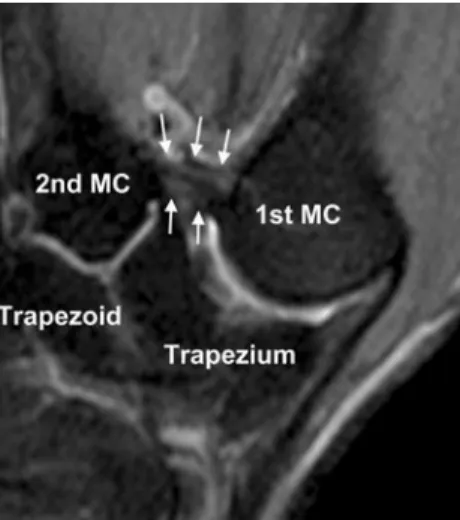
Fig. 3 a Coronal intermediate-weighted fat-saturated image (2,039/ 25 ms) shows the DRL (arrowheads), IML (white arrows), and abduc-tor pollicis longus tendon (black arrows) of the CMC joint of the thumb in a 33-year-old healthy volunteer. b Schematic drawing of
the DRL and IML at the same position as a. 1st MC=first metacarpal bone, 2nd MC=second metacarpal bone, AOL=anterior oblique liga-ment/beak, POL=posterior oblique ligament, APL=abductor pollicis longus tendon
Quantitative analysis
Thickness of the ligaments was measured at the midportion of each ligament (AOL, POL, IML, and DRL). For align-ment of the CMC joint, we measured the distance between the cartilage-covered portions parallel to the joint surface of the base of the first metacarpal bone and the trapezium (Fig. 4). This measurement was performed in the coronal and sagittal planes.
Statistical analysis
Descriptive statistics were used to report the quantitative and qualitative data of the CMC joint. Inter-rater agreement was assessed using kappa (κ) statistics for qualitative data and the intraclass correlation coefficient (ICC) for quantita-tive data. According to Landis and Koch [22], aκ value of 0–0.2 is indicative of slight agreement, 0.21−0.4 fair
agreement, 0.41−0.6 moderate agreement, 0.61−0.8 sub-stantial agreement and 0.81−1 almost perfect agreement. According to Rosner [23], the quality of inter-rater reliabil-ity by means of ICC was classified as following: > 0.75 excellent, 0.4–0.75 fair to good, and<0.4 poor.
Gender differences of the anatomical characteristics of the CMC joint were assessed using an independent t test. A p value of<0.05 was considered statistically significant. For all analyses, a statistical software (SPSS for Windows, release 17.0; SPSS, Chicago, IL, USA) was used.
Results
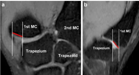
We scanned 44 % (15/34) dominant thumbs and 56 % (19/34) non-dominant thumbs. In 44 % of individuals (15/34) the thumb is particularly used in labor activities, such as typing (29 %; 10/34), physiotherapy (9 %; 3/34), Fig. 4 a Coronal (2,039/25 ms) and b sagittal (2,000/24 ms)
intermedi-ate-weighted fat-saturated images show radial subluxation (a) and dorsal subluxation (b) of the metacarpal base of the right CMC joint of the thumb in a 33-year-old healthy volunteer. The white lines reference the
cartilage-covered parts of the joint, the red lines show the measurement along the joint surfaces to assess subluxation distances. 1st MC=first metacarpal bone, 2nd MC=second metacarpal bone
Table 1 Qualitative characteristics of the carpometacarpal (CMC) ligament complex of the thumb in asymptomatic volunteers
Ligament Visibility Low SI Increased SI Striated SI Reader 1 AOL/beak 97 (33/34) 36 (12/33) 27 (9/33) 36 (12/33) POL 100 (34/34) 6 (2/34) 65 (22/34) 29 (10/34) IML 100 (34/34) 3 (1/34) 6 (2/34) 91 (31/34) DRL 97 (33/34) 33 (11/33) 58 (19/33) 9 (3/33) Reader 2 AOL/beak 97 (33/34) 42 (14/33) 27 (9/33) 30 (10/33) POL 100 (34/34) 12 (4/34) 74 (25/34) 15 (5/34) IML 100 (34/34) 12 (4/34) 12 (4/34) 76 (26/34) DRL 97 (33/34) 18 (6/33) 64 (21/33) 18 (6/33)
Data are percentage with absolute numbers in parentheses, calculated from the number of visible ligaments
AOL/beak anterior oblique ligament, POL posterior oblique ligament, IML intermetacarpal ligament, DRL dorsoradial ligament
SI Signal intensity in intermediate-weighted fat-saturated images, low as the normal low signal of ligaments and tendons, increased compared to the normal low signal of ligaments and tendons, striated with lamellar low and increased SI
and handcraft (6 %; 2/34). In 56 % of individuals (19/34), the thumb is not exposed to specific labor activities. Qualitative analysis
A summary of the qualitative results of both readers is shown in Table1.
The ligaments of the CMC joint were visible in all volunteers for the POL and IML and in all but one for the AOL and DRL with a perfect κ value of 1. On intermediate-weighted FS images the POL and DRL were commonly of increased SI. The IML had a striated appearance (Fig. 5). The AOL showed a variable SI distribution. The inter-observer agreement (κ) of the SI ranged from 0.3 to 0.5.
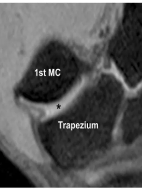
The chondral surface of the CMC joint showed in 26 %/35 % (reader 1/reader 2; 9/34 reader 1; 12/34 reader 2) an inhomogeneous SI in intermediate-weighted FS im-ages and in 15 %/29 % (5/34 reader 1; 10/34 reader 2) chondral defects were present (κ 0.4, Fig.6).
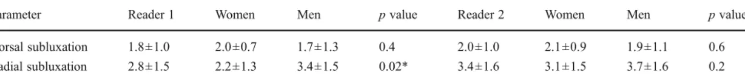
± 0.5, reader 1/reader 2) than in women (1.2 mm ± 0.6/1.4 mm ±0.6; p=0.02) for reader 1 (p=0.08 reader 2), while the IML was not substantially (p = 0.07 in both readers) thicker in men (3.2 mm ±1.1/3.3 mm±0.7) than in women (2.6 mm±0.9/2.8 mm±0.9). The POL (p=0.8/0.3 reader 1/reader 2) and DRL (p=0.2/0.4) showed no signif-icant gender differences in both readers. The overall ICC ranged from 0.2–0.8, while correlation for the AOL was good with an ICC of 0.8.
A mean dorsal subluxation of 1.8 mm ± 1.0/2.0 mm ± 1.0 and radial subluxation of 2.8 mm ± 1.5/3.4 mm ±1.6 of the metacarpal base was found (Fig. 4). Radial sub-luxation was significantly larger in men (3.4 mm ± 1.5/3.7 mm ± 1.6) than in women (2.2 mm ± 1.3/3.1 mm ± 1.5; p = 0.02/0.2) for reader 1 (Table 3). No subluxation in palmar or ulnar direction was seen. The ICC ranged from 0.5–0.8 with a good correlation for assessment of the radial subluxation (0.8).
Discussion
In our study, the ligaments of the CMC joint (AOL/beak, POL, IML, and DRL) were of variable SI ranging from low to increased SI. Even a striated appearance was observed, contrary to the low SI of the CMC ligaments reported in previous studies [3, 5]. In our study, the low SI was only evident in a minority of volunteers, with the least number of low SI cases for the POL and IML. In addition, the POL Fig. 5 Coronal intermediate-weighted fat-saturated image (2,039/25 ms)
shows the striated appearance of the IML (arrows) of a 26-year-old healthy female volunteer. 1st MC=first metacarpal bone, 2nd MC= second metacarpal bone, IML=intermetacarpal ligament
Fig. 6 a Coronal (2,039/25) and b sagittal (2,000/24 ms) intermediate-weighted fat-saturated MR images show loss of cartilage (in between the arrows) of the first metacarpal base and the trapezium in a 36-year-old asymptomatic volunteer. 1st MC=first metacarpal bone
showed an increased SI (65–74 %) and the IML a striated appearance (76–91 %) in the majority of individuals.
In previous imaging studies with MR and ultrasound [5,
17], the deep and superficial layers of the AOL could not be differentiated due to the small dimension and the close relationship without interposing tissue. A recently published study [3] distinguished these two bundles using MR arthrography. In our study, in which a dedicated extrem-ity scanner was used without administration of intraarticular contrast, we found a striated AOL in about 1/3 of the cases, which might be due to the layered architecture of this ligament (Fig. 8). The variance of ligament SI of the AOL and POL in our study com-pared to the reported low SI of ligaments might be related to the magic angle effect, as the echo times of the coronal and sagittal intermediated-balanced FS se-quences were of 25 and 24 ms, respectively. Because of the horizontal orientation of the IML, the mostly striated appearance does not assume to be related to the magic angle effect, and instead might be due to the ligament morphology [30, 31].
In MR imaging of the thumb, we were consistently able to outline four ligaments—the AOL/beak on the volar side, the POL and DRL on the dorsal side, and the IML at the ulnar aspect of the joint. Just in one female volunteer we could not visualize the AOL, which might be due to its intracapsular location or an old rupture, even though the volunteers negated a prior trauma to the thumb. The DRL was well delineated, which might be due to the superficial location adjacent to the insertion of the abductor pollicis longus tendon (Fig.3). However, in one case the DRL was not present. The IML was well visualized in all individuals due to the extracapsular location. In anatomic dissections several, ligaments of the CMC joint were described [1,
10–18]. The capsular AOL and POL and the extracapsular IML have been described as three major components [4,7,
24,25]. There is still controversy about which ligament is the key stabilizer of the CMC joint. Some believe that the AOL is the most important stabilizer [10–12,26,27] while others believe that the dorsal ligamentous structures are more important [4, 7, 13–15]. Xu et al. [28] found an equal stiffness for the AOL, POL, and DRL in a computer model study.
The extracapsular UCL is a thin, narrow structure overlapping the AOL and cannot be distinguished from the AOL in ultrasound or MR imaging [17], this is in accor-dance to our experience. Therefore, the UCL was not ana-lyzed in this study.
The thickness differed in all four ligaments, with the IML being the thickest of all with 2.9 mm to 3.1 mm and the DRL being the thinnest with 1.2 mm to 1.4 mm. The thickness of the AOL with 1.4 mm to 1.6 mm corresponded very well to the mean thickness of 1.28 mm in an ultrasound study with ten cadavers and ten volunteers [17]. Our mea-surement of the DRL thickness (1.2 mm±0.5/1.4 mm±0.5) and the AOL thickness (1.4 mm±0.6/1.6 mm±0.6) is very similar compared to an anatomy dissection of fresh-frozen cadavers (0.9 mm±0.2 and 2.0 mm±0.1, respectively) [16]. The significant gender difference in thickness of the AOL in our study might be due to general tenderness in anatomic structures in women. The thinner AOL in women might be associated with a higher prevalence of thumb CMC Fig. 7 Coronal intermediate-weighted fat-saturated MR image (2,039/25
ms) shows minor joint fluid (asterisk) of the CMC joint of the thumb in a 33-year-old asymptomatic volunteer. 1st MC=first metacarpal bone
Table 2 Quantitative analysis of the ligament thickness of the CMC joint of the thumb in asymptomatic volunteers
Parameter Reader 1 Women Men p value Reader 2 Women Men p value AOL/beak 1.4±0.6 1.2±0.6 1.7±0.6 0.02* 1.6±0.6 1.4±0.6 1.7±0.5 0.08 POL 2.3±0.6 2.3±0.6 2.3±0.5 0.8 1.9±0.5 2.0±0.5 1.8±0.5 0.3 IML 2.9±1.0 2.6±0.9 3.2±1.1 0.07 3.1±0.8 2.8±0.9 3.3±0.7 0.07 DRL 1.2±0.5 1.3±0.5 1.1±0.4 0.2 1.4±0.5 1.3±0.4 1.5±0.6 0.4 Data are mean thickness and standard deviation in millimeter
* p value<0.05 was considered statistically significant
osteoarthritis in females compared to males [32], although Imaeda [1] described a thickened AOL of severely osteoar-thritic patients in a cadaver study.
The thickness of all CMC joint ligaments varied consid-erably in our study. Therefore, the thickness of these liga-ments is not necessarily a sign of ligament injury or early osteoarthritis. Scarring of ligaments or a recent injury may lead to increased SI and thickening of the CMC ligaments. However, we also found increased SI and ligament thicken-ing in healthy volunteers, so these signs should not be misinterpreted as a clear sign of abnormality when analyz-ing MR images.
In our study, inhomogeneous SI and irregularities of the cartilage of the CMC joint were detected in one-third of the individuals. These findings occur in asymptomatic volunteers. In MR imaging obtained in the resting position of the thumb, the base of the first metacarpal bone showed a subluxation in the dorsoradial direction in relation to the trapezium in all volunteers in our study (Fig.4).
According to the literature, recognizing the dorsoradial joint incongruity is crucial in observing posttraumatic changes, and also severe osteoarthritis is associated with joint subluxation [1]. We found a mean radial subluxation of 2.8– 3.4 mm and a dorsal subluxation of 1.8–2.0 mm. This is in accordance to an X-ray study of 69 healthy volunteers with a
range of radial subluxation of 1.72–8.69 mm [29]. This amount of subluxation should not be misinterpreted as a possible sign of prior trauma or osteoarthritis in a resting thumb position. The CMC joint is only congruent in thumb opposition [4, 7]. MR imaging of the CMC joint with the thumb in opposition is not comfortable. However, this posi-tion might help to differentiate a posiposi-tion-related subluxaposi-tion from an underlying posttraumatic or disease-related subluxa-tion. A statistically significant larger radial subluxation was observed in men compared to women in our study. We found a minor amount of joint fluid in most healthy volunteers (Fig.7), so this should not be misinterpreted as an early sign of osteoarthritis or synovitis.
The following limitations of our study have to be acknowl-edged. A rather small number of 34 volunteers were evaluated in this study. We scanned the thumb in a resting position and characterized the CMC ligaments in routine thumb imaging without special angulated planes to this joint. Due to the wrapping configuration of the ligaments, the detection might be more difficult in non joint-orthogonal planes. Due to the small dimensions of the analyzed ligaments, the quantitative measurements might be prone to higher variability, although interobserver reliability was on average acceptable. As this is an anatomical study of healthy volunteers, we did not correlate our findings with surgical findings or cadaveric dissections. Fig. 8 a Sagittal
intermediate-weighted fat-saturated (2,000/24 ms) and b T1-weighted (598/14 ms) MR images show a striated appearance of the anterior oblique ligament/beak ligament of the CMC joint of the thumb, deep (short arrows) and superficial (long arrows) bundles in a 32-year-old asymptomatic volunteer. *=POL (posterior oblique ligament), 1st MC=first metacarpal bone
Detecting and characterizing the morphology of the CMC ligament complex of the thumb in MR images may be helpful for hand surgeons when planning surgery in patients with CMC joint instability. Detailed information about the ligament morphology with variable signal inten-sities and radiodorsal subluxation of the joint in healthy volunteers is presented, which may be useful for MR eval-uation of carpometacarpal diseases of the thumb.
In summary, radial and dorsal subluxation of the CMC joint can be a normal finding in a resting position at MR imaging. The CMC ligaments of the thumb showed a con-siderable variability in MR imaging in healthy volunteers; the IML is typically striated. Thickness of the AOL is typically less than 2.2 mm, and of the POL typically less than 2.9 mm. Knowledge of the ligament integrity and joint alignment is helpful in analyzing MR images of the CMC joint in thumb injuries and osteoarthritis.
Conflict of interest The authors declare that they have no conflicts of interest.
References
1. Imaeda T, An KN, Cooney WP, Linscheid R. Anatomy of trapeziometacarpal ligaments. J Hand Surg. 1993;18A:226–31. 2. Lane LB, Henley DH. Ligament reconstruction of the painful,
unstable, nonarthritic thumb carpometacarpal joint. J Hand Surg. 2001;26 A:686–91.
3. Cardoso FN, Kim HJ, Albertotti F, Botte MJ, Resnick D, Chung CB. Imaging the ligaments of the trapeziometacarpal joint: MRI compared with MR arthrography in cadaveric specimens. AJR. 2009;192:W13–9.
4. Edmunds JO. Current concepts of the anatomy of the thumb trapeziometacarpal joint. J Hand Surg. 2011;36A:170–82. 5. Connell DA, Pike J, Koulouris G, van Wettering N, Hoy G. MR
imaging of thumb carpometacarpal joint ligament injuries. J Hand Surg Br. 2004;29B(1):46–54.
6. Bosmans B, Verhofstad MHJ, Gosens T. Traumatic thumb carpometacarpal joint dislocation. J Hand Surg. 2008;33A:438– 41.
7. Edmunds JO. Traumatic dislocations and instability of the trapeziometacarpal joint of the thumb. Hand Clin. 2006;22:365– 92.
8. Fotiadis E, Svarnas T, Lyrtzis C, Papadopoulos A, Akritopoulos P, Chalidis B. Isolated thumb carpometacarpal joint dislocation: a case report and review of the literature. J Orthop Surg Res. 2010;5:16.
9. Brownlie C, Anderson D. Bennett fracture dislocation - review and management. Aus Fam Physician. 2011;40(6):394–6.
10. Pellegrini Jr VD, Olcott CW, Hollenberg G. Contact patterns in the trapeziometacarpal joint: the role of the palmar beak ligament. J Hand Surg. 1993;18a:238–44.
11. Napier JR. The form and function of the carpometacarpal (CMC) joint. J Anat. 1955;89:362–9.
12. Bettinger PC, Linscheid RL, Berger RA, Cooney WP, Kai-Nan A. An anatomic study of the stabilizing ligaments of the trapezium and trapeziometacarpal joint. J Hand Surg. 1999;24A:786–98. 13. Strauch RJ, Behrman MJ, Rosenwasser MP. Acute dislocation of
the carpometacarpal joint of the thumb: an anatomic and cadaver study. J Hand Surg Am. 1994;19:93–8.
14. Van Brenk B, Richards RR, Mackay MB, Boynton EL. A biome-chanical assessment of ligaments preventing dorsoradial subluxa-tion of the trapeziometacarpal joint. J Hand Surg. 1998;23A:607– 11.
15. Cooney W, Chao E. Biomechanical analysis of static forces in the thumb during hand function. J Bone Joint Surg. 1977;59a:27–36. 16. Nanno M, Burford WL, Patterson RM, Andersen CR, Viegas SF. Three-dimensional analysis of the ligamentous attachments of the first carpometacarpal joint. J Hand Surg. 2006;31 A:1160–70. 17. Gondim Teixeira PA, Omoumi P, Trudell DJ, Ward SR, Blum A,
Resnick DL. High-resolution ultrasound evaluation of the trapeziometacarpal joint with emphasis on the anterior oblique ligament (beak ligament). Skeletal Radiol. 2011;40:897–904. 18. Doerschuk SH, Hicks DG, Chinchilli VM, Pellegrini VD.
Histopathology of the palmar beak ligament in trapeziometacarpal osteoarthritis. J Hand Surg. 1999;24a:496–504.
19. Husarik DB, Saupe N, Pfirrmann CWA, Jost B, Hodler J, Zanetti M. Ligaments and plicae of the elbow: normal MR imaging vari-ability in 60 asymptomatic subjects. Radiology. 2010;257:185–94. 20. Mengiardi B, Pfirrmann CWA, Vienne P, Hodler J, Zanetti M. Medial collateral ligament complex of the ankle: MR appearance in asymptomatic subjects. Radiology. 2007;242:817–24. 21. Pellegrini Jr VD. Pathomechanics of the thumb trapeziometacarpal
joint. Hand Clin. 2001;17:175–84.
22. Landis JR, Koch GG. The measurement of observer agreement for categorical data. Biometrics. 1977;33(1):159–74.
23. Rosner BA. Fundamentals of Biostatistics 5th Edition. Duxbury 2000
24. Bojsen-Moller F. Osteoligamentous guidance of the movements of the human thumb. Am J Anat. 1976;147:71–80.
25. Pagalidis T, Kuczynski K, Lamb DW. Ligamentous stability of the base of the thumb. Hand. 1981;13:29–35.
26. Eaton RG, Littler JW. A study of the basal joint of the thumb, treatment of its disabilities by fusion. J Bone Joint Surg. 1969;51a:661–8.
27. Eaton RG, Littler JW. Ligament reconstruction of the painful thumb carpometacarpal joint. J Bone Joint Surg. 1973;55a(8):802–6. 28. Xu L, Strauch RJ, Ateshian GA, Pawluk RJ, Mow VC,
Rosenwasser MP. Topography of the osteoarthritic thumb carpometacarpal joint and its variation with regard to gender, age, site and osteoarthritic stage. J Hand Surg. 1998;23a:454–64. 29. Wolf JM, Oren TW, Ferguson B, Williams A, Peterson B. The carpometacarpal stress view radiograph in the evaluation of trapeziometacarpal joint laxity. J Hand Surg Am. 2009;34(8):1402– 6.
30. Hayes CW, Paralleda JA. The magic angle effect in musculoskel-etal MR imaging. Top Magn Reson Imaging. 1996;8(1):51–6. 31. Bydder M, Rahal A, Fullerton GD, Bydder GM. The magic angle
effect: a source of artifact, determinant of image contrast, and technique for imaging. J Magn Reson Imaging. 2007;25(2):290– 300.
3 2 . Badia A . Arthrosc opy of the trapez iometa carpal a nd metacarpophalangeal joints. J Hand Surg Am. 2007;32(5):707–24.