HAL Id: tel-00721774
https://tel.archives-ouvertes.fr/tel-00721774
Submitted on 30 Jul 2012
HAL is a multi-disciplinary open access archive for the deposit and dissemination of sci-entific research documents, whether they are pub-lished or not. The documents may come from teaching and research institutions in France or abroad, or from public or private research centers.
L’archive ouverte pluridisciplinaire HAL, est destinée au dépôt et à la diffusion de documents scientifiques de niveau recherche, publiés ou non, émanant des établissements d’enseignement et de recherche français ou étrangers, des laboratoires publics ou privés.
Development of a binding assay between the HIV-1
envelope protein (gp120) and coreceptors
CCR5/CXCR4 by Surface Plasmon Resonance :
Screening and optimization of viral entry inhibitors
Bridgette Janine Connell
To cite this version:
Bridgette Janine Connell. Development of a binding assay between the HIV-1 envelope protein (gp120) and coreceptors CCR5/CXCR4 by Surface Plasmon Resonance : Screening and optimiza-tion of viral entry inhibitors. Agricultural sciences. Université de Grenoble, 2012. English. �NNT : 2012GRENV013�. �tel-00721774�
THÈSE
Pour obtenir le grade de
DOCTEUR DE L’UNIVERSITÉ DE GRENOBLE
Spécialité : CHIMIE ET SCIENCES DU VIVANT (218)
Arrêté ministériel : 7 août 2006 Présentée par
Bridgette Janine CONNELL
Thèse dirigée par Hugues LORTAT-JACOB
préparée au sein du Laboratoire Structure and Activity of
GlycosAminoGlycans (SAGAG), Institut de Biologie Structurale (IBS)
dans l'École Doctorale Chimie et Sciences du Vivant
Développement d’un test d’interaction entre la
protéine d’enveloppe du VIH-1 (gp120) et les
corécepteurs CCR5/CXCR4 par résonance
plasmonique de surface: Criblage et
optimisation d’inhibiteurs de l’entrée virale
Thèse soutenue publiquement le 16 Mars 2012, devant le jury composé de :
Dr. Hugues LORTAT-JACOB
DR, CNRS, Directeur de thèse, Examinateur
Dr. Fernando ARENZANA-SEISDEDOS
DR, INSERM, Institut Pasteur, Rapporteur
Dr. Marc PARMENTIER
Proff, Université Libre de Bruxelles, Rapporteur
Dr. Winfried WEISSENHORN
Proff, Unit for Virus Host-Cell Interactions (UVHCI), UJF, Examinateur
Dr. Anne IMBERTY
DR, CNRS, Invité
Je remercie Prof. David Bonnaffé pour son jolie image de mCD4-HS12 sur la
THÈSE
Pour obtenir le grade de
DOCTEUR DE L’UNIVERSITÉ DE GRENOBLE
Spécialité : CHIMIE ET SCIENCES DU VIVANT (218)
Arrêté ministériel : 7 août 2006 Présentée par
Bridgette Janine CONNELL
Thèse dirigée par Hugues LORTAT-JACOB
préparée au sein du Laboratoire Structure and Activity of GlycosAminoGlycans (SAGAG), Institut de Biologie Structurale (IBS)
dans l'École Doctorale Chimie et Sciences du Vivant
Development of a binding assay between
the HIV-1 envelope protein (gp120) and
coreceptors CCR5/CXCR4 by
Surface Plasmon Resonance:
Screening and optimization of viral entry
inhibitors
Thèse soutenue publiquement le 16 Mars 2012, devant le jury composé de :
Dr. Hugues LORTAT-JACOB
DR, CNRS, Directeur de thèse, Examinateur
Dr. Fernando ARENZANA-SEISDEDOS
DR, INSERM, Institut Pasteur, Rapporteur
Dr. Marc PARMENTIER
Proff, Université Libre de Bruxelles, Rapporteur
Dr. Winfried WEISSENHORN
Proff, Unit for Virus Host-Cell Interactions (UVHCI), UJF, Examinateur
Dr. Anne IMBERTY
DR, CNRS, Invité
Je remercie Prof. David Bonnaffé pour son jolie image de mCD4-HS12
V
This work is dedicated to John Anthony CONNELL and George THEMISTOCLEOUS
VI “The tipping point is that moment when an idea, trend, or social behavior crosses a threshold, tips, and spreads like wildfire.”
― Malcolm Gladwell,
VII
ABSTRACT
Cell-associated Heparan Sulphate (HS) binds the V3 loop of gp120 of HIV-1 thus aiding in viral infectivity. However, a soluble polyanion (HS12) has anti-viral properties once
conjugated to CD4 (mCD4-HS12), and showed nM activity against HIV-1 in vitro. Due to the
structural complexity of HS, screening differently sulphated-oligosaccharides to improve the molecule’s activity would be too cumbersome, thus in order to obtain a more specific, higher affinity and easier to produce moiety, collaborators synthesized HS mimetic peptides. We aimed to screen these peptides and other anionic molecules for their capacity to inhibit HIV-1 entry. Thus we set-up a platform whereby solubilised CCR5 and CXCR4 were immobilized on biosensors (biacore) and used to screen for molecules that inhibited gp120-CD4 binding to the coreceptors. To control the solubilization process, CXCL12, the natural ligand of CXCR4, was injected over the immobilized CXCR4. The affinities of CXCL12 isoforms (α and γ) for CXCR4 were calculated within the ranges of previously described values with different techniques thus proving the functionality of our system. We show for the first time that HS differently regulates the binding mechanisms of these two isoforms and we propose a novel mode of action for the unusually basic C-terminal of CXCL12 γ with CXCR4. The system was subsequently used to screen the inhibitory capacity of the HS mimetic peptides. Each peptide, [S(XDXS)n], contained amino acids that mimic the hydroxyl, carboxyl and sulphate groups on HS chains. The peptide containing sulphotyrosine residues, when conjugated to mCD4 (mCD4-P3YSO3), displayed nM IC50 for simultaneously inhibiting gp120 binding to
HS, CD4, antibody, coreceptors and HIV-1 infection in vitro. This is the first bivalent entry inhibitor that targets both R5 and X4 viruses and the concept of a HS-mimetic peptide lends itself to structural-functional analysis of HS chains binding to proteins, a novel technique in this field.
La gp120 du VIH-1 se fixe aux héparane sulfate (HS) cellulaires, par le biais de la boucle V3 ce qui favorise l'infectivité virale. Cependant, une polyanion solubles (HS12), conjugués à
CD4 (mCD4-HS12) a des propriétés antivirales et a montré in vitro une activité contre le
VIH-1 à de concentrations nM. En raison de la complexité structurale des HS, le criblage d’oligosaccharides différenciellement sulfatés pour améliorer l'activité de la molécule serait trop difficile. En vue d'obtenir une molécule plus spécifique, de plus haute affinité et plus facile à produire, des peptides mimant les HS ont été synthétisés par nos collaborateurs. Notre but était de cribler ces peptides pour leur capacité à inhiber l'entrée de VIH-1. Nous avons mis en place une plateforme permettant d’immobiliser CCR5 et CXCR4 solubilisés sur des biocapteurs pour cribler des molécules qui inhibent la liaison de gp120-CD4 aux corécepteurs. Pour contrôler le processus de solubilisation, CXCL12, le ligand naturel de CXCR4, a été injecté sur CXCR4 immobilisé. Les affinités des isoformes CXCL12 (α et γ) pour CXCR4 ont été calculées dans les fourchettes de valeurs précédemment décrites avec des techniques différentes prouvant la fonctionnalité de notre système. Nous montrons pour la première fois que les HS régulent différemment les mécanismes de liaison de ces deux isoformes et nous proposons un nouveau mode d'action pour le domaine C-terminal particulièrement basique de CXCL12 γ vis-à-vis de CXCR4. Le système a ensuite été utilisé pour cribler la capacité d'inhibition des peptides mimétiques du HS. Chaque peptide, [S(XDXS)n] contient des acides aminés qui imitent les groupes hydroxyles, carboxyles et sulfates des HS. Le peptide contenant des résidus sulphotyrosines, une fois conjugué à mCD4 (mCD4-P3YSO3), montre
un IC50 de l’ordre du nM, pour l’inhibition simultanée de la liaison de gp120 aux HS, à CD4,
aux anticorps, aux corécepteurs ainsi que l’infection par VIH-1 in cellulo. Il constitue le premier inhibiteur bivalent de l’entrée qui cible à la fois les virus R5 et X4 et le concept d'un peptide mimétique des HS se prête à une analyse structurale et fonctionnelle de la liaison des chaînes HS aux protéines, une nouvelle technique dans ce domaine.
IX
ACKNOWLEDGEMENTS
Tout d'abord, je tiens à te remercier, Hugues. Merci de m'avoir acceptée dans ton équipe, de m’avoir donné cette opportunité de faire une thèse sur le VIH, l’un de mes rêves - et d’avoir eu une telle confiance en moi. Merci d’avoir toujours eu une porte ouverte pour mes questions et les sujets scientifiques dont je voulais discuter. Tu as toujours été disponible pour débattre de l'orientation scientifique de mon projet, donner ton avis sur mes expériences au laboratoire et parfois, des conseils pour mon anglais! Lorsque mes expériences ne fonctionnaient toujours pas et que la situation était désespérée, tu m’as laissée persister jusqu'à atteindre la perfection / d’or (ou platine?)! Ces expériences ont été difficiles et ont nécessité une grande quantité de consommables, donc je te remercie de m’avoir permis, malgré le coût financier, de continuer à travailler jusqu'à ce qu’elles fonctionnent. Tes idées, ta manière pédagogique de penser et d'écrire sont des compétences que je vais essayer de garder avec moi. Je pense qu’ensemble, nous avons fait une bonne équipe dans la lutte contre le SIDA!
Je tenais à remercier sincèrement les membres de mon jury. Merci Fernando Arenzana-Seisdedos et Marc Parmentier d’avoir accepté d'examiner mon manuscrit. Merci également à Winfried Weissenhorn et Anne Imberty pour votre participation à ce jury. C'est un honneur pour moi de vous compter parmi les membres de mon jury et que vous ayez examiné mon travail. Veuillez trouver ici mes sincères remerciements pour la qualité de jugement que vous portez à ce travail et soyez assurés de ma profonde reconnaissance. Grazie mille a Carlo Petosa per aver partecipato alla mia “thesis advisory commitee” per i primi due anni del mio dottorato di ricerca.
Merci beaucoup à Sidaction. Sans votre soutien, mon rêve de réaliser une thèse au cœur de la recherche sur le VIH n'aurait pas été possible. Grâce à votre action de recherche sur le VIH en France, nous avons avancé sur le développement d'un inhibiteur d’entrée du VIH-1 et j'ai noué de précieux liens dans ce domaine. Nos réunions annuelles ainsi que ma participation à l'Université des Jeunes Chercheurs (UJC) 2011 m’ont apporté énormément d’enthousiasme et de confiance. Merci pour votre soutien financier, émotionnel et psychologique. L'épidémie du SIDA touchera à sa fin avec plus de personnes et d'organisations comme la vôtre. Merci tout particulièrement à Sophie Lhuillier et Paola De Carli pour votre soutien, vos encouragements et votre aide professionnelle. J’espère que nous resterons en contact pour toujours!
Je tiens également à remercier Françoise Baleux. Merci pour ton travail remarquable dans la synthèse des isoformes CXCL12 et leurs mutants. Aussi, je voulais te remercier pour ton travail d'expert (et unique au monde !) dans la synthèse des peptides qu'aucune entreprise américaine ne pourrait –réaliser, notamment le N-terminus tyrosine sulfatée de la CXCR4. Je suis sûre que les entreprises américaines vont essayer de t’embaucher (chasser la tête !) Grâce à toi - j'ai été en mesure d'avoir des résultats très intéressants au cours de ma thèse.
Je remercie également David Bonaffé, Yves-Marie Coic et Pascal Clayette pour votre travail approfondi et de haute qualité pour nos papiers Chem Biol. David, je te remercie pour la fameuse synthèse du HS12 qui était indispensable pour mes expériences ainsi que son grand frère mCD4-P3YSO3.
X Rabia, ma maman à Grenoble! Tu as été là pour moi non seulement au niveau professionnel mais aussi personnel. Tu m’as montré tellement de techniques et tu as été très patiente avec moi au début, lorsque mon franglais était à peine compréhensible. Tu es une super enseignante et une amie merveilleuse. Tu as toujours été là, souriante, quand j'avais besoin d'une amie qui ne me juge pas. Je chérirai toujours nos discussions et nos thés ensemble. Tes délices culinaires sont si savoureux qu'il est dangereux de les manger sans modération. Rentrer dans ma robe de mariée aurait été difficile si j'avais mangé tous tes cookies, muffins et macarons! Je te souhaite bonheur et succès dans tout ce que tu fais, car tu les mérites vraiment!
Romain - Merci pour tant de choses - mais surtout je te remercie pour ton merveilleux sens de l'humour! Lorsque tu m'as fait rire, c'était un rire qui venait directement du cœur! C'est l'une des qualités les plus précieuses! Même dans les mauvais jours, tu as su me faire rire et effacer mes tracas! Merci de mettre une telle ambiance conviviale dans le labo et merci aussi pour tes conseils scientifique qui étaient « vachement » bien. Et lorsque tu chantes j’ai immédiatement envie de le chanter à tue-tête avec toi! « Je ne suis pas un héros » dans le laboratoire. Ton énergie et ton enthousiasme débordant sont contagieux! Tu es comme un membre de ma famille et il m’est difficile de te dire au revoir.
Merci à Cédric pour avoir effectué la grande expérience RMN avec 15N CXCL12γ et la N-ter de CXCR4 tyrosine sulfatée. Merci aussi pour nos discussions intéressantes sur CXCL12.
L’ours blanc (Pascal. F) - Merci aussi pour ton humour! Il n’y a jamais eu une journée maussade lorsque tu étais un gagophile! Merci d'avoir apporté ta note d'humour dans le laboratoire. Je doute que toutes les expériences fonctionnent avec un gant en latex sur la tête et un ‘pipette-man’ en guise de pistolet - mais cela fait rire et fait profiter de la vie! Merci aussi de m'avoir appris le ski de fond - je n'oublierai jamais ça!
Merci beaucoup à Nicole Thielens pour tes conseils illimités et ta disponibilité pour toutes mes questions sur le Biacore, notre ami préféré! Merci aussi pour toute ton aide et l'utilisation de la plate-forme BIAcore. Je te remercie également pour nos quelques courses à pied ensemble - même si j'aurais aimé en faire plus!
Ma balle de golfe, Julia! J'ai vraiment eu de la chance de te rencontrer! Une telle âme gentille et douce - celle que je chéris d'avoir comme amie! J’ai éprouvé quelques difficultés pour m'intégrer dans une nouvelle culture, apprendre une nouvelle langue et refaire une vie si loin de la maison et de ma zone de confort! Tu as rendu mon expérience en France pour la peine et si facile! J'espère que nous resterons amies toute notre vie! Juju, merci aussi pour toutes les corrections que tu as apportées à mon française. Je n’aurais jamais être compris sans toi! Tu es la balle de golf qui a une place toute particulière et très essentielle dans mon pot de mayonnaise!
Els, ma binôme de bureau! Wow - nous avons partagé beaucoup de grands moments ensemble - votre mariage et le mien, et l'entrée de votre belle Louane dans ce monde! Je suis toujours étonnée de la façon dont tu gères le stress et la planification de ton travail et de ta vie ! Merci pour ton soutien et tes encouragements tout au long de ma thèse et je te remercie pour tous tes dîners chez toi et tes cookies!
XI Emilie, l’autre âme douce et gentille! Merci beaucoup pour ton soutien tout au long de ma thèse! J’ai vraiment pu apprendre de ta sagesse dans la vie et la façon dont tu persévères pour obtenir ce que tu souhaites et ce qui est juste. Je te souhaite sincèrement le travail parfait ici afin que vous puissiez créez votre famille grenobloise dans votre nid parfait!
Mat-Mat (Mathieu), même si cela ne fait pas trois ans et demi que je te connais, je sais déjà que j'aurais aimé faire ma thèse avec toi! Tu es toujours de bonne humeur et tu peux me faire rire à en pleurer! Ne t’inquiète pas, tu seras en mesure d'analyser les spectres RMN dans ton sommeil à la fin de ta thèse, donc ne stresse pas si cela te semble difficile aujourd'hui : tu vas devenir le Master!
Amal, Celia, Damien et Sébastien (dans l’ordre alphabétique!), même si nous avons cohabité moins longtemps, merci pour votre soutien - toutes les petites aides et interactions avec des gens sympathique comptent beaucoup vers la fin d'une thèse! Isa, merci à toi pour ta gentillesse et pour nos discussions sur la famille et la vie! Merci de m’avoir soutenue, professionnellement pour ma thèse et personnellement pour mon mariage! Merci de si bien prendre en charge la plateforme BIAcore, et d’organiser les réunions et la planification si professionnellement! Evelyne, Monique, Pascale, Sarah et Véro, merci à vous aussi pour votre soutien! Chaque fois que je suis venue pour faire bouillir l'eau pour un café, je savais que l'une des ‘filles du LEM’ serait là pour un échange amical. Merci pour cette atmosphère conviviale et amicale, et pour votre soutien dans mon travail et ma vie privée. Vous me manquiez toutes pendant les nuits de travail à côté du Biacore lorsque je prenais une tasse de café. Merci egalement à Phillipe, toujours un sourire dans les couloirs! Mickaël, l'épine parmi les roses - expression anglaise! Merci pour ton soutien - des mots ne peuvent décrire combien je te suis reconnaissante! Je te souhaite le meilleur emploi (avec le meilleur salaire) en Suisse et un mariage très bientôt!
Mel (Mélanie) merci pour les cours de patinage (que je l'espère, tu pourras recommencer avec un dos sain et cicatrisé bientôt) et pour ton soutien pendant les semaines avant mon mariage! Flo (Florian) merci aussi pour ta gentillesse! C’était un plaisir de travailler avec toi au laboratoire. J'espère que toi et Mel trouverez du travail dans le même pays afin de ne pas subir la même séparation que Fabio et moi.
Jean-Pierre, merci pour le Jazz et le cassoulet! Ta musique de jazz était si forte que je pouvais l'entendre à l’autre bout du couloir! - mais ça m'a fait sourire! Chaque âme a besoin de bonne musique et d’un bon cassoulet au chaud!
Mes copines de ‘déjeuner en anglais’ – qui n’ont jamais duré très longtemps!’ – Blandine et Linda. Comme la vie est belle au travail avec des collègues comme vous! Merci pour votre soutien et votre humour, et pour les courses à pied! Etre si proche de vous est certainement ce qui rend difficile de quitter Grenoble et l’IBS. J'espère que nous tiendrons notre plan et que je reviendrai pour l’Ekiden au moins - une bonne excuse pour vous revoir!
Merci à Sylvie pour ton travail indispensable! Sans toi, nos expériences prendraient dix fois plus de temps et l'activité et la productivité générale de tous les travaux de l'IBS seraient moindres. Merci pour ta persévérance et le travail physique que tu fais au quotidien! Merci aussi pour tes efforts lors de la coupe du monde de foot en 2010, afin de me faire sentir comme en Afrique du Sud!
XII Chères Marie-Claire, Winnie, Adrienn et Louise. Je vous remercie beaucoup pour nos déjeuners du mercredi qui ont nourri mon âme. Votre gentillesse et votre intérêt pour de ma vie professionnelle et personnelle m'ont mise à l’aise. Marie-Claire, ta passion d’aider les autres à apprendre le français est si puissante et merveilleuse. Je te remercie aussi pour toutes tes aides de traduction pendant ma thèse. Nous avons tellement de chance de te connaître et d’avoir une enseignante comme toi! Même si mes conjugaisons ne sont pas encore parfaites - je garderai précieusement notre temps ensemble. Francesca, tu as rejoint nos déjeuners le mercredi et tu as apporté une joie de vie que je ne vois plus beaucoup! Grazie mille bella per tutto il tuo sostegno e la tua gentilezza!
Merci pour notre amitié, ton soutien et ton encouragement Iulia. B! Tu as été là depuis que je suis arrivée à Grenoble et j’ai vraiment de la chance d'avoir une amie comme toi. J'espère que nous ne perdrons jamais le contact.
Daphna, merci pour ton air de bonheur et ton sourire qui est toujours la! Nos pauses café, les dîners, soirées poker et les cours de capoeira sera toujours de précieux souvenirs pour moi!
Merci Stephanie. R pour ton soutien et ton amitié! Tu es une autre raison pour laquelle je me sens si proche des gens français et pourquoi je ne veux pas quitter la France!
Elodie, merci d’avoir initié ce grand sujet du ‘VIH’ dans le laboratoire du ‘GAGs’ - mon projet a suivi le tien et je crois que la seule recherche qui en vaut la peine est la recherche sur le VIH! Merci pour ton aide et tes conseils pendant ces années. –j’ai apprécié en sachant que je pourrais t’appeler lorsque j'en ai besoin!
Obrigardo Izabel B. Ton sourire et la gentillesse va me manquer! - et bien sûr il y a de nombreux via fetratas que nous avons encore à faire ensemble!
Alain, merci à toi pour la livraison de tous nos colis!
Merci beaucoup egalment à Fabrice. L, Georges. E, Odile. K, Isabelle. D, Didier. D et Jean-Luc. P ! Sans vous, avoir une these n'est pas possible! Je vous remercie pour votre aide avec mon ordinateur, disque dur, les annonces, les commandes et pour assurer que les pompiers m'ont sauvé quand je me suis fait piquer par un abeille! Merci beaucoup à Tony M. pour la relecture gracieuse de mes remerciements, sans cela ils auraient été moins compréhensibles!
To my ‘colocs’! Thank you SO much Harry. Y for being like my brother to me here in Grenoble! Thank you for all your guidance (both spiritual and scientific!) and thank you for our great friendship – I hope it lasts a life-time! Thank you Hedi. H for being a great friend from the day I arrived in Grenoble! Thank you for your enormous generosity and kindness that really helped me from the start. Valeeee! è sicuramente preso in giro la vita a Grenoble! Mi hai insegnato italiano (tutte le parolacce) e mi fanno sempre ridere! Proprio quello che mi serviva dopo quelle 15 ore al giorno! Thank you Ana, Anne, Juliana and Péter! Not only were you all wonderful flat-mates – but you stood by me when I was passing through the most difficult times – writing up my PhD while separated from Fabio. Thank you for putting up with my stress and bad moods and minimalistic living styles, you were so understanding and kind, when what you gave me in return was humour, comfort (food and company) and friendship! You guys made it that much easier to get through such a tough time and I hope that
XIII our time spent living together – has forged friendships for life! Muchas Graçias! Merci Beaucoup! Obrigado! Köszönöm!
Thank you too Ali, Eliza, Min, Camille, Rafael, Danielle, Aymerik, Juliette, Marion and Julianna! You are the greatest friends! When I was so new here and could not put together an understandable French phrase – you made all that lonliness and strangeness disappear! Thank you for being such great friends!
David. B! Thank you so much for sending me all your emails with juicy news bulletins, articles, videos and basically any interesting tit bit of info on HIV-1/AIDS! You kept me in the ‘science-internet-loop’ which was so appreciated due to my non-existence of time to do it myself!
Thank you to my cousin Bianca! Not only for your help with the translations, but for being a phone call and a 3 hours train trip away! Knowing that you were so close, made my experience in France much easier! Thank you for always being there to comfort me when times were hard and to discover together some of the delights of living in France!
Grazie tanto alla mia nuova famiglia italiana; Pina, Bruno, Lucia, Willy e Ivan! Probabilmente sarei morta di fame se non fosse stato per la pasta deliziosa e ravioli già preparati da te Pina che mi hanno aspettato nel congelatore! Grazie Mille! Grazie a tutti per avermi accettato come vostra figlia/sorella e per avermi sostenuto durante tutto il mio dottorato di ricerca - Sono così fortunata ad avere una famiglia come voi! To my Bro – Thank you for loving me constantly and unconditionally! I feel like the luckiest sister in the universe to have had you sing to me on our wedding day! I hope that we will live closer together in this next post-PhD phase of my life!
Dearest Grandpah! It was a true honour to have you read and correct not only my Masters thesis, but also my PhD. Your passion for biology and research must have made its way into the gene pool that got passed down to me! Thank you for your support and guidance and meticulous screening of my work over the years! You definitely helped me refine and polish my work into a handsome state that I am very proud to show to the world.
Mom and Dad. I think this is the part of the acknowledgements that I stress the most for. How is it possible to capture in a mere few lines the depth and quantity of love and gratefulness that I have for you two…For almost 30 years, you have never stopped loving , encouraging, building me up, supporting me from all sides, helping me stay focused and on the path towards my goals. Thank you Mom for proof reading my work with such a fine toothed comb and for all that over-time that you made for me when you had your own stresses and pressures from your work. Dad, thank you for all the printing and collating and time you took out from your very busy days to support me. Financially, emotionally, psychologically and physically You both were always there and You never let me lose hope nor give up…I am here because of what You gave me. So thank YOU very very much!
Amore Mio. Io sono venuta qui per essere con te - sei stato la mia priorità. E per fortuna ho trovato il dottorato perfetto. Dal giorno che ti ho incontrato - Sapevo che tu sei l’uomo giusto per me. Sposarsi è stato il mio sogno e finire il mio dotoratto era il mio obiettivo. Mi sento la ragazza più fortunata del mondo dato che il mio sogno si è avverato ed ho raggiunto il mio obiettivo! Grazie per la vita meravigliosa che abbiamo avuto insieme finora - il mio sogno ora è che duri per sempre!
XV
TABLE OF CONTENTS
ABSTRACT ... VII
ACKNOWLEDGEMENTS ... IX
TABLE OF CONTENTS ...XV
LIST OF FIGURES ... XXI
LIST OF TABLES ... XXIX
A: INTRODUCTION ... 1
Chapter 1: HIV ... 3
1.1 The Global HIV/AIDS pandemic ... 3
1.1.1 Discovery and epidemiology ... 3
1.1.2 Heterogeneity ... 4
1.1.3 Origins and Classification ... 5
1.1.4 Transmission ... 6
1.1.5 Disease Pathogenesis and Progression ... 6
1.2 The structure and Life cycle ... 7
1.2.1 Viral particle and genome ... 7
1.2.2 Gp120 ... 8 1.2.3 Structure of gp120 ... 10 1.2.4 The V3 Loop ... 12 1.2.5 CD4 ... 13 1.2.6 HIV-1 Co-Receptors ... 14 1.2.7 Viral Entry ... 16
1.2.7.1 CD4-binding site and Coreceptor binding site ... 18
1.2.8 HIV-1 Replication ... 19
1.2.9 Assembly, maturation and budding ... 20
1.2.10 Host cells ... 22
1.2.10.1 DC-SIGN ... 23
1.2.10.2 Mannose Binding Proteins (MBP) ... 24
1.2.10.3 Galactosyl Ceramide (GalCer) ... 24
1.2.10.4 Heparan Sulphates ... 24 1.2.10.5 LFA-1 / ICAM-1 ... 25 1.3 Therapeutic Strategies ...25 1.3.1 Replication Inhibitors ... 25 1.3.2 Entry Inhibition ... 26 1.3.2.1 gp120-CD4 Binding Inhibitors ... 26
1.3.2.2 Gp120-coreceptor binding inhibitors ... 27
1.3.2.3 Monoclonal Antibodies ... 28
1.3.2.4 Fusions Inhibitors ... 29
1.3.3 Neutralising Antibodies ... 31
1.3.4 Vaccine and Pre-exposure Prophylaxis ... 31
Chapter 2: The Role of Glycosaminoglycans (GAGs) in HIV-1 attachment ... 33
2.1 The Glycosaminoglycan Families ...33
2.1.1 Galactosaminoglycans and Glucosaminoglycans ... 33
2.1.2 Heparin and Heparan Sulphate ... 37
2.2 Biosynthesis and Degradation of GAGs ...38
2.2.1 Biosynthesis and organisation ... 38
XVI
2.2.1.2 Chain Elongation ... 39
2.2.1.3 Chain Maturation ... 40
2.2.2 GAG Catabolism: Remodelling and Recycling of GAGs ... 42
2.2.3 GAG degradation enzymes in the laboratory ... 43
2.3 HS-Protein Interactions ...44
2.3.1 Structure-Function Relations ... 44
2.3.1.1 Specificity ... 46
2.3.2 GAGs as coreceptors and Internalisation ... 47
2.3.3 Capture, Release and Protection of proteins ... 48
2.4 Role of HS in pathogenic Infections...48
2.4.1 Attachment of bacteria and parasites ... 48
2.4.2 Attachment of viruses ... 49
2.4.2.1 HS binding to gp120 ... 49
2.4.2.2 Characterisation of the gp120/HS interaction ... 50
2.4.2.2.1 Gp120 binding to HS is linked to Tropism ... 50
2.4.2.2.2 The CD4 induced (CD4i) domain is an HS binding site... 51
2.5 Therapeutic applications of HS in HIV infection ...54
2.5.1 Anionic Binders ... 54
2.5.2 Concept and action of CD4-HS: a glycoconjugate that inhibits HIV-1 attachment and entry ... 55
2.5.2.1 The CD4 moiety, mCD4 ... 56
2.5.2.2 The HS Moiety, HS12 ... 56
2.5.2.3 mCD4-HS12 ... 57
Chapter 3: CXCL12 / Stromal Derived Factor 1 (SDF1), natural ligand of CXCR4 ... 59
3.1 General ...59
3.2 Chemokines - Nomenclature and classification ...60
3.3 Chemotaxis ...61
3.3.1 The Chemokine side ... 61
3.3.2 The Cell side ... 61
3.4 The CXCL12 chemokine ...63
3.4.1 Gene expression of CXCL12 ... 63
3.4.2 Structure of CXCL12α ... 63
3.4.3 Physiological roles of CXCL12 and pathogenic effects ... 64
3.4.4 GAGs, CXCR4, CXCR7 and CXCL12α ... 66
3.4.5 Characterisation of the GAG-CXCL12 complex ... 67
3.4.5.1 Activities of the GAG-CXCL12 complex ... 68
3.4.5.2 The GAG component of the complex ... 69
3.4.6 Liaison with CXCR4 (Proposed Model) ... 70
3.4.6.1 Characterisation of the CXCR4 N-terminus-CXCL12 complex ... 72
3.4.6.2 Signalling Activities of the CXCR4-CXCL12 complex ... 73
3.4.7 Oligomerisation ... 74
3.4.8 CXCL12γ ... 75
Chapter 4: The Objectives of This Project ... 79
4.1 Les objectifs du projet (sommarie en français) ...79
4.2 Objectives ...81
B: EXPERIMENTAL WORK ... 83
Chapter 5: GAGs differently effect the liaison of CXCL12α and CXCL12γ with CXCR4 ... 85
XVII
5.1 L’héparane sulfate régule de façon différentielle la liaison de CXCL12 α et γ avec
CXCR4 (sommaire en français) ...85
5.2 Introduction ...87
5.3 Results ...88
5.3.1 Preparation and control of cells expressing CXCR4 ... 88
5.3.2 Alternative methods to measure binding ... 89
5.3.3 Solubilization of the CXCR4 membrane protein ... 90
5.3.4 SPR Analysis of solubilized CXCR4 ... 91
5.3.5 The role played by GAGs ... 95
5.3.5.1 Hypothesis for the Role played by GAGs in the context of signalling ... 99
5.4 Discussion ...105
Chapter 6: A synthetic heparan sulfate-mimetic peptide conjugated to a mini CD4 displays very high anti-HIV-1 activity independently of coreceptor usage ... 111
6.1 Un mimétique synthétique de l’héparan sulfate-conjugué à un peptide mini CD4 a une activité anti-VIH-1 très élevée indépendamment de l'utilisation des corécepteurs (sommaire en français) ...111
6.2 Preface ...113
6.3 Introduction and Preliminary approach ...113
6.3.1 Generation of an HS12 differently sulphate Library ... 114
6.4 Results ...116
6.4.1 Surface Plasmon Resonance Screening platform ... 116
6.4.2 A synthetic heparan sulfate-mimetic peptide conjugated to a mini CD4 displays very high anti-HIV-1 activity independently of coreceptor usage ... 118
6.5 Discussion ...169
Chapter 7: Side-Projects ... 175
7.1 Screening of small natural molecules for HIV-1 entry inhibitory capacity ...175
7.2 Molecular mechanisms underlying the increase in resistance to chemokines of R5 viruses in HIV infection ...175
Chapter 8: Methods ... 177
8.1 Materials ...177
8.1.1 Biacore reagents and antibodies ... 177
8.1.2 Lipids and detergents ... 177
8.2 Cell Culture and solubilization of co-receptors ...178
8.2.1 Cell culture ... 178
8.2.2 Preparation of liposomes ... 178
8.2.3 Coreceptor Solubilization ... 178
8.3 Protein electrophoresis and Immunodetection of Proteins ...179
8.3.1 Immunoprecipitation ... 179
8.3.2 Protein Electrophoresis ... 179
8.3.3 Immunoblotting (Western Blot) ... 179
8.4 Removal of cell surface oligosaccharides ...180
8.4.1 Na Chlorate treatment ... 180
8.4.2 Enzymatic digestion ... 180
8.5 Separation of dodecasaccharides ...180
8.5.1 Quantification of HS ... 181
8.5.2 Estimation of purity ... 181
XVIII
8.6.1 CXCL12 binding to CXCR4 ... 181
8.6.2 Screening HS mimetic peptides ... 182
8.7 Flow cytometric analysis ...183
8.8 Enzyme-Linked Immunosorbent Assay (ELISA) ...183
8.9 NMR ...184
8.10 Standard Protocols and Recipies ...184
8.10.1 SDS-PAGE ... 184
8.10.1.1 Solutions ... 184
8.10.1.2 Resolving SDS-PAGE gels ... 184
8.10.2 Western Blot ... 185
8.10.2.1 Solutions ... 185
8.10.2.2 Transfer ... 185
8.10.3 Detergents and Lipids ... 185
8.10.3.1 Detergents ... 185 8.10.4 Lipids ... 187 8.10.5 SPR ... 187 8.10.5.1 The SPR Principle ... 187 8.10.5.2 Amine Coupling ... 188 C. DISCUSSION ... 191
Chapter 9: Conclusions and Perspectives ... 193
9.1 Conclusions et Perspectives (sommaire en français) ...193
9.1.1 Etude de la liaison de CXCL12 à CXCR4 ... 193
9.1.2 L'étude d’un inhibiteur d'entrée ... 195
9.2 Setting up the GPCR Immobilization Platform ...199
9.2.1 Study of CXCL12 Binding to CXCR4 ... 201
9.2.2 The Entry Inhibitor Study ... 208
9.2.2.1 mCD4-HS12 and mCD4-P3YS03 ... 209
PUBLICATIONS AND COMMUNICATIONS ... 215
XIX
ABBREVIATIONS
AIDS Acquired Immunodeficiency Syndrome
ARV Antiretroviral
ART Antiretroviral therapy
BSA Bovine Serum Albumin
CCR5 CC Chemokine Receptor 5
CXCR4 CXC Chemokine Receptor 4
CD4 Cluster of Differentiation number 4
CD4i CD4 induced region/domain
CDR Complementarity-determining region
CRF Circulating Recombinant Forms
DC Dendritic cells
DMEM Dulbecco’s Modified Eagle’s Medium
DNA Deoxyribonucleic acid
ECL Extracellular loops
EMC Extracellular Matrix
EI Entry Inhibitor
ELISA Enzyme linked immunosorbent assay
EMEA European Medical Agency
Env Envelope glycoprotein
env Envelope gene
GAG Glycosaminoglycans Gal galactose
GalNAc N-acetyl-β-D-galactosamine Glc glucose
GlcA Glucoronic Acid
GlcN Glucosamine GlcNAc N-acetyl-β-D-glucosamine
Gp Glycoprotein
GPCR G-protein coupled receptor
HAART Highly active antiretroviral therapy
HIV-1 Human Immunodeficiency virus type 1
HIV-2 Human Immunodeficiency virus type 2
HP Heparin
HR-1 First heptad repeat region
HTLV Human T-cell leukaemia virus
HR-2 Second heptad repeat region
HS Heparan Sulphate
HSPG Heparan Sulphate Proteoglycans
IC50 Half maximal inhibitory concentration
IdoA Iduronic Acid
IN Integrase
ka Association rate constant, with units as M-1s-1
kd Dissociation rate constant, with units as s-1
XX
kDa Kilo Dalton
LAV Lymphadenopathy-Associated Virus
LC Langerhans cells
LTR Long Terminal Repeat
MA Matrix
mAbs Monoclonal antibodies
MTCT Mother to Child Transmission
MVB multivesicular body
MW Molecular weight
nM Nanomolar
NH Amino terminus
NMR Nuclear Magnetic resonance
PBMC Peripheral blood mononuclear cells
PBS Phosphate Buffered Saline
PCP Pneumocystis carinii pneumonia
PCR Polymerase chain reaction
ρg/ml Picogram per millilitre
PHA Phytohemagglutinin
PI Protease Inhibitor
PIC Pre-integration complex
PR Protease
PSSM Position Specific Scoring Matrices
R5 CCR5 utilizing HIV-1
RANTES Regulated upon activation normal T-cell expressed and
secreted
RNA Ribonucleic acid
RPMI Roswell Park Memorial Institute Medium
RT Reverse transcriptase
RTI Reverse Transcriptase Inhibitor
RT-PCR Reverse transcription polymerase chain reaction
sCD4 Soluble CD4
SDF-1α Stromal cell-Derived Factor – 1 alpha or CXCL12
SIV Simian Immunodeficiency virus
SPR Surface Plasmon Resonance
TM Transmembrane
TPST-1 Tyrosylprotein Sulphotransferase
V3 Third variable loop
V5 Fifth variable region
wt Wild type
X4 CXCR4 utilizing HIV-1
UNAIDS The Joint United Nations Programme on HIV and AIDS
V Variable Region
Vif Viral Infectivity Factor
Vpr Viral protein R
XXI
LIST OF FIGURES
Figure 1.1Diagrammatic representation of the global prevalence of HIV infected adults and children living with HIV at the end of 2009 (UNAIDS 2011). ... 4
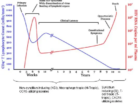
Figure 1.2 Early after primary infection there is widespread dissemination of virus and a sharp decrease in CD4+ T cells count in peripheral blood. The host launches an
immune response to HIV-1 characterised by a decrease in detectable viremia followed by a prolonged period of clinical latency. The CD4+ T-cell count continues to decrease during the following years (in un-treated patients), until it reaches a critical level below which there is a substantial risk of opportunistic infections (Pantaleo, Graziosi et al. 1993). ... 7
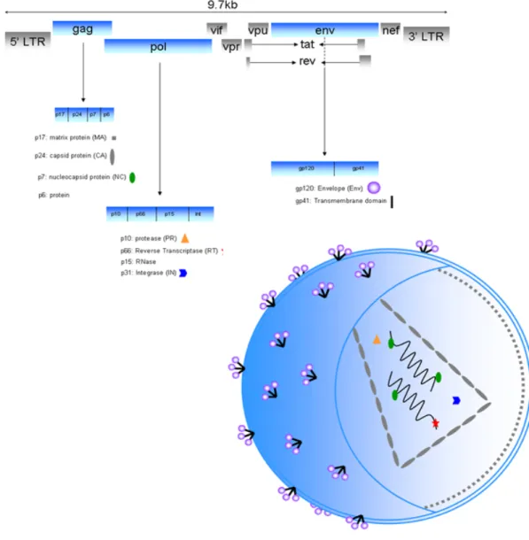
Figure 1.3 HIV structure and genome organisation: the 9 viral genes are depicted (9.7 kb) which encode open reading frames for at least 16 structural, regulatory, accessory and enzymatic proteins. The gag, pol and env genes encode protein precursors (pr55, pr160 and gp160 respectively) which require further processing by either viral or cellular proteases to generate structural proteins necessary for the formation of a mature virion. ... 8
Figure 1.4 Model of gp120 trimer from the orientation of the viral membrane. The gp120 core is a copper brown and carbohydrate core structures are blue. Picture taken from (Kwong, Wyatt et al. 2000) ... 9
Figure 1.5 Architecture and structure of gp120. (A) The gp160 protein is cleaved by furin to produce gp120 (Env, binds to CD4 and the coreceptors) and gp41 (transmembrane fusion protein). Crystal structures of unliganded (B) and liganded (C) gp120 adapted from Kwong et al. (Kwong, Wyatt et al. 1998) and Chen et al. (Chen, Vogan et al. 2005) ... 11
Figure 1.6 (Left) Fitting of the unliganded gp120 core (crystallographic structure) into a cryo-EM reconstruction of the unliganded HIV-1 spike. The outer and inner domains of the core gp120 are coloured red and gray, respectively, and the N/C extension blue. The stub of the V1/V2 loop is orange, whereas the stem of the V3 loop is green. The stems of the loops are additionally indicated by orange and green arrows. (Right) Fitting of the CD4-complexed gp120 core to cryo-EM reconstruction of the CD4 bound HIV-1 spike. CD4 binding loop (residues 364-374) are shown as spheres in yellow (left) and full length CD4 is in yellow on the right. Scale bar represents 50 Å. Adapted from Wu et al. (Wu, Loving et al. 2010) ... 12
Figure 1.7 Model taken from Wu et al., (A) This is a model of the entry complex in which the crystal structure of the CXCR4 homodimer has been placed below the structures of two gp120-CD4 complexes. V3 loops are show in magenta. (B) Close-up of the V3 loop (magenta) binding to hypothetical sulphotyrosines (circled in yellow) in the N terminus of CXCR4 at site 1 which then induces further conformational changes in gp120 allowing the V3 loop to interact with ECL2 and ECL3 at site 2. CXCR4 residues that have previously been shown to participate in gp120 binding are shown in orange and the hypothetical path of the N Terminus is shown as a blue dashed line, on the left of site 1. ... 15
XXII Figure 1.8 Schematic of the three classic stages of viral entry; initially the HIV-1 viral
particle approaches the host cell and gp120 binds to CD4, this liaison exposes/creates the CD4i coreceptor binding domain which then permits the gp120 to recognise and bind the coreceptor CCR5 and/or CXCR4. ... 17
Figure 1.9 (A) Averaged three dimensional structure of the native gp120 trimeric spike surface density map. (B) Front view of the surface density map fitted with the coordinates for gp120 core (red), the V1/V2 loops (yellow) and the V3 loop (green) derived from the complex with X5 (PDB ID 2B4C). (C and D) Front and top views of the X-ray coordinates of the ternary complex of the gp120 core (red) in complex with CD4 (yellow) and Fab fragment 17b (cyan). The arrow in C points to the likely location of the V1/V2 loops. (E and F) top view showing the change from unliganded (E) to CD4-bound (F) conformational change in the gp120 trimer, gp120, CD4, V1/V2 and V3 are shown in white, yellow, red and green respectively. (G) Schematic representation showing gp41 (blue), gp120 (red/purple) regions of the trimeric spike and the conformational changes associated with CD4 (yellow) binding. The yellow spots on the gp120 show where the CD4 potentially will bind the unliganded spike and the green dots on gp120 shown the position of the V3 loops post CD4 binding (Liu, Bartesaghi et al. 2008). ... 19
Figure 1.10 Schematic representation of the HIV-1 viral life-cycle. HIV-1 virions bind their host cell through the initial attachment to primary CD4 receptor and subsequent binding to the chemokine coreceptor CCR5 or CXCR4. Receptor binding induces fusion of viral and cellular membranes resulting in the release of the viral core and subsequent release of the viral genome into the cytoplasm of the host cell. The viral RNA genome is reverse transcribed into cDNA, transported into the nucleus where it is subsequently integrated into the host genome. The integrated provirus serves as a template for the transcription of viral genomic RNA copies as well as viral mRNA which is exported to the cytoplasm for translation. Structural and enzymatic proteins and two copies of the RNA genome assemble into nascent virion particles at the cellular membrane and bud from the cell. After their release, maturation occurs. Maturation is mediated by the protease that cleaves Gag during assembly into MA, CA, NC, SP2 an P6 proteins. ... 21
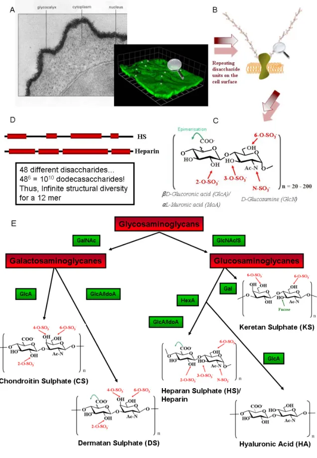
Figure 2.1 (A) This is an electron micrograph depicting a lymphocyte cell stained in ruthenium red showing the thick glycocalyx layer, which can reach up to 0.5µm. This is the interface through which the cell conducts its liaison for all biological processes (Alberts, Johnson et al. 2002). Heparan sulphates were immunostained with FITC-labelled antibodies and the image was obtained using a confocal microscope (Stevens, Hlady et al. 2007). (B) Glycosaminoglycan chains are shown covalently attached to their protein core imbedded in the cell membrane. (C) The HS disaccharide unit composed of a hexuronic acid and an N-acetylated glucosamine (4GlcA1-4GlcNAc 1) is repeated n times and can contain the following modifications: a de-acetylation of the GlcNAc and sulphation at this residue, sulphations at positions 3 and 6 on the GlcNS and on position 2 of the hexosamine and the C5 of the uronic acid can undergo epimerisation and change from a glucuronic acid (GlcA) to an iduronic acid (IdoA). (D) Domain organisation of HS and Heparin. Highly sulphated domains (NS domains - red) are the main component of heparin, and are less frequent in HS, where there is a larger occurrence of non-sulphated domains (NA domains). The domain
XXIII organisation is cell-specific and HS can be modified on so many levels, the structural diversity is vast and thus a vast number of protein binding sites exist. (E) The Glycosaminoglycan (GAG) family. GalNAc: N-acetyl Galactosamine, GlcNAc/S: N-acetyl / N-sulpho glucosamine, HexA: Hexuronic Acid, Gal: Galactose, GlcA: Glucuronic Acid, IdoA: Iduronic Acid. ... 36
Figure 2.2 The biosynthesis of CS (left chain) and HS (right chain) is initiated by the formation of the tetrasaccharide linker between the core protein ser-gly and the polysaccharide chain. Addition of the first hexosamine decides weather the chain becomes CS (GalNAc) or HS (GlcNAc). Taken from (Esko, Kimata et al. 2009) ... 39
Figure 2.3 Heparan sulphate biosynthesis involves copolymerization of N-acetylglucosamine and glucuronic acid residues. A series of modification reactions including sulphation and epimerization of glucuronic acid to iduronic acid occurs; chain polymerization and modification are thought to occur simultaneously (PAPS) 3′-phosphoadenyl–5′-phosphosulfate, the high-energy donor of sulphate groups. Taken from (Esko, Kimata et al. 2009) ... 42
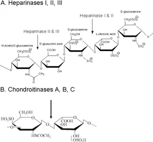
Figure 2.4 (A) The heparinases cut the oligosaccharide at the α1-4 glycosidic liaison between a glucosamine and a uronic acid (GlcA or IdoA). Heparinase I cuts between a hexosamine and a 2-O-sulphated uronic acid. Heparinase II cuts between a hexosamine and a uronic acid and heparinise III cuts between a hexosamine and a glucuronic acid. (B) Chondroitinase ABC cut between the N-acetyl hexosamine and the uronic acid. ... 44
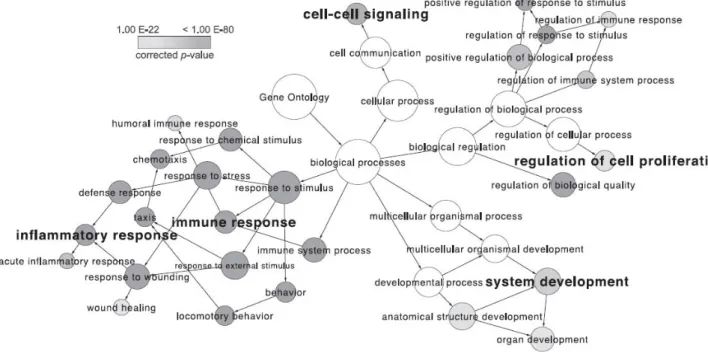
Figure 2.5 This is a gene ontology depicting the vast number of biological processes implicated in heparin/HS interactions, and thus called the ‘interactome’. Data for this map was provided from different databases of interacting proteins (e.g. NCBI Entrez GeneID). The node size is proportional to the number of heparin-binding proteins belonging to the functional category and the node shade or grey indicates the statistical significance (p value) of each pathways’ over representation (enrichment) in heparin binding proteins (HBP). I.e. the lighter the node, the stronger the enrichment in the interactome and the more studied the process is. Taken from (Ori, Wilkinson et al. 2011) ... 45
Figure 2.6 Surface Plasmon Resonance (SPR) binding curves showing the interaction between X4 (A) and R5 (B) tropic envelopes binding to a HS surface. 60nM of each envelope was injected and negative binding surface data was subtracted. Taken from (Lortat-Jacob, Fender et al. 2005). The binding responses (in RU) were recorded as a function of time (in s) – these parameters will be used for all SPR sensograms throughout the manuscript. ... 51
Figure 2.7 (A) 50nM of either gp120 alone (blue curve) or gp120 in the presence of equimolar amount of CD4 (red curve) over a Heparin surface. (B) Inhibition of gp120/CD4 (5 and 10nM respectively) complex binding to 17b on the sensor chip surface in the presence of different concentrations of heparin (0-16.7nM). 52
Figure 2.8 (A) Molecular modelling of the placement of a hexadecasaccharide of heparin onto the gp120 (HxBC2) crystallographic structure, showing that a dodeccasaccharide encompases both the V3 and CD4i binding sites. The MOLCAD surface of the gp120 is coloured according to its electrostatic potential (red for the basic residues and blue for the acidic residues). (B) Zoom up of the CD4i site with the basic amino acids involved in the GAG- binding
XXIV interaction annotated. (C) The gp120/CD4 complexes (5 and 10nM respectively) were co-incubated with different lengths of heparin oligosaccharides before injecting them over a 17b surface on the Biacore. A decasaccharide is the smallest fragment required for significant inhibition of the complex binding to 17b. Images adapted from (Vives, Imberty et al. 2005). ... 53
Figure 2.9 CD4-HS12 mode of action: the glycoconjugate binds gp120 through the
CD4 moiety which then induces the formation of the coreceptor binding domain via the synthetic CD4, followed by the high affinity interaction of the anionic HS12 domain with coreceptor binding domain. The glycoconjugate blocks both
CCR5 and CXCR4 viral entry. ... 56
Figure 3.1The association of chemokines (outer ring in grey) and their receptors (second ring from the outside in pink) and the associated disease (first three rings from the inside towards the outside in blue, green and yellow, for clinical data, human data and animal data respectively). A selection of disease associations obtained from animal models using gene deletions, neutralizing antibodies and receptor antagonists, as well as expression data in human samples and positive results from clinical trials. abbreviations: Sep, Sepsis; RA, Rheumatoid arthritis; T, Transplant; IBD, Inflammatory Bowel Disease; Onc, Oncology; SLE, Systemic Lupus; MS, Multiple Sclerosis; Ath Scl, Atherosclerosis; COPD: Chronic Obstructive Pulmonary Disease; AMD, Acute macular degeneration; NP, Neuropathic pain; Asth, Asthma; At. Derm, Atopic dermatitis; Hep, Hepatitis; Panc, Pancreatitis; Pso, Psoriasis; GVHD, Graft vs Host disease. (Garin and Proudfoot 2011) ... 60
Figure 3.2 A classical cartoon depicting the basic steps in cell migration in response to chemokine production. Chemokines are presented on the endothelial surface GAGs to chemokine receptors on leucocytes in the blood; chemokines may oligomerise on the GAGs. Whether the chemokines bind simultaneously to GAGs and chemokine receptors is not yet fully understood. Leukocyte recruitment is a multi-step process involving cytokines and chemokines driving selectin-mediated adhesion, subsequent arrest, firm adhesion, rolling and transmigration. This image was adapted from (Salanga and Handel 2011). ... 62
Figure 3.3 Alternate splicing of the two main isoforms relevant for this work, CXCL12α and CXCL12γ. The basic amino acids (K and R) that are highlighted in red in the sequences are amino acids that have been shown to be implicated in GAG-binding. ** KP signalling residues, ****** RFFESH initial contact/docking site with receptor and GAG-binding domains (BBXB) are indicated in the CXCL12γ sequence by the black brackets and the structures of each isoform are shown. Chemical shift variations upon GAG addition (dp4) are represented on CXCL12α and CXCL12γ in colour; Red residues bind the most to GAGs and orange residues bind less and yellow residues bind the least (Laguri, Sadir et al. 2007; Laguri, Arenzana-Seisdedos et al. 2008). ... 64
Figure 3.4 Model for the interaction between dimeric CXCL12α and an oligosaccharide (A) The CXCL12α is represented as a ribbon and the heparin oligosaccharide as well as the basic amino acids involved in the interaction are represented as sticks. Taken from (Sadir, Baleux et al. 2001). (B) Superimposition of 10 structures of the 13C labelled octasaccharide onto one CXCL12α ribbon structure. Taken from (Laguri, Sapay et al. 2010). ... 70
XXV Figure 3.5 The “two site” binding model for the CXCL12α-CXCR4 interaction.
Firstly, the N-loop of CXCL12α interacts with the CXCR4 N-terminus, this is followed by the N-terminus of CXCL12α binding the CXCR4 transmembrane region to trigger coreceptor activation (Crump, Gong et al. 1997). ... 71
Figure 3.6 Comparison of proposed binding of (A) the 38aa sulphotyrosine peptide binding to a CXCL12α dimer revealed by NMR. The zoomed section shows the side chains CXCL12 residues Val18, Arg47 and Val49 that are involved in binding the sTyr21 of CXCR4 (Veldkamp, Seibert et al. 2008) and in (B) a monomer CXCL12α binding to CXCR4 revealed by molecular modelling (Salanga and Handel 2011). ... 74
Figure 3.7 Analysis of CXCL12 binding to HP, HS and DS. SPR sensorgrams measured when CXCL12 was injected over HP, HS or DS activated sensorchips. The response in RU was recorded as a function of time (s) for CXCL12α (26 to 300 nM) and γ (2.6 to 30 nM). Adapted from (Laguri, Sadir et al. 2007). ... 76
Figure 5.1 Cartoon of the capture of C9 tagged GPCRs through their interaction with the 1D4 immobilized antibody and the binding of conformation specific antibodies, ligands and compounds. Adapted from (Navratilova, Dioszegi et al. 2006). ... 88
Figure 5.2 Cf2Th cells expressing either C-terminal C9 tagged CXCR4 or CCR5 (10x) and corresponding flow cytometric analysis depicting a positive stain for 12G5-FITC binding to CXCR4 expressing Cf2Th and 2D7 binding to CCR5 expressing Cf2Th cells. ... 89
Figure 5.3 The upper panels show cells that have been treated with 2% paraformaldehyde (pf) before staining with the 12G5-FITC secondary antibody and the lower panels were not fixed in pf. Panels on the left were performed with cells stored in 0.4M sucrose and panels on the right are fresh cells. Unlabelled CXCR4 expressing cells (red), pre-incubation with 5µg/ml T134 (dark and light green), pre-incubation with 12G5 (purple and pink), and cells directly labelled with FITC-12G5 (blue and orange) are shown. ... 90
Figure 5.4 (A) Representative sensorgram of EDC/NHS injection, 200µg/ml 1D4 immobilization and ethanolamine blocking on the CM4 sensor chip surfaces to ~7000 ru of 1D4. (B) Representative sensorgram of coreceptor immobilization via the C9 tag -1D4 interaction to ~3000 ru. ... 91
Figure 5.5 (A) 25nM 12G5 binding to immobilized CXCR4 in the absence (red curve) and presence (black curve) of 1µM T134 antagonist. (B) Native CXCL12α (50nM, blue curve), mutant CXCL12α 2-67 (red curve) and mutant CXCL12α 5-67 (green curve) binding to immobilized CXCR4. ... 93
Figure 5.6 (A) Binding of a range of concentrations (from top to bottom) 50, 30, 20, 10, 5nM of CXCL12α or the same range of concentrations of CXCL12γ (B) over immobilized CXCR4. The black traces correspond to the experimental data and the red traces correspond to the fitted data using a 1:1 langmuir model (A) and a 1:1 Langmuir model with mass transfer (B). ... 95
Figure 5.7 Effect of 1µg/ml HP12 (red) on binding of 50nM CXCL12α (blue) (A) and 50nM CXCL12γ (blue) (B) to CXCR4. ... 96
Figure 5.8 Systematic treatment of CEM cells with heparinase I and II and chondroitinase A, B,C prior to CXCL12 binding experiments. Cf2Th cell
XXVI digestion is shown to illustrate the efficacy of the GAG digesting enzymes as there are little to no GAGs on the CEM cell surface. For all graphs, red = unstained cells only, green = mouse FITC antibody, light blue = chondroitin-4-sulphate, orange = heparan sulphate (10E4) and pink = anti-chondroitin-4-sulphate. (A) Non-digested CEM cells, (B) GAG-digested CEM cells, (C) non-digested Cf2Th cells, (D) GAG-digested Cf2Th cells. ... 97
Figure 5.9 Effect of 1µg/ml HP12 on binding of 50nM CXCL12α and 50nM CXCL12γ to CXCR4 expressing, GAG-digested cells was tested using FACS analysis. CXCL12α (A) and CXCL12γ (B) binding to CXCR4 on CEM cells in the absence (magenta) and presence (blue) of 1µg/ml HP12. 12G5-FITC binding is shown (green) to demonstrate CXCR4 expression. The orange curve in (B) is the non-specific binding of the 1C12 antibody. ... 98
Figure 5.10 The basic amino acids (K and R) that are highlighted in red in the sequences are amino acids that have been shown to be implicated in GAG-binding. ** KP signalling residues, ****** RFFESH initial contact/docking site with receptor and GAG-binding domains (BBXB) are indicated in the CXCL12γ sequence by the black brackets and the structures of each isoform are shown. M1 is depicted, showing the BBXB HS binding motifs in the C-terminal being destroyed by mutation of certain basic residues to serines. Chemical shift variations upon GAG addition (dp4) are represented on M1 in colour; Red residues bind the most to GAGs and orange residues bind less and yellow residues bind the least (Laguri, Sadir et al. 2007). ... 98
Figure 5.11 (A) Effect of 1µg/ml HP12 (red) on binding of 50nM M1 (blue) on immobilized CXCR4 and 50nM M1 in the presence (blue) and absence (magenta) of 1µg/ml HP12 binding to CXCR4 expressing CEM cells (red). 12G5-FITC (green) is shown to demonstrate CXCR4 expression (B). ... 99
Figure 5.12 Injection of antisulphotyrosine alone over the CXCR4 surface (A [red curve], C) and injection of 50nM CXCL12γ onto CXCR4 directly followed by antisulphotyrosine (A [blue curve], E). The control injection of 4G10 onto the CXCR4 surface alone (B [red curve], D) or injection of 50nM CXCL12γ onto CXCR4 directly followed by 4G10 (B [blue curve], F). ... 100
Figure 5.13 15N labelled CXCL12γ was observed interacting with chemically synthesized peptides comprising the first 29 amino acids of the CXCR4 N-Terminus. (A) Amino acid residues that interact with both the sulphated and non-sulphated peptide (shown in red) and those that interact with the non-sulphated peptide (shown in blue). The amino acids that interact exclusively with the sulphated peptide are found both in the core domain and in the C-terminal of CXCL12γ. (B) The chemical shift perturbation observed for the CXCL12γ interacting with the non-sulphated peptide (red) overlayed with the chemical shift pertubations observed for CXCL12γ interacting with the sulphated peptide (blue) in the C-terminal region (CXCL12γ C-Ter). Single amino acids cannot be determined due to the repeating BBXB motifs in the CXCL12γ C-terminal and its non-structured mobility. ... 103
Figure 5.14 Proposed “chemokine interactome”. CXCL12α monomers signal similarly through CXCR4 in the absence (1) and presence (3) of GAGs. However, CXCL12γ signals very weakly though CXCR4 in the absence of GAGs (2) and a stronger signalling is seen in the presence of GAGs [preliminary
XXVII data] (4). Cell-surface glycosaminoglycans can induce dimer formation of the CXCL12α chemokine as can the sulphated N-terminal of CXCR4, which does not result in functional chemotaxis (5). ... 105
Figure 6.1 A Typical HPLC elution profile of HS12 showing absorbance at 232nm as a
function of time (min) and B a histogram corresponding to the quantities (in mg) of each separated HS12 library fraction. ... 115
Figure 6.2 Schematic representing the four different scenarios which were used when screening the various entry inhibitor molecules. Scenario A, B and C use streptavidin as a reference surface and scenario uses 1D4. Scenarios A has 17b as the test surface, B has biotinylated Heparan Sulphate (HSb), C has either full length CD4 or mCD4 and D has either CCR5 or CXCR4 solubilized coreceptors. For scenario C, either mCD4 or full length CD4 was immobilized and will be indicated in the text. ... 117
Figure 6.3 Binding of conformational sensitive (12G5 and 2D7) and non structural dependant antibodies (4G10 and 45502) onto solubilized CXCR4 (A) and CCR5 (B) respectively immobilized onto CD4 sensor chips through the high affinity interaction with 1D4. Cartoon diagrams represent the different coreceptors immobilized on the sensor chip surface. ... 118
Figure 8.1 Chemical Structure of DOM: MW = 510.6 g/mol, cmc = 0.17 mM (0.0087%), Aggregation number = 78-149 ... 186
Figure 8.2 Chemical structure of CHAPS: MW = 614.9 g/mol, cmc = 8 mM (0.49%), Aggregation number = 10 ... 186
Figure 8.3 Chemical structure of CHS: MW 607.9 g/mol ... 186
Figure 8.4 Chemical structure of DOPC and DOPS together with an electron micrograph taken of the extruded 100nm liposomes (DOPC:DOPS 7:3 [w/w]), 0.26mg/ml in 50mM HEPES 150mM NaCl ... 187
Figure 8.5 Binding is measured as a change in the refractive index as the sensor surface. A change of 0.001 degrees is equivalent to 1pg of protein bound per mm2. ... 188
XXIX
LIST OF TABLES
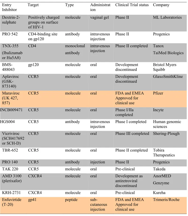
Table 1. Cell surface receptors implicated in binding HIV virions ... 23 Table 2: Overview of the different HIV-1 Entry Inhibitors (Castagna, Biswas et al.
2005; Kuritzkes 2009) ... 30 Table 3. Table showing the wide range of proteins that bind to heparin and heparan
sulphate adapted from (Capila and Linhardt 2002; Ori, Wilkinson et al. 2011) . 46 Table 4. List of physical attributes of the different CXCL12 isoforms used in this
study. ... 64 Table 5. Summary of the different buffers tested during the optimization of the
A: INTRODUCTION
Chapter 1: HIV
1.1 The Global HIV/AIDS pandemic
1.1.1 Discovery and epidemiology
Acquired Immunodeficiency Syndrome (AIDS) was first detected in May 1981 among four homosexual men in Los Angeles, United States of America who presented with infections such as Pneumocystis carinii pneumonia (PCP), Kaposi’s sarcoma, prolonged fever and Candida infections (1981; Gottlieb, Schroff et al. 1981). The apparent sexually transmitted immune deficiency in these patients was thought to be due to cytomegalovirus (CMV) infections in homosexual men, and called Gay-Related Immune Deficiency Syndrome (GRIDS) (1981; Gottlieb, Schroff et al. 1981; Hymes, Cheung et al. 1981; Masur, Michelis et al. 1981; Siegal, Lopez et al. 1981). However, this disease was not only seen in homosexual men; by 1983 groups of intravenous drug abusers, individuals receiving blood and blood products and heterosexual Haitians in America, presented with AIDS (1982; Harris, Small et al. 1983).
The causative agent of AIDS is a retrovirus that was first isolated from patients and demonstrated cytopathic effects on CD4+ T cells, which was clearly distinct from the Human T-cell leukaemia virus (HTLV) and was thus classified as a Lymphadenopathy-Associated Virus (LAV); thus a member of the T-lymphotropic retroviruses (Barre-Sinoussi, Chermann et al. 1983; Gallo, Salahuddin et al. 1984; Gallo and Montagnier 2003). This virus is now called Human Immunodeficiency Virus (HIV), the etiologic agent of AIDS. HIV-1 crossed the species barrier from chimpanzees to humans during the early twentieth century and has since infected millions of humans. Origins of HIV-1 have thus been linked to the simian immunodeficiency virus (SIV) from the genus Lentiviruses of the family Retroviridae (Chakrabarti, Guyader et al. 1987; Desrosiers and Ringler 1989; Gao, Bailes et al. 1999; Hillis 2000). Currently, one percent of the world’s population is infected with the worlds’ fastest evolving pathogen, HIV-1 (Korber, Muldoon et al. 2000).
AIDS is characterized by the progressive depletion of CD4+ T lymphocytes which play an important role in establishing and enhancing the cell-mediated and humoral immune response (Gottlieb, Schroff et al. 1981; Siegal, Lopez et al. 1981). When individuals suffer severe damage to their immune system, their vulnerability to opportunistic infections (OIs) and malignancies is heightened due to the loss of the individuals’ ability to mount an effective immune response. Ultimately death results after many years of untreated infection (Gallo, Salahuddin et al. 1984).
As worldwide efforts to create awareness, prevention and treatment programs increase, so does the total number of people living with the virus. According to the UNAIDS report on the global epidemic in 2010, there were 2.6 million newly infected people in 2009 and 1.8 million AIDS deaths, bringing the total number of people living with HIV-1 as reported at the end of 2009 to 33.3 million (UNAIDS 2011). These figures are almost equivalent to 7,123 new infections and 4.931
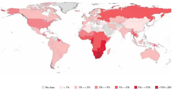
deaths per day due to AIDS. The emergence of this pandemic has arguably been the most catastrophic event in medicine in the last 30 years (Figure 1.1).
While all countries are currently fighting the impact of this disease, sub-Saharan Africa, and Southern Africa in particular continue to bear the greatest burden of people infected with and affected by HIV-1. Just over 10% of the world’s population inhabits sub-Saharan Africa, yet this region is home to 67.5% of people living with HIV-1 worldwide. In 2009, new infections in this region totalled more than those in all other regions of the world combined. In South Africa there are an estimated 5.6 million infected individuals which represents the largest number of individuals living with the virus in a single country.
Figure 1.1Diagrammatic representation of the global prevalence of HIV infected adults and children living with HIV at the end of 2009 (UNAIDS 2011).
1.1.2 Heterogeneity
Genetic diversity of HIV-1 exists along the entire length of the genome between viral isolates from different individuals and between viral quasispecies within the same individual. The unique and unstable characteristics of HIV-1 are its inherent variability and capability of generating quasispecies as a direct result of two features; lack of a proof-reading mechanism by the viral reverse transcriptase (RT) enzyme during replication (Roberts, Bebenek et al. 1988) and its rapid replication rate (Ho, Neumann et al. 1995; Wei, Ghosh et al. 1995). The error prone RT has an estimated misincorporation (insertions/deletions) rate of 1 x 10⁻⁴ - 3.4 x 10⁻⁵ per base pair per replication cycle (Preston, Poiesz et al. 1988; Roberts, Bebenek et al. 1988; Nowak 1990; Pathak and Temin 1990; Mansky and Temin 1995; Mansky 1998). This equates to about one nucleotide being miss-incorporated per replication cycle of 9.7 kb. This process is exacerbated by the high production of approximately 1 x 10¹⁰ viral particles daily and in the absence of proof-reading mechanisms, this results in extensive viral heterogeneity (Preston, Poiesz et al. 1988; Coffin 1995; Ho, Neumann et al. 1995; Wei, Ghosh et al. 1995; Perelson, Neumann et al. 1996; Zhang, Schuler et al. 1999).
Recombination between two RNA genomes also results in major gene-rearrangements and generation of diversity within the subpopulations within the host (Jung, Maier et al. 2002; Zhuang, Jetzt et al. 2002; Levy, Aldrovandi et al. 2004). Together, these features allow HIV to rapidly mutate its genome, enabling the virus to constantly evolve and increase genetic variability. This impacts on factors such as the genotypic viral diversity amongst different isolates, immune escape and emergence of Antiretroviral (ARV) drug resistance (Mansky 1998).
1.1.3 Origins and Classification
To date, two main types of HIV have been identified with origins as zoonotic lentiviruses; HIV-1 is believed to have originated from a SIVCPZ from the
chimpanzee (Pan troglodytes) population (Gao, Bailes et al. 1999; Santiago, Rodenburg et al. 2002) and HIV-2 is believed to have originated from the SIVSM
sooty mangabey (Cercocebus atys); SIV infections originated from mangabeys and ppears to be non-pathogenic, however, SIV causes AIDS-like symptoms in the Asian rhesus macaques (Gao, Yue et al. 1992; Rambaut, Posada et al. 2004). HIV-1 and 2 are transmitted in the same fashion yet HIV-2 has a lower rate of transmission, longer asymptomatic period and lower viral load; hence it is less pathogenic (Pepin, Morgan et al. 1991; Marlink, Kanki et al. 1994). HIV-2 is endemic in West Central Africa and to a lesser extent elsewhere in the world such as Europe and the West coast of India (Rubsamen-Waigmann, Briesen et al. 1991; Babu, Saraswathi et al. 1993). However, HIV-1 predominates worldwide and has a three times higher mortality rate than HIV-2 (Whittle, Morris et al. 1994). In addition to the two main types of HIV, further classification systems have been constructed from the copious phylogenetic data analyses of the many strains of HIV-1 and HIV-2 isolated and analyzed worldwide. There are four sub-classifications for HIV-1: groups, subtypes, sub-subtypes and circulating recombinant forms (CRFs). Of the groups, the Major group (Group M) is responsible for the current global pandemic (98% of HIV-1 infections worldwide) and the Outlier Group (Group O) and New group (Group N; consisting of non-O and non-M viruses) are less globally distributed. Groups O and N are genetically both highly divergent from group M and sparsely distributed in Cameroon and West Central Africa (Charneau, Borman et al. 1994; Mauclere, Loussert-Ajaka et al. 1997; Peeters, Gueye et al. 1997; Simon, Mauclere et al. 1998).
Group M is further subdivided up into 9 distinct subtypes, namely A, B, C, D, F, G, H, J, K wherein there are two sets of sub-subtypes A1, A2 and F1, F2 respectively (Louwagie, McCutchan et al. 1993; Robertson, Anderson et al. 2000). The emergence of Circulating Recombinant Forms (CRFs) has resulted from many recombination events between different HIV-1 viruses and already 34 CRFs have been described (Karlsson, Parsmyr et al. 1994; Casado, Thomson et al. 2005; 2007). These viruses share an identical mosaic structure in their genomes as they have descended from the same recombination events (Robertson, Anderson et al. 1999).
Phylogenetic analysis has revealed that the origin of HIV-1 came from four different cross-species transmissions from chimpanzees and one or two of these transmissions have been by gorillas (Sharp and Hahn 2010). It is generally
accepted that humans became infected by HIV-1 due to an inter-species transmission between SIV infected primates and humans. African people used to ingest simian meat that they hunted or acquired at "bushmeat markets". In this way, they were exposed to the contaminated meat.
1.1.4 Transmission
HIV-1 is transmitted through bodily fluids such as blood, semen and breast milk. Thus there are several pathways through which the virus can be transmitted between human beings; sexual transmission is the most common type of transmission. The epidemic in sub-Saharen Africa, which is responsible for almost 70% of the global infected population, is brought about (for the majaroity) by heterosexual transmission. However, in America and Europe, the epidemic is largly due to homosexual transmission. According to UNAIDS, the sharing of infected needles among injection drug users (IDU) is responsible for more than 80% of all HIV-1 infections in Eastern Europe and Central Asia. Another mode of HIV-1 transmission, also rife in sub-Saharan Africa, is the transmission from mother to child during natural child birth and during breast-feeding in conjunction with the use of formula milk.
1.1.5 Disease Pathogenesis and Progression
HIV-1 infection is characterised by a gradual deterioration in immune function and ultimately AIDS. Pathogenesis studies of HIV-1 explore the diverse mechanisms that lead to this immune system destruction and understanding how the virus establishes infection is essential to the identification and development of effective therapeutics and vaccines.
HIV-1 infection consists of an initial acute phase of infection followed by a period of clinical latency and finally a chronic phase. The acute phase is characterised by an increase in viral RNA (viral load) and the consequent decline in CD4+ T cells in peripheral blood (Clark and Shaw 1993). The activation of the immune system subsequently results in the suppression of viremia to a low steady state level termed the viral setpoint, and an increase in CD4+ T cells. During the clinical latency phase, viral load as well as the number of CD4+ T cells may remain constant for several years with the patient remaining largely asymptomatic. However the steady replication of HIV particles eventually overwhelms the immune system, resulting in a gradual rise in viremia and a steady decrease in CD4+ T cells until the patient is severely immunocompromised, resulting in increased susceptibility to opportunistic infections and the development of AIDS (Figure 1.2).