REVIEW
Translational medicine
Endothelial dysfunction over the course
of coronary artery disease
Enrique Gutie´rrez
1
*
, Andreas J. Flammer
2
, Lilach O. Lerman
3
, Jaime Elı´zaga
1
,
Amir Lerman
4
, and Francisco Ferna´ndez-Avile´s
1
1
Servicio de Cardiologı´a, Instituto de Investigacio´n Sanitaria, Hospital General Universitario Gregorio Maran˜o´n, Madrid, Spain;2
Cardiovascular Center, Cardiology, University Hospital of
Zurich, Zurich, Switzerland;3
Department of Nephrology and Hypertension, Mayo Clinic, Rochester, MN, USA; and4
Department of Cardiology, Mayo Clinic, Rochester, MN, USA Received 28 February 2013; revised 19 June 2013; accepted 27 June 2013; online publish-ahead-of-print 7 September 2013
The vascular endothelium regulates blood flow in response to physiological needs. Endothelial dysfunction is closely related to atherosclerosis
and its risk factors, and it constitutes an intermediate step on the progression to adverse events throughout the natural history of coronary artery
disease (CAD), often affecting clinical outcomes. Understanding the relation of endothelial function with CAD provides an important
patho-physiological insight, which can be useful both in clinical and research management. In this review, we summarize the current knowledge on
endo-thelial dysfunction and its prognostic influence throughout the natural history of CAD, from early atherosclerosis to post-transplant management.
-Keywords
Endothelial dysfunction † Coronary heart disease † Heart failure † Heart transplant † Acetylcholine
Introduction
The aim of this review is to provide a summary of the current
knowl-edge on endothelial dysfunction, particularly focused on its clinical
implications in coronary artery disease (CAD). Both coronary and
peripheral endothelial dysfunction will be discussed, as peripheral
endothelial function often serves as a surrogate for coronary vascular
function.
The vascular endothelium is a monolayer of cells covering the
in-ternal lumen of all blood vessels, thereby separating the blood
from the vascular wall and organ tissues. The vascular endothelium
serves different functions: (i) it is an anti-coagulant surface
1; (ii) it
reg-ulates fluid and molecule traffic between blood and tissues
2; (iii) it
contributes to the vascular homeostasis and repair
3; (iv) it plays a
crucial role in vascular tone and blood flow regulation.
4,5Cardiovascular risk factors produce endothelial dysfunction by
various complex mechanisms, among which oxidative stress is
con-sidered an important agent. Increased intracellular superoxide
pro-duces endothelial dysfunction by several mechanisms such as NO
inactivation and formation of peroxinitrite, NO synthase uncoupling,
prostacyclin formation inhibition, endothelin expression stimulation,
and a reduced NO signalling due to inhibition of soluble guanylate
cyclase activity.
6All these mechanisms are combined to promote a
vasoconstrictive and pro-coagulant milieu in endothelial dysfunction.
When endothelial function is altered, any of its functions could be
impaired. Yet, for practical reasons, it is the current standard to
measure endothelial function by the study of its vasomotor
regula-tion funcregula-tion. It is also customary to use the generic term ‘endothelial
function’ as a synecdoche for endothelium-dependent vascular
reactivity.
Since myocardial oxygen extraction is very high at basal conditions,
any additional metabolic demands must be met by an increase in
myo-cardial blood supply. In normal conditions, the coronary circulation is
capable of increasing its basal flow by at least three times. This
maximal increase in coronary blood flow is known as coronary
flow reserve (CFR). The main agent in this blood flow regulation is
the coronary endothelium, which produces vasodilator substances
such as nitric oxide (NO) and prostacyclin and vasoconstrictor
sub-stances, mainly endothelin-1, in response to different physiological
stimuli.
7Endothelial shear stress caused by arterial blood flow, and
autacoids such as acetylcholine, histamine, and bradykinin are the
main physiological triggers for endothelium release of NO, which
in turn produces a guanylyl-cyclase-mediated relaxation of vascular
smooth muscle (Figure
1
).
However, important, the vascular endothelium is not the only
determinant of coronary blood flow. Coronary microvascular function
can also be impaired due to other mechanisms as thrombi, debris
embolization, ventricular hypertrophy, myocardial, and vascular
*Corresponding author. Tel:+34 653992157, Fax: +34 915868275, Email:[email protected]
Published on behalf of the European Society of Cardiology. All rights reserved.
&
The Author 2013. For permissions please email: [email protected]European Heart Journal (2013) 34, 3175–3181
doi:10.1093/eurheartj/eht351
oedema, smooth muscle dysfunction, etc.
7,8When microvascular
function is impaired by an endothelium-independent factor, CFR is
reduced, limiting the overall vasodilator capacity.
Endothelium-inde-pendent microvascular dysfunction has been shown as a predictor of
adverse cardiovascular events in diverse settings from early
athero-sclerosis
9to post-stenting
10,11and acute myocardial infarction.
12Thus, although the focus of this article is set on the prognostic
import-ance of endothelial dysfunction, it has to be acknowledged that
micro-vascular endothelium-independent dysfunction can also be an
important source of flow dysregulation and adverse clinical outcomes.
Assessment of endothelial function
There are several ways to measure endothelial function in the clinical
setting. Although it is far beyond the scope of this review to discuss
the methodology in detail, we will very briefly summarize the most
common possibilities. For further reading, we refer to recent
com-prehensive reviews.
13,14All the techniques have in common that
they measure the response of the vessels to endothelial-dependent
stimuli, mainly reactive hyperaemia (shear stress) or vasoactive
substances.
The coronary vasomotion can be assessed directly and invasively
by coronary angiography.
15–17In principle, intracoronary vasoactive
stimuli, mainly acetylcholine, are infused to trigger an
endothelium-dependent vascular reaction. A functioning endothelium releases
NO in response to acetylcholine, causing vasodilation in the
epicar-dial arteries and the coronary microcirculation. Epicarepicar-dial
vasodilation is measured by quantitative angiography or intravascular
ultrasound (IVUS); vasodilation in the microcirculation is assessed
measuring the coronary blood flow with a Doppler wire, since the
microcirculation is the main determinant of coronary resistance,
and therefore of coronary blood flow. If the coronary endothelium
is dysfunctional, NO release is deficient, and paradoxical
vasocon-striction, due to direct muscarinic smooth muscle stimulation, is
observed in the epicardial arteries or the microcirculation.
Non-endothelial vasodilation can be measured using other drugs, such
as adenosine or nitroprusside for the microcirculation, and
nitrogly-cerine or nitroprusside for the epicardial arteries. Other
non-pharmacological approaches which have been described for
coron-ary endothelium-dependent vasodilation assessment are based on
flow-mediated dilation (FMD) in response to hyperaemia, like
exercise, mental stress or pacing, or on sympathetic nervous
system activation, like the cold pressor test.
The same principle of FMD can also be applied to the peripheral
vasculature
18: Briefly, ultrasound images of the brachial artery are
used to determine arterial diameter before and after reactive
hyperaemia-induced vasodilation. Reactive hyperaemia is achieved
by causing limb ischaemia with a blood pressure cuff for a few
minutes. Flow-mediated dilation is commonly expressed as per
cent change in artery diameter. This technique correlates with
inva-sively measured epicardial coronary function
19and has been widely
used in clinical research.
Peripheral arterial tonometry (PAT) is a newer technique based on
the change in finger pulse wave amplitude in response to reactive
Figure 1
Endothelium-derived vasoactive substances. Shear stress and activation of a variety of receptors leads to a release of nitric oxide by
inducing endothelial nitric oxide synthase. It exerts relaxation of vascular smooth muscle cells and exerts antiproliferative effects as well as inhibits
thrombocyte aggregation and leucocyte adhesion. Other endothelium-derived relaxing factors, including endothelium-derived hyperpolarizing
factor and prostacyclin, are also shown. ACE, angiotensin-converting enzyme; Ach, acetylcholine; AI, angiotensin I; AII, angiotensin II; AT1,
angioten-sin 1 receptor; Bk, bradykinin; COX, cyclooxygenase; ECE, ET-converting enzyme; EDHF, endothelium-derived hyperpolarizing factor; ETA and
ETB, endothelin A and B receptors; ET-1, endothelin-1;
L-Arg,
L-arginine; M, muscarinic acetylcholine receptor; PGH2, prostaglandin H2; ROS,
re-active oxygen species; S1, serotoninergic receptor; TX, thromboxane receptor; TXA2, thromboxane; 5-HT, serotonin. Reproduced with kind
per-mission from Springer Science and Business Media. Source: Springer. Pflu¨gers Arch- Eur J Physiol (2010) 459:1005 – 1013. Human endothelial
hyperaemia.
20A mini-cuff is used on the finger to record pulse
amp-litude, and another on the contralateral arm to serve as a control. This
relatively simple technique with a low observer-dependency indeed
correlates with microvascular coronary endothelial function.
21However, the observed reaction is only partly endothelial
depend-ent,
22and other factors affecting the microcirculation, such as the
sympathetic nervous system may affect this measurement.
20Endothelial dysfunction in early
asymptomatic atherosclerosis
The development of atherosclerosis is a continuous process starting
already early in life and has a long asymptomatic initial phase and a
slow progression caused and accelerated by the presence of different
risk factors.
23Importantly, endothelial dysfunction is one of the first
recognizable signs of its development and is present long before the
sometimes-devastating consequences of atherosclerosis appear.
Endothelial dysfunction has been reported in relation with most, if
not all, risk factors for atherosclerosis,
24–26such as hypertension,
27diabetes,
28hyperlipidaemia,
29and ageing.
30,31Endothelial
dysfunc-tion not only is a marker of atherosclerosis but it itself contributes
to the progression of atherosclerosis
32by various mechanisms, by
promoting coagulation, vasoconstriction, and deficient or
patho-logical vascular repair.
33Interestingly, endothelial dysfunction itself
can cause myocardial ischaemia even in the absence of relevant
cor-onary stenosis.
34Thus, it has been proposed that endothelial
dys-function constitutes a first stage of atherosclerosis, summarizing
the influence of all cardiovascular risk factors, and can itself be the
cause of cardiovascular events.
35As endothelial function is
asso-ciated with risk factors, the absence of endothelial dysfunction
might depict a particularly favourable state.
36,37In primary prevention, physicians mainly rely on classical risk
scores (e.g. Framingham or SCORE) to assess the risk for patients.
The measurement of endothelial function, however, as it depicts
the overall burden of risk and includes so far unknown factors,
might be a better measure for re-classification and personalized
decision-making.
For example, Li et al.
38recently showed a worse endothelial
func-tion, as measured by PAT, in patients with the metabolic syndrome
compared with those without it, even though both had the same
number of classical risk factors present. Interestingly, the Northern
Manhattan Study
39prospectively followed 819 subjects during a
mean of 81 months for cardiovascular events, focusing on the
incre-mental predictive value of FMD and its relation to metabolic
syn-drome. Patients with metabolic syndrome had a higher rate of
events (HR: 1.5), but patients with metabolic syndrome and
endothe-lial dysfunction had the highest rate (HR: 2.6), which shows an
incre-mental prognostic value of endothelial dysfunction. In type 2 diabetes
a normal peripheral endothelium-dependent vasodilation (as
estab-lished by an FMD .8%) has an excellent negative predictive value
for CAD.
37The cardiovascular Health Study
40evaluated the relation between
brachial FMD and cardiovascular events in a cohort of 2700 elderly
patients without known cardiovascular disease. Over a 5-year
follow-up period, FMD was a predictor of cardiovascular events, even after
adjustment for other risk factors. More recently, the multi-ethnic
study of atherosclerosis (MESA) study, by the same group, proved
the prognostic value of FMD independently of the Framingham
Risk Score in over 3000 patients followed over 5 years.
41Similar
results were obtained in a cohort of 2264 asymptomatic
postmeno-pausal women
42followed up for a mean period of 45 months, in
whom endothelial function was also evaluated by FMD. However,
in other series, as the FATE study
43and the PIVUS study,
44FMD
failed to add incremental prediction value or even correlate with
events (Table
1
). However, in the FATE study hyperaemic velocity,
a correlate of microvascular dilation, did correlate with events and
it provided additional independent prognostic value. Methodological
differences and a younger and presumably healthier population in the
FATE study may be accountable for this disparity.
Thus, the presence of endothelial function may serve as an
import-ant tool to re-classify the risk of the patients beyond the conventional
risk factors. Future studies thus have to better delineate which
patients benefit most from a measurement and which method is
best to assess endothelial dysfunction.
Endothelial dysfunction in
symptomatic patients without
obstructive coronary artery disease
Over 20% of the patients referred to a coronary angiogram due to
chest pain do not present significant obstructive coronary stenosis.
45Yet, a considerable proportion of these patients suffer from typical
angina, and often myocardial perfusion defects can be demonstrated.
Many of these patients present an abnormal coronary endothelial
function, which can result in chronic ischaemia and facilitate acute
ischaemic events, as explained below.
A study in 27 patients showed how abnormal flow response to
se-lective infusion of acetylcholine in the left anterior descending artery
(LAD) in patients with angina and non-obstructive CAD was related
to concordant exercise-induced myocardial perfusion defects on
SPECT.
46Lerman demonstrated that the perfusion defect could be
provoked at rest using the same method of selective LAD
acetylcho-line infusion.
34The results of these two studies show how coronary
vascular dysfunction without severe obstructive disease can be the
source of myocardial ischaemia due to endothelial dysfunction and
the resulting flow dysregulation.
Because the vascular endothelium not only regulates vascular tone
and flow, but has also important roles in vascular permeability and
thrombosis homeostasis, endothelial dysfunction not only may
lead to chronic stable myocardial ischaemia, but can also be the
source atherosclerosis progression
32and acute ischaemic events
(Figures
2
). One prospective study from the Mayo Clinic
17in 157
patients with mild CAD, and an invasive coronary endothelial
func-tion test by acetylcholine infusion in the LAD, showed coronary
endothelial dysfunction to be an independent predictor of
cardiovas-cular events. Schachinger et al.
47had similar results in another study
with 147 patients, using the same method. In two other studies
performed in diabetic patients without obstructive CAD, in whom
coronary endothelial function was tested by the cold pressor test,
coronary endothelial dysfunction showed a correlation with
myocar-dial perfusion defects
48and long-term adverse cardiovascular
events.
49. . . .
Table 1
Studies of cardiovascular events in patients with endothelial dysfunction and early or stable coronary artery disease
Study Population Number Method Endpoints Results
NOMAS39 Asymptomatic, .40 years old 819 FMD MACE (Stroke+ MI + CV death) OR 2.89 for patients with endothelial dysfunction and
metabolic syndrome; OR 1.64 (NS) for endothelial dysfunction alone. Cardiovascular
Health Study40
Asymptomatic, elderly (72 – 98 years old)
2792 FMD MACE, heart failure, revascularization, claudication Event free survival at 5 years 78.3 vs. 73.6 (FMD
.median vs. FMD ,median) P ¼ 0.006 MESA41
Asymptomatic 3026 FMD CV death, MI, angina, revascularization, cardiac
arrest, stroke
OR 0.79 FMD/unit SD; 29% correct reclassification of risk.
Rossi42
Asymptomatic postmenopausal women
2264 FMD MACE OR 4.42 for lower FMD tertile vs. higher FMD tertile
FATE43
Healthy, male, low-intermediate FRS 1574 FMD MACE OR 0.92, NS
Papaioannou37 Asymptomatic, type 2 diabetic 75 FMD Nuclear myocardial perfusion imaging FMD .8% had a negative predictive value of 93% for
myocardial ischaemia
PIVUS44 Asymptomatic, over 70 years old 1016 Invasive brachial—
FMD—radial tonometry
MACE Invasive brachial OR 0.72/SD (P ¼ 0.01). FMD and
tonometry no prognostic value Framingham third
generation PAT100
General population, Framingham 3rd generation
1957 PAT Relation to CV risk factors Correlation with male, BMI, total chol/HDL,
smoking, diabetes, lipid-lowering treatment Rubinhstein50 Symptomatic; low risk stress tests or
normal coronary angiogram
270 PAT MACE+ Revascularization + CV hospitalization Events 48 vs. 28% at 7 years, P ¼ 0.03
Suwaidi17 Symptomatic; no significant coronary atherosclerosis
157 Direct coronary MI+ CV death + heart failure + revascularization Only patients with severe endothelial dysfunction had events (14%)
Schachinger47 Symptomatic; no stenosis or single vessel
147 Direct coronary MI+ CV death + heart
failure+ revascularization + angina + stroke + peripheral revascularization
Patients with events had significantly poorer vascular response to acetylcholine
Halcox51
Symptomatic; with and without CAD 308 (132 with CAD; 176 without)
Direct coronary CV death+ AMI + unstable angina + stroke Patients with MACE had poorer response, both micro and macrovascular
Heitzer52
Documented CAD 281 Brachial
plethysmography
CV death, MI, stroke, revascularization Patients with events had lower responses Nitenberg49
Diabetic patients, no obstructive CAD; vs. non-diabetic controls
72 diabetics 56 controls Direct coronary, cold pressor test
Sudden cardiac death, MI, angina, stroke, TIA, revascularization
Diabetics with abnormal cold pressor test showed higher MACE rates
E.
Gutie
´rrez
et
al
.
In symptomatic patients without obstructive CAD, non-invasive
peripheral microvascular endothelial function measured by PAT is
also able to discriminate subjects at high risk for adverse
cardiovascu-lar events.
50Endothelial dysfunction in stable
coronary artery disease
In patients with stable obstructive CAD, endothelial function is an
in-dependent predictor of symptoms and cardiovascular risk. In one
study, 308 patients with and without CAD were studied by direct
coronary endothelial function testing. Abnormal response to
select-ive intracoronary acetylcholine infusion, both in the epicardial and
microvascular circulation, was an independent predictor of MACE,
even after adjusting for the presence of CAD.
51Another study in
281 patients with documented CAD showed that peripheral
endo-thelial dysfunction also identifies subjects at a higher risk for
cardio-vascular events.
52Diabetic patients constitute a high-risk population for
symptomat-ic and silent myocardial ischaemia. Two substudies of the Detection
of Ischemia in Asymptomatic Diabetics study evaluated the utility of
FMD to predict silent myocardial ischaemia
37and microvascular
damage as expressed by microalbuminuria.
53In these studies, a
cutoff value of 8% FMD showed a high negative predictive value for
silent myocardial ischaemia, and microalbuminuria correlated with
abnormal endothelium-dependent vasoreactivity.
Although endothelial dysfunction is a systemic condition, and thus
non-invasive functional tests correlate with invasive tests, endothelial
dysfunction in the epicardial arteries can also be focal. One study
showed a correlation between focal endothelial dysfunction and a
larger necrotic core, as measured by IVUS.
54This study provides a
ra-tionale for endothelial dysfunction being related to acute coronary
syndrome (ACS). Whether in this case endothelial dysfunction is
the cause or the expression of a vulnerable plaque is uncertain.
Endothelial dysfunction after
coronary artery stenting
Coronary stenting results in acute endothelial injury, sometimes even
associated with arterial dissection or haematoma, and in the
pres-ence of prosthetic metallic material in the vessel lumen; in the case
of drug-eluting stents (DES), this material is associated with an
artifi-cial polymer and a drug. A functional vascular endothelium prevents
platelet adhesion, aggregation, and activation by the secretion of
prostacyclin and NO, and maintains an adequate balance between
pro-coagulant (tissue factor) and anti-coagulant (heparin, protein
C/S) and thrombolytic factors (tissue plasminogen activator)
33; an
injured or dysfunctional endothelium loses its antiplatelet function
and may favour pro-coagulant activity, which in combination with a
reduced blood flow due to vasoconstriction can increase the risk
of stent thrombosis.
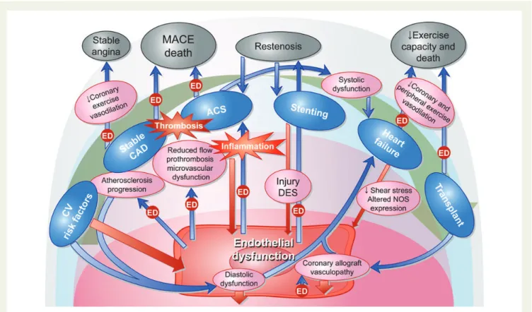
55Figure 2
Illustration of the reciprocal interaction between endothelial dysfunction, inflammation and the natural history of coronary artery
disease. Blue arrows marked with the box ‘ED’ represent processes in which endothelial dysfunction modifies the evolution or prognosis of coronary
artery disease. Red arrows represent ways in which coronary artery disease contributes to a worse endothelial function.
Several studies with DES have shown a persistently abnormal
endothelium-dependent vasoreactivity in coronary arteries after
DES implantation (Figure
3
). Both the sirolimus
56,57and the
pacli-taxel
58stents exhibit a detrimental effect extending over at least 6
months, while the second-generation zotarolimus stent seems to
have a more benign albeit not neutral behaviour.
59In the study by
Togni, endothelial response was evaluated by bicycle exercise,
while in the rest of the studies the method used was acetylcholine
infusion in the left coronary artery.
The new bioresorbable coronary scaffolds may be promising in
their ability to allow long-term restoration of coronary vasomotion,
especially at the stented segments. Data from the ABSORB stent
60,61(a polylactide bioresorbable everolimus-eluting scaffold) showed an
abnormal response of the segment distal to the scaffold at 12 months,
which improved significantly at 24 months; interestingly, the
scaf-folded segments showed improved endothelial-dependent and
inde-pendent vasomotion at 24 months of implantation, which was related
to scaffold degradation assessed by IVUS.
Lim et al. recently showed that in patients who were evaluated in
the catheterization laboratory due to chest pain (in average .1
year after stent placement) coronary vascular function was not
worse than in controls.
62Thus, although it is generally accepted
that coronary endothelial dysfunction may be impaired in the short
term, there might not be long-term relevance. Interestingly,
however, endothelial dysfunction as assessed by brachial FMD has
been reported as a predictor of in-stent restenosis.
63Endothelial dysfunction in acute
coronary syndrome
Most frequently, in ACS there is a local inflammatory status which
affects endothelial function and can make atherosclerotic plaques
more prone to rupture and platelet adhesion, vasospasm, and stasis,
which can precipitate coronary thrombosis.
64Microvascular function
is frequently impaired, but it is uncertain whether this is a contributor
or a consequence of the ACS, and it may be to a large extent
endothe-lial independent, due to thrombi and debris embolization, myocardial
oedema, and other factors. Endothelial function testing is particularly
challenging in ACS. The unpredictable nature of ACS onset, the
un-stable condition of the patient and the early dramatic impairment in
microvascular function, make it very difficult to obtain and interpret
data concerning vascular reactivity in this context.
Coronary endothelial dysfunction seems to be present in most
patients with acute coronary syndrome, and is reversible in a matter
of months.
65In patients with unstable angina, endothelium-dependent
vasoreactivity parallels inflammatory activity as expressed by
C-reactive protein levels.
66Figure 3
Illustration of the post-stent epicardial reactivity to acetylcholine in the various studies referenced in the text
56–62. The values express
the change in coronary artery lumen diameter (%) to acetylcholine or exercise, measured by angiography; horizontal short lines represent average,
and vertical bars standard deviation. The red vertical line separates drug-eluting stents from bare metal stents. As can be appreciated, patients with
drug-eluting stents exhibit a vasoconstrictive response, while patients with bare metal stents have a roughly neutral vascular response. AEES,
absorb-able everolimus eluting scaffold; BMS, bare metal stent; PES, paclitaxel eluting stent; DES, drug eluting stent; SES, sirolimus eluting stent; ZES,
zotar-olimus eluting stent.
Endothelial function after ACS, measured in the peripheral
circu-lation, has been shown as an independent predictor of MACE, and
subsequent normalization of endothelial function in these patients
predicts a lower risk of events.
67,68In patients with ST elevation
ACS treated with primary angioplasty, FMD improvement 6
months after the event also correlates with a lower end-diastolic
left ventricular volume.
69Endothelial dysfunction and heart
failure
Diastolic function is most frequently impaired in CAD, as a result of
hypertension, obesity or hypertrophy, but also due to a reduced
NO bioavailability in the myocardium. Nitric oxide plays a role in
myocardial as well as vascular relaxation,
70and there is animal
evi-dence that reduced NO availability due to reactive oxygen species
(ROS) and NOS uncoupling is a causal factor in the development of
diastolic dysfunction.
71In humans with CAD a high prevalence of
diastolic dysfunction has been found, which correlates with
endo-thelial dysfunction as assessed by FMD.
72Myocardial damage in
CAD is frequently not limited to diastolic function, but can lead
to systolic dysfunction due to myocardial necrosis and adverse
re-modelling, and it has already been mentioned how endothelial
dys-function impairs cardiac remodelling after an acute myocardial
infarction.
69Independently of the initial mechanism for heart failure,
endothe-lial dysfunction plays a major role in the progression of the disease and
has an important impact on clinical outcomes.
73–75The reduced
stroke volume produces a lower endothelial shear stress, which
causes a dysregulation in NO synthase isoforms gene expression,
76,77eventually leading to a reduced NO bioavailability.
78Furthermore,
there is an additional reduction in NO bioavailability caused by
direct NO destruction by ROS, mainly driven by an increase in
angio-tensin II and aldosterone activity, and purine metabolism.
79Both coronary and peripheral endothelial dysfunction have been
found in ischaemic as well as non-ischaemic heart failure patients,
80–82although peripheral endothelial dysfunction, measured by brachial
FMD, seems to be less important in non-ischaemic patients,
83suggesting
an incremental role of atherosclerosis in ischaemic patients.
The impaired peripheral endothelium-dependent vasodilation in
response to exercise may limit the oxygen supply to skeletal
muscles and thus have a negative impact on functional class.
Periph-eral endothelial dysfunction in heart failure is related to a worse
clin-ical outcome.
73–75Conversely, regular exercise training has proved
capable of restoring peripheral endothelial vasomotor function in
patients with heart failure,
84,85which is accompanied by a higher
ex-ercise capacity. Thus, the systemic endothelial dysfunction in heart
failure underscores the systemic nature of the disease and may be
used to assess the effectiveness of therapy and predict events.
Endothelial dysfunction in heart failure, measured by brachial FMD,
has been shown to be a pre-procedural predictor of good response
to cardiac resynchronization therapy (CRT) in one study. Also,
endo-thelial dysfunction improvement after CRT correlated with
function-al improvement.
86Two other studies have shown that CRT improves
endothelial dysfunction in heart failure, and that this improvement is
related to an increase in cardiac output, thus probably shear-stress
mediated.
87,88This may be one path by which CRT improves
symp-toms in heart failure.
Endothelial function after heart
transplant
After heart transplant the coronary endothelium is replaced by a
pre-sumably healthier one, stroke volume increases and so does
endo-thelial shear stress, one of the main inducers of NO formation.
However, the restoration of endothelial function is not immediate
or complete, probably due to the persistence of atherosclerosis
risk factors, inflammation and the use of immunosuppressive drugs
such as cyclosporine.
Indeed, a high prevalence of coronary endothelial dysfunction has
been described early after heart transplant.
89Coronary endothelial
dysfunction in early post-transplant patients has been correlated
with a higher risk of developing cardiac allograft vasculopathy
(CAV) as expressed by intimal thickening
90and clinical events.
91When allograft vasculopathy is evident, coronary endothelial
dys-function is the norm.
89Immunosuppression with cyclosporine
con-tributes to the development of endothelial dysfunction in
post-transplant patients through various mechanisms, such as a
decreased synthesis of NO,
92increased levels of endothelin
93and
increased production of ROS.
94Sirolimus seems to have a more
benign effect on endothelial function than cyclosporine.
95The evolution of peripheral endothelial function after heart
trans-plant remains controversial. Although it has been reported to
improve early after transplantation,
96other studies have not
con-firmed this observation,
97,98and indeed have found a high and
persist-ent prevalence of peripheral endothelial dysfunction in the first year
post-transplantation. Peripheral endothelial dysfunction limits the
vasodilator response to exercise, correlates with functional capacity
and may be the cause why heart transplant recipients often do not
achieve a normal functional status.
99Also, it has been found to
correlate with the risk of developing CAV in the first year.
97Conclusion
The assessment of endothelial function provides us with the ability to
obtain information on the functional significance of cardiovascular
disease at the different stages of the disease. There is a growing
body of evidence to suggest that mainly the non-invasive assessment
may intergrade into our practice to enable us to classify the risk of the
patients with CAD and assess the success of therapy.
Acknowledgements
The authors wish to acknowledge Dr Bernard Gersh for his valued
advice and encouragement.
Conflict of interest: A.L. is on the advisory board of Itamar Medical.
A.L.’s research is funded by the National Institutes of Health
(HL92954, HL77131, DK73608, AG31750) and the Mayo
Founda-tion.
References
1. de Agostini AI, Watkins SC, Slayter HS, Youssoufian H, Rosenberg RD. Localization of anticoagulantly active heparan sulfate proteoglycans in vascular endothelium: antithrombin binding on cultured endothelial cells and perfused rat aorta. J Cell Biol 1990;111:1293 – 1304.
2. van Hinsbergh WM. Endothelial permeability for macromolecules. Mechanistic aspects of pathophysiological modulation. Arterioscler Thromb Vasc Biol 1997;17: 1018 – 1023.
3. Urbich C, Dimmeler S. Endothelial progenitor cells: characterization and role in vascular biology. Circ Res 2004;95:343 – 353.
4. Furchgott RF, Zawadzki JV. The obligatory role of endothelial cells in the relaxation of arterial smooth muscle by acetylcholine. Nature 1980;288:373 – 376. 5. Griendling KK, Harrison DG, Alexander RW. Biology of the vessel wall. In: Fuster V,
Walsh R, Harrington R, (eds). Hurst’s The Heart. 13th ed. Mcgraw-hill; 2010. 6. Munzel T, Gori T, Bruno RM, Taddei S. Is oxidative stress a therapeutic target in
car-diovascular disease? Eur Heart J 2010;31:2741 – 2748.
7. Lu¨scher TF, Flammer AJ, Lerman A. Assessment of coronary vasoreactivity and the microcirculation. In: Eeckhout E SP, Wijns W, Vahanian A, van Sambeek M, De Palma R, (eds). Percutaneous Interventional Cardiovascular Medicine: The PCR-EAPCI Textbook. Toulouse, France: Europa Edition; 2012.
8. Lerman A, Holmes DR, Herrmann J, Gersh BJ. Microcirculatory dysfunction in ST-elevation myocardial infarction: cause, consequence, or both? Eur Heart J 2007;28:788 – 797.
9. Britten MB, Zeiher AM, Schachinger V. Microvascular dysfunction in angiographi-cally normal or mildly diseased coronary arteries predicts adverse cardiovascular long-term outcome. Coron Artery Dis 2004;15:259 – 264.
10. Albertal M, Voskuil M, Piek JJ, de Bruyne B, Van Langenhove G, Kay PI, Costa MA, Boersma E, Beijsterveldt T, Sousa JE, Belardi JA, Serruys PW. Coronary flow vel-ocity reserve after percutaneous interventions is predictive of periprocedural outcome. Circulation 2002;105:1573 – 1578.
11. Herrmann J, Haude M, Lerman A, Schulz R, Volbracht L, Ge J, Schmermund A, Wieneke H, von Birgelen C, Eggebrecht H, Baumgart D, Heusch G, Erbel R. Abnor-mal coronary flow velocity reserve after coronary intervention is associated with cardiac marker elevation. Circulation 2001;103:2339 – 2345.
12. Sorajja P, Gersh BJ, Costantini C, McLaughlin MG, Zimetbaum P, Cox DA, Garcia E, Tcheng JE, Mehran R, Lansky AJ, Kandzari DE, Grines CL, Stone GW. Combined prognostic utility of ST-segment recovery and myocardial blush after primary per-cutaneous coronary intervention in acute myocardial infarction. Eur Heart J 2005; 26:667 – 674.
13. Flammer AJ, Anderson T, Celermajer DS, Creager MA, Deanfield J, Ganz P, Hamburg NM, Lu¨scher TF, Shechter M, Taddei S, Vita JA, Lerman A. The assess-ment of endothelial function: from research into clinical practice. Circulation 2012;126:753 – 767.
14. Lekakis J, Abraham P, Balbarini A, Blann A, Boulanger CM, Cockcroft J, Cosentino F, Deanfield J, Gallino A, Ikonomidis I, Kremastinos D, Landmesser U, Protogerou A, Stefanadis C, Tousoulis D, Vassalli G, Vink H, Werner N, Wilkinson I, Vlachopoulos C. Methods for evaluating endothelial function: a position statement from the European Society of Cardiology Working Group on Peripheral Circula-tion. Eur J Cardiovasc Prev Rehabil 2011;18:775 – 789.
15. Ludmer PL, Selwyn AP, Shook TL, Wayne RR, Mudge GH, Alexander RW, Ganz P. Paradoxical vasoconstriction induced by acetylcholine in atherosclerotic coronary arteries. N Engl J Med 1986;315:1046 – 1051.
16. Cox DA, Vita JA, Treasure CB, Fish RD, Alexander RW, Ganz P, Selwyn AP. Ath-erosclerosis impairs flow-mediated dilation of coronary arteries in humans. Circu-lation 1989;80:458 – 465.
17. Suwaidi JA, Hamasaki S, Higano ST, Nishimura RA, Holmes DR Jr, Lerman A. Long-term follow-up of patients with mild coronary artery disease and endothelial dys-function. Circulation 2000;101:948 – 954.
18. Celermajer DS, Sorensen KE, Gooch VM, Spiegelhalter DJ, Miller OI, Sullivan ID, Lloyd JK, Deanfield JE. Non-invasive detection of endothelial dysfunction in chil-dren and adults at risk of atherosclerosis. Lancet 1992;340:1111 – 1115. 19. Anderson TJ, Uehata A, Gerhard MD, Meredith IT, Knab S, Delagrage D,
Lieberman EH, Ganz P, Creager MA, Yeung AC, Selwyn AP. Close relation of endo-thelial function in the human coronary and peripheral circulation. J Am Coll Cardiol 1995;26:1235 – 1241.
20. Kuvin JT, Patel AR, Sliney KA, Pandian NG, Sheffy J, Schnall RP, Karas RH, Udelson JE. Assessment of peripheral vascular endothelial function with finger arterial pulse wave amplitude. Am Heart J 2003;146:168 – 174.
21. Bonetti PO, Pumper GM, Higano ST, Holmes DR Jr, Kuvin JT, Lerman A. Non-invasive identification of patients with early coronary atherosclerosis by assess-ment of digital reactive hyperemia. J Am Coll Cardiol 2004;44:2137 – 2141. 22. Nohria A, Gerhard-Herman M, Creager MA, Hurley S, Mitra D, Ganz P. Role of
nitric oxide in the regulation of digital pulse volume amplitude in humans. J Appl Physiol 2006;101:545 – 548.
23. Greenland P, Alpert JS, Beller GA, Benjamin EJ, Budoff MJ, Fayad ZA, Foster E, Hlatky MA, Hodgson JM, Kushner FG, Lauer MS, Shaw LJ, Smith SC Jr, Taylor AJ, Weintraub WS, Wenger NK, Jacobs AK. 2010 ACCF/AHA guideline for assess-ment of cardiovascular risk in asymptomatic adults: a report of the American College of Cardiology Foundation/American Heart Association Task Force on Practice Guidelines. Circulation 2010;122:e584 – e636.
24. Vita JA, Treasure CB, Nabel EG, McLenachan JM, Fish RD, Yeung AC, Vekshtein VI, Selwyn AP, Ganz P. Coronary vasomotor response to acetylcholine relates to risk factors for coronary artery disease. Circulation 1990;81:491 – 497.
25. Reddy KG, Nair RN, Sheehan HM, Hodgson JM. Evidence that selective endothelial dysfunction may occur in the absence of angiographic or ultrasound atherosclerosis in patients with risk factors for atherosclerosis. J Am Coll Cardiol 1994;23:833 – 843. 26. Bonetti PO, Lerman LO, Lerman A. Endothelial dysfunction: a marker of
athero-sclerotic risk. Arterioscler Thromb Vasc Biol 2003;23:168 – 175.
27. Panza JA, Quyyumi AA, Brush JJ, Epstein SE. Abnormal endothelium-dependent vascular relaxation in patients with essential hypertension. N Engl J Med 1990; 323:22 – 27.
28. Makimattila S, Virkamaki A, Groop PH, Cockcroft J, Utriainen T, Fagerudd J, Yki-Jarvinen H. Chronic hyperglycemia impairs endothelial function and insulin sen-sitivity via different mechanisms in insulin-dependent diabetes mellitus. Circulation 1996;94:1276 – 1282.
29. Casino PR, Kilcoyne CM, Quyyumi AA, Hoeg JM, Panza JA. The role of nitric oxide in endothelium-dependent vasodilation of hypercholesterolemic patients. Circula-tion 1993;88:2541 – 2547.
30. Yasue H, Matsuyama K, Okumura K, Morikami Y, Ogawa H. Responses of angiogra-phically normal human coronary arteries to intracoronary injection of acetylcho-line by age and segment. Possible role of early coronary atherosclerosis. Circulation 1990;81:482 – 490.
31. Versari D, Daghini E, Virdis A, Ghiadoni L, Taddei S. The ageing endothelium, car-diovascular risk and disease in man. Exp Physiol 2009;94:317 – 321.
32. Lerman A, Cannan CR, Higano SH, Nishimura RA, Holmes DR Jr. Coronary vascu-lar remodeling in association with endothelial dysfunction. Am J Cardiol 1998;81: 1105 – 1109.
33. Lerman A, Zeiher AM. Endothelial function: cardiac events. Circulation 2005;111: 363 – 368.
34. Hasdai D, Gibbons RJ, Holmes DR Jr, Higano ST, Lerman A. Coronary endothelial dysfunction in humans is associated with myocardial perfusion defects. Circulation 1997;96:3390 – 3395.
35. Reriani MK, Lerman LO, Lerman A. Endothelial function as a functional expression of cardiovascular risk factors. Biomark Med 2010;4:351 – 360.
36. Li J, Flammer AJ, Nelson RE, Gulati R, Friedman PA, Thomas RJ, Sandhu NP, Reriani MK, Lerman LO, Lerman A. Normal vascular function as a prerequisite for the absence of coronary calcification in patients free of cardiovascular disease and diabetes. Circ J 2012;76:2705 – 2710.
37. Papaioannou GI, Kasapis C, Seip RL, Grey NJ, Katten D, Wackers FJ, Inzucchi SE, Engel S, Taylor A, Young LH, Chyun DA, Davey JA, Iskandrian AE, Ratner RE, Robinson EC, Carolan S, Heller GV. Value of peripheral vascular endothelial func-tion in the detecfunc-tion of relative myocardial ischemia in asymptomatic type 2 diabet-ic patients who underwent myocardial perfusion imaging. J Nucl Cardiol 2006;13: 362 – 368.
38. Li J, Flammer AJ, Lennon RJ, Nelson RE, Gulati R, Friedman PA, Thomas RJ, Sandhu NP, Hua Q, Lerman LO, Lerman A. Comparison of the effect of the meta-bolic syndrome and multiple traditional cardiovascular risk factors on vascular function. Mayo Clin Proc 2012;87:968 – 975.
39. Suzuki T, Hirata K, Elkind MS, Jin Z, Rundek T, Miyake Y, Boden-Albala B, Di Tullio MR, Sacco R, Homma S. Metabolic syndrome, endothelial dysfunction, and risk of cardiovascular events: the Northern Manhattan Study (NOMAS). Am Heart J 2008;156:405 – 410.
40. Yeboah J, Crouse JR, Hsu FC, Burke GL, Herrington DM. Brachial flow-mediated dilation predicts incident cardiovascular events in older adults: the Cardiovascular Health Study. Circulation 2007;115:2390 – 2397.
41. Yeboah J, Folsom AR, Burke GL, Johnson C, Polak JF, Post W, Lima JA, Crouse JR, Herrington DM. Predictive value of brachial flow-mediated dilation for incident car-diovascular events in a population-based study: the multi-ethnic study of athero-sclerosis. Circulation 2009;120:502 – 509.
42. Rossi R, Nuzzo A, Origliani G, Modena MG. Prognostic role of flow-mediated dila-tion and cardiac risk factors in post-menopausal women. J Am Coll Cardiol 2008;51: 997 – 1002.
43. Anderson TJ, Charbonneau F, Title LM, Buithieu J, Rose MS, Conradson H, Hildebrand K, Fung M, Verma S, Lonn EM. Microvascular function predicts cardio-vascular events in primary prevention: long-term results from the Firefighters and Their Endothelium (FATE) study. Circulation 2011;123:163 – 169.
44. Lind L, Berglund L, Larsson A, Sundstrom J. Endothelial function in resistance and conduit arteries and 5-year risk of cardiovascular disease. Circulation 2011;123: 1545 – 1551.
45. Kern MJ, Lerman A, Bech JW, De Bruyne B, Eeckhout E, Fearon WF, Higano ST, Lim MJ, Meuwissen M, Piek JJ, Pijls NH, Siebes M, Spaan JA. Physiological assessment of coronary artery disease in the cardiac catheterization laboratory: a scientific statement from the American Heart Association Committee on Diagnostic and Interventional Cardiac Catheterization, Council on Clinical Cardiology. Circulation 2006;114:1321 – 1341.
46. Zeiher AM, Krause T, Schachinger V, Minners J, Moser E. Impaired endothelium-dependent vasodilation of coronary resistance vessels is associated with exercise-induced myocardial ischemia. Circulation 1995;91:2345 – 2352. 47. Schachinger V, Britten MB, Zeiher AM. Prognostic impact of coronary vasodilator
dysfunction on adverse long-term outcome of coronary heart disease. Circulation 2000;101:1899 – 1906.
48. Nitenberg A, Ledoux S, Valensi P, Sachs R, Attali JR, Antony I. Impairment of cor-onary microvascular dilation in response to cold pressor—induced sympathetic stimulation in type 2 diabetic patients with abnormal stress thallium imaging. Dia-betes 2001;50:1180 – 1185.
49. Nitenberg A, Valensi P, Sachs R, Cosson E, Attali JR, Antony I. Prognostic value of epicardial coronary artery constriction to the cold pressor test in type 2 diabetic patients with angiographically normal coronary arteries and no other major coron-ary risk factors. Diabetes Care 2004;27:208 – 215.
50. Rubinshtein R, Kuvin JT, Soffler M, Lennon RJ, Lavi S, Nelson RE, Pumper GM, Lerman LO, Lerman A. Assessment of endothelial function by non-invasive periph-eral arterial tonometry predicts late cardiovascular adverse events. Eur Heart J 2010;31:1142 – 1148.
51. Halcox JP, Schenke WH, Zalos G, Mincemoyer R, Prasad A, Waclawiw MA, Nour KR, Quyyumi AA. Prognostic value of coronary vascular endothelial dysfunc-tion. Circulation 2002;106:653 – 658.
52. Heitzer T, Schlinzig T, Krohn K, Meinertz T, Munzel T. Endothelial dysfunction, oxi-dative stress, and risk of cardiovascular events in patients with coronary artery disease. Circulation 2001;104:2673 – 2678.
53. Papaioannou GI, Seip RL, Grey NJ, Katten D, Taylor A, Inzucchi SE, Young LH, Chyun DA, Davey JA, Wackers FJ, Iskandrian AE, Ratner RE, Robinson EC, Carolan S, Engel S, Heller GV. Brachial artery reactivity in asymptomatic patients with type 2 diabetes mellitus and microalbuminuria (from the Detection of Ische-mia in Asymptomatic Diabetics-brachial artery reactivity study). Am J Cardiol 2004; 94:294 – 299.
54. Lavi S, Bae JH, Rihal CS, Prasad A, Barsness GW, Lennon RJ, Holmes DR Jr, Lerman A. Segmental coronary endothelial dysfunction in patients with minimal atherosclerosis is associated with necrotic core plaques. Heart 2009;95: 1525 – 1530.
55. Hamasaki S, Tei C. Effect of coronary endothelial function on outcomes in patients undergoing percutaneous coronary intervention. J Cardiol 2011;57:231 – 238. 56. Togni M, Windecker S, Cocchia R, Wenaweser P, Cook S, Billinger M, Meier B,
Hess OM. Sirolimus-eluting stents associated with paradoxic coronary vasocon-striction. J Am Coll Cardiol 2005;46:231 – 236.
57. Fuke S, Maekawa K, Kawamoto K, Saito H, Sato T, Hioka T, Ohe T. Impaired endo-thelial vasomotor function after sirolimus-eluting stent implantation. Circ J 2007;71: 220 – 225.
58. Kim JW, Suh SY, Choi CU, Na JO, Kim EJ, Rha SW, Park CG, Seo HS, Oh DJ. Six-month comparison of coronary endothelial dysfunction associated with sirolimus-eluting stent vs. Paclitaxel-eluting stent. JACC Cardiovasc Interv 2008;1: 65 – 71.
59. Kim JW, Seo HS, Park JH, Na JO, Choi CU, Lim HE, Kim EJ, Rha SW, Park CG, Oh DJ. A prospective, randomized, 6-month comparison of the coronary vasomotor sponse associated with a zotarolimus- vs. a sirolimus-eluting stent: differential re-covery of coronary endothelial dysfunction. J Am Coll Cardiol 2009;53:1653 – 1659. 60. Serruys PW, Ormiston JA, Onuma Y, Regar E, Gonzalo N, Garcia-Garcia HM, Nieman K, Bruining N, Dorange C, Miquel-Hebert K, Veldhof S, Webster M, Thuesen L, Dudek D. A bioabsorbable everolimus-eluting coronary stent system (ABSORB): 2-year outcomes and results from multiple imaging methods. Lancet 2009;373:897 – 910.
61. Brugaletta S, Heo JH, Garcia-Garcia HM, Farooq V, van Geuns RJ, de Bruyne B, Dudek D, Smits PC, Koolen J, McClean D, Dorange C, Veldhof S, Rapoza R, Onuma Y, Bruining N, Ormiston JA, Serruys PW. Endothelial-dependent vasomo-tion in a coronary segment treated by ABSORB everolimus-eluting bioresorbable vascular scaffold system is related to plaque composition at the time of bioresorp-tion of the polymer: indirect finding of vascular reparative therapy? Eur Heart J 2012; 33:1325 – 1333.
62. Lim SH, Flammer AJ, Yoon MH, Lennon RJ, Gulati R, Mathew V, Rihal CS, Lerman LO, Lerman A. The long-term effect of coronary stenting on epicardial and microvascular endothelial function. Circ Cardiovasc Interv 2012;5:523 – 529. 63. Patti G, Pasceri V, Melfi R, Goffredo C, Chello M, D’Ambrosio A, Montesanti R, Di
Sciascio G. Impaired flow-mediated dilation and risk of restenosis in patients under-going coronary stent implantation. Circulation 2005;111:70 – 75.
64. Tousoulis D, Charakida M, Stefanadis C. Endothelial function and inflammation in coronary artery disease. Heart 2006;92:441 – 444.
65. Elbaz M, Carrie D, Baudeux JL, Arnal JF, Maupas E, Lotterie JA, Perret B, Puel J. High frequency of endothelial vasomotor dysfunction after acute coronary syndromes in non-culprit and angiographically normal coronary arteries: a reversible phenom-enon. Atherosclerosis 2005;181:311 – 319.
66. Fichtlscherer S, Rosenberger G, Walter G, Breuer S, Dimmeler S, Zeiher AM. Ele-vated C-reactive protein levels and impaired endothelial vasoreactivity in patients with coronary artery disease. Circulation 2000;102:1000 – 1006.
67. Fichtlscherer S, Breuer S, Zeiher AM. Prognostic value of systemic endothelial dys-function in patients with acute coronary syndromes: further evidence for the exist-ence of the ‘vulnerable’ patient. Circulation 2004;110:1926 – 1932.
68. Careri G, Nerla R, Di Monaco A, Russo G, Stazi A, Villano A, Sestito A, Lanza GA, Crea F. Clinical correlates and prognostic value of flow mediated dilation in patients with non-ST segment elevation acute coronary syndromes. Am J Cardiol 2013;111: 51 – 57.
69. Bissinger A, Grycewicz T, Grabowicz W, Lubinski A. Endothelial function and left ventricular remodeling in diabetic and non-diabetic patients after acute coronary syndrome. Med Sci Monit 2011;17:CR73 – CR77.
70. Michel T. NO way to relax: the complexities of coupling nitric oxide synthase path-ways in the heart. Circulation 2010;121:484 – 486.
71. Silberman GA, Fan TH, Liu H, Jiao Z, Xiao HD, Lovelock JD, Boulden BM, Widder J, Fredd S, Bernstein KE, Wolska BM, Dikalov S, Harrison DG, Dudley SC Jr. Uncoupled cardiac nitric oxide synthase mediates diastolic dysfunction. Circulation 2010;121:519 – 528.
72. Ma LN, Zhao SP, Gao M, Zhou QC, Fan P. Endothelial dysfunction associated with left ventricular diastolic dysfunction in patients with coronary heart disease. Int J Cardiol 2000;72:275 – 279.
73. Shechter M, Matetzky S, Arad M, Feinberg MS, Freimark D. Vascular endothelial function predicts mortality risk in patients with advanced ischaemic chronic heart failure†. Eur J Heart Fail 2009;11:588 – 593.
74. Fischer D, Rossa S, Landmesser U, Spiekermann S, Engberding N, Hornig B, Drexler H. Endothelial dysfunction in patients with chronic heart failure is inde-pendently associated with increased incidence of hospitalization, cardiac trans-plantation, or death. Eur Heart J 2005;26:65 – 69.
75. de Berrazueta JR, Guerra-Ruiz A, Garcı´a-Unzueta MT, Martı´n Toca G, Sainz Laso R, Sa´ez de Adana M, Casanova Martı´n MA, Cobo M, Llorca J. Endothelial dysfunction, measured by reactive hyperaemia using strain-gauge plethysmography, is an inde-pendent predictor of adverse outcome in heart failure. Eur J Heart Fail 2010;12: 477 – 483.
76. Damy T, Ratajczak P, Shah AM, Camors E, Marty I, Hasenfuss G, Marotte F, Samuel JL, Heymes C. Increased neuronal nitric oxide synthase-derived NO pro-duction in the failing human heart. Lancet 2004;363:1365 – 1367.
77. Marti CN, Gheorghiade M, Kalogeropoulos AP, Georgiopoulou VV, Quyyumi AA, Butler J. Endothelial dysfunction, arterial stiffness, and heart failure. J Am Coll Cardiol 2012;60:1455 – 1469.
78. Winlaw DS, Smythe GA, Keogh AM, Schyvens CG, Spratt PM, Macdonald PS. Increased nitric oxide production in heart failure. Lancet 1994;344:373 – 374. 79. Bauersachs J, Widder JD. Endothelial dysfunction in heart failure. Pharmacol Rep
2008;60:119 – 126.
80. Treasure CB, Vita JA, Cox DA, Fish RD, Gordon JB, Mudge GH, Colucci WS, Sutton MG, Selwyn AP, Alexander RW. Endothelium-dependent dilation of the coronary microvasculature is impaired in dilated cardiomyopathy. Circulation 1990;81:772 – 779.
81. Bitar F, Lerman A, Akhter MW, Hatamizadeh P, Janmohamed M, Khan S, Elkayam U. Variable response of conductance and resistance coronary arteries to endothelial stimulation in patients with heart failure due to nonischemic dilated cardiomyop-athy. J Cardiovasc Pharmacol Ther 2006;11:197 – 202.
82. Kubo SH, Rector TS, Bank AJ, Williams RE, Heifetz SM. Endothelium-dependent vasodilation is attenuated in patients with heart failure. Circulation 1991;84: 1589 – 1596.
83. Klosinska M, Rudzinski T, Grzelak P, Stefanczyk L, Drozdz J, Krzeminska-Pakula M. Endothelium-dependent and -independent vasodilation is more attenuated in is-chaemic than in non-isis-chaemic heart failure. Eur J Heart Fail 2009;11:765 – 770. 84. Belardinelli R, Capestro F, Misiani A, Scipione P, Georgiou D. Moderate exercise
training improves functional capacity, quality of life, and endothelium-dependent vasodilation in chronic heart failure patients with implantable cardioverter defibril-lators and cardiac resynchronization therapy. Eur J Cardiovasc Prev Rehabil 2006;13: 818 – 825.
85. Hambrecht R, Fiehn E, Weigl C, Gielen S, Hamann C, Kaiser R, Yu J, Adams V, Niebauer J, Schuler G. Regular physical exercise corrects endothelial dysfunction and improves exercise capacity in patients with chronic heart failure. Circulation 1998;98:2709 – 2715.
86. Akar JG, Al-Chekakie MO, Fugate T, Moran L, Froloshki B, Varma N, Santucci P, Wilber DJ, Matsumura ME. Endothelial dysfunction in heart failure identifies
responders to cardiac resynchronization therapy. Heart Rhythm 2008;5: 1229 – 1235.
87. Enomoto K, Yamabe H, Toyama K, Matsuzawa Y, Yamamuro M, Uemura T, Morihisa K, Iwashita S, Kaikita K, Sugiyama S, Ogawa H. Improvement effect on endothelial function in patients with congestive heart failure treated with cardiac resynchronization therapy. J Cardiol 2011;58:69 – 73.
88. Tesselaar E, Schiffer A, Widdershoven J, Broers H, Hendriks E, Luijten K, Creusen J. Effect of cardiac resynchronization therapy on endothelium-dependent vasodilata-tion in the cutaneous microvasculature. Pacing Clin Electrophysiol 2012;35:377 – 384. 89. Fish RD, Nabel EG, Selwyn AP, Ludmer PL, Mudge GH, Kirshenbaum JM, Schoen FJ, Alexander RW, Ganz P. Responses of coronary arteries of cardiac transplant patients to acetylcholine. J Clin Invest 1988;81:21 – 31.
90. Davis SF, Yeung AC, Meredith IT, Charbonneau F, Ganz P, Selwyn AP, Anderson TJ. Early endothelial dysfunction predicts the development of transplant coronary artery disease at 1 year posttransplant. Circulation 1996;93:457 – 462.
91. Hollenberg SM, Klein LW, Parrillo JE, Scherer M, Burns D, Tamburro P, Oberoi M, Johnson MR, Costanzo MR. Coronary endothelial dysfunction after heart trans-plantation predicts allograft vasculopathy and cardiac death. Circulation 2001;104: 3091 – 3096.
92. Sudhir K, MacGregor JS, DeMarco T, De Groot CJ, Taylor RN, Chou TM, Yock PG, Chatterjee K. Cyclosporine impairs release of endothelium-derived relaxing factors in epicardial and resistance coronary arteries. Circulation 1994;90:3018 –3023. 93. Edwards BS, Hunt SA, Fowler MB, Valantine HA, Anderson LM, Lerman A. Effect of
cyclosporine on plasma endothelin levels in humans after cardiac transplantation. Am J Cardiol 1991;67:782 – 784.
94. Diederich D, Skopec J, Diederich A, Dai FX. Cyclosporine produces endothelial dysfunction by increased production of superoxide. Hypertension 1994;23(6 Pt 2):957 – 961.
95. Raichlin E, Prasad A, Kremers WK, Edwards BS, Rihal CS, Lerman A, Kushwaha SS. Sirolimus as primary immunosuppression is associated with improved coronary vasomotor function compared with calcineurin inhibitors in stable cardiac trans-plant recipients. Eur Heart J 2009;30:1356 – 1363.
96. Kubo SH, Rector TS, Bank AJ, Tschumperlin LK, Raij L, Brunsvold N, Kraemer MD. Effects of cardiac transplantation on endothelium-dependent dilation of the periph-eral vasculature in congestive heart failure. Am J Cardiol 1993;71:88 – 93. 97. Roig E, Cuppoletti A, Masotti M, Kianco R, Vallejos I, Sitges M, Ortiz J, Perez-Villa F.
Assessment of peripheral endothelial-dependent vasodilatation within the first year after heart transplantation. J Heart Lung Transplant 2009;28:299 – 304. 98. Cuppoletti A, Sitges M, Perez Villa F, Orus J, Magrina J, Roig E. Impairment in forearm
endothelium-dependent vasodilation after heart transplantation. Transplant Proc 2003;35:2011 – 2013.
99. Andreassen AK, Kvernebo K, Jorgensen B, Simonsen S, Kjekshus J, Gullestad L. Ex-ercise capacity in heart transplant recipients: relation to impaired endothelium-dependent vasodilation of the peripheral microcirculation. Am Heart J 1998;136: 320 – 328.
100. Hamburg NM, Keyes MJ, Larson MG, Vasan RS, Schnabel R, Pryde MM, Mitchell GF, Sheffy J, Vita JA, Benjamin EJ. Cross-sectional relations of digital vascular function to cardiovascular risk factors in the Framingham Heart Study. Circulation 2008;117: 2467 – 2474.
101. Flammer AJ, Luscher TF. Human endothelial dysfunction: EDRFs. Pflugers Arch 2010; 459:1005 – 1013.