Cite this article as: Pozzoli A, Taramasso M, Colombo DF, Ancona F, Cianflone D, Bella PD et al. Electrophysiological efficacy of temperature-controlled bipolar radiofrequency. Eur J Cardiothorac Surg 2015;47:e188–e92.
Electrophysiological ef
ficacy of temperature-controlled
bipolar radiofrequency
†
Alberto Pozzoli
a,*, Maurizio Taramasso
a, Daniele Filippo Colombo
b, Francesco Ancona
c,
Domenico Cianflone
c, Paolo Della Bella
d, Ottavio Alfieri
aand Stefano Benussi
aa Department of Cardiothoracic Surgery, San Raffaele University Hospital, Milan, Italy bFriedrich Miescher Institute for Biomedical Research, Basel, Switzerland
c Cardiac Rehabilitation Unit, San Raffaele University Hospital, Milan, Italy
dArrhythmia Unit and Electrophysiology Laboratories, San Raffaele University Hospital, Milan, Italy
* Corresponding author. San Raffaele University Hospital, Via Olgettina 60, Milan 20132, Italy. Tel: +39-02-26435316; fax: +39-02-26437125; e-mail: [email protected] (A. Pozzoli).
Received 26 September 2014; received in revised form 9 December 2014; accepted 19 December 2014
Abstract
OBJECTIVE: Clinical success of atrialfibrillation (AF) ablation depends on persistent block of electrical conduction across the ablation lines. The fate of ablations performed with temperature-controlled bipolar radiofrequency (RF) is unknown. The purpose of this study was to val-idate the electrophysiological (EP) efficacy of these lesions, recording pulmonary vein isolation (PVI) after open chest ablation, in the human being.
METHODS: Ten consecutive mitral patients (mean age: 53 ± 12 years) with concomitant AF were treated with the Cobra Revolution (Estech, San Ramon, CA, USA) bipolar RF device were enrolled for EP assessment. During surgery, pairs of additional temporary wires were positioned on the right PVs (RPV) and on the roof of the left atrium (RLA), before ablation. Pacing thresholds (PTs) were assessed before, after a single encircling ablation and at chest’s closure. EP study was repeated before discharge and at 3 weeks. RLA wires served as control. RESULTS: Baseline PTs were 0.83 ± 0.81 mA (range 0.2–3 mA) from RPV and 1.13 ± 0.78 mA (range 0.3–3 mA) from RLA. PVI was reached in all patients acutely, and was maintained at 1 week. At 3 weeks, the PTs were 14.3 ± 4.3 mA from RPV (range 7–20 mA) and 3.1 ± 1.3 mA (range 1.5–7 mA) from RLA. All patients were discharged in sinus rhythm.
CONCLUSIONS: Cobra Revolution temperature-controlled bipolar RF provides complete PVI after a single ablation up to 1 week. This not-withstanding, only 30% of patients were completely isolated (exit block validation) at 3 weeks.
Keywords:Arrhythmia• Conduction block • Atrial fibrillation • Temperature-controlled bipolar radiofrequency ablation • Cardiac surgery
INTRODUCTION
Durable transmurality of the ablation lines is considered the gold standard for the current treatment of atrialfibrillation (AF). One of the key aspects of AF treatment consists in ablating the pulmonary vein (PV) ostia, generally one of the thickest areas of the left atrium, obtaining their electrical disconnection [1–3]. Many technological refinements were introduced in recent years to ameliorate the efficacy of ablative energy sources, in particular bipolar radiofrequency (RF) lesions proved to be reproducibly transmural and contiguous on the cardiac muscle [4,5]. It is fundamental for surgeons to know and report which are the electrophysiological (EP) properties of the ablative platforms
available in the operating room, to compare and to share these results with the ablations performed in the EP laboratories. The Cobra Revolution (Estech, San Ramon, CA, USA) ablative platform uses temperature-controlled bipolar RF. The concept of this technology is to maintain the ablated myocardial tissue at safe yet effective temperatures, to ensure reproducible transmural (full-thickness) endo-epicardial lesions, either on the beating or on arrested heart. The purpose of this study was to validate the (EP) fate of these lesions, by assessing the evolution of the conduction across the right PV ablation, after open chest ablation. This was obtained using the same protocol used by our group to evaluate two other energy sources in 2009 and 2012, by pacing from add-itional strategically positioned atrial temporary wires, repeating the EP studies till the third week after ablation [6–7]. The EP prop-erties of this device after one single ablation have never been studied.
†Presented at the 28th Annual Meeting of the European Association for Cardio-Thoracic Surgery, Milan, Italy, 11–15 October 2014.
© The Author 2015. Published by Oxford University Press on behalf of the European Association for Cardio-Thoracic Surgery. All rights reserved. doi:10.1093/ejcts/ezv016 Advance Access publication 1 February 2015
MATERIALS AND METHODS
Patients
Ten patients underwent mitral valve surgery and concomitant AF treatment with temperature-controlled RF, from December 2012 to February 2014. Indication for ablation was paroxysmal and per-sistent AF. Preoperative data were similar to those described in [7], details of all patients undergoing the coronary angiography, trans-oesophageal echocardiography (also to rule out atrial appendage thrombi) and 24-h Holter monitoring are summarized in Table1. The study protocol was approved by our Institutional Review Board, and informed written consent was signed by each patient before surgery. All enrolled patients agreed to stay at our institu-tion until completing the postoperative rehabilitainstitu-tion period. At follow-up, a 12-lead electrocardiogram, 24-h Holter monitoring and transthoracic echocardiography were performed at 3, 6 and 12 months after surgery.
System description and surgical technique
All the ablations were performed in the same way, by the same surgeon (Stefano Benussi). The single encircling ablation around the right PV (RPV) and then on the left PV orifices were per-formed epicardially, on cardiopulmonary bypass before aortic cross-clamping and before opening the left atrium, using the Cobra Revolution (Estech) temperature-controlled bipolar RF ab-lation platform. This was the lesion set performed for the parox-ysmal AF patients. The 3 patients with early persistent AF had a left lesion set done under complete cardioplegic arrest. The left atrium was opened with an extended incision parallel to the Waterston groove. A mitral ablation line was then carried out by clamping the left atrial wall in an endo-epicardial fashion with bipolar RF, doubled thereafter with cryoenergy. Cryoablation of the AV groove is adopted to adhere to the original maze
technique, as described by Coxet al. [8]. It is in fact a mainstay of all known iterations of the maze operation, adopted to have a better penetration of the ablations in the thickest and fattiest area of the left atrium—namely, the AV groove—where RF might not penetrate adequately. Then, two endo-epicardial inter-pulmonary lines were performed to connect the two encirclings. One last left atrial appendage (LAA) connecting endocardial ab-lation was performed by clamping the device with one jaw in the appendage and one in a left PV [9]. LAA was excluded from systemic circulation in all patients with a double-layer suture, except one patient who had his LAA excluded with a TigerPaw™ System II (Maquet, Inc.). Thereafter, the mitral and tricuspid disease was addressed.
Electrophysiological and clinical assessment
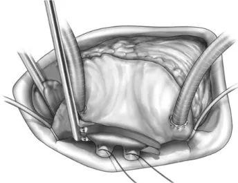
Using the same protocol of research described in 2009 and in 2012 [6–7], after cardiopulmonary bypass was instituted, pairs of additional temporary wires (6494F, Medtronic, Inc., Minneapolis, MN, USA) were transfixed strategically into two points. The first on the RPVs, was positioned close to their atrial ostium (Fig.1), to obtain the diagnostic value. The second pair, as control, was posi-tioned on the roof of the left atrium (RLA) in proximity to the Bachmann bundle, outside the left posterior atrial ablation box, in order to have a control EP value. Pacing thresholds (PTs) from all atrial wires were assessed at baseline and immediately after one single encircling ablation of the RPVs, using an external temporary pulse generator (5833, Medtronic, Inc.).
Further, the same assessment was repeated after chest closure and after removal of chest drains, to rule out possible dislodge-ment of the wires, before discharge from the division and 3 weeks after surgery, before leaving the rehabilitation unit of our hospital.
By definition, exit block was considered significant when no capture was obtained at 20 mA, the maximum output of our pulse generator [10]. After completing the 3 weeks control, the wires were removed and a transthoracic echocardiogram was per-formed the following day to rule out pericardial collection, before discharge.
Table 1: Preoperative data
Variables N Number of patients 10 Age (years) 53 ± 12 Female gender 5 (50%) NYHA class I 1 (10%) II 8 (40%) III 1 (10%)
Duration of atrial fibrillation (months) 30.7 ± 5.1 Atrial fibrillation type
Paroxysmal 7 (70%)
Persistent 3 (30%)
Valvular heart disease
Mitral 10 (100%)
Tricuspid 3 (30%)
Degenerative/rheumatic valve disease 7/3
Left atrial diameter (mm) 47 ± 5
Left atrial volume (mm) 80 ± 47
Telediastolic left ventricular diameter (mm) 63 ± 6
Ejection fraction (%) 55 ± 4
Values are expressed as number and (percentage) for discreet variables and as mean ± standard deviation for continuous variables.
Figure 1:Right pulmonary vein ablation performed by temperature-controlled bipolar radiofrequency device after positioning of a pair of temporary wires on the right pulmonary veins.
AD UL T C A R D IA C A. Pozzoli
Data analysis
No formal sample size calculation was performed. The study was designed as a pilot study as no previous data were available. Statistical analysis was conducted using the JMP 8.0 package (SAS Institute, Inc., Cary, NC, USA). Shapiro–Wilk test was used to assess the normality of all the data samples. All results are generally expressed as mean ± standard deviation (range) for continuous variables and as proportion (%) for categorical variables. Patients had been followed for the follow-up through 15 March 2014.
RESULTS
Clinical
As performed in our previous report on the EP efficacy of high-intensity focused ultrasound (HIFU) energy source [7], 10 patients were enrolled in this pilot study. Mean age was 53 ± 12 years, 5 (50%) patients were male. No patient had previous transvenous ablations. There were 7 patients with paroxysmal AF (70%) and 3 patients with persistent AF (30%), who underwent a complete left atrial maze lesion set with cryoablation of the mitral annulus. Concerning the mitral pathology, degenerative disease affected 7 patients, who underwent mitral valve repair (central edge-to-edge technique in 2 cases, quadrangular resection of the posterior leaflet in 5 cases with positioning of neochordae on the anterior leaflet in 2 cases) with implantation of a partialflexible ring in all of them; in 3 patients concomitant tricuspid tri-dimensional annuloplasty was associated. Three patients required mitral valve replacement with mechanical prosthesis, due to extensivefibrotic degeneration of the valve apparatus for previous rheumatic disease. The mean extracor-poreal time was 97 ± 24 min, mean cross-clamping time was 74 ± 21 min. Median postoperative hospitalization was 7 days (range 5–13 days) and all patients were discharged in sinus rhythm.
At a mean follow-up of 12 ± 5 months, 10 patients (100%) were in sinus rhythm. Of these, 4 (40%) were free from antiarrhythmic medications. Two patients experienced AF within 3 months after the operation, treated successfully with direct current (DC) shock. No permanent pacemaker implantation occurred after surgery.
Adverse events
No early in-hospital death and late adverse events were registered.
Electrophysiological study
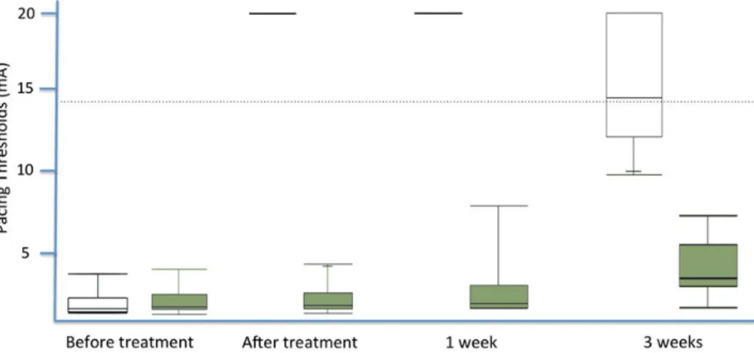
During surgery, the mean PTs considered as baseline were 0.83 ± 0.81 mA (range 0.2–3 mA) on the RPV and 1.13 ± 0.78 mA (range 0.3–3 mA) on the RLA.
Every patient was isolated (PTs = 20) immediately after ablation and at chest closure and after removal of chest drains (Fig.2). At pre-discharge EP study (6 ± 2 days after surgery), the mean RPV PTs was 20 ± 0 mA (range 1–7.8 mA), complete isolation persisted in all patients. At the same time, the mean PTs on RLA was 2.7 ± 1.9 mA (range 0.75–7.7 mA). At 3 weeks (23 ± 2.4 days after surgery), the mean PTs were 14.3 ± 4.3 mA from the RPV and 3.1 ± 1.3 mA from the RLA (range 7–20 and 1.5–7 mA, respectively).
Complete isolation, identified by absence of ventricular capture pacing from the RPV leads (PT or exit block equal to 20 mA), was maintained in 3 patients, the other 7 patients lost the complete isolation between thefirst and the third week.
After surgery, a slight increase of the PTs from the RLA was observed, progressively passing from 1.4 mA at baseline to 6.8 mA at 3 weeks. Likewise, a positive trend in the same time span was observed for the RLA leads, PTs grew from 1.7 to 6.3 mA. These results are obtained after one single encircling ablation with the Cobra Revolution temperature-controlled bipolar RF ablation platform.
DISCUSSION
Complete electrical isolation of the PVs should be the main goal in every ablation strategy targeting the left atrium. Its achieve-ment requires, at least, the assessachieve-ment and demonstration of conduction block across the lesions. Following the protocol of EP investigation conducted at our institution to validate energy sources [6–7], this is thefirst study to systematically check the EP properties of the PVs after a single ablation performed with the Cobra Revolution bipolar temperature-controlled RF. Acute con-duction block (PTs of the ablation or exit block >20 mA) was always achieved with this energy source, after a single ablation. Complete electric block of the PVs was maintained during thefirst week, as shown at the EP study (Fig.2). At 3 weeks, only 3 patients (30%) demonstrated complete pulmonary vein isolation (PVI), while the other 7 patients (70%) outlined partial PV reconnection (PTs between 9 and 15 mA). Adopting the same protocol of
investigation, Benussi et al. documented that multiple ablations with bipolar irrigated RF around the RPV couple reached com-plete acute isolation in all patients, and it persisted till the third week in only 62% of patients [6]. The post-acute fate of temperature-controlled bipolar RF ablations has never been reported in the clinical setting. Therefore, our results provide sig-nificant comprehension on the incidence, evolution and charac-teristics of recovery of PV conduction after one single ablation with this type of energy. Initially, the inflammation masked the block, which disappeared in most patients during the second to third week, showing reconnection. Even if different forms of nec-rotic cell death can be distinguished, based on their initiating mechanisms (necrosis, necroptosis and secondary necrosis), the later aspects of the death process that happens after ablation take place exactly in the same way during the following few hours [11].
Based on the current biological and molecular evidence, the ablative lesions can only decline thereupon; no described mech-anism could explain a delayed myocardial cell death, in which the event occurs during the ablation and the outcome cannot be detected after 3 weeks. Indeed,fibroblasts should replace only the necrotic myocytes, while the not-necrotic cells will recover withoutfibrosis, leaving a gap (e.g. the inner cells of the thickest parts of the myocardial muscle). The knowledge of the acute con-duction properties of ablation platforms registered in the oper-ation theatre is fundamental, since the electrical disconnection of the PVs maximizes the chance of ablation success [12,13]. After percutaneous ablations, the major predictor of failure is the lack of PV electrical disconnection after the procedure, indicating recon-nection of PV conduction or ineffective PV isolation [14, 15]. Moreover, it is demonstrated that repeated ablations in the cath-eter laboratory improve the success rate of paroxysmal and per-sistent AF [15–18]. This relationship fosters the central role of durable isolation of the ablations in the cure of AF. Indeed, the data on transmurality of beating-heart epicardial ablations with unipolar devices are all but satisfactory. Although acutely effective in most cases, epicardial microwave ablation on the beating heart at times fails to accomplish electrical isolation even after repeat ablation [19]. This is true also for unipolar RF ablation, due to the composition and thickness of the atrial wall. The major obstacle to the progression of the lesion through the subendocardial layer is the convective cooling exerted by blood flow through the atrial chamber [20]. Finally, a systematic assessment of post-acute block after ablation with non-clamping epicardial HIFU, showed a radic-ally different outcome. In fact, the postoperative EP study revealed the complete absence of electrical isolation of HIFU ablations, in both the acute and post-acute phase [7].
To the contrary, bipolar systems are specifically designed to work epicardially. The clamping mechanism abates the endocar-dial convective cooling, and reduces tissue thickness by compres-sion, at the same time improving contact. In our first series reporting EP acute outcomes of this energy source, PT assessment showed a complete conduction block in 92% (22/24) of the PV couples after a single ablation and in 100% after one repeat abla-tion. This fact suggested that acute conduction block was not con-stantly maintained in the post-acute phase, even after multiple parallel ablations in patients with particularly thick tissue around the PV ostia [21]. Both impedance-guided and temperature-guided bipolar devices require more than one ablation to achieve acute conduction block.
The slight increase in PTs occurring in the unablated leads on the RLA, along the first 3 weeks, was consistent with a normal foreign body reaction around the electrodes. Although not all
patients were completely isolated, all patients were in sinus rhythm during follow-up. This could be explained by the fact that the valve was repaired and an adequate ablative lesion set was performed, so, no other factor should virtually limit the persist-ence of sinus rhythm. Notably, 4 patients needed AAD to stay in sinus rhythm. The study was not designed, due to the nature of valvular patients affected by paroxysmal and early persistent AF, to do inferences between EP properties and clinical freedom from arrhythmia. This study has several limitations. First of all, a small cohort of patients was enrolled. Further, the EP analysis was performed only on the right PVs and the complete PVI on both the PV cuff was not available up to 3 weeks after temperature-controlled RF ablation; moreover, we are unaware of any further changes in the EP conduction possibly occurring beyond this period.
In conclusion, Cobra Revolution temperature-controlled bipolar RF provides complete PVI after a single ablation up to 1 week. This notwithstanding, only 30% of patients were completely isolated (exit block validation) at 3 weeks.
ACKNOWLEDGEMENTS
This work would not have been the same without the contribution of Leonardo Mandile, to whom we owe his ardent support and valuable suggestions.
REFERENCES
[1] Cheema A, Dong J, Dalal D, Vasamreddy CR, Calkins H, Marine JEet al. Long-term safety and efficacy of circumferential ablation with pulmonary vein isolation. J Cardiovasc Electrophysiol 2006;17:1080–5.
[2] Verma A, Kilicaslan F, Pisano E, Marrouche NF, Fanelli R, Brachmann Jet al. Response of atrialfibrillation to pulmonary vein antrum isolation is direct-ly related to resumption and delay of pulmonary vein conduction. Circulation 2005;112:627–35.
[3] Todd DM, Skanes AC, Guiraudon G, Guiraudon C, Krahn AD, Yee Ret al. Role of the posterior left atrium and pulmonary veins in human lone atrial fibrillation: electrophysiological and pathological data from patients undergoing atrialfibrillation surgery. Circulation 2003;108:3108–14. [4] Bugge E, Nicholson IA, Thomas SP. Comparison of bipolar and unipolar
radiofrequency ablation in an in vivo experimental model. Eur J Cardiothorac Surg 2005;28:76–80.
[5] Aupperle H, Doll N, Walther T, Ullmann C, Schoon HA, Mohr WF. Histologicalfindings induced by different energy sources in experimental atrial ablation in sheep. Interact Cardiovasc Thorac Surg 2005;4:450–5. [6] Benussi S, Galanti A, Zerbi V, Privitera YA, Iafelice I, Alfieri O.
Electrophysiologic efficacy of irrigated bipolar radiofrequency in the clin-ical setting. J Thorac Cardiovasc Surg. 2010;139:1131–6.
[7] Pozzoli A, Benussi S, Anzil F, Taramasso M, Privitera YA, Cianflone D et al. Electrophysiological efficacy of Epicor high-intensity focused ultrasound. Eur J Cardiothorac Surg 2012;42:129–34; discussion 134.
[8] Cox JL, Jaquiss RDB, Scheussler RB, Boineau JP. Modification of the maze procedure for atrialflutter and atrial fibrillation. II. Surgical technique of the maze III procedure. J Thorac Cardiovasc Surg 1995;110:485–95. [9] Benussi S, Nascimbene S, Galanti A, Fumero A, Dorigo E, Zerbi Vet al.
Complete left atrial ablation with bipolar radiofrequency. Eur J Cardiothorac Surg 2008;33:590–5.
[10] Gerstenfeld EP, Dixit S, Callans D, Rho R, Rajawat Y, Zado Eet al. Utility of exit block for identifying electrical isolation of the pulmonary veins. J Cardiovasc Electrophysiol 2002;13:971–9.
[11] Vanden Berghe T, Vanlangenakker N, Parthoens E, Deckers W, Devos M, Festjens Net al. Necroptosis, necrosis and secondary necrosis converge on similar cellular disintegration features. Cell Death Differ 2010;17:922–30. [12] Mokadam NA, McCarthy PM, Gillinov AM, Ryan WH, Moon MR, Mack MJ
et al. A prospective multicenter trial of bipolar radiofrequency ablation for atrialfibrillation: early results. Ann Thorac Surg 2004;78:1665–70.
AD UL T C A R D IA C A. Pozzoli
[13] Gersak B, Kiser AC, Bartus K, Sadowski J, Harringer W, Knaut Met al. Importance of evaluating conduction block in radiofrequency ablation for atrialfibrillation. Eur J Cardiothorac Surg 2012;40:1191–6.
[14] Ouyang F, Bänsch D, Ernst S, Schaumann A, Hachiya H, Chen Met al. Complete isolation of left atrium surrounding the pulmonary veins: new insights from the double-Lasso technique in paroxysmal atrialfibrillation. Circulation 2004;110:2090–6.
[15] Nanthakumar K, Plumb VJ, Epstein AE, Veenhuyzen GD, Link D, Kay GN. Resumption of electrical conduction in previously isolated pulmonary veins: rationale for a different strategy? Circulation 2004;109:1226–9. [16] Callans DJ, Gerstenfeld EP, Dixit S, Zado E, Vanderhoff M, Ren JFet al.
Efficacy of repeat pulmonary vein isolation procedures in patients with re-current atrialfibrillation. J Cardiovasc Electrophysiol 2004;15:1050–5. [17] Weerasooriya R, Khairy P, Litalien J, Macle L, Hocini M, Sacher Fet al.
Catheter ablation for atrialfibrillation: are results maintained at 5 years of follow-up? J Am Coll Cardiol 2011;57:160–6.
[18] Tilz RR, Rillig A, Thum AM, Arya A, Wohlmuth P, Metzner Aet al. Catheter ablation of long-standing persistent atrialfibrillation: 5-year outcomes of the Hamburg Sequential Ablation Strategy. J Am Coll Cardiol 2012;60: 1921–9.
[19] Maessen JG, Nijs JF, Smeets JL, Vainer J, Mochtar B. Beating-heart surgical treatment of atrialfibrillation with microwave ablation. Ann Thorac Surg 2002;74:1307–11.
[20] Santiago T, Melo J, Gouveia RH, Neves J, Abecasis M, Adragao Pet al. Epicardial radiofrequency applications: in vitro and in vivo studies on human atrial myocardium. Eur J Cardiothorac Surg 2003;24:481–6. [21] Benussi S, Nascimbene S, Calori G, Denti P, Ziskind Z, Kassem Set al.
Surgical ablation of atrialfibrillation with a novel bipolar radiofrequency device. J Thorac Cardiovasc Surg 2005;130:491–7.
APPENDIX. CONFERENCE DISCUSSION
Scan to your mobile or go tohttp://www.oxfordjournals.org/page/6153/1
to search for the presentation on the EACTS library
Dr T. Weimar (Stuttgart, Germany): Your study perfectly shows that you cannot always translate animal studies to human beings. There have been animal
studies showing conduction block with one application even chronically on the beating heart, which you proved is probably not true for the human being. However, I don’t think that any company or any surgeon would recommend applying those clamps only once on the pulmonary veins if you use a beating heart.
I think radiofrequency as well as cryoenergy are well proven and reliable energy sources; however, the big difference is that extracting the heat by cryoe-nergy does not really require an algorithm, while this is needed for radiofre-quency. Again, it does not prove transmurality, it just indicates to the surgeon changes of conductance or temperature or whatever is used as algorithm.
However, reading through your paper, and you touched most of those points during your discussions, I would have two questions. Can you comment on the algorithms, temperature-controlled versus conductive-controlled? Which algorithm we should go with,first of all?
Secondly, I was a little surprised that you had such excellent results. You had 100% sinus rhythm at 12 months, although not all of them were off drugs, which means our cornerstone PVI seems not as important as we thought? Your data suggests good results with only 30% of PVs being isolated. Do we overesti-mate PVI as cornerstone?
Dr Pozzoli: The first question was about the temperature control. I cannot comment very deeply on the physical aspect of temperature-controlled bipolar radiofrequency, but I can say that the jaws of this clamp are different in respect to other ablators, withflexible serpentine electrodes. So, in an optic of com-parison with other clamps available in arrhythmia surgery, it’s a different way of delivering the energy. So in the thickest regions of the pulmonary veins it could be challenging to reach the complete transmurality or, in other words the elec-tric isolation.
Concerning the other question, I think this study was not designed to study the correlation between the EP properties and the clinical outcomes. So I think that, if we want to do this inference, we should select patients with early persist-ent atrialfibrillation, we need different patients from those of the current series. Most of our patients were paroxysmal with mitral regurgitation. After repair it is quite easy that they are actually on sinus rhythm, with the use of antiarrhythmic drugs. So this will be in the future another study. I am also curious in under-standing how much the complete EP isolation translates in successful clinical outcomes. From the cath lab we know that if we repeat ablations and we close gaps, we have better results. In fact, patients who are re-ablated three times normally obtain better results. So, I think it’s not reliable to use this study, this data, as answer to your question.