Ph.D. THESIS
At Agrocampus-Ouest
Under the seal of European University of Brittany
To obtain the degree of:
Docteur de L’Institut Supérieur Des Sciences
Agronomiques, Agro-alimentaire, Horticoles Du Paysage
Specialisation: Food ScienceDoctoral College: VAS (Vie-Agro-Santé)
Presented by:
Solène LE MAUX
β-lactoglobulin/linoleate complexes:
binding properties and biological functions
Teagasc Food Research Centre, Moorepark, Fermoy, Co. Cork, Ireland
INRA-Agrocampus Ouest, Science et technologie du lait et de l’œuf (STLO), Rennes, France
Public defence March 21, 2013 by Examination Committee
JURY
Yves Le Roux, Director of Research, ENSAIA, Nancy, France President
Philippe Cayot, Professor, AgroSup Dijon, France Reviewer
Claire Gaudichon, Professor, AgroParisTech, France Reviewer
Denis Renard, Director of Research, INRA, Nantes, France Member
Thomas Croguennec, Professor, Agrocampus-Ouest, Rennes, France Ph.D. Supervisor
Saïd Bouhallab, Director of Research, INRA, Rennes, France Ph.D. Co-Supervisor
André Brodkorb, Senior Scientist, Teagasc Moorepark, Fermoy, Co. Cork, Ireland Ph.D. Co-Supervisor Linda Giblin, Senior Scientist, Teagasc Moorepark, Fermoy, Co. Cork, Ireland Ph.D. Co-Supervisor
A c k n o w l e d g m e n t s | iii
ACKNOWLEDGMENTS
The work reported in this thesis was undertaken in collaboration between the
Teagasc Food Research Centre, Moorepark, Fermoy in Ireland and
INRA-Agrocampus Ouest, STLO, Rennes in France. This project was supported by a
Teagasc Walsh Fellowship and by the Department of Agriculture, Fisheries and Food
(FIRM project 08/RD/TMFRC/650). I would also like to acknowledge funding from
IRCSET-Ulysses Travel Grant and the doctoral school VAS (Vie-Agriculture-Santé)
grant.
This PhD thesis has allowed me to work in many different laboratories, and
therefore given me the chance to collaborate with so many people that I want to
thank for their contribution in the success of this project.
I first would like to thank my supervisors Thomas Croguennec, André
Brodkorb, Linda Giblin and Saïd Bouhallab for their encouragement, support and
continued guidance. It is thanks to you all that I love science and research more and
more each day.
I am grateful to Claire Gaudichon, Philippe Cayot, Denis Renard and Yves Le
Roux for accepting to be in my Jury.
Thanks to Vincent Rioux, my VAS tutor, for his time and the scientific
discussions.
I would like to thank Didier Dupont for his help, his time and his guidance
through my PhD, especially with regard to the in vitro digestion work.
Thanks to Ken H. Mok, John O’Brien, Marie-Noelle Madec, Mark Auty, Julien
Jardin, and Valérie Briard-Bion for bringing me to the world of NMR, confocal and
MS. Thank you for your help and the scientific discussion.
Special thanks to Rachel Boutrou, the ISFPL team and the TFRC in
Moorepark for their support and for all the discussion that helped me through my
PhD.
Thanks to Amélie Deglaire, Valérie Lechevalier, Catherine Guerin, Claire
Bourlieu, Stéphane Pezennec, Marie-Hélène Famelard and Romain Jeantet for all
the advice in the last stages of my PhD.
Thank you to the Moorepark staff and students for allowing me to spend my
PhD in such a such good environment, for their help, their smiles and all the good
laughs. Special thanks to my friends Sandra, Viv, Grace, Noel, Tony, Pa and Eoin.
Thank you so much for making my Irish life so amazing (with an “A” like Adam).
Un grand merci au personnel et aux étudiants du STLO pour leur aide et la
bonne ambiance qui règne dans ce laboratoire. Je remercie particulièrement notre
côté de la passerelle, pour m’avoir accueillie à plusieurs reprises et aussi pour les
gâteaux (enfin, lorsque Thomas ne mange pas tout…), “big up” à Céline, Kéra, Gui,
Melanie, Claire, Maryvonne, Marie-Claude, Dominique…
I would like to thanks my housemates, from Beechfield Estate, to Clancy
Street and Boulevard J. Cartier, for all the good moments we had.
iv | A c k n o w l e d g m e n t s
Merci à mes amis, particulièrement à Fanny, Alex, Aurélie et Benjamin, pour
tous ces moments de joie et de fou rire. Merci également au chemin neuf et aux
copains Lyonnais qui ont égayé mes retours dans la meilleure ville au monde.
Je
remercie
très
chaleureusement
le
clan
Le
Maux/Lin-Wee-Kuan/Badoinot/Nicolas pour tous les moments de détentes. J’ai la chance d’avoir
une famille où on ne s’ennuie jamais : des wantans au charsiou, de Platon à Bob ou
encore de Collonges City à Anglet.
Pour toute la nouvelle génération, Chloé, Quentin, Gaby, Maxime, Louise,
Lisa, Maxence, Korben et Lilly, merci d’apporter cette petite étincelle de bonheur à
chaque fois que je vous vois.
Mon Alan, I will never thank you enough, for the GC help, the reviewing, the
support, pour tout et le reste. You are the one for me formidable!
Merci à mes parents et ma sœur, pour leur amour, leur soutien, leurs
encouragements, leurs conseils et leur patience. Nous avons des parents
extraordinaires qui nous ont permis de faire des études et qui ont toujours été à nos
côtés, c’est pourquoi je leur dédie cette thèse.
T h e s i s O u t p u s | v
THESIS OUTPUTS
PublicationsLe Maux S., Giblin L., Croguennec T., Bouhallab S., Brodkorb A., (2012). β-lactoglobulin as a molecular carrier of linoleate: Characterisation and effects on intestinal epithelial cells in vitro. J. Agric. Food Chem. 60, 9476–9483.
Le Maux S., Brodkorb A., Croguennec T., Hennessy A. A., Bouhallab S., Giblin L. β-lactoglobulin/linoleate complexes: in vitro digestion and role of the protein in fatty acids uptake. Accepted in Journal of Dairy Science.
Le Maux S., Bouhallab S., Brodkorb A., Giblin L., Croguennec T. Complexes between linoleate and native or aggregated β-lactoglobulin: Interaction parameters and in vitro cytotoxic effect. Accepted in Food Chemistry.
Review
Le Maux S., Bouhallab S., Brodkorb A., Giblin L., Croguennec T. β-lactoglobulin/fatty acids complexes: Binding, structural and biological properties. In Preparation.
Oral presentations
Le Maux S., Giblin L., Croguennec T., Bouhallab S., Brodkorb A., (2011). Complexes β-lactoglobuline/Acide Linoléique-Formation, Stœchiométrie et Capacité Cytotoxique. Rencontres de Biologie-Physique du Grand Ouest 5, Rennes, France.
Le Maux S., Giblin L., Croguennec T., Bouhallab S., Brodkorb A., (2011). β-lactoglobulin/Linoleic Acid Complexes - Formation, Binding Stoichiometry and Cytotoxic Capability. Irish Area Section Biochemical Society Annual Meeting 2011, Maynooth, Ireland.
Poster
Le Maux S., Giblin L., Croguennec T., Bouhallab S., Brodkorb A., (2012). Fat Absorption in the Gut - Can Milk Protein Change it? Walsh fellowship seminar.
T a b l e o f C o n t e n t s | vii
TABLE OF CONTENTS
ACKNOWLEDGMENTS ... III THESIS OUTPUTS ... V LIST OF ABBREVIATIONS ... IX GENERAL INTRODUCTION ... 11PART 1 LITERATURE REVIEW ...7
1 WHEY PROTEINS ... 10
2 Β-LACTOGLOBULIN, THE MAJOR WHEY PROTEIN IN BOVINE MILK ... 11
2.1 Structure of β-lactoglobulin ...11
2.2 Influence of pH and temperatures on β-lactoglobulin structure ...14
2.3 Enzymatic digestion of β-lactoglobulin and its transport in the intestinal barrier ...16
2.4 Biological functions of the β-lactoglobulin ...17
3 INTERACTION BETWEEN Β-LACTOGLOBULIN AND LIGANDS ... 18
3.1 Binding sites localisation ...18
3.2 Binding properties: stoichiometry and association constant ...20
3.3 Impact of the fatty acid/protein binding on the biological function of the fatty acids ...23
3.4 Impact of the fatty acid/protein binding on the structural function of the protein ...25
3.5 Impact of the non-native protein structure on binding properties of the fatty acid/protein complex ..26
4 LONG CHAIN FATTY ACIDS PROPERTIES, EXAMPLES OF LINOLEIC ACID AND CONJUGATED LINOLEIC ACID ... 27
4.1 Fatty acids properties ...28
4.2 Linoleic acid, an essential long chain fatty acids ...29
4.3 Conjugated linoleic acid, isomer of linoleic acid ...29
4.4 Impact on heath ...30
4.5 Lipids digestion ...31
5 CONCLUSION ... 33
6 REFERENCES ... 33
PART 2 MATERIALS AND METHODS... 43
1 MATERIALS ... 45
2 PROTEIN SAMPLE PREPARATION ... 45
2.1 Covalent dimers of β-lactoglobulin ...45
2.2 β-lactoglobulin nanoparticles ...46
2.3 Protein sample reconstitution ...46
3 FORMATION OF THE PROTEIN/LIGAND COMPLEXES ... 46
4 IN VITRO DIGESTION ... 47
5 BIOLOGICAL PROPERTIES ANALYSIS ... 48
5.1 Caco-2 cell line ...48
5.2 STC-1 cell line ...48
5.3 Cytotoxicity assay ...48
5.4 Real-Time Cell Analyzer ...49
5.5 Caco-2 transepithelial transport ...50
5.6 Cyclic AMP assay ...52
5.7 Gene expression by messenger RNA levels ...52
5.8 Determination of cholecystokinin secretion using ELISA ...55
6 METHODS FOR STUDYING PROTEIN/LIGAND INTERACTIONS ... 56
6.1 Isothermal titration calorimetry ...56
6.2 Intrinsic fluorescence ...57
7 BIOCHEMICAL AND PHYSICOCHEMICAL ANALYSIS ... 59
7.1 Gas chromatography ...59
7.2 Gel Permeation-HPLC ...60
7.3 Fourier transform infrared spectroscopy ...61
7.4 Polyacrylamide gel electrophoresis (SDS-PAGE) ...61
7.5 Dynamic light scattering ...62
7.6 Determination of linoleate pKa ...62
7.7 Determination of the critical micelle concentration of linoleate ...62
8 MICROSCOPIC ANALYSIS ... 63
8.1 Confocal laser scanning microscope...63
viii | T a b l e o f C o n t e n t s
9 REFERENCES ... 64
PART 3 RESULTS AND DISCUSSION ... 67
CHAPTER 1 Β-LACTOGLOBULIN AS A MOLECULAR CARRIER OF LINOLEATE: CHARACTERISATION AND EFFECTS ON INTESTINAL EPITHELIAL CELLS IN VITRO ... 69
1 INTRODUCTION ... 71
2 PAPER 1: Β-LACTOGLOBULIN AS A MOLECULAR CARRIER OF LINOLEATE: CHARACTERISATION AND EFFECTS ON INTESTINAL EPITHELIAL CELLS IN VITRO ... 72
2.1 Abstract ...72
2.2 Introduction ...73
2.3 Materials and methods ...74
2.4 Results ...79 2.5 Discussion ...87 2.6 Abbreviations used...90 2.7 Acknowledgements ...90 3 ADDTITIONAL DATA ... 91 3.1 Linoleate properties...91
3.2 Linoleate/β-lactoglobulin molar ratio determined by gel permeation-HPLC ...93
3.3 Cytotoxicity of linoleate at different time points ...93
3.4 Formation of complexes, protein aggregation at the different stages ...95
3.5 FTIR ...96
3.6 FA transporters PCR ...97
4 REFERENCES ... 97
CHAPTER 2 Β-LACTOGLOBULIN/LINOLEATE COMPLEXES: IN VITRO DIGESTION AND ROLE OF THE PROTEIN IN FATTY ACIDS UPTAKE ... 103
1 INTRODUCTION ... 105
2 PAPER 2: Β-LACTOGLOBULIN/LINOLEATE COMPLEXES: IN VITRO DIGESTION AND ROLE OF THE PROTEIN IN FATTY ACIDS UPTAKE ... 106
2.1 Abstract ...106
2.2 Introduction ...106
2.3 Materials and methods ...108
2.4 Results ...114 2.5 Discussion ...120 2.6 Abbreviations used...123 2.7 Acknowledgements ...123 3 ADDITIONAL DATA ... 124 4 REFERENCES ... 125
CHAPTER 3 COMPLEXES BETWEEN LINOLEATE AND NATIVE OR AGGREGATED Β -LACTOGLOBULIN: INTERACTION PARAMETERS AND IN VITRO CYTOTOXIC EFFECT ... 129
1 INTRODUCTION ... 131
2 PAPER 3:COMPLEXES BETWEEN LINOLEATE AND NATIVE OR AGGREGATED Β-LACTOGLOBULIN: INTERACTION PARAMETERS AND IN VITRO CYTOTOXIC EFFECT ... 132
2.1 Abstract ...132
2.2 Introduction ...133
2.3 Materials and methods ...134
2.4 Results ...141
2.5 Discussion ...148
2.6 Abbreviations ...151
2.7 Acknowledgements ...151
3 ADDITIONAL DATA ... 151
3.1 Atomic force microscopy ...151
3.2 Conjugated linoleic acids interaction with β-lactoglobulin and bioaccessibility ...153
3.3 Copper detection and impact on linoleate cytotoxicity ...159
3.4 Differences in isothermal titration calorimetry data at 25°C and 60oC ...160
4 REFERENCES ... 161
PART 4 GENERAL CONCLUSION AND PERSPECTIVES ... 165
REFERENCES ... 175
L i s t o f A b b r e v i a t i o n s | ix
LIST OF ABBREVIATIONS
αla α-lactalbuminAFM Atomic force microscopy ANOVA Analysis of variance
ANS Anilino naphthalene sulfonic acid Asp Aspartic acid
ATR Attenuated total reflectance AU Arbitrary units
βlg β-lactoglobulin
BAMLET Bovine Alpha-Lactalbumin Made LEthal to Tumor cells BSA Bovine serum albumin
cAMP Cyclic adenosine 3’,5’-monophosphate CCK Cholecystokinin
CLA Conjugated linoleic acid
CLSM Confocal laser scanning microscopy CMC Critical micelle concentration
CMP Caseinmacropeptide Cys Cysteine
DHA Docosahexaenoic acid DLS Dynamic light scattering
DMEM Dulbecco’s modified Eagle medium cDNA Complementary deoxyribonucleic acid ε Extinction coefficient
EC50 Effective concentration 50 EDTA Ethylenediaminetetraacetic acid EGCG Epigallocatechin gallate
ELISA Enzyme-linked immunosorbent assay ESI-MS Electrospray ionization mass spectrometry FA Fatty acid
FABPpm Plasma membrane-associated fatty acid-binding protein FAME Fatty acid methyl ester
FAO Food and Agriculture Organization of the United Nations FATP4 Fatty acid transport protein 4
FBS Foetal bovine serum
FTIR Fourier transform infrared spectroscopy GC Gas chromatography
GLP1 Glucagon-like peptide-1
GP-HPLC Gel permeation high performance liquid chromatography HAMLET Human Alpha-Lactalbumin Made LEthal to Tumor cells
x | L i s t o f A b b r e v i a t i o n s
IBMX 3-Isobutyl-1-methylxanthine
ISO International Organization for Standardization ITC Isothermal titration calorimetry
Ka Association constant LA Linoleic acid
LCFA Long chain fatty acid LD50 Lethal dose 50
MCFA Medium chain fatty acid
MTS 3-(4,5-dimethylthiazol-2-yl)-5-(3-carboxymethoxyphenyl)-2-(4-sulfophenyl)-2H-tetrazolium mRNA Messenger ribonucleic acid
Mw Molecular weight n Reaction stoichiometry
NATA N-acetyl-tryptophanamide NMR Nuclear magnetic resonance PAGE Polyacrylamide gel electrophoresis PBS Phosphate buffered saline
PEPT1 Peptide transporter 1 PYY Peptide YY
rh Hydrodynamic radius
RP-HPLC Reversed-phase high performance liquid chromatography RT-PCR Real time polymerase chain reaction
RTCA Real time cell analyzer
SANS Small-angle neutron scattering SAXS Small-angle X-ray scattering SCFA Short chain fatty acid SD Standard deviation
SDS Sodium dodecyl sulphate SEC Size exclusion chromatography TEER Transepithelial electrical resistance TEM Transmission electron microscopy TFA Trifluoracetic acid
Trp Tryptophan
VLCFA Very long chain fatty acid WHO World Health Organization
G e n e r a l I n t r o d u c t i o n | 3
The nutritional quality of food is a major issue for the global food industry. A non-equilibrated diet has been associated with major health problems such as nutrient deficiencies, obesity, diabetes and other food related diseases. As such, an adequate and balanced intake of high quality nutrients is important for health. Food structure and digestion impact on the nutritive and bioactive properties of the individual components, which could have a high effect on health. For instance, the presence of grapefruit in the diet has been shown to impact on intestinal absorption of certain drugs such as cardiovascular medicines. This occurs as components present in the fruit, especially bergamottin, alter the metabolism of the drugs in the enterocyte cells changing their transport through the intestinal barrier. These phenomena can be dangerous as the control of the dose response of these medicinal products is essential, therefore the consumption of grapefruit by patients on certain medicinal products is contraindicated. Recent studies also show that the antioxidant properties of green tea may be reduced by the presence of milk. This is attributed to the interaction of the catechin epigallocatechin-3-gallate (EGCG) with milk proteins, which decreases its accessibility to intestinal cells. These examples highlight how the biological and nutritional properties of food components can be affected not only by the composition of food, but also by interactions between the ingested components.
Milk is extensively used in the food industry because of its palatibility, nutritional attributes and diversified functional properties such as gelling, emulsifying and foaming properties. Milk proteins caseins and whey proteins (β-lactoglobulin, βlg; α-lactalbumin, αla; and bovine serum albumin, BSA) are widely used ingredients. βlg, the major whey protein in bovine milk is an extensively studied protein, and is known to bind hydrophobic ligands such as fatty acids (FA) or vitamins. Nevertheless, its biological function beyond nutrition is unknown. The interaction of native βlg with some ligands such as retinol or palmitic acid are well described in the literature, but the impact of these interactions on the ligands biological function are weakly described. Additionally, βlg structure is highly sensitive to processing conditions used in food manufacture, especially heat treatments, which are routinely used to change food textures or reduce microbial load. Such treatments denature the protein, leading to the formation of non-native monomers, oligomers and higher aggregates of βlg in food products. Information regarding the interactions of hydrophobic ligands with these process-induced species of βlg are weakly described in the literature.
The interaction between milk protein and FA appears to increase FA digestion (Perez et al., 1992). Moreover, the binding of hydrophobic ligands to milk proteins can protect the ligands against oxidation (Futterman and Heller, 1972). It can also modify the kinetics of the enzymatic hydrolysis of milk proteins (Puyol et al., 1993; Mandalari et al., 2009). Complexes between human or bovine α-lactalbumin and oleic acid, called HAMLET/BAMLET (Human/Bovine Alpha-lactalbumin Made LEthal to Tumor cells), increase the FA cytotoxicity
4 | G e n e r a l I n t r o d u c t i o n
to various cancerous cell line (Knyazeva et al., 2008), probably due to an increase in the FA bioaccessibility. In contrast palmitic acid bioaccessibility decreases when bound to βlg (Puyol et al., 1995; Riihimäki-Lampén, 2009).
In this thesis, we investigated the interaction between βlg and the essential n-6 FA linoleic acid or its conjugated isomers, conjugated linoleic acids (CLA), and the effect of binding on the biological properties of the complexes. This multidisciplinary project was performed between the Teagasc Food Research centre in Moorepark, Ireland, and INRA-Agrocampus Ouest STLO in Rennes, France. Teagasc has expertise in milk protein/FA interaction, impact of FA and peptides on gut signals, and the efficacy of dietary bioactives in food matrices. STLO has expertise in whey protein denaturation/aggregation, properties evaluation of the non-native protein structure and digestion. In this thesis, we combined the expertise of both laboratories to study the impact of βlg structure on its interaction properties with FA (linoleate and CLA) and the biological properties of the complex (bioaccessibility and satiety). Hence, the objectives of the project were to answer a number of key research questions, including:
- What are the binding properties of the βlg/FA complexes?
- Does the protein structure affect ligands interaction and biological properties? - What is the impact of such complexes on the protein structure?
- Is the protein digested differently when βlg forms a complex with linoleate?
- How does the protein impact on linoleate bioaccessibility and transport in cell models?
Therefore, the thermodynamic properties of the complexes made with native βlg and linoleate were investigated. The impact of linoleate on the protein properties were studied, focusing on the structural changes of the protein. The biological functions of the FA, especially its bioaccessibility for intestinal absorption, in presence of βlg, were analysed. βlg aggregates of controlled size were used to elucidate the importance of the protein structure on linoleate binding parameters and the biological properties of the complexes. Finally, another complex made with βlg and CLA allowed us to understand the effect of the ligands properties on the complexes formation and properties (bioaccessibility and satiety).
The present PhD thesis is presented in four distinct parts:
- The literature review describes general aspects of milk and whey proteins, with a focus on pH and temperature structural changes of βlg. The thermodynamic properties of ligands/βlg interaction and the impact of the interaction on the ligands and protein properties are reported. Finally, biological properties of FA, which make them valuable nutritional components, are described. The FA/protein interaction section of
G e n e r a l I n t r o d u c t i o n | 5
the literature review (Part 1, 3-Interaction between β-lactoglobulin and ligands) is currently being prepared for publication.
- Materials and methods used during the project are presented in the second part.
- Results and discussion are presented in three different chapters, each written in the form of publication complemented with supplementary data.
The first chapter presents the binding properties of linoleate with native βlg and the impact of this interaction on the protein structure. It also describes the effect of the interaction on linoleate cytotoxicity and transport in vitro. The content of this chapter has been published in Journal of Agricultural and Food Chemistry, 2012, 60, 9476– 9483.
Chapter 2 focuses on the in vitro gastro-intestinal digestion of native βlg complexed with linoleate. The linoleate transport was determined using a Caco-2 cell monolayer, which mimics the intestinal barrier. The impact of the interaction on the ability of the FA to reach the cell membrane was also studied by detection of the hormone cholecystokinin (CCK). The result obtained in this chapter has been submitted to the Journal of Nutritional Biochemistry.
In Chapter 3, the impact of βlg structure on the FA binding properties was evaluated. Different states of βlg aggregation were investigated, namely: native βlg, covalent dimers, and nanoparticles. The effect of such complexes on the FA cytotoxicity was assessed. This chapter is currently submitted for publication in Food Chemistry. - Finally, a general conclusion summarises the main findings of this thesis and proposes
PART 1
L i t e r a t u r e R e v i e w | 9
Milk is a white liquid secreted by the mammary glands of all female mammals, which provides the nutrients for the growth and development of their infants. It is a complete food for the neonate supplying it with carbohydrates, proteins, fatty acids (FA), vitamins, inorganic elements, and water.1 Milk composition varies amongst the species according to the physiological and nutritional needs of the neonate.2 However, humans are the exception, consuming milk well past infancy. Global production of milk, primarily bovine, was 749 million tonnes in 2011.3 Milk is consumed as liquid milk, butter, butter oil, cheese, whole milk powder, skim milk powder and as food ingredients.
Dairy proteins are used for their nutritional and techno-functional properties such as their ability to form stable foam or emulsion. Protein concentrations in milk vary with species from 1 % in human, to 23.7 % for white-tailed jack rabbit.2 Bovine milk has an average protein concentration of 3.4 %. This concentration varies depending on cow breed, animal feeding strategies and lactation stage. Dairy proteins are divided into two categories based on their solubility at pH 4.6. The insoluble fraction is referred to as casein, whereas the soluble fraction is called whey or serum protein. Precipitation, centrifugation, coagulation or filtration can be used to separate casein proteins from whey proteins.2
Milk is a complex solution at pH 6.7 containing lactose, fat, caseins, whey proteins and ash (Table 1 and 2). Milk fat, mainly composed of triacylglycerols, exists as fat globules and is mainly composed of palmitic and oleic acids (22-35 % w/w and 20-30 % w/w in milk fat, respectively4). Different casein structures called αs1-, αs2-, β- and κ-casein differ in primary structure, type and degree of post-translational modification.5 Associated caseins in complexes with calcium phosphate form a supramolecular organization called casein micelles. The exact structure of the casein micelles have been subject to controversy. Because of their large diameter (up to 400 nm6), casein micelles diffract light making the milk opaque and white.
Table 1: General composition of mature bovine milk. Modified fom Smithers (2008)7.
Component g/L Total protein Casein 28 Whey protein 7 Lactose 49 Fat 37 Ash 7
10 | L i t e r a t u r e R e v i e w
Table 2: Protein composition of bovine milk and some of their properties. Modified from Farrell et al (2004)8.
Variant/class Molecular weight A % (1 cm at 280 nm) Caseins αs1-casein B 23,615 10.05 C 23,542 10.03 αs2-casein A 25,226 - β-casein A1 24,023 - A2 23,983 4.6, 4.7 B 24,092 4.7 κ-casein A 19,037 - B 19,006 10.5 Whey proteins β-lactoglobulin A 18,363 9.6 B 18,277 10, 9.6 α-lactalbumin B 14,178 20.1 - 20.9 Serum albumin A 66,399 6.3 - 6.9 Immunoglobulin G1 161,000 13.6 G2 150,000 13.6 A 385,000-417,000 12.1 M 1,000,000 12.1 Lactoferrin - 76,110 9.91 1 WHEY PROTEINS
A significant portion of the milk produced globally (~35 %) is used for cheese production. Cheese manufacture results in the production of whey, as a by-product. This accounts for more than 80 % of the whey available in the world.3 Additionally, whey production comes from the manufacture of casein ingredient.3 In 2011, the United States and the European Union produced around 0.5 and 1.9 million tonnes of whey powder, respectively.3 Whey proteins represent about 20 % (w/w) of the whole milk protein.9 The major whey proteins are β-lactoglobulin (βlg), α-lactalbumin (αla) and bovine serum albumin (BSA). Minor whey proteins are immunoglobulins and lactoferrin.6,8 If the whey comes from casein rennet coagulation, it may contain peptides coming from caseins called proteose peptone and caseinmacropeptide (CMP).
Historically, whey was considered a waste product of cheese manufacture and was used primarily for animal feeding. However, whey is now valued for its nutritional and functional properties and is added to infant milk formula, dietary supplements, sports foods, and animal food products.7,10 Their amino-acid composition (high proportion of cysteins, tryptophan and branched amino acids), their ability to bind ligands and the presence of bioactive peptides within their sequence has resulted in the re-evaluation of whey as a functional food. Indeed, whey proteins have also important techno-functional properties in
L i t e r a t u r e R e v i e w | 11
food systems such as their ability to act as emulsifiers or to form gels.7 Unlike caseins, which lack secondary structure, whey proteins have highly organized structures and most are globular proteins.
The α-lactalbumin (αla) is the second major protein of bovine whey proteins and accounts for about 20 % of the total whey proteins. It is the principle protein in human milk because of the absence of βlg. αla has 123 amino acids and a molecular weight of 14.2 kDa. It contains eight cysteine Cys residues, all engaged in disulfide bonds (Cys6-Cys120; Cys28-Cys111; Cys61-Cys77; Cys73-Cys91) that stabilize the tertiary structure of the protein. This protein has an important role in lactose synthesis, regulating the lactose synthase enzyme.2,11 Moreover, αla can bind one calcium ion per protein.12 In absence of calcium, αla can bind FA such as oleic acid, with these complexes being highly studied for their biological functions, especially their action against tumor cells.13,14
Bovine serum albumin (BSA), the third major protein of bovine whey proteins, constitutes about 10 % of total whey proteins. It consists of 582 amino acids with molecular weight of 66.4 kDa.6 It is a monomeric protein containing one sulfhydryl group and 17 disulfide bonds. BSA binds large amounts of hydrophobic molecules. By its ability to bind free FA, BSA has an important role in milk fat digestion.
2 β-LACTOGLOBULIN, THE MAJOR WHEY PROTEIN IN BOVINE MILK
2.1 Structure of β-lactoglobulin
β-lactoglobulin (βlg) is the major whey protein in bovine milk. It is present in the milk of ruminants, but also in the milk of non-ruminants such as the pig, horse, dog, cat, dolphin, and marsupials.15,16 Interestingly, it is absent from human, lagomorph and rodent milks.16 With a concentration in bovine mature milk of 3.2 g/L, βlg accounts for about 10 % of the total milk protein and about 50 % of the total whey proteins. βlg was first isolated by Palmer in 193417; since that time, several methods have been used for its purification.18
There are several genetic variants of βlg, A and B variants being the most common.19 These two variants both contain 162 amino acids, but differ by two amino acids at positions 64 and 118. Variant A has an aspartic acid residue in position 64 and a valine residue in position 118, while variant B has glycine and alanine in these positions, respectively. The molecular weight of a monomeric βlg variant A is 18,362 Da and B is 18,277 Da (Table 3).8 βlg contains five Cys, located at positions, 66, 106, 119, 121 and 160 (Figure 1). These cysteines form two disulphide bonds, between Cys66 and Cys160, and between Cys106 and
12 | L i t e r a t u r e R e v i e w
Cys119.20,21 Cys121 is a free thiol, however, it is unavailable for reactions under physiological conditions as it lies buried in the centre of βlg structure.22,23 Cys121 participates in protein stability and when exposed, it forms reactive monomers able to form aggregates with other proteins.24-26
Figure 1: Primary structure of βlg variant B. Disulphide bonds are shown with blue line. Swiss-Prot accession number P02754.
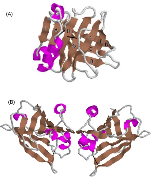
The secondary structure of the βlg shows that it is composed of 15 % α-helix, 50 % β-sheet, 15-20 % reverse turn.16,27 The nine strands labelled from A to I, which form two β-sheets, and the three turns α-helix are arranged to form the βlg globular structure.
Tertiary structure of the βlg is shown in Figure 2A. In aqueous solution and at neutral pH, the β-sheets form a flattened and conical barrel, called a calyx.20,21 This barrel is connected by strand A on one side, while a secondary connection is formed between strands D and E. The α-helix is stored between the strands A and H, and is followed by the ninth β-strand called I.16 In the native protein, disulfide bonds link strands G to H, and strand D to the C-terminal. The free thiol group is located in the central cavity, inaccessible to solvent in the native protein structure.22,23 This calyx is closed at one end by the N-terminal loop and at the other by the EF loop. βlg belongs to the lipocalin protein family, which typically contain a β-barrel, inside which they can bind small hydrophobic molecules.28 Variant A and B of βlg have similar tertiary structure at neutral pH as the amino acids that are different on the two variants are in a mobile surface loop and in the hydrophobic core.16,29 However, these
1 11
Leu Ile Val Thr Gln Thr Met Lys Gly Leu Asp Ile Gln Lys Val Ala Gly Thr Trp Try
21 31
Ser Leu Ala Met Ala Ala Ser Asp Ile Ser Leu Leu Asp Ala Gln Ser Ala Pro Leu Arg
41 51
Val Tyr Val Glu Glu Leu Lys Pro Thr Po Glu Gly Asp Leu Glu Ile Leu Leu Gln Lys
61 71
Trp Glu Asn Gly Glu Cys Ala Gln Lys Lys Ile Ile Ala Glu Lys Thr Lys Ile Pro Ala
81 91
Val Phe Lys Ile Asp Ala Leu Asn Glu Asn Lys Val Leu Val Leu Asp Thr Asp Tyr Lys
101 111
Lys Tyr Leu Leu Phe Cys Met Glu Asn Ser Ala Glu Pro Glu Gln Ser Leu Ala Cys Gln
121 131
Cys Leu Val Arg Thr Po Glu Val Asp Asp Glu Ala Leu Glu Lys Phe Asp Lys Ala Leu
141 151
Lys Ala Leu Pro Met His Ile Arg Leu Ser Phe Asn Pro Th Gln Leu Glu Glu Gln Cys
161 His Ile
L i t e r a t u r e R e v i e w | 13
variants have different properties (ie. surface hydrophobicity and pI) making them dissociable using methods such as reverse phase-HPLC or ion exchange chromatography.
βlg has a quaternary structure which varies with pH, temperature, ionic strength or protein concentration.30-32 At natural milk pH (pH 6.7) and at concentrations greater than 50 µM, βlg exists as dimers in equilibrium with monomers33 (Figure 2B) with dissociation constants around 10-6 M as described in Table 3. Therefore, increasing βlg concentration increases the dimer content of the solution, the dimer interface being formed by the I β-sheet and the AB loop.20-22,34 βlg variant A is also able to associate as octamers, at ambient temperature and at pH between pH 3.5 and 5.5, because the additional carboxyle Asp64 can be ionized.31,32,35
Figure 2: Schematic view of the main-chain fold of bovine βlg. (A) Native monomer with the strands names, (B) Native dimer. RCSB PDB code 1BEB.
(A)
14 | L i t e r a t u r e R e v i e w
Table 3: Some basic molecular properties of bovine βlg, modified from Sawyer and Kontopidis (2000)16 and Saywer (2013)35.
Number of amino acids 162
Monomeric Mw (A genetic variant) 18,362
Monomeric Mw (B genetic variant) 18,277
Isoelectric point
B genetic variant, native 5.407
reduced and denaturing conditions 4.968 Extinction coefficient: 1 mg/ml at 280 nm 0.961
Monomer radius, Rg 1.75 nm
Axial ratio (dimer) 2:1
Dimer Kd (A genetic variant)
pH 3.0 3.07 х 10-3 M
pH 6.5 4.93 х 10-6 M
pH 8.2 1.96 х 10-5 M
Dimer Kd (B genetic variant)
pH 2.7 5.08 х 10-5 M
pH 6 7 7.04 х 10-6 M
pH 7.5 7.94 х 10-6 M
Octamer dissociation constant pH 4.7, 274 K 1.58 х 10-12 M3
2.2 Influence of pH and temperatures on β-lactoglobulin structure
The structure of βlg changes with the pH. Structural changes of βlg can lead to the exposure of the free sulphydryl group Cys121 on the protein surface, thereby increasing its reactivity.33 It is responsible for sulphydryl/disulphide exchange reactions and sulphydryl oxidation reactions leading to protein aggregation.33,36
At room temperature, pH plays an important role in the quaternary structure of βlg. Below pH 2, the protein is in a compact monomeric form. Between pH 2 and 3, βlg is dimeric, but becomes an octamer above pH 3. Around the natural pH of milk (between pH 5.1 and 7), the protein is in its dimeric form. Above pH 7 the protein undergoes the Tanford transition with βlg in the R-state. At pH above 9, the protein irreversibly unfolds and is able to form covalent aggregates.6,16,18,32 The pH has an effect on the tertiary structure too. At low pH, the EF loop is in the closed position and ligand binding is inhibited, whereas at high pH, it is open allowing ligands to penetrate into the hydrophobic binding site.37 Changes in the pH would allow the association or dissociation of the ligand in the calyx.22
Thermal denaturation of βlg at neutral pH is a multistep mechanism illustrated in Figure 3.33,36,38-41 Native dimer dissociates to form native monomers at temperatures above 40°C. Between 40°C and 55°C, the globular monomer slightly unfolds yielding a looser conformation, this change being referred to as the Tanford transition. This conformation of
L i t e r a t u r e R e v i e w | 15
βlg (R-state) allows the formation of aggregates in small quantities. On heating to temperatures between 60°C and 70°C, βlg reversibly unfolds into a molten globule, exposing the sulphydryl group and making it available for the formation of irreversible aggregates. Irreversible unfolding of the monomers occurs above 70°C; these unfolded proteins are able to form aggregates. Different types of aggregates are formed depending on temperature.33 Heating βlg at acidic pH predominantly leads to non-covalent aggregates as the thiol group is weakly reactive at these pH. The temperature of denaturation (irreversible formation of molten globule) varies with pH. Indeed, at pH close to the pI of βlg, the temperature of denaturation is highest. This is explained by a stabilisation of βlg structure through its oligomerisation under these conditions.
Controlling physico-chemical conditions, to which βlg is exposed, makes it possible to control the formation of specific types of βlg aggregates (ie. fibers, oligomers, nanoparticles, etc.).33,42-44 For example, copper has a strong affinity toward sulfhydryl groups in a deprotonated state and promotes disulfide bond formation through oxidation reactions.45 By heating a βlg solution at 80°C for 30 min in the presence of copper, βlg proteins unfold, exposing their free sulfhydryl group, which are oxidized by copper, resulting in stable covalent dimers.43 Schmitt et al. (2009)42 highlighted the importance of the balance between attractive and repulsive interactions occurring between unfolded βlg molecules. These interactions are strongly affected by environmental conditions such as pH and ionic strength. These authors42 observed that by heating βlg at 85°C for 15 min at pH 5.8, βlg can form nanoparticles with a size of around 160 nm, stable to sedimentation and a ζ-potential more negative than native βlg.
Figure 3: Mechanism of the thermal denaturation of βlg, modified from Tolkach and Kulozik (2007)41.
16 | L i t e r a t u r e R e v i e w
2.3 Enzymatic digestion of β-lactoglobulin and its transport in the intestinal barrier
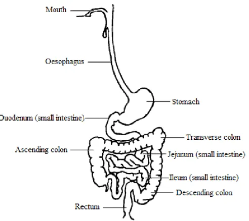
Following ingestion, proteins undergo digestion starting in the mouth and then continuing in the stomach, duodenum, and ileum (Figure 4). Digestion results in the release of small peptides and amino acids. It occurs through a combination of biological (enzymes), chemical (acidic pH) and mechanical (peristaltic) means. The main entero-enzymes responsible for protein hydrolysis are pepsin, trypsin, chymotrypsin, elastase, carboxypeptidase A and B, as well as peptidases from the enterocyte brush border of the intestine, which complete the digestion process.46,47 Degradation products are absorbed by the intestinal barrier primarily in the jejunum, but also in the ileum. Such absorption is facilitated through the enterocytes using peptide transporters such as PEPT1 and HTP1.48 However, peptides or even proteins can be absorbed using paracellular transport or by endocytosis. Indeed, Caillard and Tome (1995)49 showed that 10 to 20 % of βlg can be absorbed intact by rabbit enterocytes.
Digestion and transport of proteins through the gut are influenced by digestive processes such as gastric emptying, enzyme secretion, bile salt, pH, and intestinal microflora. Additionally, digestion is also influenced by the protein structure and the food matrix. For example, Puyol et al. (1993)50 demonstrated that the binding of palmitic acid to βlg has a protective effect on the protein against hydrolysis probably by increasing the amount of hydrophobic interactions. All these factors combine to influence the protein conformation, the activity of digestive enzymes and the binding of the enzyme to its catalytic site.50-56
βlg is more resistant to enzymatic hydrolysis in its native conformation than in an unfolded state.52,57 In humans, Mahe et al. (1996)58 have shown that 64 % of βlg is intact in the jejunum 30 min after ingestion in in vivo studies. In vitro studies have also indicated that βlg is highly resistance to pepsin hydrolysis, but as the conditions used differed substantially (ie. pH, concentrations, enzymes used), comparisons can be difficult. Moreover, the aim of several of these studies was not to mimic the intestinal conditions, but to show a decrease in the allergenicity of βlg and the production of bioactive peptides that could be achieved following hydrolysis. These studies did however all highlight the resistance of native βlg to pepsin hydrolysis possibly due in part to its compact globular form.51 The protein is more extensively hydrolysed during intestinal digestion than gastric digestion, an observation that can be explained by the opening of the protein structure at higher pH.22,57
L i t e r a t u r e R e v i e w | 17
Figure 4: Schematic representation of the gastrointestinal tract. Modified from Liu et al. (2003)59.
2.4 Biological functions of the β-lactoglobulin
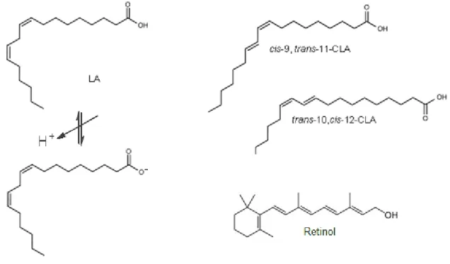
βlg is the only major whey protein whose biological role, other than nutritional value, is still unknown. The most important biological properties attributed to βlg to date are related to its ability to (a) bind small hydrophobic ligands and (b) influence FA digestion. Indeed, Perez et al. (1992)60 demonstrated that βlg participates in the digestion of milk lipids, during the neonatal period, by enhancing the activity of pregastric lipase. It does this by sequestering FA that inhibit this enzyme. In vitro, βlg also binds retinol.61 It was thought for a time that βlg could have a role in the transport of retinol from the mother to the neonate because of the proteins homology to serum retinol binding protein. However, βlg was shown to bind mainly FA but not retinol in milk (retinol structure versus FA structure on Figure 7).60,62 As the protein is not expressed in the milk of all species, it is unlikely that its ability to bind FA is its primary function. Indeed Perez and al. (1993)63 described that pig and horse βlg could not bind FA. This failure to bind FA is attributed to structural modifications particularly at the C-terminus and the inability of porcine βlg to form native dimers.64 βlg has also been demonstrated to bind other hydrophobic ligands such as vitamin D, cholesterol, curcumin, FA and their derivates.64-70
18 | L i t e r a t u r e R e v i e w
Digestion of βlg can form peptides with bioactivities such as antioxidant, antimicrobial activities, hypocholesterolemia benefits, antihypertensive and immunomodulating.71-73 βlg or its peptides may also have satiating properties. Whey proteins are known to be more satiating that casein proteins or milk proteins.74,75 In general, ingestion of protein induces secretion of satiety hormones by specialized enterendocrin cells that line the gut. Such satiety hormones include Cholecystokinine (CCK), glucagon-like peptide-1 (GLP1) and peptide YY (PYY). Hydrolyzed protein gives a higher CCK response compared to undigested protein.76,77 However, other studies demonstrated a lower CCK response to hydrolysed protein or amino acids than undigested proteins, which could be due to the difference of the protein degree of hydrolysis.78,79 Nevertheless, as βlg is resistant to hydrolysis in its native form, its value as a satiety enhancing protein is questionable.
3 INTERACTION BETWEEN β-LACTOGLOBULIN AND LIGANDS
In 1949, McMeekin et al.80 described the binding of sodium dodecyl sulphate (SDS) to βlg. Since, βlg has been demonstrated to bind numerous hydrophobic ligands such as retinol, vitamin D, cholesterol, curcumin, FA and their derivates, polycyles (protoporphyrine IX), aromatic compounds, catechin and cations (Ca2+).66-68,81-90 Different methods have been used to study these interactions such as partition equilibrium, isothermal titration calorimetry (ITC), mass spectrometry, affinity chromatography, fluorescence, circular dichroism or nuclear magnetic resonance.
In this review, particular attention was given to binding of FA with bovine βlg. Molar ratio of free FA to βlg in milk is about 10 FA:βlg. After triglyceride hydrolysis, the molar ratio of linoleic acid (LA) to βlg in milk is about 10 LA:βlg.
3.1 Binding sites localisation
The main binding site of βlg for hydrophobic ligands is formed by the calyx of the protein (Figure 5).37,91,92 Wu et al. (1999)91 showed, by crystallography, the binding of palmitate in the calyx. Several studies have also indicated a secondary binding site on the protein monomer. Indeed, Dufour et al. (1990)89 and Narayan & Berliner (1998)93 observed that βlg can simultaneously bind two different ligands at two different sites, describing the binding of retinol and protoporphyrine IX and the binding of palmitic acid and retinoid, respectively. However, Puyol et al. (1991)61 found that palmitic acid and retinol have the same binding site. To determine the competitivity between two ligands, these studies used
L i t e r a t u r e R e v i e w | 19
the difference of binding constants in presence of one or two ligands. They however did not use the same methods (ultrafiltration and fluorescence), which could explain the different results between these studies. The second binding site of βlg was hypothesized to be located in the hydrophobic pocket formed by the α-helix and the β-barrel, next to the dimer interface.37,91,94 This was confirmed experimentally in 2008 when Yang et al. (2008)94 studied the crystal structure of βlg with vitamin D3. These authors described the second binding site between the α-helix and the I β-strand. Another possible binding site was suggested to be located at the dimer interface.34 Using an ultrafiltration based methodology, Wang et al. (1998)34 found that the binding of palmitate to βlg was affected by protein concentration. Indeed, an increase in protein concentration from 1 to 200 mM, which increased dimer content, resulted in a decrease of the association constant for a palmitic acid binding site. This binding site was described by the authors on the βlg dimer interface, binding 2 moles of palmitate per dimer of βlg. Another binding site with weaker affinity was described on the surface of the monomer, binding 24 moles of palmitate per mole of βlg. It is important to highlight that the same study found only one binding site by fluorescence at low protein concentration (20 µM). Similarly, Forrest et al. (2005)65 studied βlg/vitamin D3 binding by fluorescence at pH 6.6 and lower. This study showed that at low pH, when the EF loop is closed, binding can occur only at an external binding site. By varying pH and ionic strength, these authors showed that vitamin D3 binds to the protein in the calyx and at the surface of the protein. This latter site exhibited a low affinity when the protein is monomeric, whereas affinity increased significantly when βlg is dimeric.
Location of binding sites and the binding properties remain controversial in the literature. This may simply be a methodological issue with methods such as fluorescence, partition equilibrium or ITC following different phenomena. For example, small association constants, similar association constants for different sets of binding sites, or other phenomena such as protein oligomerisation and change in ligand solubility may distort data and cause artefacts.95
20 | L i t e r a t u r e R e v i e w
Figure 5: A schematic view of the main-chain fold of bovine βlg in interaction with linoleic acid in its central cavity. RCSB PDB code 4DQ496.
3.2 Binding properties: stoichiometry and association constant
An association between two molecular species, for instance a protein and its ligands, is characterized by its stoichiometry (molar ratio of ligand bound to the protein, n) and its association constant (Ka in M-1). The constant Ka determines the ratio of protein/ligand (PL) to free protein (P) and free ligand (L) and is expressed by:
↔
Perez et al. (1989)15 demonstrated that bovine βlg binds FA in milk, at a molar ratio of one mole of FA per mole of βlg dimer. Palmitic acid and oleic acid, which are the major FA in milk were found to be the main lipids bound to βlg.
Table 4 is a non-exhaustive list of known FA that bind to native βlg but does not include binding data for βlg mutants or chemically unfolded βlg. Several studies showed that βlg does not bind hydrophobic ligands at low pH. Frapin et al. (1993)64 demonstrated that the binding observed at pH 7 was not detectable at pH 3 for myristic, palmitic and oleic acids. Ragona et al. (2000)92 demonstrated that the interaction between βlg and palmitic acid was reversible from pH 2.4 to 7.3. Similarly, Dufour et al. (1994)97 highlighted that the interaction of βlg/retinol was pH-dependent in the range 3 to 8. Using fluorescence, Dufour et al. (1992)88 found that the very long chain fatty acid (VLCFA) cis-parinaric acid binds βlg at pH 3 with a high Ka but low n (n of 2 and Ka of 4.7 × 10⁷ M-1). By increasing the pH to 7, these authors observed an increase in n values with no change in Ka. Interestingly, Collini et al. (2003)98 showed an increase of Ka by increasing pH, from 6 to 8, for short, medium and long chain FA.
L i t e r a t u r e R e v i e w | 21
Collini et al. (2003)98 and Loch et al. (2012)99 demonstrated that the binding affinity of short chain fatty acids (SCFA) to βlg is low, probably due to the higher solubility of FA in aqueous solution and to the low number of hydrophobic interactions.100,101 Association constant of FA to βlg increases with increasing chain length: between 102 M-1 to 104 M-1 for SCFA, between 103 M-1 to 106 M-1 for medium chain fatty acids (MCFA) and between 104 M-1 to 107 M-1 for LCFA and VLCFA. Frapin el al. (1993)64 and Spector & Fletcher (1970)102 studied binding of LCFA, showing the highest Ka for palmitic acid. This demonstrated that the βlg calyx is better adapted for a 16 length chain carbon. Frapin et al. (1993)64 also observed that the structural constraints imposed by the number and position of double bonds within FA, only weakly affects the interaction of FA with βlg. Wang et al. (1998)34
showed that the decrease of dimeric βlg, with decreasing protein concentration, increased the affinity constant.
Most authors agree that the number of ligands bound to the molecule (n) is close to one at the main binding site. Usually the n value was less than one but Loch et al. (2012)99, who found a value of 0.7 by ITC, showed that the calyx contained one ligand per βlg monomer when examined by crystallography. A second binding site was observed in several studies resulting from the external binding site. This second binding site has a lower affinity (103 to 104 M-1) but n can vary from 2 to 24. As Frapin et al. (1993)64 clearly demonstrated, determination of both Ka and n are highly dependant on the fitting procedure.
Table 4: Parameters of the interactions between βlg and FA: Caprylic acid, Octanoic acid; Capric acid, Decanoic acid; Lauric acid, Dodecanoic acid; Myristic acid, Tetradecanoic acid; Myristoleic acid, cis-9-Tetradecenoic acid; Palmitic acid, Hexadecanoic acid; Palmitoleic acid, Hexadecenoic acid; Stearic acid, Octadecanoic acid; Oleic acid, cis-9-Octadecenoic acid; Elaidic acid, trans-9-cis-9-Octadecenoic acid; Rumenic acid (CLA), cis,trans-9,11-Octadecadienoic acid; Linoleic acid, cis,cis-9,12-Octadecadienoic acid; Linolelaidic acid, trans,trans-9,12-Octadecadienoic acid; γ-Linolenic acid, cis,cis,cis-6,9,12-Octadecatrienoic acid; Linolenic acid, cis,cis,cis-9,12,15-cis,cis,cis-6,9,12-Octadecatrienoic acid; Cis-Parinaric Acid, cis,trans,trans,cis-9,11,13,15-Octadecatetraenoic acid; Arachidic acid, Eicosanoic acid; Arachidonic acid, trans,trans,trans,trans-5,8,11,14-Icosatetraenoic acid; Docosahexaenoic acid, all cis-4,7,10,13,16,19-Docosahexaenoic acid. Different methods were used to study the interactions: F, fluorescence; ITC, isothermal titration calorimetry; EP, equilibrium partition; ESI-MS, Electrospray ionization mass spectrometry; NMR, nuclear magnetic resonance; UF, ultrafiltration; FTIR, fourier transform infrared spectroscopy. n1 Ka1 and n2 Ka2 being the binding constant for the first and second binding sites, respectively.
Common name pH [βlg] (µM) method n1 Ka1 M-1 n2 Ka2 M-1 Reference
Caprylic acid C8:0 6 5 to 10 F (ANS) <3 102 Collini et al., 2003
Caprylic acid C8:0 7 5 to 10 F (ANS) <7 102 Collini et al., 2003
22 | L i t e r a t u r e R e v i e w
Common name pH [βlg] (µM) method n1 Ka1 M-1 n2 Ka2 M-1 Reference
Caprylic acid C8:0 7.5 20 F 0.7 1.1 104 Loch et al., 2010
Capric acid C10:0 7.5 20 F 0.6 6 103 Loch et al., 2010
Lauric acid C12:0 6 5 to 10 F (ANS) 2.4 104 Collini et al., 2003
Lauric acid C12:0 7 5 to 10 F (ANS) 1.6 105 Collini et al., 2003
Lauric acid C12:0 8 5 to 10 F (ANS) 1.4 105 Collini et al., 2003
Lauric acid C12:0 7 10 to 60 F 0.9 1.4 106 Frapin et al., 1993
Lauric acid C12:0 7.5 50 to 100 ITC 0.7 1.7 105 Loch et al., 2012
Lauric acid C12:0 7.4 200 EP 1 5.2 104 2 1.1 103 Spector and Fletcher, 1970
Lauric acid C12:0 7.4 200 EP 1 4 104 24 0.1 103 Spector and Fletcher, 1970
Myristic acid C14:0 3 10 to 60 F 0 Frapin et al., 1993
Myristic acid C14:0 7 10 to 60 F 0.3 3 106 Frapin et al., 1993
Myristic acid C14:0 8.5 12 ESI-MS 1.9 105 Liu et al., 2011
Myristic acid C14:0 7.5 50 to 100 ITC 0.6 7.8 105 Loch et al., 2012
Myristoleic acid C14:1 7 10 to 60 F 0.8 6.3 106 Frapin et al., 1993
Palmitic acid C16:0 6 5 to 10 F (ANS) 6.6 104 Collini et al., 2003
Palmitic acid C16:0 7 5 to 10 F (ANS) 4.7 105 Collini et al., 2003
Palmitic acid C16:0 8 5 to 10 F (ANS) 5.1 105 Collini et al., 2003
Palmitic acid C16:0 3 10 to 60 F 0 Frapin et al., 1993
Palmitic acid C16:0 7 10 to 60 F 0.9 1 107 Frapin et al., 1993
Palmitic acid C16:0 8.5 12 ESI-MS 3.8 105 Liu et al., 2011
Palmitic acid C16:0 7.5 40 to 60 ITC 1.1 20 105 Loch et al., 2012
Palmitic acid C16:0 6.5 10 to 150 F 1 2 106 Narayan and Berliner, 1998
Palmitic acid C16:0 7 10 to 150 F 1 1.7 106 Narayan and Berliner, 1998
Palmitic acid C16:0 8.5 10 to 150 F 1 2.5 106 Narayan and Berliner, 1998
Palmitic acid C16:0 7.2 41.5 EP 1 4.2 106 Perez et al., 1992
Palmitic acid C16:0
8.4 to
2.1 1500 NMR 1 Ragona et al., 2000
Palmitic acid C16:0 7.4 200 EP 1 6.6 105 6 7.1 103 Spector and Fletcher, 1970
Palmitic acid C16:0 7.4 200 EP 1 7 105 24 1.6 103 Spector and Fletcher, 1970
Palmitic acid C16:0 7 1 to 200 UF 1 2.3 105 23 0.4 104 Wang et al., 1998
Palmitic acid C16:0 7 20 F 1.2 2 106 Wang et al., 1998
Palmitic acid C16:0 8 5 F 0.8 2.3 107 Yang et al., 2009
Palmitoleic acid C16:1 7 10 to 60 F 0.8 3.9 106 Frapin et al., 1993
L i t e r a t u r e R e v i e w | 23
Common name pH [βlg] (µM) method n1 Ka1 M-1 n2 Ka2 M-1 Reference
Stearic acid C18:0 8.5 12 ESI-MS 1.6 106 Liu et al., 2011
Stearic acid C18:0 7.4 200 EP 1 1.6 105 2 4.2 103 Spector and Fletcher, 1970
Stearic acid C18:0 7.4 200 EP 1 1.8 105 4 17 103 Spector and Fletcher, 1970
Stearic acid C18:0 7.4 200 EP 1 1.6 105 6 12 103 Spector and Fletcher, 1970
Stearic acid C18:0 7.4 200 EP 1 1.5 105 12 5.9 103 Spector and Fletcher, 1970
Stearic acid C18:0 7.4 200 EP 1 1.9 105 24 2.5 103 Spector and Fletcher, 1970
Oleic acid C18:1 3 10 to 60 F 0 Frapin et al., 1993
Oleic acid C18:1 7 10 to 60 F 0.8 7.7 106 Frapin et al., 1993
Oleic acid C18:1 7.4
163 and
1630 FTIR ~8 Lišková et al., 2011
Oleic acid C18:1 7.2 41.5 EP 1 2.3 106 Perez et al., 1992
Oleic acid C18:1 7.4 200 EP 1 0.4 105 24 1.3 103 Spector and Fletcher, 1970
Elaidic acid C18:1 7 10 to 60 F 0.8 6.7 106 Frapin et al., 1993
Rumenic acid
(CLA) C18:2 7 1 F 2.5 3.7 10
6
Jiang and Lui, 2010
Linoleic acid C18:2 7 10 to 60 F 0.8 5.3 106 Frapin et al., 1993
Linoleic acid C18:2 7.4 163 F 0.6 2.7 105 5.8 5.9 103 Le Maux et al., 2012
Linolelaidic acid C18:2 7 10 to 60 F 0.9 3.3 106 Frapin et al., 1993
γ-Linolenic acid C18:3 7 10 to 60 F 0.9 7.7 106 Frapin et al., 1993
Linolenic C18:3 7 10 to 60 F 0.9 5.9 106 Frapin et al., 1993
Cis-Parinaric Acid C18:4 3 1 to 20 F 0.2 4.8 107 Dufour et al., 1992
Cis-Parinaric Acid C18:4 7 1 to 20 F 0.8 3.6 107 Dufour et al., 1992
Arachidic acid C20:0 7 10 to 60 F 0.9 2.5 106 Frapin et al., 1993
Arachidonic acid C20:4 7 10 to 60 F 0.8 3 106 Frapin et al., 1993
Docosahexaenoic
acid C22:6 7 1 F 2.7 6.8 10
5
Zimet and Livney, 2009
3.3 Impact of the fatty acid/protein binding on the biological function of the fatty acids
Binding FA to proteins such as βlg, can modify the bioaccessibility of FA for cells. This can be measured by looking at the FA transport into cells or by measuring FA cytotoxic effect when bound to a protein compared to that of free. LCFA are known to be cytotoxic, but need to be in the cells to have this effect.103,104 The cytotoxic effect of different FA increase or
24 | L i t e r a t u r e R e v i e w
decrease when bound to βlg compared to unbound. Jiang et al. (2010)69 demonstrated that when conjugated linoleic acid (CLA) is part of a CLA/βlg complex, it is 30 % more cytotoxic at a concentration of 100 μM after 48 h on Caco-2 cells. Similarly, the toxic effect of oleate/oleic acid was increased when it was bound to βlg or αla.13,68 Oleic acid/αla complex also exhibited a ~40 % increase in cytotoxic effect on human larynx carcinoma cells compared to free oleic acid.13 However, we have shown that at a concentration of 58 µM linoleate, free linoleate decreases Caco-2 viability by 50 %, whereas linoleate bound to βlg had no effect on the cell viability (see Part 3, Chapter 181).
Jiang et al. (2010)69showed an increase of the CLA uptake from 45.8 µM to 85.9 µM when bound to βlg compared to free after 4 h of Caco-2 cells exposure to 100 µM of FA, in agreement with their viability tests on 2 cells described above. However, using a Caco-2 monolayer that mimics the intestinal barrier in combination with confocal imaging, we have shown that linoleate transport into Caco-2 cells decreases in the presence of βlg, in agreement with the cytotoxic study (see Part 3, Chapter 2). Riihimäki-Lampén (2009)105 and Puyol et al. (1995)106 showed that palmitic acid was transported more efficiently across the monolayer in its free form than in complex with βlg. Riihimäki-Lampén (2009)105 showed similar results for retinol, although no change was observed for cholesterol transport by Puyol et al. (1995)106. Interestingly, Levin et al. (1992)107 observed that oleic acid in lipidic micelles with taurocholate was absorbed more efficiently than oleic acid bound to BSA. In this instance, the emulsifying properties of taurocholate may function to increase the solubility of the free FA, which explains the difference with the cytotoxicity assays previously presented.
Recent studies based on direct measurement of oleic acid in solution would argue that oleic acid alone or involved in a complex has comparable cytotoxic effects towards various cells, with the protein alone having no effect.68,108 The measured cytotoxic effect observed with oleic acid/βlg or oleic acid/αla complexes maybe related to the solubility of the FA. Oleic acid has a poor solubility in aqueous solution, its critical micelle concentration (CMC) being at 20 and 69 μM at 17 and 45°C, respectively, for a pH of 8.3, 1 mM EDTA and no salts.13 Therefore in the absence of protein, the amount of oleic acid available (soluble) for the cells would be low. The binding of oleic acid to βlg or other proteins increased the solubility of oleic acid on its own and possibly its bioaccessibility. Solubility of FA increases with the number of C=C double bond along the FA aliphatic chain.101 Therefore, the solubility of LA (C18:2) is higher than that of oleic acid (C18:1). Under the experimental conditions of Collin et al. (2010) study109, linoleate has a CMC of 2 mM. The impact of the FA solubility was highlighted by Norman et al. (1988)110 who found a higher cytotoxic effect of linoleate, the soluble salt form of linoleic acid, on the epithelial mouse cells, Ehrlich Ascites Tumor, compared to linoleic acid. Study from Spector and Fletcher (1970)102 supported the solubility
L i t e r a t u r e R e v i e w | 25
hypothesis demonstrating that palmitate bound to βlg was taken up twenty times faster by Ehrlich Ascites Tumor cells at a faster rate than palmitate bound to BSA for a palmitate/protein molar ratio of 0.8. This is due to the stronger binding of palmitate to albumin compared to βlg; therefore, the FA was less bioaccessible to the cells when bound to albumin. Hence, the FA bioaccessibility is altered by the presence of protein, which modifies its solubility in function of the binding properties of the FA/protein complex.
We have shown that even if the linoleate uptake by cells was decreased by its binding with βlg, the protein itself could help a small amount of FA to be transported into a Caco-2 cell monolayer.81 This kinetic of transport would be much lower than the kinetic of transport of free FA.
Zimet and Livney (2009)111 demonstrated that compared to docosahexaenoic acid (DHA) alone, the combination of DHA with βlg, protects DHA against oxidation at pH 7 and at 40°C. Similarly, Futterman and Heller (1972)85 demonstrated that retinol was protected against enzymatic oxidation by BSA and βlg. It has also been shown that Retinol and β-carotene were protected against degradation by heating, oxidation and irradiation.112
3.4 Impact of the fatty acid/protein binding on the structural function of the protein
The βlg calyx closes at below pH 6.2 (Tanford transition). At this acidic pH, the EF loop region moves to a closed position, preventing the binding of a ligand.22 Therefore, interaction of FA to βlg can only impact on the protein structure at non-acidic pH. Interestingly, Puyol et al. (1993)50 demonstrated that by increasing the protein stability with the increase of hydrophobic interactions, the binding of palmitic acid to βlg has a protective effect on the protein against hydrolysis, but that the binding of retinol did not. Mandalari et al. (2009)113 also observed that the binding of retinol to βlg did not have a protective effect toward hydrolysis. However, these authors demonstrated that the protective effect of the phosphatidylcholine binding to βlg could be due to the binding of this ligand next to the proteases cleavage sites.113 Furthermore, Mandalari et al. (2009)113 demonstrated that this protective effect was not effective if the protein was not in its native form.
Barbioli et al. (2011)24 showed that the binding of palmitate to βlg protects the protein against temperature and chaotrope denaturation. It does this by stabilizing the calyx at the hydrophobic interface between the barrel itself and the long helix where the thiol group of Cys121 is buried. Other studies showed that binding of ligands such as myristic acid, CLA, SDS, anilino naphthalene sulfonic acid (ANS) or retinol to βlg, stabilised the protein against denaturation by heating or high pressure treatment.114,115
26 | L i t e r a t u r e R e v i e w
Binding of linoleate or phosphatidylcholine to βlg does not modify the protein secondary structure.81,116 However, Tavel et al. (2008)117 and Kanakis et al. (2011)87 observed changes in the βlg amide I band with the presence of aromatic compounds and tea polyphenols, respectively. Kanakis et al. (2011)87 showed that this was due to big size of ligands. However, protein aggregation has been reported for βlg and other protein systems in the presence of lipids.118 CLA/βlg binding formed aggregates complexes with a size around 170 nm at pH 7.69 Liskova et al. (2011)68 and Le Maux et al. (201281, see Part 3 Chapter 1) described the formation of covalent dimers and trimers when the interaction between βlg and linoleate or oleate was formed under heating conditions at 60°C.
3.5 Impact of the non-native protein structure on binding properties of the fatty acid/protein complex
Modification of the Cys121 in the βlg protein weakens or totally eliminates the FA binding capacity of the βlg due to steric hindrance, showing the importance of the protein structural integrity to bind ligands.93 βlg structure is influenced by the protein environment such as pH, temperature or pressure. Dufour et al. (1992)88 studied the impact of pressure and demonstrated an irreversible dissociation of βlg/cis-parinaric acid complexes at 350 MPa. βlg denatured by urea, acetylation or unfolded by temperature, have a weaker affinity for palmitate.102 Dufour et al. (1992)88 showed that chemically modified βlg has a higher Ka for benzo(α)pyrene and ellipticine, whereas a higher n was observed for the binding of cis-parinaric acid to this denatured βlg. O’Neil and Kinsella (1998)84 showed that binding affinity of the 2-nonanone for βlg was reduced but n was increased when the protein was heated at 75°C for 10 and 20 min. In this thesis, Part 3 Chapter 3 shows similar association bindings but an increase of linoleate bound to βlg when the protein size increased. Because of this binding difference, we found that linoleate uptake by Caco-2 cells was faster for FA bound to non-native βlg compared to the native protein (Part 3, Chapter 3). Similarly, Yang et al. (2009)119 showed that heated βlg decreased the binding constant to vitamin D3 and therefore decreased the uptake of FA in mouse. By heating βlg at 100°C for 16 min, Yang et al. (2008)94 demonstrated that binding of vitamin D3 to the calyx was ineffective but that the external binding site remained thermally stable.
With the exception of raw milk, βlg is often found as aggregates in food products. However, binding of FA to βlg aggregates has not been extensively reported yet. We have shown that βlg aggregates (covalent dimers and nanoparticles) bind a larger number of linoleate than native βlg (Part 3, Chapter 3). CLA binding to native βlg, covalent dimers or nanoparticles was also shown to differ (Part 3, Chapter 3). Shpigelman et al. (2010)120