ORIGINAL ARTICLE
Ultrafast assessment of left ventricular dyssynchrony
from nuclear myocardial perfusion imaging on a new
high-speed gamma camera
Aju P. Pazhenkottil&Ronny R. Buechel&Bernhard A. Herzog&Rene N. Nkoulou&
Ines Valenta&Ursula Fehlmann&Jelena-Rima Ghadri&Mathias Wolfrum&
Lars Husmann&Philipp A. Kaufmann
Received: 8 December 2009 / Accepted: 19 May 2010 / Published online: 17 June 2010 # Springer-Verlag 2010
Abstract
Purpose To validate the ultrafast assessment of left ventricular (LV) dyssynchrony by phase analysis using high-speed nuclear myocardial perfusion imaging (MPI) on a new gamma camera with cadmium-zinc-telluride (CZT) solid-state detector technology.
Methods In 46 patients rest MPI with 960 MBq 99m Tc-tetrofosmin was acquired on a dual-head detector SPECT camera (Ventri, GE Healthcare) and an ultrafast CZT camera (Discovery NM 530c, GE Healthcare) with acquisition times of 15 and 5 min, respectively. LV dyssynchrony was assessed using the Emory Cardiac Toolbox with established values for histogram bandwidth (male <62.4°; female <49.7°) and standard deviations (male <24.4°; female <22.1°) as the gold standard. Evaluating CZT scan times of 0.5, 1, 2, 3 and 5 min (list mode) in 16 patients revealed the preferred scan time to be 5 min, which was then applied in all 46 patients. Intraclass correlation and the level of agreement in dyssynchrony detection between the CZT and Ventri cameras were assessed.
Results In LV dyssynchrony the mean histogram bandwidths with the CZT camera (n=8) and the Ventri camera (n=9)
were 123.3±50.6° and 130.2±43.2° (p not significant) and 42.4±13.6° vs. 43.2±12.7° (p not significant). Normal bandwidths and SD obtained with the CZT camera (35.9± 7.7°, 12.6±3.5°) and the Ventri camera (34.8±6.6°, 11.1± 2.1°, both p not significant) excluded dyssynchrony in 38 and 37 patients, respectively. Intraclass correlation and the level of agreement between the CZT camera with a 5-min scan time and the Ventri camera were 0.94 (p<0.001, SEE 14.4) and 96% for histogram bandwidth and 0.96 (p<0.001, SEE 3.9) and 98% for SD.
Conclusion This ultrafast CZT camera allows accurate assessment of LV dyssynchrony with a scan time of only 5 min, facilitating repeat measurements which would potentially be helpful for parameter optimization for cardiac resynchronization therapy.
Keywords Left ventricular dyssynchrony . Phase analysis . Heart failure . Cadmium-zinc-telluride technology . Myocardial perfusion imaging
Introduction
Congestive heart failure is one of the leading causes of morbidity and mortality in western countries. Cardiac resynchronization therapy (CRT) in addition to standard pharmacological therapy is an established method for the treatment of patients with moderate to severe heart failure (New York Heart Association functional class III/IV, reduced left ventricular (LV) ejection fraction (LVEF ≤35%), and a broad QRS complex (>120 ms) [1–3]. However, when selecting patients based on these standard criteria, 20–30% of all patients do not respond to CRT [4,5]. Therefore, great effort has been applied to the search for
A. P. Pazhenkottil
:
R. R. Buechel:
B. A. Herzog:
R. N. Nkoulou
:
I. Valenta:
U. Fehlmann:
J.-R. Ghadri:
M. Wolfrum
:
L. Husmann:
P. A. Kaufmann (*)Cardiac Imaging, University Hospital Zurich, Ramistrasse 100,
8091 Zurich, Switzerland e-mail: [email protected] P. A. Kaufmann
Zurich Center for Integrative Human Physiology (ZIHP), University of Zurich,
optimized pacing parameters and better selection criteria for CRT. Following the recognition that patients with LV dyssynchrony respond better to CRT [6–8], attention has shifted toward imaging methods for the assessment of LV dyssynchrony. Among these, blood-pool ventriculography with multigated acquisition (MUGA) has for a long time been the most accurate and reproducible technique [9]. Recently, phase analysis on gated single photon emission CT myocardial perfusion imaging (SPECT-MPI) using the Fourier harmonic function was introduced as a reliable alternative for the assessment of LV dyssynchrony [10,11]. Unfortunately, as with MUGA, the reported long scan times (up to 23 min) [11] prevent the use of SPECT phase analysis for multiple repeat measurements in one session to optimize CRT pacing parameters.
The latest advance in SPECT imaging, namely the introduction of the gamma camera with cadmium-zinc-telluride (CZT) detector technology, offers an improved count sensitivity resulting in a substantial reduction in scan time down to 2–3 min for MPI [12]. Whether such short scans would also allow accurate assessment of LV dyssynchrony is unknown.
The purpose of the present study was to validate ultrafast LV dyssynchrony assessment by gated SPECT-MPI acquired on a high-speed cardiac gamma camera with CZT solid-state detector technology in comparison to a standard scan on a dual-head SPECT gamma camera.
Materials and methods
A total of 46 patients who underwent adenosine stress/rest SPECT-MPI on a standard dual-detector SPECT camera (Ventri; GE Healthcare) were included in this study. Exclusion criteria were: nonsinus rhythm, haemodynamic instability and large fixed defects. The study protocol was approved by the institutional review board (local ethics committee of the University Hospital Zurich) and written informed consent was obtained from each patient.
The rest study was performed 90 min after injection of
99m
Tc-tetrofosmin at rest (956.1 ±89.1 MBq) using the Ventri camera (15 min scan duration), immediately followed by acquisition using the CZT camera (Discovery NM 530c, GE Healthcare) (5 min scan duration).
SPECT-MPI image acquisition and reconstruction
The first acquisition was performed using the Ventri dual-head camera with a low-energy high-resolution collimator, a 20% symmetrical window at 140 keV, a 64 × 64 matrix, and an elliptical orbit with step-and-shoot acquisition at 3° intervals over a 180° arc (45° right anterior oblique to 45° left posterior oblique) with 30
steps (60 views). The scan time was set to 25 s per frame for stress and rest, resulting in a total acquisition time of 14 min 52 s (including interstep rotation time) for each scan as recommended by the American Society of Nuclear Cardiology (ASNC) [13]. Images were recon-structed on a dedicated workstation using a standard iterative reconstruction algorithm with ordered subset expectation maximization (OSEM) with two iterations and ten subsets into a standard short axis as well as vertical and horizontal long axes, and polar maps of perfusion encompassing the entire left ventricle without using resolution compensation or attenuation correction.
CZT image acquisition and reconstruction
The second scan was acquired on a CZT camera with pinhole collimation [14], in which the conventional sodium iodide crystals had been replaced by CZT semiconductor technology, improving the sensitivity by a factor of almost four compared to a dedicated cardiac gamma camera (Ventri, GE Healthcare) as previously reported in detail [12]. In brief, the new CZT technology is extremely compact and this miniaturization has enabled a geometry with a stationary array of 19 small gamma cameras packed closely around the heart. As a consequence, this device simultaneously acquired all the views necessary for tomographic reconstruction saving the time needed by conventional cameras for rotating around the subject. The simultaneous acquisition of all angles allowed list-mode data files to be obtained. These files were divided into scan durations of 0.5, 1, 2, 3 and 5 min. The pinhole geometry has several advantages, such as a reduction in the contribution of background organs and tissues to the cardiac images which facilitates reliable three-dimensional iterative reconstruction.
Images were reconstructed on the same workstation as above using a new dedicated iterative algorithm with integrated collimator geometry modelling using maximum penalized likelihood iterative reconstruction to obtain perfusion images in standard axes with 50 iterations. A Butterworth post-filter (frequency 0.37; order 7) was applied to the reconstructed slices.
Phase analysis
The gated SPECT images obtained using the Ventri and CZT cameras were evaluated by phase analysis with the Emory Cardiac Toolbox software (Emory University/ Syntermed, Atlanta, GA) [10]. The phase analysis tech-nique measures the first Fourier harmonic phase of regional LV count changes throughout the cardiac cycle, which is approximately linear to the myocardial wall thickness and therefore related to the time interval when a region in the
LV myocardial wall starts to contract. It provides information on the regularity of the distribution of these time intervals for the entire LV, i.e. it is a measure of LV synchrony or dyssynchrony [10, 11, 15, 16] (Fig. 1). We used the two parameters obtained from the phase distribution of the cardiac cycle that have been shown to best identify LV dyssynchrony [11, 17], as follows: (1) phase histogram bandwidth, and (2) phase histogram standard deviation (SD). The previously established normal values [10] are 38.7±11.8 (males) and 30.6±9.6 (females) for histogram bandwidth and 14.2±5.1 (males) and 11.8±5.2 (females) for SD. The cut-off for identifying LV dyssynchrony was defined as greater than mean + two standard deviations. LV dyssynchrony was considered to be present if both parameters were above the cut-off value. By contrast, skewness and kurtosis were not included in the present analysis as these two parameters are of limited value for predicting LV dyssynchrony using tissue Doppler imaging as the gold standard [11]. Each image was analysed independently from the equivalent image acquired with the other camera and by a reader who was blinded to the history of the patient. If necessary, the automatically determined landmarks such as apex and base were manually corrected.
Statistical analysis
SPSS software (SPSS 15.0, SPSS) was used for statistical testing. Quantitative variables are expressed as means±SD and categorical variables as frequencies, means or
percen-tages. For each scan duration values of the two parameters histogram bandwidth and histogram SD from the CZT camera were compared by intraclass correlation with the data from the Ventri camera, while agreement in diagnosing LV dyssynchrony was assessed by sensitivity, specificity, negative and positive predictive values, and accuracy. The minimal required CZT scan duration was assessed in the first 16 patients by increasing the scan intervals stepwise until one of the parameters (phase histogram bandwidth or phase SD) reached 100% agreement with standard data from the Ventri camera. Intraclass correlation coefficient and Bland-Altman limits of agreement were calculated for this duration with regard to the two parameters. P values less than 0.05 were considered statistically significant and the 95% confidence intervals are presented.
Results
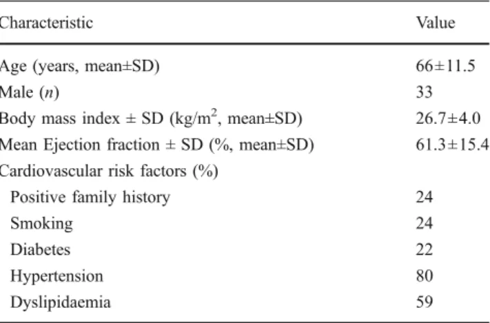
All 46 patients successfully underwent MPI on both cameras. The baseline characteristics of the study population are given in Table1.
As the subanalysis of the first 16 patients revealed 5 min as the minimal required scan duration, all the following results refer to the 5-min dataset obtained on the CZT camera.
The Ventri camera identified LV dyssynchrony in 9 patients with a mean histogram bandwidth of 130.2±43.2° and a mean SD of 43.2±12.7°. Normal LV synchronicity
Fig. 1 Phase polar map (left panels) and phase histograms (right panel) of a patient without LV dyssynchrony (a) and a patient with LV dyssynchrony (b). In the patient with LV dyssynchrony the histogram bandwidth and SD are greater
was found in 37 patients with a mean bandwidth of 34.8± 6.6° and a mean SD of 11.1±2.1°. The CZT camera identified 8 patients with LV dyssynchrony (all among the 9 identified by the Ventri camera). The mean histogram bandwidth was 123.3±50.6° and the mean histogram SD was 42.4±13.6°. Patients with normal findings with the CZT camera showed a mean histogram bandwidth of 35.9°±7.7° and a mean SD of 12.6±3.5°. Of note, three additional patients had a pathological histogram bandwidth but a normal SD, and were therefore categorized as normal (Fig.2).
MPI detected perfusion defects in a total of 14 patients and LV dysfunction (LVEF ≤55%) [18] in 13 patients. All patients with LV dyssynchrony were in the group of patients with LV dysfunction and had a perfusion defect.
The intraclass correlation coefficients comparing the histogram bandwidth and phase SD data from the CZT camera with those from the Ventri camera were 0.94 (CI 0.90–0.97, SEE 14.4, p<0.001) and 0.96 (CI 0.93–0.98, SEE 3.9, p<0.001), respectively (Fig.3).
Sensitivity, specificity, and positive and negative predictive values were 89% (8/9), 100% (37/37), 100% (8/8) and 97% (37/38), respectively, for datasets from the CZT camera compared to those from the Ventri camera as the standard of reference. The agreement between the two methods was 98%.
Discussion
We compared in our study scans from a new gamma camera with CZT semiconductor detector technology acquired with different scan times from 0.5 to 5 min with scans from a conventional dual-head gamma camera with a 15-min standard acquisition time. The data from 16 patients revealed the optimal scan time with the new CZT camera to be 5 min. Applying this acquisition time to the whole patient population showed an excellent correlation and agreement between the results with the two gamma cameras, and thus the use of the CZT camera could be used which would result in a reduction in the total acquisition time by two-thirds.
As heart failure is associated with a high morbidity and mortality in western countries, improvements in diagnostic assessment and treatment of heart failure are of great importance. Until now, CRT in addition to medical therapy is used in patients with severe heart failure with the following selection criteria [1–3]: New York Heart
Associ-Fig. 2 Histogram bandwidth (left panel) and phase SD (right panel) showing normal and pathological distributions in all patients, documenting the excellent clinical agreement between the two
techniques. The cut-off value used was the average value for males and females
Table 1 Baseline characteristics of the 46 included patients
Characteristic Value
Age (years, mean±SD) 66±11.5
Male (n) 33
Body mass index ± SD (kg/m2, mean±SD) 26.7±4.0
Mean Ejection fraction ± SD (%, mean±SD) 61.3±15.4
Cardiovascular risk factors (%)
Positive family history 24
Smoking 24
Diabetes 22
Hypertension 80
ation functional class III/IV, reduced LVEF (≤35%), and a broad QRS complex (>120 ms). Recently, CRT has been used in patients with less severe heart failure [19, 20], suggesting that patients with milder symptoms and evi-dence of LV dyssynchrony may benefit from CRT implantation by improvements in quality of life, exercise capacity, morbidity and mortality. As one-third of all patients receiving a device usually do not respond, efforts have been made to more accurately assess LV mechanical dyssynchrony.
Different imaging modalities have been introduced to accurately predict mechanical dyssynchrony, such as echocardiography, especially with tissue Doppler imaging and strain imaging [21, 22], magnetic resonance imaging (MRI) and MUGA radionuclide ventriculography.
Although accurate, MRI has the disadvantage that it cannot be used in patients with implanted devices. Tissue Doppler imaging is one of the most widely used techniques,
but may be subject to interobserver variability in part because of differences in observer experience [23]. MUGA radionuclide ventriculography has overcome both of these disadvantages, but requires a long scan time, which makes it less useful for repeat measurements to adjust pacing parameters after device implantation.
Recently, assessment of LV dyssynchrony by phase analysis of gated SPECT-MPI scans has been reported [10]. Gated SPECT-MPI scans has several potential benefits, such as automated and reproducible assessment and the possibility to gather information on myocardial perfusion at the same time. As one of the most common causes of chronic heart failure is coronary artery disease [24], a widespread and available diagnostic tool such as SPECT-MPI to assess both pathologies, i.e. ischaemia and dyssynchrony, at the same time would seem to be of great advantage. However, assessment of LV dyssynchrony by SPECT phase analysis has so far been confined to evaluation before device
Fig. 3 Linear regression analysis (upper panels) and Bland-Altman plots (lower panels) for histogram bandwidth (in degrees; left panel) and phase SD (in degrees; right panel) with a 5-min scan time
acquired on the CZT camera compared with 15-min acquisition time on the Ventri camera
implantation, as the long acquisition time prevents repeat measurements needed for objective optimization of pacing parameters once the device has been implanted. The reduction in the acquisition time of SPECT-MPI from 15 min with the conventional gamma camera to 5 min with the new high-speed CZT gamma camera means that repeat measurements have now become clinically realistic allowing pacing parameters to be adjusted. These results support those of previous studies showing the benefits of the new CZT technology, highlighting the importance of the substantial reduction in scan time [12,25].
We acknowledge the following limitations. First, this was a single-centre study with only a limited population of 46 patients due to the pilot nature of the study. Second, the use of manual base and apex placement may lead to bias. However, it has been reported previously that repeatability of phase analysis is better with manual placement than with an automated technique [26], justifying the manual place-ment tool for phase analysis. Finally, in the present study CZT scans showed a 98% accuracy in the detection of LV dyssynchrony with a scan duration of 5 min, and the accuracy therefore could not be further improved by prolonging the scan. By contrast, for assessing precision, longer scan durations may be needed. This, however, was beyond the scope of this present pilot study of accuracy.
In conclusion, the current study showed that LV dyssynchrony can be accurately assessed with phase analysis of the Emory Cardiac Toolbox from gated SPECT-MPI using the new CZT gamma camera requiring a scan time of only 5 min.
Acknowledgments The study was supported by a grant from the
Swiss National Science Foundation and by the ZIHP (Zurich Center for Integrative Human Physiology, University of Zurich, Switzerland). We are grateful to Ennio Mueller, Edlira Loga, Mirjam De Bloeme, Verena Weichselbaumer, Désirée Beutel and Josephine Trinckauf for their excellent technical support.
Conflicts of interest None
References
1. Abraham WT, Fisher WG, Smith AL, Delurgio DB, Leon AR, Loh E, et al. Cardiac resynchronization in chronic heart failure. N Engl J Med 2002;346:1845–53.
2. Bristow MR, Saxon LA, Boehmer J, Krueger S, Kass DA, De Marco T, et al. Cardiac-resynchronization therapy with or without an implantable defibrillator in advanced chronic heart failure. N
Engl J Med 2004;350:2140–50.
3. Cleland JG, Daubert JC, Erdmann E, Freemantle N, Gras D, Kappenberger L, et al. The effect of cardiac resynchronization on morbidity and mortality in heart failure. N Engl J Med
2005;352:1539–49.
4. Bax JJ, Abraham T, Barold SS, Breithardt OA, Fung JW, Garrigue
S, et al. Cardiac resynchronization therapy: part 2– issues during
and after device implantation and unresolved questions. J Am Coll
Cardiol 2005;46:2168–82.
5. Yu CM, Fung WH, Lin H, Zhang Q, Sanderson JE, Lau CP. Predictors of left ventricular reverse remodeling after cardiac resynchronization therapy for heart failure secondary to idiopathic
dilated or ischemic cardiomyopathy. Am J Cardiol 2003;91:684–8.
6. Leclercq C, Faris O, Tunin R, Johnson J, Kato R, Evans F, et al. Systolic improvement and mechanical resynchronization does not require electrical synchrony in the dilated failing heart with left
bundle-branch block. Circulation 2002;106:1760–3.
7. Bax JJ, Bleeker GB, Marwick TH, Molhoek SG, Boersma E, Steendijk P, et al. Left ventricular dyssynchrony predicts response and prognosis after cardiac resynchronization therapy. J Am Coll Cardiol 2004;44:1834–40.
8. Achilli A, Sassara M, Ficili S, Pontillo D, Achilli P, Alessi C, et al. Long-term effectiveness of cardiac resynchronization therapy
in patients with refractory heart failure and“narrow” QRS. J Am
Coll Cardiol 2003;42:2117–24.
9. Hesse B, Tagil K, Cuocolo A, Anagnostopoulos C, Bardies M, Bax J, et al. EANM/ESC procedural guidelines for myocardial perfusion imaging in nuclear cardiology. Eur J Nucl Med Mol
Imaging 2005;32:855–97.
10. Chen J, Garcia EV, Folks RD, Cooke CD, Faber TL, Tauxe EL, et al. Onset of left ventricular mechanical contraction as determined by phase analysis of ECG-gated myocardial perfusion SPECT imaging: development of a diagnostic tool for assessment of
cardiac mechanical dyssynchrony. J Nucl Cardiol 2005;12:687–
95.
11. Henneman MM, Chen J, Ypenburg C, Dibbets P, Bleeker GB, Boersma E, et al. Phase analysis of gated myocardial perfusion single-photon emission computed tomography compared with tissue Doppler imaging for the assessment of left ventricular dyssynchrony. J Am Coll Cardiol 2007;49:1708–14.
12. Herzog BA, Buechel RR, Katz R, Brueckner M, Husmann L, Burger IA, et al. Nuclear myocardial perfusion imaging with a cadmium-zinc-telluride detector technique: optimized protocol for
scan time reduction. J Nucl Med 2010;51:46–51.
13. Hansen CL, Goldstein RA, Akinboboye OO, Berman DS, Botvinick EH, Churchwell KB, et al. Myocardial perfusion and function: single photon emission computed tomography. J Nucl
Cardiol 2007;14:e39–60.
14. Blevis I, Tsukerman L, Volokh L, Hugg J, Jansen F, Bouhnik J. CZT gamma camera with pinhole collimator: spectral measure-ments. Nuclear Science Symposium Conference Record IEEE;
2008. p. 4931–2.
15. Henneman MM, Chen J, Dibbets-Schneider P, Stokkel MP, Bleeker GB, Ypenburg C, et al. Can LV dyssynchrony as assessed with phase analysis on gated myocardial perfusion SPECT predict response to CRT? J Nucl Med 2007;48:1104–11.
16. Cooke CD, Garcia EV, Cullom SJ, Faber TL, Pettigrew RI. Determining the accuracy of calculating systolic wall thickening using a fast Fourier transform approximation: a simulation study
based on canine and patient data. J Nucl Med 1994;35:1185–92.
17. Boogers MM, Van Kriekinge SD, Henneman MM, Ypenburg C, Van Bommel RJ, Boersma E, et al. Quantitative gated SPECT-derived phase analysis on gated myocardial perfusion SPECT detects left ventricular dyssynchrony and predicts response to
cardiac resynchronization therapy. J Nucl Med 2009;50:718–25.
18. Lang RM, Bierig M, Devereux RB, Flachskampf FA, Foster E, Pellikka PA, et al. Recommendations for chamber quantification:
a report from the American Society of Echocardiography’s
Guidelines and Standards Committee and the Chamber Quantifi-cation Writing Group, developed in conjunction with the European Association of Echocardiography, a branch of the European Society of Cardiology. J Am Soc Echocardiogr 2005;18:1440–63.
19. Cleland JG, Freemantle N, Daubert JC, Toff WD, Leisch F, Tavazzi L. Long-term effect of cardiac resynchronisation in patients reporting mild symptoms of heart failure: a report from
the CARE-HF study. Heart 2008;94:278–83.
20. Linde C, Abraham WT, Gold MR, St John Sutton M, Ghio S, Daubert C. Randomized trial of cardiac resynchronization in mildly symptomatic heart failure patients and in asymptomatic patients with left ventricular dysfunction and previous heart failure symptoms. J Am Coll Cardiol 2008;52:1834–43. 21. Bax JJ, Abraham T, Barold SS, Breithardt OA, Fung JW, Garrigue
S, et al. Cardiac resynchronization therapy: part 1– issues before
device implantation. J Am Coll Cardiol 2005;46:2153–67.
22. Suffoletto MS, Dohi K, Cannesson M, Saba S, Gorcsan 3rd J. Novel speckle-tracking radial strain from routine black-and-white echocar-diographic images to quantify dyssynchrony and predict response to
cardiac resynchronization therapy. Circulation 2006;113:960–8.
23. Chung ES, Leon AR, Tavazzi L, Sun JP, Nihoyannopoulos P, Merlino J, et al. Results of the Predictors of Response to CRT
(PROSPECT) trial. Circulation 2008;117:2608–16.
24. Gheorghiade M, Bonow RO. Chronic heart failure in the United States: a manifestation of coronary artery disease. Circulation 1998;97:282–9.
25. Buechel RR, Herzog BA, Husmann L, Burger IA, Pazhenkottil AP, Treyer V, et al. Ultrafast nuclear myocardial perfusion imaging on a new gamma camera with semiconductor detector technique: first clinical validation. Eur J Nucl Med Mol Imaging
2010;37:773–8.
26. Trimble MA, Velazquez EJ, Adams GL, Honeycutt EF, Pagnanelli RA, Barnhart HX, et al. Repeatability and reproducibility of phase analysis of gated single-photon emission computed tomography myocardial perfusion imaging used to quantify cardiac