)OCUMENT OFFICE 2---T RM00 36-412 RESEARCH LABORATORY OF ELECTRONICS VASSACHUSETTS INSTITUTE OF TECHNOLOGY
'AMBRIDGE, MASSACHUSETTS 02139, U.S.A.
ACOUSTICALLY EVOKED POTENTIALS IN THE
DURING CONDITIONING
ROBERT D. HALLROGER GREENWOOD MARK
ad
RAT
64
TECHNICAL REPORT 455NOVEMBER 30, 1966
MASSACHUSETTS INSTITUTE OF TECHNOLOGY
RESEARCH LABORATORY OF ELECTRONICSCAMBRIDGE, MASSACHUSETTS
The Research Laboratory of Electronics is an interdepartmental laboratory in which faculty members and graduate students from numerous academic departments conduct research.
The research reported in this document was made possible in part by support extended the Massachusetts Institute of Tech-nology, Research Laboratory of Electronics, by the JOINT SERV-ICES ELECTRONICS PROGRAMS (U.S. Army, U.S. Navy, and U.S. Air Force) under Contract No. DA36-039-AMC-03200 (E); additional support was received from the National Science Founda-tion (Grant GK-835), the NaFounda-tional Institutes of Health (Grant 2 PO1 MH-04737-06), and the National Aeronautics and Space
Ad-ministration (Grant NsG-496).
Reproduction in whole or in part is permitted for any purpose of the United States Government.
MASSACHUSETTS INSTITUTE OF TECHNOLOGY
RESEARCH LABORATORY OF ELECTRONICS
Technical Report 455 November 30, 1966
ACOUSTICALLY EVOKED POTENTIALS IN THE RAT DURING CONDITIONING
Robert D. Hall and Roger Greenwood Mark
(Manuscript received August 26, 1966)
Abstract
Acoustically evoked potentials were recorded from unanesthetized rats in a series of experiments designed to study changes in sensory evoked potentials during condi-tioning. It is shown that when clicks are established as conditional stimuli (CS) in con-ditioned emotional response (CER) situations, click-evoked potentials recorded from central auditory structures and from mesencephalic reticular formation exhibit appre-ciable amplitude increases. Similar increases were found with Sidman avoidance con-ditioning. These changes in evoked potentials during aversive conditioning were not related to acquired discriminative or conditional properties of the acoustic stimulus, since similar changes in click-evoked potentials were found when a CER was brought under control of a photic CS. These alterations in click-evoked potentials were shown to be independent of movement or movement-related variables. Potentials evoked in central auditory structures by electrical stimuli applied to the cochlear nucleus or within the cochlea also revealed increases in amplitude during acquisition of a CER. In one experiment nearly all movement was eliminated in both CS and control conditions through methods of behavioral control. Data-sampling techniques provided a further control for residual differences in amount of movement in the two periods. These pro-cedures did not eliminate increases in amplitudes of click-evoked potentials during aversive conditioning.
In general, whenever behavioral measures indicated that rats were frightened, acoustically evoked potentials evidenced increased amplitudes, whether or not a CS was present. In all experiments only changes in late components of acoustically evoked potentials were consistently related to observed behavioral changes. It is concluded that changes in sensory evoked potentials observed during conditioning are not related to what may be considered the neural substrate of conditioning, but, in aversive condi-tioning situations at least, they are associated with fear elicited initially as an uncondi-tioned response to noxious stimulation and later as a condiuncondi-tioned response.
a
-TABLE OF CONTENTS
I. INTRODUCTION 1
1.1 Changes in Sensory Evoked Potentials Observed in Classical
Aversive Conditioning Studies 1
1. 2 Changes in Sensory Evoked Potentials Observed during
Avoidance Conditioning 6
1. 3 Changes in Evoked Potentials Observed with Appetitive
Conditioning Procedures 9
1.4 Experimental Background of the Present Investigation 10
II. GENERAL EXPERIMENTAL METHODS AND PROCEDURES 13
2.1 Subjects 13
2.2 Electrodes 13
2. 3 Procedures for Implanting Electrodes 13
2. 4 Histological Verification of Electrode Placements 14
2. 5 Recording and Processing Neural Potentials 14
2.6 Conditioned Emotional Response Situation 15
2. 7 Behavioral Apparatus 16
2. 8 Acoustic Stimuli 17
2. 9 Presentation of Data 17
III. CLICK-EVOKED POTENTIALS RECORDED FROM CENTRAL
AUDITORY STRUCTURES 19
IV. EXPERIMENTAL ANALYSIS OF CHANGES IN ACOUSTICALLY
EVOKED POTENTIALS DURING CONDITIONING 23
4.1 Experiment I 23 4. 2 Experiment II 28 4.3 Experiment III 33 4. 4 Experiment IV 40 4. 5 Experiment V 52 4. 6 Experiment VI 57
V. SUMMARY OF EXPERIMENTAL WORK AND DISCUSSION 70
5.1 Early components of Auditory Evoked Potentials 70 5. 2 Late components of Auditory Evoked Potentials 71 5. 3 Comments on Reported Changes in Evoked Potentials Related
to "Attention" 75 5.4 Closing Remarks 77 VI. SUMMARY 78 Acknowledgment 79 References 80 iii
I. INTRODUCTION
The search for neuroelectric correlates of conditioning may be traced to the first report of a conditioned alpha block by Durup and Fessard,Z0 in 1935, but with a few notable exceptions this endeavour belongs to the last decade. Experimental work with animals had to wait upon adequate techniques for the permanent implantation of elec-trodes. General improvements in electrophysiological methods and instrumentation have also helped to make this work feasible. At last, though hardly least, the computer, within very recent years, has added new dimensions to brain research with the behaving organism.
A review of the entire literature concerned with neuroelectric correlates of condi-tioning is clearly beyond the scope of this experimental report. For a most comprehen-sive and relatively recent review the reader may wish to consult Morrell.6 2 The
published proceedings of several international symposia also provide interesting and representative cross sections of research on the electrical activity of the brain during conditioning. 1 8 24,37 50 Our attention here will be confined to changes in sensory evoked potentials observed during conditioning.
1.1 CHANGES IN SENSORY EVOKED POTENTIALS OBSERVED IN CLASSICAL AVERSIVE CONDITIONING STUDIES
Galambos and Morgan32 describe an experiment by two Russian workers, Artemyev and Bezladnova,3 which to the best of our knowledge is the first report of alterations in evoked potentials related to conditioning. (We make a distinction between sensory activ-ity evoked by "flickering" stimuli that may "drive" neural potentials, and evoked responses to stimuli presented at sufficiently low repetition rates to preclude appreci-able interactions between successive evoked responses.) Artemyev and Bezladnova employed tone bursts of 1. 3-sec duration as conditional stimuli (CS) for a leg flexion response in cats. The unconditional stimulus (UCS) was an electric shock to the paw. The potentials evoked by the tone bursts were monitored on an oscilloscope, and electro-myograms from the leg muscles provided a measure of the conditioned response (CR). As the CR developed, it was accompanied by an increase in the percentage of evoked responses that were detectable in single oscilloscope traces, and thus signified an increase in amplitude of these potentials. With extinction the potentials reverted to preconditioning levels.
The first report of similar findings from American laboratories was that of Galambos, Sheatz, and Vernier.3 0 In this study, electrodes were permanently implanted in cochlear nucleus, auditory and visual cortex, septal area, hippocampus, amygdala, and caudate nucleus of cats. During a preconditioning period the subjects were habit-uated to click stimuli presented day and night at a rate of 1/3 sec for "many days or weeks." In the conditioning phase of the experiments that followed, approximately
10-20 electric shocks were presented to the chest contiguously with random clicks.
1
Evoked potentials recorded during this procedure were compared with potentials recorded before conditioning and with those recorded during an extinction period that followed. No systematic behavioral measures were reported, but crouching, snarling, twitching or similar responses to the click CS were regarded as evidence of conditioning. It was found that amplitudes of click-evoked potentials decreased during the long
habit-uation period, increased when the clicks were "paired" with shock, and fell to precondi-tioning levels during extinction. Additional experiments were performed with cats paralyzed with Flaxedil in order to determine if the changes in evoked potentials were
related to movement. Similar changes were found in the paralyzed cats.
Following this initial report, Galambos and various co-workers have published a series of papers confirming the original findings. 7' 28, 34, 41, 59,63 Both cats and mon-keys were employed as subjects in this series of experiments. In all of these studies trains of clicks or tone bursts were used as conditional stimuli. The CS was followed
by shock or, in the more recent experiments, by puffs of air to the subject's face. The subjects were always exposed to the auditory stimuli for long periods preceding the conditioning phase of an experiment; and in general, evoked potentials were found to undergo appreciable reductions in amplitude during these habituation periods. Pairing of the acoustic stimulus with a noxious one consistently led to increases in the ampli-tudes of acoustically evoked potentials. This was true for potentials recorded from
sev-eral locations in the classical auditory projection and for potentials recorded from other CNS locations. The latter included hippocampus, caudate nucleus, reticular formation, dorsal midbrain tegmentum, habenula, cingulate cortex, and field of Forel. Auditory structures that yielded larger evoked responses with conditioning included cochlear nucleus, trapezoid body, superior olivary complex, inferior colliculus, medial genicu-late body, and auditory cortex.
In the study by Moushegian, Rupert, Marsh, and Galambos,63 changes in amplitudes of click-evoked cortical potentials during habituation and conditioning were found in four cats with severed middle-ear muscles. A report by Hugelin, Dumont and Paillas4 7 had suggested that middle-ear muscles might play a role in the modification of acoustically evoked potentials during attentive behavior. In encephale isol6 cats it had been found that electrical stimulation of the reticular formation led to reductions in amplitudes of auditory cortical potentials. This effect could not be reproduced in animals with sev-ered middle-ear muscles. The report by Moushegian et al. and the earlier one by Galambos, Sheatz, and Vernier seem to rule out middle-ear muscle activity as the explanation for changes in acoustically evoked potentials during conditioning, since the alterations were found in animals with severed middle-ear muscles and in animals para-lyzed with Flaxedil.
Galambos and Sheatz3 4 have noted that acoustically evoked potentials recorded from many sites in the central nervous system, auditory and "nonauditory" alike, assume essentially the same waveform when the acoustic stimulus has been established as a conditioned one. They have described it as a triphasic response: a positive potential
2
-followed by a negative wave and a second positive wave. Increased similarity in wave-forms effected through conditioning has also been reported by John, Ruchkin, and Villegas.51
Among the earliest reports of alterations in sensory evoked potentials related to conditioning was a paper by Jouvet and Hernindez-Peon,5 3 first presented in 1955 at the Fifth Marseille Colloquium of the International Federation of Electroencephalography
and Clinical Neurophysiology. The conditioning phase of this study was a logical exten-sion of the authors' work on changes in sensory evoked potentials during habituation and attention, also treated in the same paper, and described in other publications of the same period.4 4' 45 We shall defer discussion of the work on habituation and attention and consider only that part of the study concerned with conditioning.
Jouvet and Hernindez-Peon employed a tone burst of 2500 cps and 2. 0-sec duration as a CS. This was followed by the UCS, a shock to the paw. The subjects were cats with permanently implanted electrodes in cortical and subcortical structures. These included primary auditory cortex, reticular formation, and that part of somatic, sen-sorimotor cortex serving the limb involved in the conditioned response. Electromyo-grams from the subject's leg provided a measure of the CR. With acquisition of the CR, amplitudes of evoked potentials recorded from auditory cortex increased. Moreover, potentials evoked by the auditory CS were also recorded from somatic cortex. With extinction, evoked potentials from auditory cortex diminished, while those recorded from somatic cortex could no longer be discerned in the EEG. Reconditioning returned the potentials to amplitudes seen during the initial conditioning.
The report by Hernindez-Peon, Jouvet and Scherrer,4 5 concerned mainly with habit-uation of evoked potentials, also described a conditioning experiment with cats in which
amplitudes of acoustically evoked potentials increased when a tone-burst CS was paired with shock to the paw, Other reports by Hernindez-Peon and his co-workers have described imilar changes in evoked potentials recorded from the visual pathway and
reticular formation when photic stimuli were employed as conditional stimuli in classi-cal aversive conditioning situations.4 3' 66
.- 11
An early report by Buser, Jouvet, and Hernindez-Peon1 described a variation on the modification of sensory evoked potentials during conditioning. In this experiment with three unanesthetized cats, the "excitability cycle" of mesencephalic reticular formation was altered by conditioning procedures. Potentials evoked by pairs of clicks were recorded before, during, and after a conditioning procedure in which click pairs were regularly followed by shock to the paw. The second click of each pair typically followed the first by 300-400 msec. Before the introduction of shock, the response evoked by the second click was appreciably smaller than the response evoked by the first. The difference in amplitudes was reduced when shocks to the paw were presented after each pair of clicks. Responses to both clicks were enhanced, but the enhancement was greater for potentials evoked by the second click. The change was interpreted as a decrease in the subnormal excitability of the reticular formation that ordinarily
3
followed a response to the first click of each pair. Omission of the shock provided little evidence of an expected extinction effect. Interestingly, a pseudoconditioning control procedure had ambiguous effects. This control consisted of shock presentations that were "random" with respect to the acoustic stimuli. One subject evidenced changes in evoked potentials similar to those observed during conditioning; another subject did not. This is one of very few experiments that have employed any controls of this kind.
In one of the few experiments to employ rats as subjects, Macadar, Gin6s, Bove, and Garcia-Austt5 7 have described changes in photically evoked potentials recorded from visual cortex during conditioning. The conditioning procedure was one in which shocks were presented at either the beginning or the end of 40-sec periods in which light flashes were presented at 1/sec. Photic stimulation periods alternated with 40-sec periods of no stimulation. Flash-evoked cortical potentials evidenced increased ampli-tudes when shocks were presented during a train of flashes. It apparently made no dif-ference whether the shocks were delivered at the beginning or the end of the flash series.
From the same Montevideo laboratory, Buno, Velluti, Handler, and Garcia-Austt have described changes in round-window potentials recorded from guinea pigs during
conditioning. Acoustic stimuli, clicks or tone pips were in some cases presented directly to the middle ear through a tube fixed in place at the time round-window elec-trodes were implanted. Parts of the ossicular chain in the middle ear were also removed at the same time. Electric shocks delivered to the contralateral pinna were paired with acoustic stimuli in the following way: Clicks or tone pips presented at 1/sec were each followed by a shock for a period of three minutes. No evoked potentials were
recorded during these shock periods. The shock periods alternated with three-minute periods in which no shocks were presented. During the latter, round-window potentials were recorded. Cochlear microphonics evoked by tone pips were found to increase in
amplitude with the commencement of shocking, but with continued shocking underwent reductions which the authors regarded as evidence of "rehabituation." When shocks were discontinued this reduction was accelerated. Similar changes were found in the
N1 response to click stimulation. Buno et al. believe that the way in which stimuli were presented, i. e., directly into the middle ear through a tube, rules out an explanation of the changes in terms of uncontrolled stimulus parameters. Removal of the ossicles
eliminated the possibility that changes in round-window potentials were due to contrac-tions of middle-ear muscles. In view of the potential significance of the findings, the
appreciable variability in the data presented is disturbing. We can only wish that addi-tional systematic data from a number of subjects had been presented.
To the best of our knowledge, Beck, Doty, and Kooi6 have been the only workers to report that sensory evoked potentials did not change when acoustic stimuli were made conditional stimuli in a classical aversive conditioning situation. Their experiments were concerned primarily with conditioned cortical arousal responses. Cats
immobi-lized with bulbocapnine were employed as subjects. Cortical arousal was elicited by 2-sec tone bursts after the acoustic stimulus had been paired with shock to the paw, but
evoked cortical responses to tone onset showed no systematic changes during condi-tioning. For one subject, a series of four clicks was employed as the CS, and the
click-evoked potentials did not appear to change either. Whether or not these findings can be attributed to the use of bulbocapnine is difficult to say.
Behavioral measures of a conditioned response have been conspicuously absent in most of the published reports reviewed above. In many instances there has been neither
definition nor measurement of the response that presumably has been conditioned. Justi-fication for use of the term "conditioning" has been that the relevant sensory stimulus was "paired" in some more or less systematic way with another stimulus, usually elec-tric shock. The so-called "pairing of stimuli" is not, however, a sufficient operation to define a conditioning situation, including that of "sensory-sensory conditioning." The
conditioning process is influenced by a number of important variables, and there are conditions under which the pairing of stimuli does not lead to the occurrence of condi-tioned responses. To assume that the temporal contiguity of two stimuli has led to some sort of conditioning would seem to be poor practice in a scientific endeavour struggling with such complex problems. We believe, and will attempt to show, that repeated
failures to obtain careful systematic measures of behavior have from the outset led to a misunderstanding about the nature of changes in evoked potentials during conditioning. To assume that alterations in sensory evoked potentials are a sign that conditioning has
occurred would seem to beg the question, at least if we are talking about conditioned changes in behavior. The phrase 'neural correlates of conditioning' will be meaningful only when systematic alterations in neuroelectric activity are related to orderly changes in measures of a conditioned response.
It may not be unreasonable to regard a change in evoked potentials as a conditioned response, quite independently of any measurable changes in behavior, be it muscular or glandular. If, however, such changes are to be viewed within a Pavlovian conditioning paradigm (and this seems to have been the model that has dictated the "pairing" of stimuli in studies employing such procedures), then the UCS, shock in most cases, must be regarded as a stimulus that itself is capable of eliciting the changes in evoked poten-tials. The essential role of the unconditional stimulus in classical conditioning para-digms revolves around its capacity to elicit the response that is to be conditioned. Briefly, this implies that in classical aversive conditioning situations, a shock UCS should elicit changes in evoked potentials similar to those that have been reported as a function of conditioning, independently of any associative processes. No one seems to have considered this possibility, but in fact it turns out to be so. The changes are not, however, independent of measurable and correlated changes in behavior.
In summary, it would seem unwise to consider changes in sensory evoked potentials as neuroelectric correlates of conditioned changes in behavior when it is not shown that orderly changes in behavior accompany the recorded alterations in evoked potentials. On the other hand, if changes in evoked potentials are themselves to be regarded as conditioned responses, then some substitute must be found for the Pavlovian conditioning
5
paradigm (certainly the operant one is not appropriate) or we must recognize the capac-ity of the UCS to elicit similar changes in evoked potentials.
Although many of the experiments reviewed above have serious methodological short-comings, the cumulated data strongly suggest that when impulsive physiological stimuli are employed as conditional stimuli in classical aversive conditioning paradigms, there are appreciable changes in the potentials evoked by these stimuli during the course of conditioning. Although this finding, on the face of it at least, seems clear enough, the interpretations afforded it have been rather less than clear. There is in all of these studies, however, the implication that the alterations in sensory evoked potentials are somehow intimately related to the neural substrate of conditioning. This notion we shall have ample reason to question.
1.2 CHANGES IN SENSORY EVOKED POTENTIALS OBSERVED DURING AVOIDANCE CONDITIONING
Changes in sensory evoked potentials during avoidance conditioning have proved to be more complex than those seen in situations employing unavoidable noxious stimuli. Pickenhain and Klingberg,6 7 for example, have described a complex series of changes in visual cortical potentials during several phases of avoidance conditioning. Electrodes were implanted in rats over olfactory bulbs, visual cortex, and other cortical areas. Following a short habituation period, subjects were trained to avoid shocks to the feet by climbing upon a vertical rod. The discriminative stimulus signaling shock consisted of a train of 5 or 10 brief light flashes presented at a rate of 1. 5/sec. In the analysis of the neuroelectric and behavioral data, conditioning and extinction periods were sub-divided according to several criteria. The conditioning period was first divided into two major subperiods. The first, called the period of reinforcement, included all trials before the occurrence of the first CR. The second, the conditioning period, included all trials from the first trial on which a CR occurred to the trial preceding the first unreinforced failure to respond during extinction. The extinction period consisted of the trial marked by the first unreinforced failure to respond and the trials that followed. The two conditioning periods and extinction period were further subdivided when the data appeared to delineate three phases common to each of them. In this regard there has been a modification of the analysis offered in the 1965 publication, and we shall consider only the later findings. These were described by Dr. Pickenhain in a talk before the
Communications Biophysics Group, Research Laboratory of Electronics, M. I. T., on March 10, 1966. The first phase in each of the three major periods was called the
"phase of disturbance." It was characterized by general excitability, increases in respiratory rate (measured from recordings of olfactory bulb activity), strong desynchronization of the electrocorticogram, and decreases in the amplitudes of flash-evoked potentials. The second phase, called the "phase of adaptation," was marked by arrest reactions, less general excitability, and goal-directed behavior. During this period, photically evoked potentials evidenced increases in amplitude and
6
prominent afterdischarges. The last phase, the phase of well-adapted behavior, was characterized by quiet, orderly behavior, and the "automatic" occurrence of conditioned responses during the conditioning period. Flash-evoked potentials were relatively small in this period, and afterdischarges were not conspicuous.
Pickenhain and Klingberg have interpreted their findings in terms of changes in the level of vigilance, or level of arousal. They do not view the alterations in evoked poten-tials as evidence of neural mechanisms underlying the conditioned avoidance behavior. Jasper4 9 has reached a similar conclusion in a brief report presented during a discus-sion at the Pavlovian Conference on Higher Nervous Activity held at the New York Acad-emy of Sciences. Jasper described an experiment in which a conditioned leg withdrawal was established in cats. The CS consisted of a train of clicks presented at a rate of 5/sec. Measures of evoked potentials were reported only for electrodes on primary auditory cortex. During the first 10 days of the experiment, the clicks were not followed by shock, and the cortical potentials decreased in this period to approximately 50% of their original amplitudes. During the first few days of conditioning, the potentials con-tinued to show reductions in amplitude. But around the third day, still before the occur-rence of many avoidance responses, amplitudes increased and continued to do so until the percentage of avoidance responses became appreciable. At this point, evoked poten-tials again diminished. Jasper noted the poor correlation between measures of avoid-ance behavior and amplitudes of evoked potentials. He suggested that the changes in auditory potentials seemed more related to alerting reactions.
A similar suggestion has been made by Gerken and Neff,3 8 following the analysis of data from a study in which several conditioning procedures were employed. Evoked potentials were recorded from auditory cortex of cats under four conditions: (i)
pre-conditioning, essentially a habituation procedure, (ii) pseudopre-conditioning, in which acoustic stimuli and shocks were presented in a "random" manner, (iii) classical condi-tioning, and (iv) avoidance conditioning. Two kinds of acoustic stimuli were employed: the CS consisted of a 4-sec burst of clicks presented at 4000/sec, and a test stimulus
consisted of a single click. The potentials evoked by these two stimuli were found to be similar. Four separate amplitude measurements were made on evoked responses recorded under each experimental condition. All subjects were not exposed to all of these procedures. Some, for example, did not receive the pseudoconditioning treatment before one kind of conditioning or the other. During preconditioning, evoked potentials,
especially the later components, tended to increase, while the early components showed some evidence of reduced amplitudes. The patterns of change shown in the published records are marred, however, by considerable variability from subject to subject. Evidence of increases in the amplitudes of cortical potentials was also found during pseudoconditioning, again primarily in the later components. Curiously, the changes were sometimes seen in potentials recorded from one electrode in a given subject, but not from other cortical electrodes in the same subject. The potentials also showed increased amplitudes during both classical and avoidance conditioning - but not when
7
-conditioning had been preceded by the pseudo-conditioning procedure. On the basis of these and other findings, Gerken and Neff concluded that the alterations in evoked poten-tials did not appear to be related to the learning process, but rather to the emotional state or alertness of the subjects.
Hearst, Beer, Sheatz, and Galambos4 1have also studied acoustically evoked poten-tials during avoidance conditioning. Only one subject was employed, a monkey with elec-trodes implanted in cochlear nucleus, medial geniculate body, caudate nucleus, and hippocampus. Bar-pressing behavior was maintained on a multiple schedule of rein-forcement in which clicks, presented at approximately 1/sec, were correlated with a Sidman avoidance component. The results are rather perplexing. During periods when the monkey was clearly responding discriminatively to the clicks, no appreciable click-evoked activity could be seen in any of the brain sites monitored. With removal of the lever, the animal continued to slap his hand at the place where the lever had been. When this behavior finally weakened (after 18 consecutive hours during which no shocks were presented) potentials recorded from medial geniculate, caudate nucleus, and hippo-campus were larger than they had been during any previous phase of the experiment. When the avoidance procedure was resumed, click-evoked potentials were once more difficult to detect in the EEG.
John, Ruchkin, and Villegas5 1 ' 5 have described an avoidance conditioning study with cats in which 4/sec flashes were established as discriminative stimuli. Many elec-trodes, 14-30, were implanted in each subject, in both specific sensory pathways and nonspecific structures. Average evoked responses were computed for potentials recorded from all electrodes in each subject. Correlation coefficients, Pearson's r, were also computed for all possible pairs of average responses. This was the first step
in the factor analysis of the evoked potentials. The correlation coefficients (not reported) and the subsequent factor analysis suggested that waveforms of the average responses tended to become more similar with the establishment of the conditioned avoidance
response. Functional groupings of some neural loci were also indicated by similar changes in factor loadings for potentials recorded from these structures at different stages of the experiment. We must confess we find the data that have been presented unconvincing on both counts. The finding of increased similarity in waveforms of evoked potentials during conditioning has also been described by Galambos and Sheatz,3 4 as noted above.
In summary, the data from avoidance conditioning experiments reveal some incon-sistencies, and, at the very least, some rather complex changes in sensory evoked potentials. Some of these inconsistencies and complexities are more apparent than real.
This should become apparent in the work to be reported here. One idea of consequence does emerge from the three studies of Jasper, Pickenhain, and Klingberg, and Gerken and Neff: Changes in evoked potentials seen during avoidance conditioning are not related to the conditioning process. They appear, rather, to be associated with some more general change, specifically with a change in arousal level or emotional "state. "
1.3 CHANGES IN EVOKED POTENTIALS OBSERVED UNDER APPETITIVE CONDITIONING PROCEDURES
Most of the reported studies of sensory evoked potentials during conditioning have employed aversive conditioning techniques. Of the few that have employed positive rein-forcement, only one, to the best of our knowledge, has made use of a Pavlovian para-digm, though even in this one no measures of a behavioral respondent were obtained. This was described by Hearst, Beer, Sheatz, and Galambos4 1 in their study of evoked
potentials in four different conditioning situations. The one subject, a monkey, had permanently implanted electrodes in hippocampus, caudate nucleus, cerebellar white matter, and medial geniculate body. The CS consisted of 400-cps tone pips, 0. 5 sec in duration, presented every 1.5 sec for 15 seconds. This was followed by the delivery of a sugar pellet that the subject, reportedly, ate each time. A conditioned respondent was was not defined. A habituation period was followed by conditioning, extinction, and reconditioning periods. Evoked potentials recorded from hippocampus increased during conditioning, diminished during extinction, and grew again with reconditioning. Evoked potentials from other electrodes evidenced no changes during the experiment.
Hearst et al. also measured click-evoked potentials during an operant discrimina-tion procedure employing the same sugar reinforcement. Potentials were recorded from
the hippocampus, caudate nucleus, medial geniculate body, and cochlear nucleus. The monkey, again the only subject, was trained to press a lever only during presentations of a click stimulus. Reinforcement in the SD periods was presented on a 1-minute Fixed Interval schedule. With the establishment of the discriminative behavior, amplitudes of evoked potentials from all electrodes were smaller than those recorded in precondi-tioning sessions. Removal of both the lever and food cup from the situation led to an
increase in the amplitudes of evoked responses, while a return to the normal discrimina-tion procedure again resulted in diminished evoked potentials. It will be recalled that the avoidance conditioning experiment described in the same report also indicated that evoked potentials decreased in amplitude with the acquisition of a discriminative operant.
Somewhat different findings have been reported by Worden7 5 in a study of acoustically evoked potentials during operant discrimination training with a single cat. The animal had electrodes implanted in nearly all projection sites of the classical auditory system, but emphasis has been placed on potentials recorded from the trapezoid body or infe-rior olivary complex. (Histological verification of electrode placements had not been accomplished at the time of the report.) Short tone bursts, presented at a rate of 2/sec, were established as discriminative stimuli for lever-pressing. Before the acquisition of the discrimination, only small evoked responses, if any, were recorded. By the third day of training, when the cat appeared to be "waiting" for the acoustic signal, large potentials appeared in the trapezoid body. On the sixth day, potentials from all recording sites were large. More complex changes were seen in later stages of the
9
--experiment; this suggested that evoked potentials do not remain large as long as discrim-inative behavior is maintained, but revert to smaller amplitudes when discriminations are well established.
Brazier, Killam, and Hance8 have found changes of still another kind in flash-evoked potentials recorded from lateral geniculate body during acquisition of an operant discrim-ination. Again only one subject was used, a cat. The animal was trained to press a lever in the presence of light flashes presented at 10/sec. No changes in the lateral geniculate potentials were seen until the animal was required to respond within 15 sec following the onset of the flashes. Under these conditions, an increase in the amplitudes of both primary and secondary responses was seen. The discriminative behavior was further differentiated by establishing 6/sec flashes as an SA signalling no food reinforce-ment for lever-pressing. The acquisition of this discrimination led to further increases in amplitude of the lateral geniculate responses, as well as to changes in waveform of the later components.
In several reports, Freeman2 5' 26 has also described increases in amplitudes of evoked potentials recorded from prepyriform cortex when cats were trained to discrim-inate electrical stimuli applied to lateral olfactory tracts or prepyriform cortex. In one experiment, animals were trained to traverse a runway on the presentation of electrical stimuli to prepyriform cortex. In the other, stimulation of the lateral olfactory tracts was employed as a discriminative stimulus for bar-pressing. Food and milk reinforcers were employed in these experiments. Amplitude-frequency functions for evoked
poten-tials were obtained by systematic manipulation of stimulus repetition rates at various stages of the experiments. In general, it was found that these functions had sharper peaks when the stimuli had become discriminative. The functions were flatter during extinction of the discriminative response and in preconditioning, habituation sessions. Increases in amplitudes of some components of evoked prepyriform potentials during acquisition of the discrimination were also reported.
Data from the few conditioning studies that have employed positive reinforcing stim-uli are, at best, fragmentary. With the exception of the studies of Freeman, none employed more than a single subject, and it is not surprising that the results have been rather inconsistent. Changes in amplitudes of evoked potentials as a function of condi-tioning procedures have been described in each report, but increases were found in
some experiments, decreases in others, and nonmonotonic alterations in the rest. This was how things stood when our own investigations began. It seemed clear at that time that much work had yet to be done in-the study of sensory evoked potentials during condi-tioning, especially in operant situations in which discriminations were established with positive reinforcement.
1.4 EXPERIMENTAL BACKGROUND OF THE PRESENT INVESTIGATION
Findings from the classical aversive conditioning studies reviewed above certainly suggest that changes in evoked potentials occur when a stimulus is established as a
conditioned one in a Pavlovian paradigm. At the outset of the present investigation, in 1961, we had hoped to extend this finding to the case of operant discriminations estab-lished with positive reinforcers. At that time, none of the studies employing operant conditioning techniques, aversive or appetitive, had provided convincing evidence of changes in sensory evoked potentials correlated with the acquisition of a discriminative operant. In our opinion, this statement is still correct. But we had hoped to find such changes and set about in earnest to do so. For it seemed to us, as certainly it has to others, that an unequivocal demonstration of changes in sensory activity during the establishment of some discriminative behavior would provide a convenient starting point for the analysis of neural mechanisms underlying conditioning.
Our first attempts were rather clumsy, both in design and execution. It was not especially alarming, therefore, to find no changes in potentials evoked by either visual or auditory stimuli as the stimuli were made discriminative. But repeated attempts did not alter the picture. None revealed any systematic and reproducible changes in evoked
responses. The last two experiments of that series deserve some brief mention, for they appeared optimal in their design and were reasonably executed.
Rats were employed as subjects, and for the most part our concern was with corti-cal evoked potentials. In two essentially parallel experiments, click stimuli were employed with one group of animals and photic stimuli with another group. Evoked potentials were recorded from the primary cortical projection areas. The experimental situations were quite analogous to a simple reaction time situation employed with human subjects. Animals were trained to respond rapidly, by releasing a lever, to a single click or photic stimulus presented on each trial. Rats were first trained to hold the lever depressed until a reinforcement was presented. Activation of the feeder and other stimulus changes served as stimuli for release of the lever in this initial stage of the experiment. The discrimination was then transferred to either the click or photic stim-ulus, the latter consisting of a two-second illumination of a circular target immediately in front of the subject's face. Food reinforcement was contingent upon release of the lever within 2 seconds of stimulus onset, and in final stages of training within 1 second. The discriminative stimulus was presented at some variable interval, 3-8 sec, following initiation of the bar-holding response. Evoked potentials were averaged across trials to obtain an average response for each daily session. It was also possible to obtain separate averages for trials on which animals responded correctly and for trials on which responses were incorrect. When the discrimination had become well established, the situation was reversed to the original conditions in which activation of the feeder
served as signal for the bar-release, and then reversed again.
Throughout the course of these experiments, there was no evidence of any change in cortical evoked potentials that could be related to the conditioned changes in behavior. With sufficient training the rats responded correctly on 90-95% of the approximately
250 daily trials. Response latencies had modal values of approximately 0. 5 sec. There was no doubt that the bar-release was under control of the auditory or visual stimulus,
11
but there was no indication of any change in evoked potentials as this behavior was estab-lished. Nor did the average evoked potentials associated with correct or incorrect behav-ioral responses show any differences. We could only conclude that the establishment of
a stimulus as a discriminative one with positive reinforcement procedures does not alter evoked potentials recorded from primary sensory cortex. It also seemed unlikely that
changes would occur at lower stations in the classical sensory pathways and not be reflected in the cortical responses.
It was at this point that we began to question seriously the fact that consistent and convincing changes in evoked potentials had been reported only for the classical aversive conditioning paradigm. Our question had a two-fold nature: (i) Could we reproduce those changes in rats in our laboratory? (ii) What could be unique about classical
aver-sive conditioning with respect to alterations in sensory evoked potentials? Was it the use of noxious stimulation? No one had demonstrated that changes in evoked potentials were dependent upon the acquisition of a conditioned response. Few had even measured a conditioned response. Nor had anyone determined whether the changes might be a sensitization or pseudoconditioning effect. To answer these and other questions, the experiments described below were undertaken. It was a relatively simple matter to
show that alterations in evoked potentials found during classical aversive conditioning were not directly related to the conditioning process. It was somewhat more difficult to show that they were related to an emotional response dependent upon the use of noxious stimulation.
II. GENERAL EXPERIMENTAL METHODS AND PROCEDURES
Many of the experimental methods and procedures employed in the experiments described below were common to most, if not all, of the individual experiments. It will be economical, therefore, to describe them once here, and consider only critical dif-ferences in later descriptions of particular experiments.
2.1 SUBJECTS
Thirty-four albino rats, descendants of the Sprague-Dawley strain, were employed in this series of experiments. Rats were purchased from the Charles River Breeding Laboratories. In most cases only three or four subjects were used in each experiment. Although this number may seem small, it will become apparent that many of the experi-ments served to a considerable degree as replications of earlier experiexperi-ments. Weights of the animals ranged from approximately 300 gm to 450 gm.
2.2 ELECTRODES
All electrodes were made of stainless steel. Cortical electrodes, unless noted other-wise, had ball tips approximately 0. 5 mm in diameter and were placed on the dural sur-face. Deep electrodes were in most cases made from Teflon-insulated wire, 125 L in diameter. In a few instances we used 250-pL wire insulated with a 4-ply enamel coating. In the earlier experiments the recording tip of a deep electrode was simply the trans-verse section of the wire. In later experiments exposed tips were electrolytically etched to points, 0. 5 mm long. In all but a few cases monopolar derivations were employed. Reference electrodes consisted of stainless-steel screws. These were placed in one of several locations: over olfactory bulbs, frontal cortex or cerebellum. We have not been able to detect any differences in click-evoked potentials associated with these dif-ferent placements of reference electrodes. Each subject was adequately grounded by means of a large neck electrode which consisted of a loop of 250-p. wire, approximately 1 cm long, laid next to the occipital bone. Neck muscles on the posterior aspect of the skull were retracted, the neck electrode was fixed in place, and the muscles were then laid over it. Smaller electrodes for recording neck-muscle activity were employed in some instances.
2.3 PROCEDURES FOR IMPLANTING ELECTRODES
The rats were anesthetized with Nembutal during the implanting operations. Initial doses of 50 mg/kg of body weight were used, and supplementary doses were administered as needed. Three or four screws were threaded into the skull to serve as anchoring screws for the electrode assembly or as reference electrodes. Recording electrodes were inserted through trephine holes, 1. 5 mm in diameter.
Deep electrodes were placed stereotaxically with the aid of coordinates calculated from DeGroot's atlas of the rat brain.1 7 Click-evoked potentials were monitored during
13
the placement of each electrode as an additional aid in placing the electrode correctly. When the position was considered satisfactory, the hole in the skull was filled with bone wax and the electrode was cemented in place. Electrode wires were then soldered to either 9- or 15-pole connectors of the Cannon Electric Company's Micro-D series. 2.4 HISTOLOGICAL VERIFICATION OF ELECTRODE PLACEMENTS
At the end of each experiment the animals were anesthetized and perfused first with normal saline and then with 1Q% formalin. The head was cut off, all extracranial tissue was removed, and the head was placed in 10% formalin over night. In order to remove the electrodes carefully, without having to grind away the dental cement, the head was mounted in a jig that held the skull and electrode connector rigidly with respect to each other. The jig and head were then placed in Carnoy' s fixative which is 30% chloroform. After two or three days in this solution, the dental cement was completely dissolved by the chloroform. The electrodes could then be easily removed while the entire assembly and skull were still held rigidly.
Brains were imbedded by using the hot celloidon technique of E. C. Clayden. 5 When electrodes had been implanted in the cochlear nuclei, the tympanic bullae were left in place when the brain was removed from the skull. The bone was then decalcified before the celloidon imbedding. The brains were cut, typically in 50-p sections, and stained with either Weil or cresyl violet stains. The identification of neural structures in which electrode tips were located was accomplished with the aid of K6nig and Klippel's atlas of the rat brain.5 6 The revised edition of Craigie's atlas by Zeman and Innes7 7 was also employed in the identification of brain-stem auditory structures.
2.5 RECORDING AND PROCESSING NEURAL POTENTIALS
Electric potentials were amplified by Grass P-5 or P-5 11 AC amplifiers. The amplified signals were then recorded on FM, 7-channel magnetic tape. Ampex FR 1100 or Sanborn Ampex 2000 tape recorders were employed for this purpose. The variation of the frequency response of the entire recording system depended upon the source of
the potentials. Channels used in recording potentials from eighth nerve and cochlear nucleus had a frequency response that was flat between 1.5 cps and 2500 cps. High-frequency limits (half-amplitude) were 2000 cps for medial geniculate and inferior
colliculus channels, and 500 cps for cortical recordings.
Evoked potentials were averaged with the aid of the Average Response Computer, ARC, 14, 16 or a PDP-4 computer (Digital Equipment Corporation). An X-Y plotter was used to obtain permanent records of the average responses, and amplitude measurements were made from these plots.
Records of brain potentials, muscle activity and other signals were sometimes obtained on a Grass Model 3 EEG. All electric signals were routinely monitored on oscilloscopes. Special details of recording procedures will be considered in descrip-tions of the appropriate experiments.
14
2.6 CONDITIONED EMOTIONAL RESPONSE SITUATION
We have employed the conditioned emotional response (CER) situation of Estes and Skinner2 2 and Brady and Hunt7 in many of the experiments that will be described. In this situation a bar-pressing response is first established and maintained at relatively constant rates on an intermittent schedule of reinforcement. A reinforcement in the present experiments consisted of a single 0. 045-gm food pellet. In place of the Vari-able Interval (VI) schedule of reinforcement usually employed in CER experiments, we have used a tandem Variable Interval Fixed Ratio schedule, typically a tand. VI 30 sec
FR 4. This kind of schedule tends to generate higher rates of responding than the simple VI schedule, but has some of the conveniences associated with the latter, e. g., long
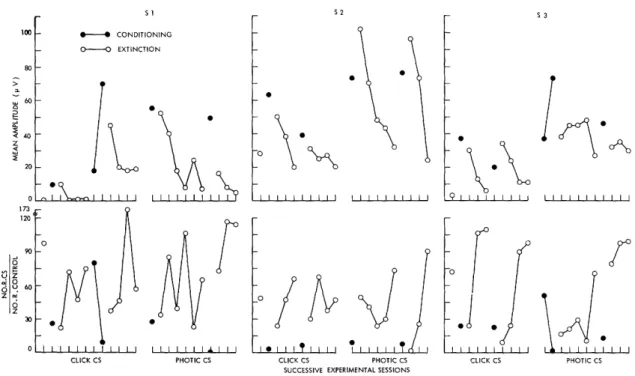
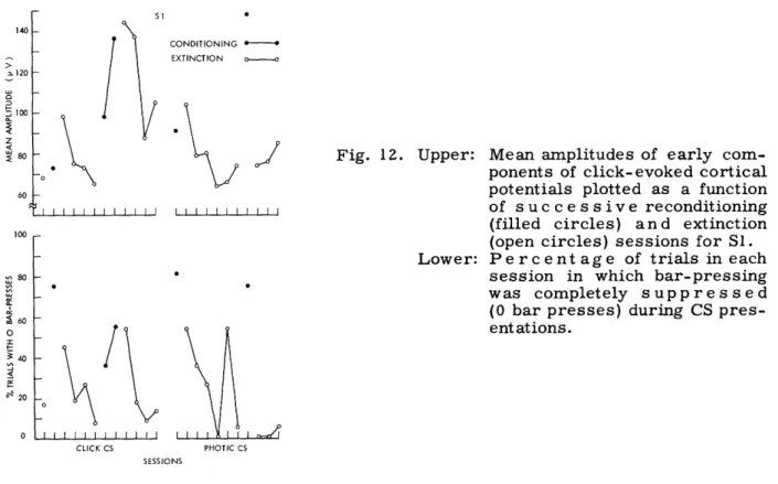
programmed intervals of no reinforcement. Conditioning of an emotional response is initiated when bar-pressing has become stable. This is accomplished by presenting a conditional stimulus (CS), for 1-minute periods in our experiments, which is followed on each occasion by an unavoidable noxious electric shock. Acquisition of the conditioned anxiety or fear can readily be traced in the increasing suppression of bar-pressing during CS presentations. The ease with which this conditioning can be followed in measures of bar-pressing, and the ease with which the behavior is established make the CER situation a particularly convenient aversive conditioning paradigm. An example of the behavior generated in such situations can be seen in Fig. 1. The cumulative response record shows a relatively high rate of bar-pressing in the absence of the CS, but vir-tually no responding at all during CS presentations.
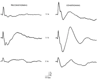
Fig. 1. Cumulative response record of bar-pressing in conditioned emotional response (CER) situation. First pip of each pair on the response curves indicates onset of CS. Second pip indicates termination of CS and presentation of shock UCS. Food reinforcements are in-dicated by pips on the bottom line.
15
2.7 BEHAVIORAL APPARATUS
In all but two experiments, the subjects were not restrained. They worked in long narrow boxes made of black Bakelite, 12 in. long, 3 in. wide, and 15 in. high. The food cup was located 3 in. above the lever at one end of the box. Dimensions of the box and location of the food cup minimized the amount of circling behavior and, therefore, the troublesome twisting of electrode leads. The food cup was also made of Bakelite, and the lever which the animals pressed was covered with a gravel and epoxy mixture. The use of nonconducting materials for food cup and lever eliminated noise arising from
con-tact potentials. The gravel-epoxy compound on the lever was the only nonmetallic mate-rial we could find that was hard enough to discourage or withstand chewing. Photoelectric
switches were used on levers to avoid the noise associated with mechanical switches, and the limiting stops were cotton-cushioned for the same reason.
The box contained a grid floor consisting of only 4 bars. Shock stimuli were deliv-ered to the subjects' feet through this grid. Two methods were employed (in different experiments) to eliminate the possibility that subjects might escape shock by standing on isopotential bars. In one, adjacent bars were connected through 10-kQ resistors so that the grid was a simple voltage divider. The second method employed a special
scrambling circuit designed by Richard J. Clayton. A variac provided a variable shock source of 60 cps AC. This current was usually chopped by means of an oscillating relay and then led through an isolation transformer to the grid or scrambling circuit. Shock duration in each instance was 0. 5 sec.
The rat boxes were housed in sound-attenuating, electrically shielded chambers, 22 in. wide, 21 in. deep, and 45 in. high. The chambers were located in a separate room to insure acoustic isolation from the control and recording equipment. The experi-ments, except for the changing of subjects, were completely automated. This was accomplished with the aid of conventional relay circuitry plus some solid-state devices. The latter were also designed by Richard J. Clayton.
In several cases, subjects were placed on food-deprivation schedules and were handled.daily for a week or more before electrodes were implanted, but in most experi-ments these procedures were not initiated until 5-7 days following surgery. Body weights were maintained at 75-80% of the animals' ad libitum feeding weights. It has been our experience that bar-pressing behavior in CER situations is best when animals are required to work for their entire daily food ration in the experimental situation, and when this allotment is ample. Experimental sessions, therefore, usually lasted 2. 5-3. 0 hours, and the animals received approximately 300-350 reinforcements, i. e., 13.
5-15. 75 gm of food. Water, with an added vitamin supplement, was always available to subjects in their home cages.
Bar-pressing was established under continuous reinforcement. Intermittent rein-forcement was introduced with schedules of low values. These were gradually increased until the final values were reached in order to maintain relatively high rates of
16
responding. No acoustic stimuli were ever introduced until rats had achieved high stable response rates. For at least 2-3 days before the first recording session, animals were run with electrode leads attached so that adaptation to the leads would not confound any effects related to the introduction of auditory stimuli.
2.8 ACOUSTIC STIMULI
Evoked potentials of concern in these experiments were all evoked by clicks. The clicks were generated by applying 0. 15-msec square pulses across a loudspeaker. The loudspeaker was located 37 in. above the floor of the experimental chamber. Walls of the chamber were lined with acoustic tile to reduce the amount of reflected sound. Click intensities were generally moderate, approximately 30-35 db above the rat's threshold. One of us determined the approximate threshold for click stimuli under the conditions of our experiments from both behavioral and evoked-potential measurements. 5 8 Click stimuli were always presented against a low-level background masking noise that was present throughout experimental sessions. In all experiments clicks were presented at a rate of 1/sec.
It was often the case that a food reinforcement was presented during a train of clicks, simply because such presentations were determined by the behavior and reinforcement schedule. On these occasions, there was no interruption of the click train. The stimuli presented during the 5-6 sec immediately following a reinforcement were not marked, however, on the magnetic tape. Consequently, click-evoked potentials recorded in post-reinforcement periods were not included in the average responses. This was done to eliminate the masking effects of chewing. It was clear from the muscle activity seen on cortical electrodes that ingestion of a food pellet was nearly always accomplished within 6 seconds.
2.9 PRESENTATION OF DATA
Our primary concern in this investigation has been with correlated changes in sen-sory evoked potentials and behavior in the individual organism. At this time there would appear to be no good justification for combining data from individual subjects, for at this stage of our inquiry into neuroelectric correlates of conditioning a model that might jus-tify the use of group measures is clearly lacking. Moreover, group means or other measures of central tendency often obscure important features of the data, and only
rarely do they present a more convincing summary of experimental findings than do data from individual subjects. The presentation of data from individual subjects is not
with-out its own problems. If all data are to be presented there are clearly problems of econ-omy. If the "typical case" is the adopted solution to these problems, one runs the risk of serious sampling errors. Throughout this report we have tried to find some com-promise, but in all cases each subject is represented in data presented for the several
experiments.
Habituation of evoked potentials in unanesthetized subjects has been described in
17
43 - 36
many reports (see, for example, Hernindez-Peon ; Garcia-Austt ). This habitua-tion refers to a more or less systematic reduchabitua-tion in the amplitudes of sensory evoked potentials associated with repeated presentations of the stimulus. The nature of these
changes is still a matter of dispute. The conditions under which it occurs, and where in the nervous system evoked potentials show such changes are problems that have not been resolved.
In order to obtain stable baseline measures of evoked potentials, we have routinely employed habituation procedures before any conditioning operations. These procedures were often in effect for 10 days or more. Although we have found evidence of habitua-tion in click-evoked potentials, these data will not be considered here. A discussion of
this problem would only lengthen this very long report and detract from its principal thesis. Habituation data from these and other experiments will be described elsewhere.
18
III. CLICK-EVOKED POTENTIALS RECORDED FROM CENTRAL AUDITORY STRUCTURES
Electric potentials evoked by impulsive sensory stimuli and recorded with macro-electrodes are summations of the electric responses in relatively large populations of cells. Different cell populations and several kinds of neural potentials, e. g., unit
"spikes" and postsynaptic potentials, may contribute to these summated responses. In this report we shall employ the term 'sensory evoked potential' in its narrower sense to mean the summated responses recorded by means of macroelectrodes. Such poten-tials often assume complex waveforms that are difficult to describe and difficult to quan-tify in some physiologically meaningful way. These difficulties are due in large measure to our inadequate understanding of the nature of these potentials. Three decades or more of experimental work have yet to provide a generally accepted and reasonably pre-cise account of evoked responses from primary projection areas of the cortex, perhaps the most extensively studied evoked potentials in the central nervous system. The anal-ysis of evoked potentials from most subcortical stations of specific sensory systems has
been only rudimentary. Moreover, the analysis has barely dealt with the potentials recorded from unanesthetized organisms, potentials that are admittedly more complex than those recorded from anesthetized preparations. But in spite of these difficulties, we pursue the study of evoked potentials, for it is clear that much has been learned
about functions of the C. N. S. through these efforts.
In the work reported here it was often necessary to proceed in considerable igno-rance regarding evoked potentials to be found in subcortical structures of unanesthetized rats. Moreover, the present report will not permit a detailed analysis of such activity. In some instances we cannot be sure that all components of evoked responses recorded from electrodes within a given structure have their origins in the activity of that struc-ture. Current spread from nearby structures is an ever present hazard when recording evoked potentials in the C. N. S. by means of so-called monopolar derivations. The prob-lem of interpretation is somewhat ameliorated by the use of "bipolar" electrodes, but this technique introduces its own problems. We have found monopolar recording desir-able for two reasons: (i) Electrodes are smaller than bipolar types and inflict less dam-age on neural tissue. (ii) In our experience, the reproducibility of evoked potentials from one subject to the next, and the correlation of these potentials with electrode loca-tions have proved much easier with monopolar derivaloca-tions. In the rest of this section, evoked potentials recorded in these experiments from cortical and subcortical auditory structures are described. It can be understood that these potentials were recorded from electrodes within or on the surface of the several structures. It cannot be assumed that these potentials necessarily have their origins in the same structures. An experi-mental analysis of some of these potentials is in progress, but until it is complete no definite statements can be made regarding the sources of at least some components of the evoked responses. We emphasize this problem for it will become clear that changes
19
in acoustically evoked potentials during conditioning are complex changes that may or may not involve particular response components. A component analysis of the wave-forms is therefore critical for any fundamental understanding of the changes.
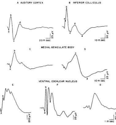
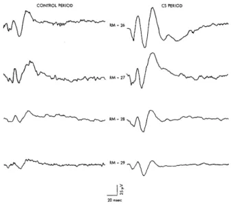
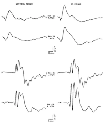
Figure 2 shows average click-evoked responses that are representative of evoked potentials recorded in this study from the several auditory structures. Evoked responses that deviated markedly from these potentials were generally excluded from the analysis.
A AUDITORY CORTEX B INFERIOR COLLICULUS
b
O nsec
10 m sec
MEDIAL GENICULATE BODY
b C D
b
1.
VENTRAL COCHLEAR NUCLEUS 10 m sec
E F G
b
a a
I m sec
Fig. 2. Average click-evoked potentials from auditory cortex, inferior colliculus, medial geniculate body, and ventral cochlear nucleus. Response of medial geniculate on the left is from anterior portions of the nucleus; on the right, from more posterior portions. See text for description of electrode placements for responses from cochlear nucleus. Averages were computed from 500-600 evoked potentials. Note different time and voltage calibrations. In this and succeeding figures, positive changes of poten-tial are indicated by downward deflections.
The cortical response in Fig. 2A is similar to click-evoked cortical potentials in unanesthetized cats described by other workers.3 3' 38, 74 An initial positive deflection (labeled a) is followed by three other peaks of alternate polarities (peaks b, c, and d). Respective latencies of the four peaks are 7-12, 12-17, 25-29, and 40-45 msec. (All latencies reported here have been corrected for the approximately 3 msec required for the sound-pressure wave to reach the subject.) The late negative wave, d, may occa-sionally peak as late as 65-70 msec, although it is by no means clear that such late
20
...
I
._
_
b
I