Adult articular cartilage culture system and
effects of IL-10
by
Wei Wang
Submitted to the Department of Electrical Engineering and
Computer Science
in partial fulfillment of the requirements for the degree of
Master of Science
at the
MASSACHUSETTS INSTITUTE OF TECHNOLOGY
May 1996
( Massachusetts Institute of Technology 1996. All rights reserved.
A uthor
...
...
Department of Electrical Engineering
d
Certified by...
...
Computer Science
May 17, 1996
Martha L. Gray
Associate Professor
Thesis Supervisor
Accepted by ...
Chairman, Department 1 Committee
I
A3OACGHU3oTTS
INoS:.U
i
OF TECHNOLOGY.
.
.
. ..
..L .Lthaler
on Graduate Students
JUL
16 1996
LIBRARIESAdult articular cartilage culture system and effects of IL-10
by
Wei Wang
Submitted to the Department of Electrical Engineering and Computer Science on May 17, 1996, in partial fulfillment of the
requirements for the degree of Master of Science
Abstract
Previous research in this lab has established a calf epiphyseal cartilage culture system and looked at effects of a particular cytokine, interleukin 10 on cartilage. In addition to the findings of increased tissue degradation and decreased tissue regeneration, evi-denced by accelerated release of matrix components and reduced uptake of essentials, it is also observed that as IL-1/3 is allowed to diffuse into the extracellular matrix from the periphery, observed degradation of tissue nevertheless centers around the blood vessels embedded inside the matrix. Although vasculature is regularly present in young cartilage tissues, mature cartilage has no blood vessels. Therefore an older culture system without vasculature will help elucidate effects of IL-11 on cartilage specific chondrocytes, with no complication from other cell types.
The first purpose of this research, therefore, was to define such a culture system. This task involves modifying several experimental protocols, and conducting control experiments to define parameters for such a system. It was found that both chon-drocyte and GAG concentrations are much lower in mature cartilage than in younger tissues, in addition to the much smaller overall ECM volume. Although less populous, chondrocytes in adult bovine articular cartilage have comparable synthetic capability as calf of new glycosaminoglycan (GAG) molecules. Furthermore, in culture with 20% FCS, adult cartilage loses proportionally much more GAG every day than calf, averaging about 5% of overall GAG content.
Given the average parameter values and their variances, the statistical paired-t test was adopted. As sample size is directly related to the width of confidence intervals, minimum sizes were calculated for an experiment to be able to detect any possible significant changes due to treatment. From one of the control experiments, it was found that at least 40 cylindrical cartilage disks have to be included in analysis to see a deviation in bulk GAG content from control on the order of 10% due to treatment. Such numbers were obtained for other parameters as well. Treatment protocols of sufficient sample sizes were then designed to ensure conclusive results can be drawn. The second purpose, after the control experiments, was to study the effects of IL-10 on the metabolism of cartilage proteoglycans. It was found that at the con-centration of 500 ng/ml, IL-10 has significant impact on mature cartilage matrix,
both severely repressing new generation and promoting increased loss of tissue GAG molecules. Accelerated degradation was detectable less than 20 hours after the first introduction of IL-13. GAG synthesis is only 13% of control after 3 days of IL-10 treatment.
The third purpose was to understand the correspondence of spatial distribution between introduced IL-1f and GAG loss, through histological staining. It was found that although the spatial pattern of GAG loss was as expected, no clear difference was observable between control and treatment. Unlike younger cartilage with localized GAG loss following IL-10 treatment, mature ECM releases GAG over the entire volume, making it harder to discern from histological stains.
Thesis Supervisor: Martha L. Gray Title: Associate Professor
Acknowledgments
First of all, I would like to thank my thesis advisor, Prof. Martha Gray, without whom this work would not have been nearly as coherent as it seems right now. Her encouragement, suggestions, and understanding to me, along with examples of her own dedication and commitment, educated me over the last two years on how to become a better researcher and a better person. I feel lucky in the sense that not every one gets to work for someone that they admire.
My appreciations and gratitude to Minerva Garcia for always being there when I needed help, and for her sense of humor that made long nights of running assays much more bearable. Many times, she was the source of both knowledge and lab supplies. Finally I will quit bothering her and say a word of 'glacias', excuse my Spanish. I would also like to thank Greg Allen for generous offerings of his experiences and friendship, from showing me how to cut open a cow the very first time, to suggesting optimal films for photomicroscopy. Ann Black also deserves much thanks. Without her the lab would at least be in chaos. With her around, I can rest assured that the DMB bottle usually won't be empty, the old radiolabels always get used up first, and that I would have nothing dirty left out on the bench for long.
I would like to thank the other people in the lab, Shelly, Arthur, Adil, Dan and Nimjak for sharing the space and laughs with me for the past two years; and all of those friends, students, colleagues and professors in this great institute, for making
my stay at MIT a truly enjoyable and memorable one.
Finally I would like to thank my beloved family, my parents and my dear little sister, without whom I would not have been here in the first place, and without whose support I wouldn't have gone nearly as far as I did. I'm just so happy that they will get to see me getting my diploma in person.
Contents
1 Introduction 13
1.1 Cartilage Extracellular Matrix ... . . . . . .. . . . . 14
1.2 Previous Research ... ... 15
1.3 Thesis Objectives ... 17
2 Cartilage Biochemistry and Mechanics 18 2.1 Major matrix components ... 20
2.1.1 Collagen . . . . . . . .. 20
2.1.2 Proteoglycan ... 21
2.2 Cartilage Mechanics ... 24
2.2.1 Force Balance in ECM ... 24
2.2.2 Cartilage Degradation ... 26
2.2.3 Interleukin 1 ... . .... ... .. 27
2.3 Tissue Culture Systems . ... . ... . 29
2.3.1 Calf Epiphyseal System ... .. 29
2.3.2 Adult Articular System ... 31
2.3.3 Other Cartilage Culture Systems . ... 33
2.4 Effects of IL-1, on Cartilage ... 33
3 Materials and Methods 36 3.1 M aterials . . . .. . 36
3.2 Cartilage Explant and Culture Condition . ... 37
3.3.1 Wet and Dry weights ... 38
3.3.2 Tissue DNA and GAG ... 38
3.4 Measurements of radiolabel incorporation . ... 39
3.5 Measurements of media GAG ... 41
3.6 Tissue Fixation and Staining ... ... . . ... 42
4 Results and Discussion 43 4.1 Control Studies and Data Analysis Methods . ... 43
4.1.1 Statistical Analysis ... 43
4.1.2 DNA and GAG Concentration in Adult Cartilage ... 49
4.1.3 Sensitivities of DNA and GAG Measurements . ... 51
4.1.4 Changes of DNA and GAG content during culture ... 60
4.1.5 Synthetic Rates ... 61
4.1.6 Rate of GAG Release ... 62
4.2 Effects of IL-1/ on Adult Cartilage . ... 69
5 Summary and Future Work
5.1 Thesis Objectives and Summary ...
5.2 Future W ork . . . .. . . . . .... .. .. . .. .. .. . .. .
A Effects of axial confinement on adult cartilage explants
77
77 82
List of Figures
2-1 Major Components of Articular Cartilage Extracellular Matrix . . .. 19
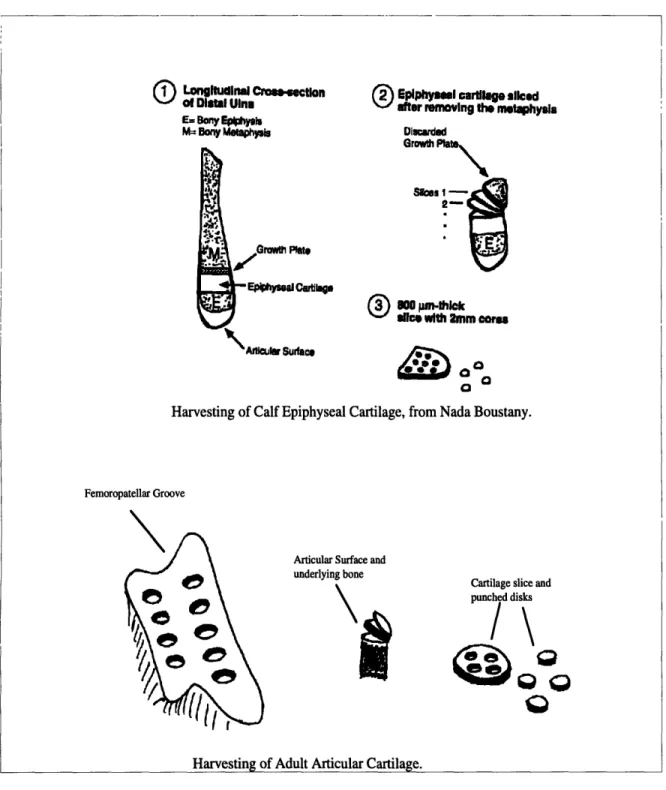
2-2 Schematic of Aggrecan Structure and interaction with hyaluronan. Many chondroitin sulfate and keratan sulfate groups are covalently as-sociated with a core protein. Each core protein is then noncovalently associated to a long chain hyaluronic acid molecule stabilized by link proteins . . . . 22 2-3 Schematic of cartilage tissue harvest. The three steps are named
cor-ing, sliccor-ing, and punching. Usually multiple slices of 0.8-2 mm thick
can be obtained from each epiphyseal cartilage core, but only one slice of 0.6-0.8 mm from adult articular surface. In order to obtained cylin-drical disks with parallel surfaces, the first few pm's are discarded. For articular cartilage tissue harvest, only sources of the femoropatel-lar groove is shown. Cartilage explants are also taken from the two condyles and the patellae. ... 30 2-4 Schematic of adult bovine femur-tibia joint used for harvesting
carti-lage tissues. Articular carticarti-lage is taken from femoropatellar groove, femoral condyles and the patellae (areas shaded). . ... 32
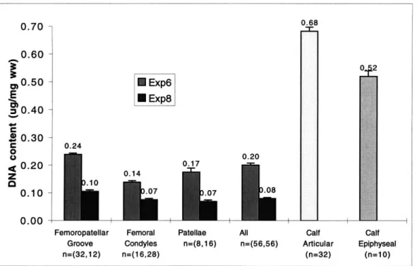
4-1 Average DNA contents normalized to wet weight, mean±SEM. Exp6 and Exp8 were experiments with adult articular cartilage; calf artic-ular cartilage was cultured from the femoropatellar groove; and calf epiphyseal data was adopted from Greg Allen. . ... 50
4-2 DNA contents for each disk (Exp8), normalized to wet weight. From left to right, the dotted lines separate the femoropatellar groove, the femoral condyles and the patellae in that order, and four disks in each slice are arranged adjacent to each other. . ... 50 4-3 Average GAG contents normalized to wet weight, mean+SEM . . .. 52 4-4 GAG contents for each disk (Exp8), normalized to wet weight. From
left to right, the dotted lines separate the femoropatellar groove, the femoral condyles and the patellae in that order, and four disks in each slice are arranged adjacent to each other. . ... 52 4-5 Two-sample CIs and sensitivities for DNA contents, columns and left
axis show sensitivities. Lines and right axis show ranges of CI for
•ci - Xc2, tickmarked at the means. FPG=Femoropatellar Groove;
FC=Femoral Condyles; and P=Patellae. . ... 54 4-6 CIs and sensitivities for DNA contents, columns and left axis show
sensitivities. Lines and right axis show ranges of CI (Xc1 - Xc2 for
two-sample and dc,-c2 for paired-sample), tickmarked at the means.
P=with pairing; P&A=with pairing and local averaging; otherwise two-sample. FPG=Femoropatellar Groove. . ... 56 4-7 Paired-sample CIs and sensitivities for DNA contents, columns and
left axis show sensitivities. Lines and right axis show ranges of CI for dcl-c2, tickmarked at the means. FPG=Femoropatellar Groove;
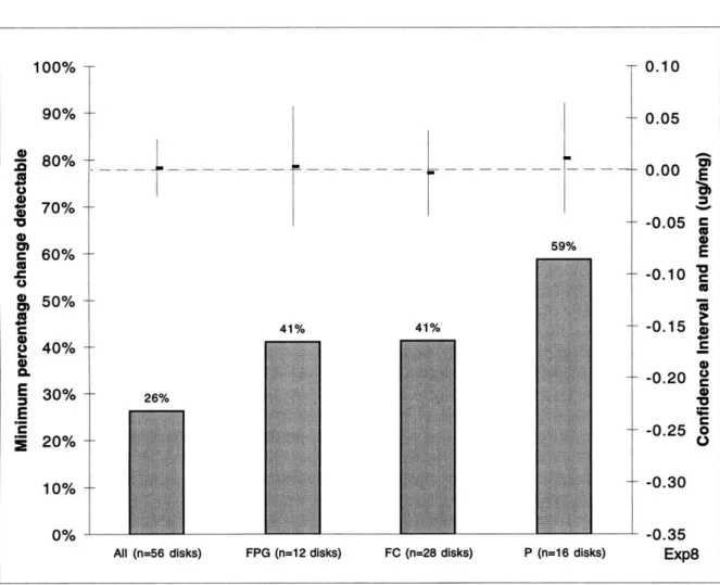
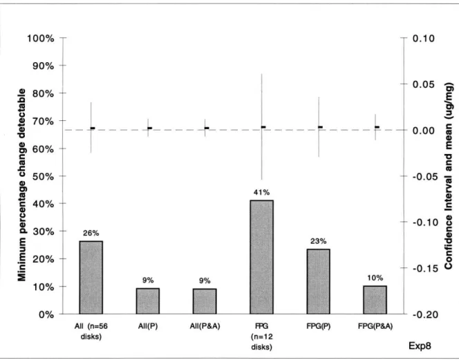
FC=Femoral Condyles; and P=Patellae. . ... 57 4-8 CIs and sensitivities for GAG contents, columns and left axis show
sensitivities. Lines and right axis show ranges of CI (Xc, - Xc2 for two-sample and dc,-c2 for paired-sample), tickmarked at the means.
P=with pairing; P&A=with pairing and local averaging; otherwise two-sample. FPG=Femoropatellar Groove. . ... 58
4-9 Paired-sample CIs and sensitivities for GAG contents, columns and left axis show sensitivities. Lines and right axis show ranges of CI for dcl-c2, tickmarked at the means. FPG=Femoropatellar Groove; FC=Femoral Condyles; and P=Patellae. . ... 59 4-10 Average 3H- and 35S incorporation rates in adult articular, calf
ar-ticular, and calf epiphyseal cartilage explants, in nmol/24h/fg DNA
(mean±SEM). Numbers in parenthesis indicate sample size. ... 63 4-11 Average remaining GAG content in cartilage disks as percentage of
GAGt, mean±SEM. For adult tissues, measurements of two randomly
selected groups (C1 and C2) are shown. For comparison, release data from calf articular (n=32) and calf epiphyseal cartilage (n=4) are also show n . . . .. . . . ... 64 4-12 Amount of GAG released during four days of culture for adult
car-tilage, and 6 days of culture for calf articular cartilage; and com-pared to amount of GAG synthesized on the last day of experiment, in Mg/24h/mg wet weight ... ... 65 4-13 Comparison of GAGo, GAGf, GAGt and S8 (mean±SEM) in adult
cartilage explants, where GAGo and GAGf are the measured GAG contents on days 0 and 8; GAGt is the calculated sum of GAGf and 8 days of release; and S8 is the GAG amount synthesized on day 8. . . 67 4-14 Comparison of GAGo, GAGf, GAGt and S6 (mean±SEM) in calf
carti-lage explants, where GAGo and GAGf are the measured GAG contents on days 0 and 8; GAGt is the calculated sum of GAGf and 6 days of release; and S6 is the GAG amount synthesized on day 6. ... . 68
4-15 Average DNA contents in adult articular cartilage with and without IL-10 treatment, mean±SEM. Both experiments were cultured for a total of 8 days, with IL-1 treatment starting on day 4... .. 70 4-16 Average GAG contents in adult articular cartilage with and without
IL-10 treatment, meaniSEM. Both experiments were cultured for a total of 8 days, with IL-1 treatment starting on day 4... . 71
4-17 Average GAG contents in adult articular cartilage with and without IL-1, treatment, mean±SEM. Both experiments were cultured for a total of 8 days, with IL-1 treatment starting on day 4... .. 73 4-18 Average remaining GAG content in adult cartilage disks with and
with-out IL-10 treatment, mean±SEM normalized to GAGt. Data taken from Expl6 . . . . 74 4-19 Average daily GAG loss in adult cartilage disks as percentage of bulk
GAG content of the previous day, with and without IL-1P, treatment, mean±SEM. Data taken from Expl6 and normalized to amount per 24 hour. . . . . 75
5-1 Average remaining GAG content in adult and calf articular cartilage disks with and without IL-13 treatment, mean±SEM normalized to
GAGE. Data taken from Expl6 and Expl7. . ... 81
A-1 Average DNA contents of adult articular cartilage in freeswelling and
axially confined cultures, for 8 hours (Expl5,n=14) and 86 hours (Expl2,n=36), meaniSEM. Both experiments included 4 days of freeswell culture
be-fore applying compression. The estimated strain of compression is 1%. 86 A-2 Average GAG contents of adult articular cartilage in freeswelling and
axially confined cultures, for 8 hours (Expl5,n=14) and 86 hours (Expl2,n=36), m ean±SEM ... 87 A-3 Average 35S-incorporation rates of adult articular cartilage in freeswelling
and axially confined cultures, for 8 hours (Expl5,n=14) and 86 hours (Expl2,n=36), mean±SEM. ... 88 A-4 Average 3H-incorporation rates of adult articular cartilage in freeswelling
and axially confined cultures, for 8 hours (Expl5,n=14) and 86 hours (Exp12,n=36), mean±SEM. ... 89 A-5 Average remaining GAG content in adult cartilage disks between freeswelling
and axially confined disks, mean±SEM normalized to GAGt. Data taken from Expl2. ... 90
A-6 Average daily GAG loss in adult cartilage disks as percentage of bulk GAG content of the previous day, from freeswelling and axially confined disks, mean±SEM. Data taken from Expl2 and normalized to amount every 24 hour .. . . . . 91
List of Tables
2.1 Collagen Types Present in Articular Cartilage t ... 21
2.2 Cartilage Sources and Tissue Handling in Selected Literature ... 34
3.1 Sulfate Sources in Radiolabeling Media ... 40
4.1 Minimum numbers of cartilage disks to have low than 10% sensitivity, using both pairing and local averaging. . ... . . . 60 5.1 Average values of DNA, GAG, 3H- and 35S-incorporation rate, and
water percentage for cartilage disks in selected experiments without IL-1 treatment, in mean±SEM. All cartilage samples were cultured in 20% FCS. Bulk DNA, GAG and radio-incorporations rates were measured on the last day of each experiment. Sample sizes are included in parentheses. ... 77 5.2 Average changes due to IL-13 treatment, as percentage of control.
Both experiments had sample size of 32 disks each for both control and treatment, and were cultured identically except days of IL-13 treatment. Numbers in parenthesis indicate sensitivities following non-conclusive testing ... ... 80
Chapter 1
Introduction
Articular cartilage is a major connective tissue type providing low-friction and wear-resistant bearing surfaces for load redistribution of the underlying bones. It is avascu-lar and aneural in nature with less than 5% cell volume. The resident cells, chondro-cytes, are loosely distributed throughout the extracellular matrix (ECM). Although sparse, these cells are mostly responsible for maintaining and regulating a rather complex network of macromolecules, cytokines and growth factors. This task is ac-complished through the art of intricate balancing and feedback systems. For example, chondrocytes adjust rates of synthesis for new matrix macromolecules according to the current status and need of ECM. At the same time, they synthesize and release degradative enzymes to break down the macromolecular structure for growth and development. In healthy joints, these two processes take place simultaneously and synergistically to fulfill mechanical supporting functions of articular cartilage [52, 49]. Another balance exists through harmony of contributing forces in ECM. With close to 80% of wet weight in water and under physiological pH, the numerous negative charge groups on the proteoglycans (PG) dissociate to generate strong electrostatic repulsion as they exist in close proximity. The resulting swelling pressure relates directly to the compressive resistance of cartilage critical to its function. At the same time, the integrity of the matrix structure is maintained by tension in the collagen network, holding the swelling PG's together. Therefore, the well-being of cartilage as a functional mechanical buffer relies on sustained balances of many different factors
of biological, mechanical and chemical in nature [40].
In joint diseases such as degenerative osteoarthritis (OA) and inflammatory rheuma-toid arthritis (RA), accelerated loss of charge bearing components out of ECM from heightened protease activities results in joint swelling and decreased stiffness. In this case, unfortunately articular cartilage behaves much like a unstable system. Once degradation prevails, chondrocytes seem to have little power to regain the balance, and the state of deterioration can only get worse.
Through in vitro explant culture studies, many events occurring during healthy and diseased cartilage have been characterized. Chondrocytes have been shown to modify their biosynthetic behaviors following changes in matrix environment, in terms of new macromolecule synthesis and matrix degradation. In controlled explants, el-evated levels of chondrocyte synthesis, even net deposition of matrix materials fol-lowing biological and/or mechanical stimulation have been achieved. It is the long term goal that through better characterization of cartilage remodeling mechanisms,
possible approaches will be adopted to intervene joint degradation processes [28]. As joint modulating factors are vast and interconnected, explant cultures are de-veloped to offer more defined systems and more meaningful interpretation of cartilage behavior with limited sources. This research defines an adult articular cartilage cul-ture system as an alternative to the existing young tissue system in this lab. The non-vascular nature of more mature cartilage offers the advantage of no complication from multiple cell types, a difficulty encountered in interpreting behavior of younger cartilage. This explant culture is subsequently treated with interleukin 1, an impor-tant cytokine believed to be a major player in matrix degeneration. Responses are characterized in terms of changes in bulk material properties, cellular activities, and spatial distribution profiles.
1.1
Cartilage Extracellular Matrix
Articular cartilage contains two major structural components, proteoglycan and colla-gen. The collagen network (mainly type II) renders shape and tensile strength to the
ECM, while proteoglycan provides the ability to undergo reversible deformation and to withstand compressive load bearing. The well being of the matrix depends upon both the integrity of collagen network and the quality and quantity of embedded pro-teoglycan. The responsibility to maintain the matrix rests almost entirely on the few resident chondrocytes. As the cells are distant from each other and away from direct stimulation, there must exist signalling mechanisms by which chondrocytes respond to changes within the cartilage matrix during growth and development. Cytokines and growth factors are likely to be important modulators in cartilage because of their presence in ECM, their receptors on chondrocytes, and the correlation of their activ-ities to changes in matrix properties. Equally important are changes in mechanical loading in modifying material properties of ECM, as cartilage is constantly under loading and changes in loading conditions (such as loss of gravity) result in changes in ECM.
1.2
Previous Research
Among the many different types of cytokines and growth factors, interleukin 10
(IL-10) is believed to be a key player in cartilage degeneration as it is involved in both
reduced synthesis and accelerated degradation, through two independent pathways [34]. As a local modulator, IL-10 can be synthesized by chondrocytes [7] and have receptors on them [5].
IL-1, suppresses synthesis of cartilage specific collagen types [27], and has in-hibitory effects on proteoglycan synthesis [19]. This lab has used radioactive precur-sor incorporation as means of monitoring chondrocyte synthesis and their responses to IL-1i3 and other changes in the environment [50].
IL-10 is also capable of up-regulating the production and activation of various enzymes while simultaneously down-regulating various enzyme inhibitors. With ele-vated concentration and activity, these enzymes, notably stromelysin and the yet un-known 'aggrecanase', proceed to chop off structural and functional ECM components. Observable consequences include increased release of macromolecule
glycosaminogly-can (GAG) fragments out of the ECM, and increased matrix swelling.
Besides being affected by cytokines like IL-1P, the primary function of cartilage as load-bearing material requires that chondrocytes monitor and adjust to changing mechanical environments. Alterations in biosynthesis due to compressive loading have been studied under many protocols [28]. Generally, static compression reduces both protein and GAG synthesis, while dynamic loading can enhance, reduce, or have no effect on cartilage explant depending on loading conditions. However, mechanical effects to matrix degradation are not well known, neither is the extent of chondrocyte involvement [3].
This thesis was motivated by results of an ongoing study in this lab. The ob-jective of the study was to examine the combined effects of loading and IL-10 on
cartilage metabolism, as arthritic joints are simultaneously under the influence of both. Using an in vitro immature cartilage culture system, this study showed that IL-10 induced degradation is dramatically reduced under concurrent static compres-sion, suggesting loading status is a very important determinant in how biological factors are interpreted. Also, it was found that the diffusion of IL-1, through ECM is the rate-determining step of PG degradation, despite its relatively small size [47]. The much lower diffusivity compared to comparably sized macromolecules implies possible binding of IL-13 to matrix components, and have significant implication in understanding how cytokine-mediated cartilage degradation occurs and how mechan-ical loading could alter this process.
The spatial profile of PG degradation was also studied. It was found that GAG loss occurs progressively from the perivascular regions in this younger cartilage cul-ture, contrary to the progression of introduced IL-10. This result suggests possible involvement of multiple cell types from the vasculature. Blood vessels, while present in immature cartilage, are absent in mature cartilage tissue, hence the interest in studying adult articular cartilage explants, and effects of IL-1I induced degradation in such culture.
1.3
Thesis Objectives
The specific objectives of this thesis are:
1. What are the biochemical composition and metabolic behavior of adult bovine articular cartilage explants, and how is it different from immature cartilage?
2. What are the animal-to-animal and site specific variances for the above prop-erties, and the implication of such variation on methods of analysis?
3. Whether this culture system can be used for cartilage studies, judging from the availability of tissue and the required sample size for conclusive results?
4. How does treatment of IL-1,3 affect joint properties, such as biochemical com-position, biosynthesis and degradation, and whether such changes correspond to previous observed roles of IL-10?
5. What is the spatial degradation profile following IL-10 treatment, and how does it compare to those seen in younger tissue?
Chapter 2
Cartilage Biochemistry and
Mechanics
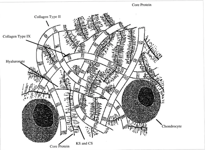
Articular cartilage is a specialized connective tissue covering the ends of long bones in synovial joints functioning as load bearing materials. The mechanical properties of articular cartilage come from the biochemical composition and metabolic behaviors of the chondrocytes. Although major structural constituents of have been characterized long ago, understanding of cartilage biomechanics requires knowledge of interactions of numerous contributing forces in a very complicated network. The major component of adult cartilage is water, about 70% of total wet weight. Almost equal amounts of collagens and proteoglycans comprise most of the remaining mass, each contributing about 10-20% of wet weight [49]. Cartilage specific cells, chondrocytes, occupy only 2-10% of tissue volume. Due to the lack of blood vessels, nerve fibers and direct cell-cell contact, cellular communications occur through diffusion of signalling factors through ECM. The same mechanism is also used for nutrient and waste transport.
Core Protein
'hondrocyte
Figure 2-1: Major Components of Articular Cartilage Extracellular Matrix
-7-2.1
Major matrix components
2.1.1
Collagen
Collagen is a major protein type in the body, comprising more than 30% of all protein mass. It is organized into insoluble fibers of great tensile strength to function as the
major stress bearing component of the underlying tissue [60].
Like other proteins, the primary structure of collagen consists of amino acids. However, almost one third of the amino acids in collagen are glycines, the smallest amino acid with only a single H atom as the side chain. About 1000 amino acid groups are aligned to form one left-handed a chain, with the typical Gly-X- Y repeats. In this triplet, X is most likely proline, and Y 4-hydroxyproline. Three such a chains intertwine into a right-handed superhelix of molecular weight on the order of 300 kDa. This hierarchy of fiber bundles alternately twisted in opposite directions is thought to be one of the primary sources of high tensile strength critical to collagen function, as it is able to convert longitudinal tension to a much easily supported lateral compression on an almost incompressible triple helix. Glycine has to occur every third position because that is where the a helices go through the triple helical center, too congested for any larger amino acid type. Many collagen amino acid side chains are hydroxylated to facilitate interchain hydrogen bonding, critical at stabilizing the tertiary structure. For example, proline is modified by prolyl hydroxylase to hydroxyproline, in which ascorbic acid is required for enzymatic activity [38].
Several collagen types are present in articular cartilage (Table 2.1). Type II col-lagen is the major structural colcol-lagen in cartilage, comprising 95% of total colcol-lagen in adult hyaline cartilage [38]. It further assembles through quarter-staggering and crosslinking to form collagen fibrils. Fibrils then interact with proteoglycan to define the material properties of extracellular matrix. Unlike in other connective tissues, single collagen fibrils are scattered seemingly randomly in cartilage. Several other collagen types are also present in cartilage. Type IX (1%) is very important in joint remodeling. Its covalent association with type II collagen molecules strengthens and stabilizes the network, and is broken down during normal matrix growth, repair and
Table 2.1: Collagen Types Present in Articular Cartilage t
Collagen Formula % total Properties or Possible Functions
Type II [al (II)]3 80-90% Forms fibrils and provides tensile strength to cartilage collagen network.
Type VI al(VI)a2(VI)a3(VI) 1-2% Forms network of fine fibrils between
larger fibrils.
Type IX a i(IX)a2(IX)a3 (IX) 10% Present on surface of type II collagen
fibrils. May play a role in cross-linking fibrils to each other or other ECM molecules. Type XI al(XI)a2 (XI)a3 (XI) 5% May play a role in determining type
II collagen fibril diameter. tAdopted from D.D.Dean [5]
also abnormal cartilage destruction [6, 31].
The biosynthesis of type II collagen involves first the formation of procollagen, which has propeptides of about 100 residues on both the N- and C- terminals of the collagen molecule. The presence of propeptides are necessary for the proper assembly of the triple helix. The Pro and Lys residues are hydroxylated to Hyp, 3-hydroxy-Pro, and 5-hydroxy-Lys in RER, as part of the post-translational chemical modification. This event precedes the folding of the three polypeptides, which requires the involvement of Hyp for stability.
2.1.2
Proteoglycan
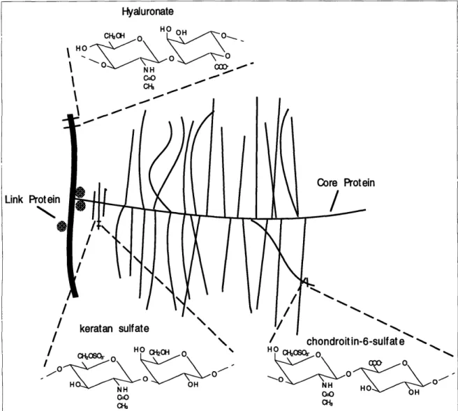
Proteoglycan (PG) is the other major structural component in cartilage. While col-lagen defines the major framework, proteoglycan gives matrix the ability to undergo reversible deformation. Proteoglycan is a subcategory of glycoproteins, the nomencla-ture for compounds associating both protein and carbohydrates. In this case, PG is the covalent association of a polypeptide core protein with one or more glycosamino-glycans (GAG) to form a group of complex heterogeneous macromolecules. Most of the heterogeneity comes from affiliating different GAG chains and varying numbers of them. These GAG chains usually account for over 90% of PG's total mass [49]. There are several types of PG found in cartilage, but the most abundant and structurally important one is aggrecan, the aggregating proteoglycan (Figure 2-2).
Hyaluronate o 0 N H C 00 C •o Link Proteil
NI%-I \ H N. OH NH 0:0 / chondroitin-6-sulfate o o HO cosor 0 NH H COH
Figure 2-2: Schematic of Aggrecan Structure and interaction with hyaluronan. Many chondroitin sulfate and keratan sulfate groups are covalently associated with a core protein. Each core protein is then noncovalently associated to a long chain hyaluronic acid molecule stabilized by link proteins.
A
The core protein of aggrecan is a 210 kDa polymer with three globular domains (G1, G2 and G3) and two extended segments (El and E2) [25]. G1 is the first domain at the N-terminal, and the site of attachment to hyaluronan, a independent long chain GAG. G2 has a similar structure to G1, and is separated from it by a 21 nm extended region called El. The long extended domain following G2 is called E2. This region of roughly 260 nm long is where the majority of GAG chains are covalently attached. At the C-terminal lies the G3 domain, a distinctive structure with unknown functions. In aged cartilage matrix, this domain is usually lost due to cleavage by proteases within E2. Varying lengths of E2 is another sources of heterogeneity for PG sizes in mature cartilage.
A glycosaminoglycan molecule is defined as a unbranched polysaccharide chain usually of alternating uronic acid and hexosamine residues. There are two major GAG types in cartilage, both of which are covalently linked to the core protein. Chondroitin sulfate (CS) is the major GAG type in ECM. It consists of repeating D-glucuronic acid and N-Acetyl-D-galactosamine disaccharide units. Each hexosamine residue is also sulfated, either at the 4' or 6' positions. Under physiological pH, chondroitin sulfate chains are highly anionic, resulting from deprotonation of both carboxyl and sulfate residues. Keratan sulfate (KS) is also a negative charge contributing source. The repeating disaccharides are D-galactose and N-Acetyl-D-galactosamine instead. However, since D-galactose is neutral at physiological pH, KS contributes one charge per disaccharide.
Aggregates of core proteins and GAG's are not randomly distributed in the ECM. They are noncovalently connected to each other through another important GAG, the hyaluronic acid (HA). It is the backbone for up to 200 aggrecan units to bind at their G1 domain, alternatively named the hyaluronic acid binding region (HABR). Neither of the disaccharide units in hyaluronate, D-glucuronic acid and N-Acetyl-D-glucosamine, are sulfated [51]. The association of aggrecans to HA is facilitated and enhanced by link proteins, the resulting conglomerate has mass on the order of 5 x
107 to 5 x 10⁸ [49].
Varia-tions in its amino acid sequence are results of alternative splicing of exons on a single gene product. Then the addition and modification of GAG chains occur. Having no standard template or sequence to follow, glycosylation is rather inhomogeneous. GAG chains are added one at a time through UDP-sugar intermediates. For example, in the case of CS chain addition, xylose is first attached to serine (or threonine) by xylose transferase (through UDP-xylose), followed by the addition of two galactose residues, and then alternating addition of N-acetyl galactosamine and glucuronic acid. Sulfate incorporation occurs after the synthesis of the unsulfated GAG chains, and in the Golgi Apparatus through a 3'-phosphoadenosine-5'-phosphosulfate (PAPS) inter-mediate. In the case of CS, both the 4 and 6 positions of N-acetyl galactosamine can be sulfated, and it has been shown that older cartilage synthesizes more 6-sulfated CS chains.
Since HA is not synthesized on an existing core protein, a different mechanism is used. An HA molecule is initiated in the inner plasma membrane, and the growing chain is extruded into the extracellular space. The precursors are still the UDP-sugars, but since HA doesn't go through Golgi complex, no sulfate groups are added.
2.2
Cartilage Mechanics
Articular cartilage biomechanics, i.e. how this important joint support material works, depend very much on the existence and maintenance of numerous contributing forces. On the one hand, these opposing forces provide functionality and allow remod-eling. On the other hand, excessive imbalance results in loss of material properties and joint destruction.
2.2.1
Force Balance in ECM
The first order force balance in ECM is ionic in nature. Both collagen and proteo-glycan are electrolytes capable of association/dissociation of their charge groups at different pH. For collagen, as well as protein cores of PG, the ionizable groups come from the amino and carboxyl residues (roughly 250 each in every collagen molecule).
Although most of them have pK's far away from the physiological pH, the entire collagen molecule is pretty much net isoelectric [52]. Therefore, collagen is not a significant contributor in ionic force balance. The story is totally different for pro-teoglycans. Their polysaccharide components account for almost all the net charges in many connective tissues. As mentioned above the charge groups in PG are the carboxyl groups (HA and CS) and sulfate groups (KS and CS), both capable of de-protonating to become negative charge carriers. Since the pK's for these groups are very low (between 2 and 4 observed), all of them are ionized under physiological con-ditions, resulting in the large net negative fixed charge density in ECM. The name 'fixed' comes from the fact that these charges are immobilized on the long GAG chains and in a network of collagen fibrils. The 'free' mobile charges, for example the much smaller hydronium (H30+) and sodium ions (Na+), then partition themselves across
ECM and synovium boundaries to maintain bulk electroneutrality. As we can see, the densely packed negative charges create a strong swelling pressure to expand, only to be limited by the framework of collagen network and any imposing mechanical loading. It has been shown that if allowed, the PG network can swell up to 5 times its size in ECM [25]. This strong swelling pressure is the source of compressive resis-tance offered by ECM. One thing to note though. Articular cartilage has an active matrix, not only in the sense that its components are constantly regenerated, but also that the water content can change under loading or stress relaxation. As water content is changed, as under compression, the concentration of ions changes, so does their interaction, or the characteristic Debye length. The mobile charges change their partition accordingly. Therefore, dynamic equilibrium and ionic charge balance are keys to matrix function.
Aside from ionic interactions, other noncovalent forces, such as hydrogen bonding are also very important. For example, bridged water molecules between amino acid hydroxyl groups, in particular between 4-hydroxyprolyl residues, associate to help stabilize the triple helix. But much stronger covalent cross-linking is believed to be ultimately responsible for the tensile strength of collagen and the structure it forms. Since there is no cysteine residues in the fibril-forming collagen polypeptides, the
usual means of cross-linking - disulfide bonding is not present in collagen. Rather a different mechanism is employed involving lysyl oxidase. With the help of this Cu-containing enzyme, lysyl residues are converted to their corresponding aldehydes: allysines, which subsequently undergo aldol or aldimine condensation to covalently link structural components. The extent of cross-linking increases with age, which is why single collagen molecule is only obtainable in very young animals.
Various ionic, covalent and noncovalent forces interact and balance to give artic-ular cartilage its unique material properties and function. Indirectly but ultimately, chondrocytes are responsible for producing macromolecules and enzymes to maintain such a balance. In growth or disease, one or more processes become dominant to allow
changes to occur. However, if balance is not restored, matrix integrity and function will be undermined, leading to destruction.
2.2.2
Cartilage Degradation
In articular cartilage, collagen types II and IX are major sources of structural integrity of the ECM, by containing adequate amount of water and by resisting tensile force and osmotic pressure of proteoglycans. Type IX collagen acts as the interfibrillar "glue" that stabilizes and lends cohesion to collagen network of normal articular cartilage [6]. This cohesive framework physically traps aggregating proteoglycans, and provide binding surfaces for small non-aggregating proteoglycans like decorin and fibromodulin. However, this network is not covalently constructed, i.e. there is no covalent linkage between two of the most important cartilage components, collagen and proteoglycans. Therefore, extensive cleavage is not required to break down a system under constant swelling pressure. Little alteration in the meshwork (loosening of interfibrillar association) by limited collagenolysis, or alternatively through cleavage at the El region of proteoglycan, can result in significant loss of GAG diffusing out of the matrix and compromised material properties [30].
Matrix degradation can be enzymatic or non-enzymatic. Among enzymatic agents, matrix metalloproteinases (MMPs) have been the focus in literature as they are very much active under physiological pH levels present in ECM [48, 11]. Other proteases
and glycosidases, as well as nonenzymatic agents such as oxidative radicals can also be involved in matrix degradation [57].
It has been shown in OA cartilage, the content of natural type II collagen re-duces significantly [10], presumably following increased hydration and denaturation by MMPs. Also, electron microscopic studies have demonstrated damage to collagen fibrils in RA [53]. Among the metalloproteinases, interstitial collagenase (MMP-1), gelatinase (MMP-2) and neutrophil collagenase (MMP-8) [59] have all been shown to cleave collagen [30], specifically at a single locus on the 3 a-chains of type II collagen. Another important endogenous enzyme is stromelysin (MMP-3), which can cleave at the N-telopeptide cross-linking sites of type II collagen, but not within the triple helix [6]. However, it can cut all three a chains of type IX collagen. Beyond direct actions, stromelysin is also involved in the activation processes of other proteases [18, 1].
Cleavages on PG occurs mostly on the protein core between G1 and G2 [13], and in the E2 region where CS and KS chains are attached [44]. Once the peptide bonds are broken, the entire structure not attached to HA is free to diffuse, and many ends up outside of ECM. Several types of MMPs, including stremolysin and collagenase have been shown responsible for many cleavage products [5]. However, one of the major cleavage site within El is still not accounted for [8].
Activities of proteases are very low in healthy joints [5], and there exist many protective measures to prevent cartilage from excessive degradation. Requirements of cytokine stimulation and activation from proforms are two examples. IL-1 discussed below has been shown to involve in both processes.
2.2.3
Interleukin 1
Interleukin 1 (IL-1) is a key member of the cytokine family, a collection of polypeptide hormones that mediate boy's response to various changing conditions. Similar to other cytokines, IL-1 level is negligible unless activated by some specific process, such as the immune response; and it conveys different messages depending on the types of target cells [7]. IL-1, and several other growth factors are particularly important in adult articular cartilage, as it has no means of communication through blood vessels
or nerve fibers, and cells are remote to each other.
The term IL-1 refers to two biochemically distinct but structurally related mem-bers, IL-la and IL-1L. Although both share the same set of receptors with almost identical affinity, IL-10 is believed more dominant in extracellular space with pI near 7 [12]. Although IL-10 is active in extracellular compartment, it has no signal peptide sequence, common to proteins secreted out of cell membranes [7]. The proforms are converted to be biologically active through the actions of serine proteases, particu-larly elastase and plasmin. The mature polypeptide of IL-10 has molecular weight of 17.5 kDa.
Chondrocytes have been shown to produce IL-10, as well as a host of other cell types [54]. Although small amounts of IL-10 may be present in cartilage under nor-mal conditions, its production and presence is dramatically increased in degenerative pathology of the joint such as OA [21, 19]. Several endogenous sources capable of in-ducing IL-10 production have been categorized [55]. Cartilage and bone debris, either from a traumatic events or from age-related accumulation, may cause the secretion of IL-10 and initiate catabolic processes. in intro evidences have also shown that
macromolecules of cartilage such as PG and different collagen types, when exceeding
a concentration limit, can initiate IL-13 production. Immune complexes, such as im-munoglobulins, have been detected in increased concentration in OA than in healthy joints, implying potential role in the induction of IL-10. Other exogenous sources, such as synovial tissue and neutrophil leukocytes, may also introduce IL-10 into ECM [9].
IL-10 has many target cell types, including chondrocytes [54]. The structure and post-binding signal transduction have not been fully characterized. One possibility is that the IL-10/receptor complex gets translocated to the nucleus upon binding, where it interact on the level of transcription [56]. Another possibility is the use of a signal transducing complex in the plasma membrane that in turn activates a second messenger system like cAMP. In general, cells stimulated with IL-10 demonstrate increasing cytosolic calcium levels, higher sodium/potassium ion fluxes, and increased protein kinase activities.
2.3
Tissue Culture Systems
In order to study cartilage and chondrocyte behaviors and properties, tissue culture systems are used to better isolate and characterize individual responses. Here tissue culture system refers to controlled in vitro culturing of native tissue to preserve tissue and cell viability and presumed similar behavior as is in vivo. In cartilage research, many aspects of tissue property and function have been studied through explant cultures. Following sterile harvesting, live tissue can be obtained and cultured in defined media containing essential nutrients, including amino acids, vitamins and minerals. It was found that for long term cultures, some growth factors and proteins in serum, e.g. fetal calf serum (FCS) are essential at keeping metabolic rates comparable to those in vivo [46]. FCS is more effective in stimulating metabolism than adult animals [46], and insulin-like growth factor is believed to be the active component [14]. After harvesting, it takes four to five days in culture before cartilage samples reach a constant metabolic state. In literature, for PG, this state is defined as constant PG levels in ECM as the result of the rates of synthesis and catabolism of PGs being equal [43].
2.3.1
Calf Epiphyseal System
The calf cartilage culture system in this lab takes samples from the distal ulnae of newborn calves (A. Arena Co., Hopkinton MA). Epiphyseal cartilage in between the growth plate (metaphysis end) and the bony epiphysis is sectioned to desired thickness (0.8 to 2 mm) (Figure 2-3). Then 2 to 3 mm-diameter disks are punched from the slices and incubated in culture media under 37°C and 5% CO2. The culture
medium consists of low glucose Dulbecco's Modified Eagles Medium supplemented with additional 0.4 mM proline and 0.1 mM non-essential amino acids, with daily supplements of 1% fetal calf serum, 4 mM L-glutamine and antibiotics.
It was found that calf epiphyseal cartilage contains by volume, about 7% blood vessels and 10% cells, with the remaining space occupied by water and the macro-molecules [2]. Chondrocyte density is estimated at 133,000 cells per mm3 [58].
Mea-LonglIUdlnal Croawsectlon of Distal UlMi
E= Bary E•ayam MW Bony MetaphiWis
®
Eplphyail cartilage silcedafter removing the metaphysis
8fts I
-WlPg
0
OW pn-thick slice with 2mm cora00
Harvesting of Calf Epiphyseal Cartilage, from Nada Boustany.
Femoropatellar Groove
Articular Surface and underlying bone
\ 9
Cartilage slice andpunched disks/\
Q
a
Harvesting of Adult Articular Cartilage.
Figure 2-3: Schematic of cartilage tissue harvest. The three steps are named coring,
slicing, and punching. Usually multiple slices of 0.8-2 mm thick can be obtained
from each epiphyseal cartilage core, but only one slice of 0.6-0.8 mm from adult articular surface. In order to obtained cylindrical disks with parallel surfaces, the first few pm's are discarded. For articular cartilage tissue harvest, only sources of the femoropatellar groove is shown. Cartilage explants are also taken from the two condyles and the patellae.
0
sured DNA concentration has mean of 0.015 pg/mg wet weight. Measured GAG density varies according to anatomical locations, increasing closer to the epiphysis. The values range from 40 to 80 mg/mL tissue water. However, because of the rela-tively large volume taken by vessels and cells, this calculation of GAG concentration underestimates the real ECM GAG density by about 15-20% [2].
This epiphyseal cartilage culture system was selected mainly for reasons of tissue abundance. Furthermore, the cartilage has relatively higher chondrocyte density than mature cartilage, providing metabolically more active specimens. The explants are relatively easy to harvest and culture, and are typical of hyaline cartilage in terms of metabolism and material properties. However, as mentioned above, young cartilage is generally vascularized regardless of the site of explant, with possibilities of complication from multiple cell types.
2.3.2
Adult Articular System
Mature articular cartilage is presumably more representative of aging tissue and more prone to arthritis. Although bovine articular cartilage has been successfully harvested and cultured in this lab, difficulties in handling did not make it a routine practice (Figure 2-4). Such difficulties include it being less cellular and consequently less metabolically active; harder to harvest due to less tissue abundance and harder bones; and harder to culture due to higher rate of GAG loss. As a consequence of difficulty in harvesting, the longer time tissue needs to be exposed, and the more likely to to have an infection. Moreover, variations both in terms of different joints in one animal and from one animal to another are rather high, making large sample size a necessity before making meaningful conclusions.
From the literature, mature cartilage have chondrocyte density on the order of 47,000 cells per mm3 [58], 4-5 times lower than young cartilage, a number confirmed later in this study. The effects of serum in culture have been studied. Without serum, chondrocytes settle at a much lower level of synthesis. With an optimal 20% of FCS, synthetic rates have been observed similar to those in vivo [46]. However, cellular outgrowth in long term cultures have been observed with 20% addition of
Figure 2-4: Schematic of adult bovine femur-tibia joint used for harvesting cartilage tissues. Articular cartilage is taken from femoropatellar groove, femoral condyles and the patellae (areas shaded).
32
FCS [16, 32]. On the other hand, GAG concentration is not significantly different in the two systems, although PG turnover is much faster in mature cartilage, with mean half life of 10 days compared to over 20 days in younger tissue [26].
2.3.3
Other Cartilage Culture Systems
Bovine adult articular and calf epiphyseal cartilage cultures are not the only ones studied. Many other sources of cartilage are reported in literature. Selected few are summarized in the following table. Most people use DMEM or Ham's F12 media, with supplements of serum, non essential amino acids (NEAA), 1-glutamine, ascorbate, proline and antibiotics. Although these supplements enhance explant culture, their compositions are important in quantitative characterization of cartilage metabolic behavior, as discussed in the next chapter.
2.4
Effects of IL-1/3 on Cartilage
IL-1, is a multifunctional hormone, mostly in the up-regulation of cellular metabolism and increased expression of several genes coding for biologically active molecules. In cartilage, it is believed to be involved in both the inhibition of synthesis and the pro-motion of degradation. The inhibitory effect of IL-1 on the synthesis of proteoglycan is well established. The limited amount of proteoglycan still synthesized appears to have the same structure as those under normal conditions [21]. At sufficient concen-tration, IL-1 is able to bring about a complete inhibition of PG synthesis. Similarly, IL-1, reduces the rate of synthesis of major cartilage collagen types (II and IX), while promoting the synthesis of some non-cartilage collagen types (II and III) [21]. Elevation of degradation occurs through activation of proteases by IL-13, particularly neutral metalloproteases stromelysin and collagenase [22]. During OA, where IL-13 level is higher, increased cartilage hydration and ultrastructural changes of collagen fibers are observed, indicating alterations in the collagen fiber network. The release of free proteoglycan fragments also increases significantly, causing loss of matrix com-ponents and compromised ability to withstand compressive loads.
Table 2.2: Cartilage Sources and Tissue Handling in Selected Literature.
Joint Tissue Principle Major Comments
Source Size Investigators Interests
porcine N/A T.E.Hardingham [45] PG studies Cartilage obtained fresh from
laryngeal the slaughterhouse, dissected
cartilage free of adhering tissue and
perichondrium, then shaved with a Stanley Surform. Immediately used for extraction of PG. Referenced by many.
bovine N/A C.J.Handley [29] PG studies extracted in 4M guanidine
nasal HCl for 48 hours.
septa
carpo- full V.C.Mow [42] mechanical havested from central region
metacarpal thickness compression of joint within 3-4 h after
joints of discs; studies death. DMEM supplemented
4-5 month 1-1.5mm with BSA, NEAAs, glu, and
old calves thick, 5mm antibiotics.
diameter
bovine 50-150mg C.J.Handley; PG Studies Cartilage obtained within 2 h of
metacarpo- wet weight V.C.Hascall [46] death. Articular surfaces exposed
phalangeal for each under sterile conditions with rinses of
joint, 1-2 sample sterile media to prevent drying. Incubated
year old at 370C with DMEM, 1g/l glucose
steer and organic buffers and amino acids.
Paper referenced by many.
bovine complete G.H.Korver [17] Culture medial sesamoid bones from
sesamoid cartilage system for MCP joints obtained within 2 h
bones from with bone anatomically after slaughter, and washed with
metacarpo- attached intact GBSS. Ham's F12 medium,
phalangeal articular supplemented with 10%FBS and
joints of cartilage antibiotics.
6 month old calves
porcine N/A J.T.Dingle [24] Synovial Explants of articular cartilage
metacarpo- catabolic from the metacarpals of young
phalangeal factor pigs (5-9 months old). Cultured
joints of study in DMEM with 15% (v/v) heat
adult pigs inactivated normal rabbit serum.
bovine full length D.Heinegard [35] Mechanical Cartilage explants obtained
metacarpal punched Studies within 2 h of sacrifice.
of 6 month explants, Ham's F12 medium, no serum,
old calves 4 mm in with HEPES and supplemented
diameter with ascorbate and antibiotics.
bovine 8mm cores, J.Steinberg [23] Tissue culture Bovine snouts obtained fresh
nasal 1mm thick model of RA, at the time of slaughter.
cartilage discs, PG studies Nasal septum cleaned with
highly betadine/soap solution,
uniform then placed in GBSS. Found
(60-70 mg) microscopic vascular channels.
2 year 2mm D.Heinegard [36] PG studies cartilage obtained at the time
old bovine diameter, of slaughter, and rinsed with
nasal 25-35 mg PBS. Most avascular region taken.
septa Ham's F12 medium with glu,
HEPES and antibiotics.
2 year 2 to 3mm R.Sah [32] and Growth factors 1-2 slices from each core. Disks
old bovine diameter, A.J.Grodzinsky and mechanical punched from the central area of
femoro- 500 pm compression slices. DMEM with NEAA, pro,
patellar thick ascorbate, and choices of sera or
grooves growth factors.
1-2 year five 8mm J.J.Parkkinen [20] Cyclic loading Knee opened under sterile conditions,
old bovine cores, on cartilage cores obtained and attached
knee joint, mean PG synthesis to petri dishes with a tissue adhesive.
both sides thickness Explants cultured for 2 days before
of the 1.5mm experiment. Eagle's MEM with Earle's
patellar salts, supplemented with antibiotics,
surface of ascorbate and glu.
The recovery of cartilage after an IL-1 attack has also been studied. The in-crease in proteoglycan synthesis is very slow, requiring weeks to recover. In contrast, degradation induced by IL-1 returned to the normal level relatively quickly (3 days) [44].
It has become clear that the ultimate state of cartilage matrix does not depend upon a single factor, but the balance/imbalance of a variety of forces, including cy-tokines and their antagonists, procycy-tokines and their activators, proteinases and their activators/inhibitors, and activator inhibitors, to just name a few. For example, IL-1 has been shown to be able to inhibit the production of tissue inhibitors of metallo-proteases (TIMP) [5], altering the balance in metallometallo-proteases and their inhibitors to favor degradation. It is also able to up-regulate (down-regulate) plasminogen activa-tor (plasminogen activaactiva-tor inhibiactiva-tor) to favor the activation of metalloproteases and itself [15]. The total balance of the entire complex network of hormones, enzymes and structural molecules are required for the well being of the matrix, and the lack of it, combined with limited ability of chondrocytic regulation, is the ultimate source joint degenerative diseases, like OA.
Chapter 3
Materials and Methods
3.1
Materials
Hanks Balanced Salt Solution (HBSS) and Dulbecco's Modified Eagle's Medium (DMEM) with 25 mM HEPES were purchased from GIBCO (Grand Island, NJ). DMEM was also purchased from JRH (Lenexa, KS), as was fetal calf serum (FCS). Non-essential Amino Acids (NEAAs), l-glutamine, antibiotics (10,000 units penicillin, 10 mg streptomycin, 25 pg/ml amphotericin B in 0.9% NaC1) and papain were bought from Sigma (St.Louis, MO). Hoechst 33258 dye was from Hoefer Scientific Instru-ments (San Francisco, CA). Calf thymus DNA standards and shark chondoitin sul-fate standards were also from Sigma. Radioactive sulsul-fate was from New England Nuclear (Boston, MA), and radioactive proline from Amersham (Arlington Heights, IL). Ruthenium Hexammine Trichloride (RHT) and 8% glutaraldehyde were from Polysciences (Warrington, PA), and sodium cacodylate was from Fluka Chemie AG (Buchs, Switzerland). There are two sources of recombinant interleukin 113, one cour-tesy of Professor Lee Gehrke, and the other from Cistron Biotechnology (Pine Brook, NJ).
3.2
Cartilage Explant and Culture Condition
Intact femur-tibia joint of an adult cow were obtained from a local abattoir (A. Arenas Co., Hopkington MA) on the day of slaughter. Under sterile conditions, the joint was opened, and cartilage tissues were exposed from three types of joint surfaces, femoropatellar groove (FPG), two femoral condyles (FC), and the patellae (P). The four joint surfaces and underlying bones were separated with a hand saw to be later mounted on the drilling vise. During this process (20-30 min) rinsing solution was applied continuously, prepared from HBSS and 1% (v/v) antibiotics. Once separated, cartilage surfaces were immediately covered with plastic wrap and placed on ice. On the vise, 3/8 inch diameter cores were drilled perpendicular to the articular surface. They were 2-3 mm long with less than 1 mm of cartilage at one end. Visibly damaged or arthritic cartilage were avoided. Cores were placed in culture dishes filled with HBSS and antibiotics and labeled of their relative positions. After drilling, Thickest possible (600 Mm-800 Am) cylindrical cartilage slices were cut out with parallel surfaces using a sledge microtome (American Optical, Buffalo NY), and again placed in HBSS with antibiotics. The first few Am from the articular surfaces were discarded, so were those close to the tide mark. Four cylindrical disks of 3 mm in diameter were then obtained from each slice using a dermal punch (Miltex, Lake Success NY). Each disk was immediately transfered to a 24-well culture dishes containing exactly 0.5 ml supplemented media, and pre-incubated at 370C with 5%
CO2 for at least 30 min. Culture media was prepared by from base media stock
and daily supplements. Base media stock was prepared immediately before each experiment and includes DMEM, 0.1 mM NEAAs and 0.4 mM additional -proline. Daily supplements include 20% FCS [43, 32] and 1% each of 1-glutamine, ascorbic acid, and antibiotics.
The entire process of harvesting, from exposure of articular cartilage to disks freeswelling in media, took 7-9 hours to complete for about 100 disks, during which time cartilage surfaces were either continuously rinsed with or immersed in HBSS and antibiotics. Generally about 10 slices can be obtained from the two faces of FPG; 6-8
slices each from the two FCs; and 5-6 slices from the patellae. During microtoming and punching, some slices (mostly from FCs) showed dramatic curling. Disks from such slices usually showed relatively large changes in wet weights from day 1 to the last day of experiment, and many of them yielded minimal chondrocyte activity at the end of experiments.
3.3
Measurements of Bulk Properties
3.3.1
Wet and Dry weights
Approximately 15-18 hours after cartilage harvest, all disks were weighed as sterile as possible on a microbalance (Mettler, Highstown NJ) to obtain initial wet weights. Each disk was then immediately transfered to another culture dish filled with 0.5ml newly prepared supplemented media. The process of weighing for each disk took less than 2 minutes. On the last day of the experiment, each disk was again weighed to obtain its final wet weight. Following lyophilization of 18-24 hours, dried disks were measured for their solid mass.
3.3.2
Tissue DNA and GAG
Papain was used to dissolve cartilage disks in preparation of DNA and GAG measure-ments. 125 pg/ml papain solution was prepared with 5 mM cysteine hydrochloride, 0.1M phosphate buffered saline and papain. Each disk was added exactly 0.5 ml of the above solution, and left in 600C water bath for at least 24 hours or until all tissues
were dissolved.
The DNA content of each disk was determined using the bisbenzimidazole (Hoechst 33258) dye binding assay [37]. A SPF-500 spectrofluorometer (SLM Instruments, Ur-bana IL) was used for photometric measurements. This assay is based on enhanced fluorescence in high salt solution following binding of bisbenzimidazol with double stranded DNA. Duplicate 50pl cartilage digest solutions were aliquated for each disk, to which 2 ml Hoechst dye solutions were added. DNA dye solution was prepared