HAL Id: hal-00379796
https://hal.archives-ouvertes.fr/hal-00379796 Submitted on 29 Apr 2009
HAL is a multi-disciplinary open access
archive for the deposit and dissemination of sci-entific research documents, whether they are pub-lished or not. The documents may come from teaching and research institutions in France or abroad, or from public or private research centers.
L’archive ouverte pluridisciplinaire HAL, est destinée au dépôt et à la diffusion de documents scientifiques de niveau recherche, publiés ou non, émanant des établissements d’enseignement et de recherche français ou étrangers, des laboratoires publics ou privés.
mHDA1/HDAC5 histone deacetylase interacts with and
represses MEF2A transcriptional activity.
Claudie Lemercier, André Verdel, Bertrand Galloo, Sandrine Curtet, Marie-Paule Brocard, Saadi Khochbin
To cite this version:
Claudie Lemercier, André Verdel, Bertrand Galloo, Sandrine Curtet, Marie-Paule Brocard, et al.. mHDA1/HDAC5 histone deacetylase interacts with and represses MEF2A transcriptional activity.. Journal of Biological Chemistry, American Society for Biochemistry and Molecular Biology, 2000, 275 (20), pp.15594-9. �10.1074/jbc.M908437199�. �hal-00379796�
J Biol Chem, Vol. 275, Issue 20, 15594-15599, May 19, 2000
mHDA1/HDAC5 Histone Deacetylase Interacts with and
Represses MEF2A Transcriptional Activity
*Claudie Lemercier , André Verdel
§, Bertrand Galloo, Sandrine Curtet, Marie-Paule
Brocard, and Saadi Khochbin
¶From the Laboratoire de Biologie Moléculaire et Cellulaire de la Différenciation, INSERM U309, Equipe, Chromatine et Expression des Gènes, Institut Albert Bonniot, Faculté de Médecine, Domaine de la Merci, 38706 La Tronche Cedex, France
Abstract
Recently we identified a new family of histone deacetylases in higher eukaryotes related to yeast HDA1 and showed their differentiation-dependent expression. Data presented here indicate that HDAC5 (previously named mHDA1), one member of this family, might be a potent regulator of cell differentiation by interacting specifically with determinant transcription factors. We found that HDAC5 was able to interact in vivo and in vitro with MEF2A, a MADS box transcription factor,and to strongly inhibit its transcriptional activity. Surprisingly,this repression was independent of HDAC5 deacetylase domain. TheN-terminal non-deacetylase domain of HDAC5 was able to ensure an efficient repression of MEF2A-dependent transcription. We thenmapped protein domains involved in the HDAC5-MEF2A interaction and showed that MADS box/MEF2-domain region of MEF2A interacts specifically with a limited region in the N-terminal part of HDAC5which also possesses a distinct repressor domain. These data showthat two independent class II histone deacetylases HDAC4 and HDAC5 are able to interact with members of the MEF2 transcription factor family and regulate their transcriptional activity, thus suggesting a critical role for these deacetylases in the control of cellproliferation/differentiation.
Introduction
Acetylation of chromatin proteins and transcription factors is part of a complex signaling system that is largely involved in the control of gene expression (1). Thus far, the specific involvement of histone acetyltransferases and deacetylases inthe control of individual gene expression has been clearly established(2, 3). One major role of these enzymes is the control ofcell differentiation in response to specific signals. Evidenceexists for the participation of the RPD3-related members in this process (4). Recently, however, a new family of higher eukaryotic histone deacetylases, distinct from the already characterized RPD3-related members, has been identified (5-7). These enzymes are related to yeast HDA1 histone deacetylase and within the cloned members, two show sequence homology and the same domain organization and are called HDAC4 and HDAC5 (5, 6). Despite this homology, HDAC4 and HDAC5 are probably capable of exerting distinct functions, since immunoprecipitation experiments showed that in cells they can be associated with different partners (6). A member, namedmHDA2/HDAC6, shows unique features within deacetylases, in that it possesses two HDA1 homology domains (5, 6). In contrast to the RPD3-related members, the expression of these genes is not ubiquitous. HDAC5 and mHDA2/HDAC6 expression is activatedupon cell differentiation (5). These observations suggest thatmembers of the class II histone deacetylases may play a specific role in the regulation of cell differentiation. Looking for potentialpartners of HDAC5, we obtained evidence of interaction
between HDAC5 and MEF2 transcription factors. We therefore focused our efforts on investigating this issue. The MEF2 family of transcriptionfactors belongs to the large family of MADS box transcriptional regulators present from yeast to humans (8). Besides their established role in myogenesis (9), MEF2 family members have been implicated in gene activation in response to mitogenic signaling(10, 11; for review, see Ref. 9). Very recently, it has beenshown that one member of the class II histone deacetylase, HDAC4,can interact with two different members of MEF2 family, MEF2A(12) and MEF2C (13). Here we show that HDAC5, a distinct memberof class II deacetylase, is also able to interact specificallywith the MEF2A transcription factor and to repress its transcriptional activator capacity. Data presented here and the fact that theinduction of various differentiation programs was found to beassociated with an up-regulation of HDAC5 gene expression (5),suggested that HDAC5 might be a general regulator of cell differentiation. We discuss here the possibility that HDAC5 might control the early stages of various differentiation programs by inhibiting the precociousactivity of determinant transcriptionfactors.
Materials and Methods
DNA Constructs-- MEF2-Luc, L8G5-Luc, and L8-Luc reporter plasmids were generated from
MEF2-CAT1 (14), L8G5-CAT, and L8-CAT (15), by replacing the CAT reporter gene by
luciferase (respective promoter regions were cloned in the pGL2 basic vector, Promega). PMT2-MEF2A (14), pcDNA-mHDA2/HDAC6(5), and LexA-VP16 (15) expression vectors have been described before. Deletions in HDAC5 were obtained by polymerase chain reaction.The resulting DNA fragments were cloned in pcDNA3.1 (Invitrogen)in-frame with a N-terminal HA-tag. GAL4 DNA-binding domain fusionprotein constructs were generated by polymerase chain reaction.DNA fragments coding for HDAC5 amino acids 123-673 and 673-1113were cloned in pcDNA-GAL4 DB, in-frame with the DNA-binding domain(amino acids 1-147) of GAL4. Mouse HDAC4 and human MITR cDNAs have been obtained by polymerase chain reaction from mouse embryo and human skeletal muscle cDNA libraries, respectively (Marathonready cDNA, CLONTECH).
Transfection-- HeLa cells were seeded in 6-well dishes (105 cells/well) and transfected 2 days
later with Exgen 500 (Euromedex) accordingto the supplier's protocol. In each transfection, DNA amount waskept constant with pcDNA3 empty vector. Cell extracts were prepared24 h post-transfection. Luciferase and -galactosidase activities were measured using the "Luciferase Assay System" (Promega) and the Luminescent -Gal Detection kit (CLONTECH),respectively.
In Vitro Interaction-- Glutathione S-transferase (GST) pull-down assays were performed as
described (16). GST fusion proteins were produced in BL21 Escherichia coli transformed with pGEX-5X-3 plasmid (Amersham Pharmacia Biotech), encoding GST alone, or GST fused to variousdomains of MEF2A protein. 35S-Labeled proteins were produced in rabbit
reticulocyte lysate from pcDNA plasmids using the TNT transcription/translation kit (Promega) and [35S]methionine (Amersham PharmaciaBiotech).
Co-immunoprecipitation, Immunofluorescence, and Western Blot-- HeLa cells were
transfected with Exgen500 as described above using 5 µg of pMT2-Mef2A or pMT2 vector and 5 µg of pcDNA-HDAC5(HA tagged) or pcDNA empty plasmid per 100-mm plate. 24 h post-transfection, cells were washed in cold phosphate-buffered saline and lysed in 50 mM
Tris, pH 8.0, 150 mM NaCl, 5 mM EDTA, 0.5% Nonidet P-40,1 mM phenylmethylsulfonyl fluoride for 20 min on ice. After centrifugation(12,000 × g, 5 min, 4 °C) extracts were diluted
with 1 volume of lysis buffer without Nonidet P-40 and incubated with 4 µg of polyclonal anti-MEF2 antibody for 1 h at 4 °C. 30-µl aliquotsof protein G-Sepharose were added and the incubation was continuedfor 2 h. Precipitates were washed in lysis buffer containing 0.25% Nonidet P-40 and finally re-suspended in SDS-polyacrylamide gel electrophoresis loading buffer. Western blot analysis was performed using standard procedures and immunocomplexes were detected by chemiluminescence (ECL+, Amersham Pharmacia Biotech). Immunofluorescence was performed as described (17). The following primary antibodies have been used: anti-MEF2 (C-21, Santa Cruz), anti-HA (3F10, RocheMolecular Biochemicals), and anti-HDAC5 (rabbit polyclonal antibodies raised against a peptide localized at the C-terminal region oftheprotein).
Results
MEF2A Transcription Factor and HDAC5 Are Found in a Complex in Vivo-- In our previous
work we reported the domain organization of HDAC5 histone deacetylase (5). The histone deacetylase domainof this protein was found to be located at the C-terminal halfof the protein and the N-terminal non-deacetylase domain did not show any significant homology with sequences present in the database. In order to have insight into the function of HDAC5, we periodically searched the data bank for new sequences homologousto the N-terminal domain of HDAC5. This approach allowed us to identify a Xenopus cDNA presenting significant sequence homology with the non-deacetylase N-terminal domain of HDAC5 (accession number, Z97214, the putative protein encoded by this cDNA is now known as MITR, Ref. 18). The GenBank sequence information presented MITR as a potential partner of Xenopus
MEF2 homologues. The sequence homology between HDAC5 and MITR suggested that HDAC5 could also be a partner of MEF2 transcription factors. In orderto investigate this possibility, we first expressed HDAC5 andMEF2A in HeLa cells and examined the formation of complexes bearingboth proteins in vivo by co-immunoprecipitation. Extracts obtainedfrom cells transfected with HA-tagged HDAC5, MEF2A, and both expression vectors were immunoprecipitated with anti-MEF2 antibodies. Thepresence of MEF2A and HA-HDAC5 in immunoprecipitated material was then monitored using anti-HA and anti-MEF2 antibodies. Fig.1 shows that anti-MEF2 antibodies are able to co-immunoprecipitateefficiently HDAC5 when both proteins are expressed in cells (Fig.1, anti-HA panel, far right). The endogenous MEF2 in HeLa cellscan also interact with HDAC5 since ectopically expressed HDAC5was also found associated with endogenous MEF2, in the absence of exogenous MEF2A expression (Fig. 1). It is interesting to note that HeLa cells transfected with HDAC5-expression vector producedtwo HA-tagged HDAC5 proteins (Fig. 1, input panel) and within these two proteins only the full-length one could interact with MEF2A.
HDAC5 Interacts Directly with MEF2A-- In order to investigate the possibility of direct
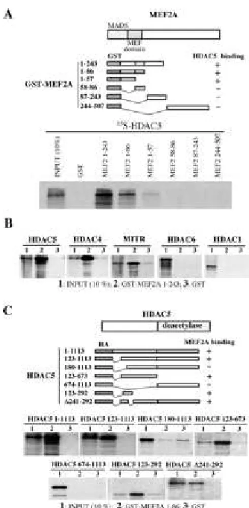
interaction between HDAC5 and MEF2A, we prepared bacterially expressed fusionproteins containing GST fused to different regions of MEF2A andexamined the interaction of these proteins with 35S-labeled full-length HDAC5. Fig. 2A shows that a GST fusion protein
harboring the N-terminal half of MEF2A is able to interact efficientlywith HDAC5 (Fig. 2A, GST-MEF2A 1-243), while the C-terminal half and the middle part of the protein did not recognize HDAC5 (Fig. 2A, GST-MEF2A 244-507 and GST MEF2A 87-243). This observation suggested that the region encompassing MADS box and the so-called MEF2 domain, has the ability to interact with HDAC5. To know whether MADS box or MEF2 domain or both are involved in the interactionwith HDAC5, we fused both or each of these domains to GST and analyzed the interaction with HDAC5 as above. These experiments
showed that MADS box/MEF domain is sufficient to direct an efficient interaction with HDAC5 (Fig. 2A, GST MEF2A 1-86). However, theMADS box alone was found to be able to interact weakly with HDAC5(Fig. 2A, GST MEF2A 1-57) while the MEF2 domain was not able todo so (Fig. 2A, GST MEF2A 58-86). The specificity of this interactionwas shown by considering the capacity of MEF2A N-terminal region to interact with two other deacetylases, HDAC1 and mHDA2/HDAC6. In this experiment we also used HDAC4 and MITR as known partnersof MEF2. Data show in Fig. 2B confirmed the interaction of these proteins with MEF2A as it has been reported very recently (12, 13, 18). However, no interaction between MEF2A and HDAC1 or mHDA2/HDAC6 was observed in these conditions (Fig. 2B), showing the specific interaction between HDAC5 and MEF2A in our assays.
A Limited Region in the N-terminal Non-deacetylase Domain of HDAC5 Is Involved in the
Interaction with MEF2A-- To determine regions in HDAC5 involved in the interaction with
MEF2A, expression vectors encoding HDAC5 deletion mutants wereprepared and used to obtain corresponding 35S-labeled proteins. These proteins were incubated with a GST-MEF2A
MADS box/MEF2 domain fusion protein and a pull-down was performed. A truncated HDAC5 molecule lacking the first 123 amino acids wascapable of interacting with MEF2A, while the removal of additional 57 amino acids completely abolished the binding (Fig. 2C, compareHDAC5 123-1113 and 180-1113 proteins). This observation showedthat the region of 57 amino acids, located between amino acids123 and 180 plays a major role in interaction with MEF2A. In order to confirm these findings, we expressed 35S-labeled polypeptides
corresponding to the 123-292 and 123-673regions of HDAC5 and found that both interact efficiently with MEF2A (Fig. 2C, HDAC5 123-292 and HDAC5 123-673, respectively). In these assays the deacetylase domain of HDAC5 could not interact with MEF2A (Fig. 2C, HDAC5 674-1113). These experiments showed that a limited region of HDAC5 encompassing amino acids 123-180 plays an essential role in the direct interaction with MEF2A.
HDAC5-MEF2A Interaction Represses the Transactivator Capacity of MEF2A-- A transient
transfection assay was designed to evaluate the role of HDAC5-MEF2A interaction on the transcriptional activity of MEF2A. The reporter system used consisted of the minimal promoterregion of myosin heavy chain gene flanked by two MEF2-bindingsites (14), cloned upstream of a luciferase gene. In HeLa cells, the expression of MEF2A led to a 20-fold activation of reporter gene expression (Fig. 3A). Co-expression of MEF2A and HDAC5 showeda very efficient inhibition of the activator capacity of MEF2A.Indeed, the use of as little as 1 or 5 ng of HDAC5 expression vector in these co-transfection assays, repressed significantly MEF2A activity (Fig. 3A). We then showed that the repression of MEF2A activity by HDAC5 was as efficient as that observed by HDAC4(12). However, the capacity to repress MEF2A was not sharedby all members of class II histone deacetylases, since the expressionof the third member of this family (mHDA2/HDAC6) did not alterMEF2A activity (Fig. 3B).
We then tried to show whether HDAC5-MEF2A interaction is necessary to repress MEF2A activity. HeLa cells were co-transfected with the MEF2 responsive reporter, a MEF2A expression vector and vectors expressing different deletion mutants of HDAC5. As shown before, the expression of full-length HDAC5 repressed efficientlyMEF2A activity (Fig. 3D, 1-1113 construct). Interestingly, this repression was found to be independent of the
deacetylase domain.Indeed a truncated HDAC5 bearing only the non-deacetylase domainof the protein was almost as efficient in repressing the MEF2Atranscriptional activity as the full-length HDAC5 (Fig. 3D, compare1-1113 and 123-673 constructs). This repression was found to be dependent on MEF2A-HDAC5 interaction, since a deletion mutant lacking the N-terminal region, defined to be the site of interactionwith MEF2A, was absolutely inefficient in repression (Fig. 3D,180-1113 construct). However, HDAC5/MEF2A interaction alone was not sufficient to repress transcription since two constructs expressingtruncated versions of the N-terminal non-deacetylase domain ofHDAC5, able to interact with MEF2A in vitro, were unable to represstranscription (Fig. 3D, 123-292 and 241-292 constructs, respectively).This experiment showed that the N-terminal region of HDAC5 possessesin addition to the MEF2-binding domain, a distinct repression domain. This region is localized C-terminal to the MEF2-binding domain and covers a region that encompasses amino acids 241-292 of HDAC5. Indeed, the deletion of this region abolished HDAC5repressive activity. However, amino acids located C-terminal tothis region are also important for this repressive activity, sincethe 123-292 version of HDAC5 were unable to repress transcription.The expression of the deacetylase domain alone did not show any repressive activity (Fig. 3D, 674-1113 construct). Western blots presented in Fig. 3, C and E, show that the vectors used in these experiments express efficiently all the expected proteins. However,it appeared that most of the constructs used, besides the expectedprotein (Fig. 3E, arrows), produced at least another protein.We could show that these products are not a result of degradation but are generated after the splicing of a fraction of the messengertranscribed from the plasmid. Indeed the use of different antibodiesand the cloning of a mRNA generated after transfection, showedthat the full-length mRNA transcribed from the plasmid can undergo splicing and generate variants (not shown and see Fig. 5B).
HDAC5 Possesses Multiple Transcriptional Repression Domains-- Since the deacetylase
domain of HDAC5 was found to be unnecessary to repress MEF2A transcriptional activity, one could questionthe ability of this domain to repress transcription. In orderto investigate this issue, we targeted the HDAC5 deacetylase domain into a promoter containing GAL4-binding sites. The reporter systemused contained eight copies of the binding sites for LexA immediatelyadjacent to five copies of the binding site for GAL4 (15), allcloned upstream of a luciferase gene. In the presence of LexA-VP16 fusion co-activator and the GAL4 DNA-binding domain alone (GAL4-DB), this reporter was activated to high levels of expression (Fig.4, GAL4-DB). An expression vector was prepared expressing a fusionprotein containing the deacetylase domain of HDAC5 (amino acids674-1113) fused to the GAL4 DNA-binding domain. Co-expression of LexA-VP16 and GAL4-HDAC5 showed that the HDAC5 deacetylasedomain could inhibit by 10-fold the transcriptional activity ofLexA-VP16 (Fig. 4,
HDAC5 674-1113 construct). We showed also thatthe non-deacetylase domain of HDAC5
possesses an independent repression domain capable of repressing MEF2A transcriptional activity (Fig.3D). To confirm this finding we also fused HDAC5 N-terminal non-deacetylase domain (amino acids 123-673) to the GAL4 DNA-binding domain. The expression of this fusion protein together with LexA-VP16 led also to an efficient repression of LexA-VP16 transcriptional activity(Fig. 4, HDAC5 123-673 construct). In order to show the specificityof this repressive activity, we prepared an expression vector encoding GAL4 DNA-binding domain fused to the MEF2-binding domainof HDAC5 (amino acid 123-292). This region of HDAC5 possesses the ability to interact with MEF2A (Fig. 2C) but does not repress its transcriptional activity (Fig. 3D). Therefore, it should not be able to repress the VP16-dependent transcriptional activation.As expected, the expression of this protein did not affect the ability of LexA-VP16 to direct an efficient transcription of the reporter gene (Fig. 4,
HDAC5 123-292). These data showed that the HDAC5 deacetylase domain, although unnecessary to repress MEF2A transcriptional activity, is an active repressor domain. They also confirmed that besides thedeacetylase domain, HDAC5 possesses another repression domainlocated in its N-terminalregion.
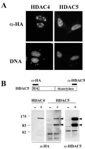
HDAC4 and HDAC5 Are Two Distinct Proteins-- The present report and those published
recently indicate that HDAC4 and HDAC5 are able to interact with members of MEF2 transcription factors. These two members of class II deacetylases, although sharing similar domain organization and sequence homology, may be able to ensure different functions in cells, as they have been found to be associated with distinct partners (6). In orderto obtain more evidence of functional differences between thesetwo deacetylases, we transfected HeLa cells with HA-tagged HDAC4 and HDAC5 expression vectors and examined the cellular distribution of these proteins using an anti-HA antibody. In cells transfectedwith HDAC4 expression vector a significant proportion of cellsshowed foci of HDAC4 accumulation in the nucleus (Fig. 5A, topleft), as it has been previously shown (7, 12) while noneof the cells transfected with HA-HDAC5 expression vectors showedthis particular pattern of expression (Fig. 5A, top right). Inthe majority of these cells, a homogenous distribution of HA-HADC5 was observed in the nucleus (Fig. 5A, HDAC5 panel). Moreover,Western blot analysis of cells transfected with HA-HDAC5 expression vectors showed the expression of two major forms of the protein, one presenting the expected size and the other was found to have a molecular mass around 83 kDa (Fig. 5B, HDAC5 panel, -HA, arrows). The use of an antibody raised against the very C-terminal regionof HDAC5 revealed the same bands (Fig.
5B, HDAC5 panel, -HDAC5,arrows). This experiment showed that the smaller band is not
theresult of proteolysis but rather is an altered form of the protein,possessing both the N-terminal HA-tag and the C-N-terminal epitope.This observation suggests that a fraction of the messenger producedfrom the vector undergo splicing giving rise to HDAC5 variants.Indeed, we cloned and sequenced a cDNA produced after transfection and confirmed the above hypothesis (not shown). Interestingly, the smaller HDAC5-related protein is not able to interact withMEF2A in vivo (Fig. 1, compare, input and IP -MEF2). It appearedtherefore that transfected cells are capable of generating atleast one form of HDAC5 that is unable to interact with MEF2A transcription factor. We are currently analyzing the presenceof such HDAC5 variants produced from the endogenous HDAC5 gene.Western blot analysis of cell transfected with HA-HDAC4 showedthe presence of a single band at the expected size. These datashow that HDAC4 and HDAC5, although both interacting with MEF2Aand modulating its activity, are different proteins and are probably involved in distinct regulatory pathways. Discussion
Within class II histone deacetylases, HDAC4 and HDAC5 encoded by two independent genes, show similar domain organization andsequence homology (6). However, the analysis of associatedproteins showed that in cells these deacetylases might enter functionallydistinct complexes. Indeed, HDAC4, but not HDAC5 was found associatedwith RbAp48 (6). Data presented here further support this ideaand show that when expressed in cells, HDAC4 and HDAC5 presentvery different patterns of nuclear localization. Moreover, analtered form of HDAC5, unable to interact with MEF2A could be generated in cells after transfection. Differentially splicedforms of HDAC5 have been already observed in different tissues and cell lines (5).2 All these observations suggest that HDAC4 and HDAC5 exert distinct
functions in cells and are probably involved in different regulatory pathways. Our data showed, however, that HDAC5, like HDAC4, is capable of interacting specifically with the MEF2A transcriptionfactor. This interaction completely abolished the transcriptionalactivity of MEF2A. We were surprised to find that this repressionwas independent of the deacetylase activity of HDAC5. Indeed, the N-terminal region of HDAC5 was found to interact with MEF2Aand repress its transcriptional activity. Moreover, we found thatMEF2A interaction and transcriptional repression are ensured by distinct regions of HDAC5 N-terminal non-deacetylase domain. Therepression domain in this region of HDAC5 might recruit cellular histone deacetylases to ensure its repressive activity. Indeed,it has been shown that MITR, which shows significant sequencehomology with the non-deacetylase domain of HDAC5 can interact with HDAC1 (18) and moreover, both HDAC4 and HDAC5 were found to be associated with HDAC3 (6). One may question the roleof the histone deacetylase domain of HDAC5 in the activity ofthis protein. We showed here that this domain is fully functionalin repressing transcription when it is targeted to a promotervia the GAL4 DNA-binding domain. We believe therefore that HDAC5is a multifunctional repressor; it is capable of a repressive activity, independent of or dependent on its own deacetylase domain. However, one should keep in mind that the deacetylase activity of HDAC5 may be required to repress transcriptional activity of endogenous MEF2A-responsive genes. MEF2A, and other transcriptionfactors, may target HDAC5 into chromatin where the deacetylasedomain could participate in a chromatin remodeling activity. Moreover, HDAC5 like RPD3-related members, may enter distinct multifunctionalcomplexes via "adaptors." Indeed, evidence does exist that HDAC4can form a complex when expressed in HeLa cells (7). The wayHDAC5 and HDAC4 are used in the regulation of gene expressionappears therefore to be similar to that of HDAC1 and HDAC2. Indeed, these RPD3-related deacetylases are capable of interacting directly with transcription factors, i.e. YY1, Sp1, etc. (19, 20) and/orthey may enter various complexes via interaction with moleculessuch as Sin3. Sin3 seems to play a pivotal role in targeting RPD3-related HDACs into various and distinct complexes (4). However, Sin3was not found associated with HDAC4 and HDAC5 (6) indicatingthat other molecules may accomplish a Sin3-like function in targeting HDAC5 and HDAC4 into different complexes. Interestingly, HDAC4, HDAC5, and a new member of the class II histone-deacetylase, HDAC7, were found to interact directly with corepressors N-CoR and SMRT(21, 22), confirming the fact that these new deacetylasescan target various complexes in a Sin3-independent fashion. The fact that RbAp48 was found to form a complex specifically with HDAC4 (6) provides some hints concerning the nature of complexes containing this deacetylase. Indeed, RbAp48, besides its involvement in NRD and Sin3 complexes (23, 24), where HDAC4 was not found(6), is associated with chromatin assembly machinery (25, 26). A role for HDAC4 in histone metabolism is therefore possible. The inhibition of MEF2A and MEF2C by HDAC4 (12, 13), as wellas that of MEF2A by HDAC5 reported here, indicate the possible involvement of these proteins in various regulatory pathways.Since MEF2 transcription factors regulate the activity of numerousmuscle-specific genes and play an important role in myogenesis, HDAC4/HDAC5 could be considered as potential regulators of myogenesis. However, a different role for these two proteins in myogenic differentiationcould be envisaged since we have observed spliced forms of HDAC5 messenger (but not HDAC4) in myoblasts.3 Moreover, since MEF2 members are also
involved in the regulationof cell proliferation, one might also propose a connection between HDAC4/HDAC5 activity and MEF2 function in this process. Previously,we showed that the expression of HDAC5 was up-regulated during a variety of differentiation programs (5). A careful examinationof the timing of HDAC5 expression during the induced differentiationof murine erythroleukemia cells, showed a transient activationof HDAC5 gene expression (5). This accumulation occurs between6 and 12 h of induction and is followed by decay in mRNA
content.This period precedes the commitment and the induction of globin gene expression (17). The same transient induction of HDAC5 gene expression was observed during the in
vitro differentiationof myoblasts to myotubes.3 It is therefore tempting to think that HDAC5
plays a role inthe pre-commitment period of various differentiation programsand functions as an early and transient inhibitor of specifictranscriptionfactors.
Acknowledgements
We are grateful to Dr. Jean Jacques Lawrence, the head of INSERM U309 for encouraging this work, to Dr. S. F. Konieczny forMEF2 plasmids and Dr. Mary Callanan for the critical reading of this manuscript. We thank Dr. Duncan Sparrow and Xiang-Jiao Yang for communicating results prior to publication and Amandine Jaulmesfor technicalassistance.
Footnotes
Contributed equally to the results of thiswork.
§ Recipient of a fellowship from the "Ligue Nationale Contre le Cancer, comité de la Haute
Savoie."
¶ To whom correspondence should be addressed. Fax: 95; Tel:
33-4-76-54-95-83; E-mail: khochbin@ujf-grenoble.fr.
2 C. Lemercier, A. Verdel, B. Galloo, S. Curtet, M.-P. Brocard, and S. Khochbin, unpublished
observations.
3 C. Lemercier, A. Verdel, B. Galloo, S. Curtet, M.-P. Brocard, and S. Khochbin, unpublished
data.
Abbreviations
The abbreviations used are: CAT, chloramphenicol acetyltransferase; GST, glutathione S -transferase.
References
1. Turner, B. M. (1999) Cell Dev. Biol.
10
, 165-1672. Kuo, M. H., and Allis, D. (1998) BioEssays
20
, 615-626 3. Struhl, K. (1998) Genes Dev.12
, 599-6064. Johnson, C. A., and Turner, B. M. (1999) Cell Dev. Biol.
10
, 179-188 5. Verdel, A., and Khochbin, S. (1999) J. Biol. Chem.274
, 2440-24456. Grozinger, C. M., Hassig, C. A., and Schreiber, S. L. (1999) Proc. Natl. Acad. Sci. U. S. A.
7. Fischle, W., Emiliani, S., Hendzel, M. J., Nagase, T., Nomura, N., Voelter, W., and Verdin, E. A. (1999) J. Biol. Chem.
274
, 11713-117208. Theissen, G., Kim, J. T., and Saedler, H. (1996) J. Mol. Evol.
43
, 484-516 9. Black, B. L., and Olson, E. N. (1998) Annu. Rev. Cell Dev. Biol.14
, 167-19610. Marinissen, M. J., Chiariello, M., Pallante, M., and Gutkind, J. S. (1999) Mol. Cell. Biol.
19
, 4239-430111. Yang, S. H., Galanis, A., and Sharrocks, A. D. (1999) Mol. Cell. Biol.
19
, 4028-4038 12. Miska, E. A., Karlsson, C., Langley, E., Nielsen, S. J., Pines, J., and Kouzarides, T. (1999)EMBO J.
18
, 5099-510713. Wang, H. A., Bertos, N. R., Vezmar, M., Pelletier, N., Crosato, M., Heng, H. H., Th'ng, J., Han, J., and Yang, X. J. (1999) Mol. Cell. Biol.
19
, 7816-782714. Yu, Y-T., Breitbart, R. E., Smoot, L. B., Lee, Y., Mahdavi, V., and Nadal-Ginard, B. (1992) Genes Dev.
6
, 1783-179815. Hollenberg, S. M., Sternglanz, R., Cheng, P. F., and Weintraub, H. (1995) Mol. Cell. Biol.
15
, 3813-382216. Hagemeier, C., Casewell, R., Hayhurst, G., Sinclair, J., and Kouzarides, T. (1994) EMBO J.
13
, 2897-290317. Gorka, C., Brocard, M. P., Curtet, S., and Khochbin, S. (1998) J. Biol. Chem.
273
, 1208-121518. Sparrow, D. B., Miska, E. A., Langley, E., Reynaud-Deonauth, S., Kotecha, S., Towers, N., Spohr, G., Kouzarides, T., and Mohun, T. J. (1999) EMBO J.
18
, 5085-509819. Yang, W. M., Inouye, C., Zeng, Y., Bearss, D., and Seto, E. (1996) Proc. Natl. Acad. Sci.
U. S. A.
93
, 12845-1285020. Doetzlhofer, A., Rotheneder, H., Lagger, G., Koranda, M., Kurtev, V., Brosch, G., Wintersberger, E., and Seiser, C. (1999) Mol. Cell. Biol.
19
, 5504-551121. Kao, H. Y., Downes, M., Ordentlich, P., and Evans, R. M. (2000) Genes Dev.
14
, 55-66 22. Huang, E. Y., Zhang, J., Miska, E. A., Guenther, M. G., Kouzarides, T., and Lazar, M. (2000) Genes Dev.14
, 45-5423. Tong, J. K., Hassig, C. A., Schnitzler, G. R., Kingston, R. E., and Schreiber, S. L. (1998)
Nature
395
, 917-92124. Zhang, Y., LeRoy, G., Seelig, H. P., Lane, W. S., and Reinberg, D. (1998) Cell
95
, 279-28925. Parthun, M. R., Widom, J., and Gottschling, D. E. (1996) Cell
87
, 85-94Fig. 1.
HDAC5 and MEF2A are found in a complex
in vivo.
HeLa cells were transfectedwith 5 µg of either HA-tagged HDAC5 or MEF2A or both expression vectors. 24 h post-transfection cell extracts were prepared and subjected to immunoprecipitation using an anti-MEF2 antibody (IP panel). The immunoprecipitated material was then used to obtain a Western blot and the presence of HA-HDAC5 and MEF2 was monitored using anti-HA and anti-MEF2 antibodies ( -HA and -Mef2, respectively). A fraction of the extracts was used to control the presence of the proteins of interest in the extracts before immunoprecipitation
Fig. 2.
Direct interaction between MEF2A and HDAC5.
A, mapping of MEF2A-HDAC5 interacting domains. The indicated regions of MEF2A (schemes in upper panel) were fused to GST and bacterially expressed fusion proteins immobilized on glutathione beads were incubated with 35S-labeled HDAC5. After pull-down, bound material was subjected toSDS-polyacrylamide gel electrophoresis and autoradiography. B, MEF2A interacts specifically with HDAC5, HDAC4, and MITR but not with HDAC1 and HDAC6. Indicated 35S-labeled
proteins were incubated with GST-MEF2A (1-243 fragment) immobilized on glutathione beads and subjected to pull-down and analyzed as above. C, a limited region of the N-terminal non-deacetylase domain of HDAC5 is involved in interaction with MEF2A. GST-MEF2A
(MADS-box+MEF2 domain, 1-86) was used in a pull-down experiment as above using
Fig. 3.
Repression of MEF2A transcriptional activity by HDAC5.
A, HDAC5 represses the transcriptional activity of MEF2A. One µg of a plasmid containing a luciferase reporter gene cloned downstream of myosin heavy chain minimal promoter flanked by two MEF2-binding sites was used to transfect HeLa cells. Co-transfections were carried out with 200 ng of a MEF2A expression vector alone (lane 0) or together with the indicated amounts of HDAC5 expression vectors. 100 ng of a -galactosidase expression vector were also used in each transfection. 24 h post-transfection, luciferase activity was measured and normalized with respect to that of -galactosidase. 100% represents mean values of three independent experiments performed in the absence of HDAC5 expression, standard deviations are indicated. B, HDAC5 is able to repress specifically MEF2A transcriptional activity. A transfection experiment was set up using the MEF2-responsive luciferase reporter gene, MEF2A expression vector (200 ng) without ( ) or with (+) 20 ng of the indicated expression vectors. Luciferase and -galactosidase activities were measured and normalized as above. C,HeLa cells were transfected with 10 µg of expression vectors encoding the indicated HA-tagged proteins and a Western blot was obtained and probed with an anti-HA antibody to show the expression of the proteins. D, HDAC5-dependent repression of MEF2 activity depends on HDAC5-MEF2A interaction. The MEF2-responsive luciferase reporter gene was used to transfect cells together with MEF2A expression vector, without ( ) or with (+) 20 ng of a vector expressing the indicated deletion mutants of HDAC5. Numbers (amino acids), refer to the regions of HDAC5 analyzed (see also Fig. 2C). 241-292, represents a HDAC5 mutant bearing a deletion in the indicated region. 674-1113 construct expresses the deacetylase domain of HDAC5. Luciferase and -galactosidase activities were measured, normalized, and presented as above. E, a Western blot obtained and analyzed as in B shows the expression of HA-tagged HDAC5 mutants. Arrowheads indicate the position of the expected full-length proteins.
Fig. 4.
HDAC5 possesses two distinct repressor domains.
HeLa cells were transfected with 400 ng of L8G5-luc reporter (scheme) and 50 ng of vectors expressing either the GAL4 DNA-binding domain (GAL4-DB) or the indicated regions of HDAC5 fused to GAL4 DNA-binding domain, in the absence ( ) or presence (+) of 100 ng of Lex-A VP16 expression vector (black histograms). In the control experiment, cells were transfected with the a reporter gene lacking the GAL4-binding sites (L8-Luc) and expression vectors encoding the Lex-A VP16 activator as well as the indicated GAL4-HDAC5 fusion proteins (open histograms). Luciferase and -galactosidase activities were measured, normalized, and presented as described in the legend to Fig. 3.Fig. 5.
HDAC4 and HDAC5 are two different proteins.
A, different distribution of HDAC4 and HDAC5 in HeLa nuclei after transfection. HeLa cells were transfected with 4 µg of expression vectors encoding HA-tagged HDAC4 and HDAC5. 24 h post-transfection cells were fixed and subjected to immunofluorescence detection of proteins using an anti-HA antibody ( -HA panel). DNA panels show nuclei of cells expressing HDAC4 and HDAC5 stained with DNA-specific dye Hoechst 33258. B, HDAC5 encoding vector expresses two different forms of HDAC5. HeLa cells were transfected with either HDAC4 or HA-HDAC5 expression vectors (+) or the corresponding empty vectors ( ). Extracts prepared from these cells were subjected to Western blot analysis using an anti-HA antibody ( -HApanel) or antibodies raised against the C-terminal region of HDAC5 ( -HDAC5 panel). Note
that the two HDAC5-related bands are revealed by both anti-HA anti-HDAC5 antibodies


