Design and Evaluation of a Device for Trapping Hepatitis C Viral
Particles at Ultra Low Concentrations
by Gregory J. Ekchian B.S. Biomedical Engineering Boston University, 2009
ARCHIVES
MASSACHUSETS INSTITUTfE OF TECHNOLO3YJAN 0 4
LIBRARIES
SUBMITTED TO THE DEPARTMENT OF MATERIALS SCIENCE AND ENGINEERING IN PARTIAL FULFILLMENT OF THE REQUIREMENTS FOR THE DEGREE OF
MASTER OF ENGINEERING IN MATERIALS SCIENCE AND ENGINEERING AT THE
MASSACHUSETTS INSTITUTE OF TECHNOLOGY SEPTEMBER 2010
C 2010 Gregory J. Ekchian. All rights reserved. The author hereby grants to MIT permission to reproduce
and to distribute publicly paper and electronic copies of this thesis document in whole or in part
in any medium now known or hereafter created.
Signature of Author:
'I
(A'
/1/i
Certified by:
t
t
Department of Materials Science and Engineering
4
) August 13, 2010Michael J. Cima Sumitomo Electric Industries Professor of Engineering Thesis Supe igor Accepted by:
Christopher Schuh Chair, Departmental Committee on Graduate Students
Design and Evaluation of a Device for Trapping Hepatitis C Viral Particles at Ultra Low Concentrations
by
Gregory J. Ekchian
Submitted to the Department of Materials Science and Engineering on August 13th, 2010 in Partial Fulfillment of the
Requirements for the Degree of Master of Engineering in Materials Science and Engineering
ABSTRACT
A new method to quantify hepatitis C (HCV) viral particles when present in ultra low
concentrations is being developed. Hepatitis C is a viral infection that affects the liver. There are 3.2 million people in the United States with an active hepatitis C infection. Untreated HCV can lead to cirrhosis, liver failure and liver cancer. HCV treatments can be very costly and physically taxing for patients; the side-effects of treatment are comparable to persistent flu-like symptoms. Physicians are looking to shorten the duration of the standard treatment, typically 24 to 48 weeks, for patients who respond quickly. Physicians must have more sensitive testing equipment to truly know when a patient has been cured and be able to successfully shorten the length of treatment. Current diagnostic tests are insufficiently sensitive when the patient begins to positively respond to treatment and the amount of the virus present in his/her blood
dramatically decreases. This limitation can be overcome by employing an in-vivo sampling technique, where a device is placed in a vein to trap HCV viral particles present in the blood.
These particles are then subsequently quantified with a commercially available test. This technique allows at least 40,000 times more blood to be sampled in 30 minutes than with a traditional blood draw, greatly increasing the effective sensitivity of the test. The approach provides significant medical benefit to the patient being treated and a strong financial incentive to the entity paying for the treatment.
Thesis Supervisor: Michael J. Cima
Acknowledgements
I would like to thank Professor Michael Cima for his invaluable guidance,
feedback and advice over the last year as I worked on my thesis. I would also like
to thank everyone in the Cima Lab for giving me guidance and assistance during
my thesis and always being very helpful. I would especially like to thank Vincent
Hok Liu, Maple Hongye Ye and Chris Vassiliou who were always willing to take
the time to meet with me and discuss this project throughout the year.
Table of Contents
Introduction ... 7
Statistics of the Hepatitis C Infected Population ... 7
Transm ission of Hepatitis C... 8
Diagnosis of Hepatiti s C ... 9
Current RNA Testing Equipment ... 10
Patient Classifications in Response to Treatment ... 11
Drugs to Treat Hepatitis C and Their Side-effects... 11
Duration and Cost of Treatm ent... 12
Em erging H CV Treatments ... 12
Funding for H CV Research ... 13
The Unmet Need...13
The Solution ... 14
Anti body/Antigen Interactons ... 15
Initial Design Concept ... 16
Trapping Capabilities of "Twisted Coil " Design ... 19
Im plantation Location and Procedure ... 21
Implantation Location... 21
Implantation Procedure ... 22
Blood Sam ple Preparation ... 24
Current Sample Preparation ... 24
Sample Preparation When Using Trapping Device ... 24
M anufacturing ... 24
M aterials Selection ... 24
M anufacturing of Nitinol Backbone ... 25
Polymer Coating ... 26
Application ofAntibodies ... 26
Packaging and Storage ... 26
M anufacturing Cost ... 27
Supply Chain ... 29
Distribution Chain ... ... 0
Insurance Companies, M edicare and M edicaid ... ... ... 30
Potential Customers and Incentives for Adoption ... 31
M arket for the HCV Trapping Device ... ... ... 32
Prim ary M arkets...32
Secondary M arkets...32
Cost of the Device and Procedure ... 34
M arket with New Therapies ... 35
6-M onth Follow-up ... 38
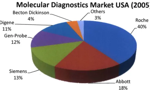
Current Diagnostics M arket...38
M arket Size...38
Profit M argin of the Current M arket Leaders ... 39
Profit M argin ofthe Proposed D evice ... 40
Intellectual Property ... 41
Prior Art...41
Future Patent Portfolio... 43
FDAApproval ... 44
Approval Process ... 44
Business M odel ... 44
Teaming with Companies Producing New H C V Treatments ... 46
Future Experiments...47
In- Vitro Testing ... 47
In-Vivo Animal Testing ... 47
Funding - Next Two Years...48
Conclusion ... 48
Appendix ... 50
Design #1 - Diffusion of Viral Particles through Polymer Membrane... 50
Design #2 D ifusion of Viral Particles in Blood ... 50
Blood Sampling Calculations - "Twisted Coil Design ... 52
Two Year Research Budget ... 54
Y ear 1 ... 5 4 Y e ar 2 ... 5 4 References ... 55
Introduction
Hepatitis C (HCV) is a viral infection that causes inflammation of the liver (location of liver shown in Figure 1)1. The disease is usually spread through contact with the blood of an infected person.
Treatments for hepatitis C range from 24 weeks to 72 weeks depending on genotype and the patient's
response to treatment. Hepatitis C can be classified into two groups. Acute hepatitis C describes the virus when it is within six months of the initial infection. Chronic hepatitis C describes the virus when it progresses beyond the initial six month time window. Acute
hepatitis C becomes chronic hepatitis C for 75%-85% of Figure 1 shows the location of the liver, the site of replication for people infected. The remaining 15%-25% of people are hepatitis C.
able to clear the virus from their bodies without the
need for treatment; it is unclear why this happens for some people and not others.
Statistics of the Hepatitis C Infected Population
Each year about 17,0002 to 35,0003 people are infected with HCV; not all of these people are aware they are infected. Roughly 65,0004 people are treated yearly for an HCV infection in the United States. This includes some new infections as well as past infections that are newly diagnosed. There are currently 4.1 million people in the United States who have HCV antibodies, as well as another 170 million worldwide. Antibodies are an indication that one currently has an active HCV infection or has had one in the past. HCV antibodies will remain even if a patient no longer has an active infection. It has been estimated that 3.2 million people in the antibody positive population have an active infection. One study has found that as many as 80%5 of people with an active infection do not know that they are infected, presenting a
serious global health problem because many people unknowingly expose others to hepatitis C. It has been suggested that the currently available HCV statistics may underestimate the problem
because many high risk populations are often not counted. The HCV infection rate has been observed to be higher than average with prisoners, intravenous drug users and the homeless. A study from 2005 estimated that 42% of homeless veterans are infected with HCV; this is much higher than the roughly 1.5% of the general population.6 Another study shows that the prison
population was observed to have an infection rate between 15% and 41%. The complications of hepatitis C can be severe if the disease goes untreated. HCV was the leading cause of liver cancer and liver transplants in the United States as of 2009.7 The need for a liver transplant imposes two serious burdens on society. It limits the supply of healthy livers available for transplant for non-HCV related patients and costs roughly $400,000 (transplant and follow-up
8
care) .
There are six genotypes of hepatitis C, numbered 1 through 6. Genotype 1 accounts for roughly 75% of all cases in the United States and 60% worldwide. Genotypes 2 and 3 account for about 10%-20% of all cases, while the remaining cases are classified as genotypes 4, 5 and 6. Most research in the United States focuses on genotypes 1 through 3 because of their prevalence in the US.
It is expected that by 2020, one million HCV patients will have cirrhosis.9 Each year 10,000-12,00010 deaths are attributed to hepatitis C and this number is expected to triple by 2020.11 Projections show that the risk of cirrhosis, liver cancer and liver related deaths can be reduced by 16%, 31% and 36% respectively with the use of currently available drugs to treat patients." The development of new therapies and diagnostics will only help to improve these numbers.
Transmission of Hepatitis C
There are many sources for contracting the hepatitis C virus, and it is not always possible to determine how a patient contracted the virus. However, the majority of HCV cases are a result of injection drug use (at least 60 %); 13 even if it was infrequent or well in the past, it can still be
the source of the infection. Many medical procedures that are considered routine today have, in the past, exposed patients to HCV because there were not sufficient tools to screen blood transfusions and organ transplants. Patients who had blood transfusions before 1992 have an increased chance of being infected with HCV. Procedures such as receiving clotting factors, specifically prior to 1987, and hemodialysis for kidney failure can also increase a patient's
chance of contracting HCV. Birth to an infected mother remains a source of transmission, but accounts for less than 5% of total cases. Accidental needle sticks from an HCV infected person can increase one's chance of being HCV positive and pose a particular risk to healthcare
workers. While they are at a higher risk for becoming infected, healthcare workers typically have a high rate of successful treatment because treatment begins almost immediately after the initial infection.
Diagnosis of Hepatitis C
There are three types of tests that are key to the treatment of hepatitis C. The first test
administered to a patient who is suspected of having an HCV infection is an HCV antibody test. This is typically the first test because of its low cost compared to other diagnostic tests available (about a third to half the price). A positive antibody test is indicative of a past or present HCV infection. A follow-up test is required for anyone testing positive for HCV antibodies to determine if the infection is still active. Confirmation is done with an HCV RNA test. If a patient is positive for HCV RNA, it is an indicator of an active infection. There are two critical results obtained from an RNA test. First, the HCV genotype is determined. This helps to predict the success of treatment and dictates the duration of treatment. The second use for the HCV RNA test is to determine the concentration of virus present in a patient's blood, clinically termed viral load. The viral load prior to treatment, like the genotype, is also a strong indicator of the potential for a successful course of treatment. If the viral load is low (below 2 million viral particles/mL), the success of treatment is higher than if it is high (above 2 million viral particles/mL). The last test used is a liver biopsy. Liver biopsies are used to determine the extent of liver damage. Liver biopsies provide useful information regarding the extent of liver damage the patient has experienced. It is important to note that successful eradication of the virus does not undo existing damage to the liver. HCV RNA testing is repeated as frequently as each week during treatment to monitor a patient's response to the drug regimen.14 It is
recommended that the same HCV RNA test be used throughout treatment to avoid the inherent variations of different tests.15
Current RNA Testing Equipment
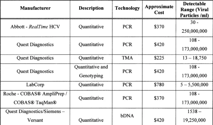
The table below shows the sensitivities, costs and manufacturers of current HCV tests. Because the proposed device is only used to trap the viral particles, one of the tests listed in Table 1 will need to be used to quantify the viral load. These HCV tests range between $225 and $780, depending on whether the physician is trying to quantify a high viral load or a low viral load. The first step of any testing regimen will be to use a traditional blood draw to quantify the viral load.
I
DetectableManufacturer Description Technology Approximate Range (Viral
S
Cost Particles /ml)30-Abbott -RealTime HCV Quantitative PCR $370
250,000,000 108
-Quest Diagnostics Quantitative PCR $420
173,000,000
Quest Diagnostics Quantitative TMA $225 13 - 18,750
Quantitative
and 108-Quest Diagnostics PCR $420
Genotyping 173,00000
LabCorp Quantitative PCR $780 5 - 5,500,000
Roche -COBAS@ AmpliPrep / 108
-Quantitative
PCR $370COBAS@ TaqMan@ 173,000,000
Quest Diagnostics/Siemens- bDNA
1538-Versant Quantitative $420 19,250,000
Patient Classifications in Response to Treatment
Patients undergoing treatment are split into four groups based on how they respond to the treatment: patients who have a rapid virological response (RVR), those who have an early virological response (EVR), those who have an end-of-treatment response (EOTR) or non-responders. Patients who have an RVR develop an undetectable level of the HCV virus at the 4-week point of treatment. Patients with an EVR have a detectable level of the virus at the 4-4-week point, but develop an undetectable level of the virus by the 12-week point of treatment. End-of-treatment response is defined as patients with an undetectable level of the virus at the end of treatment (48 weeks for genotypes 1, 4, 5, and 6, and 24 weeks for genotypes 2 and 3). Non-responders are patients who have a detectable level of the virus or who have not achieved a 2-log drop in viral load at the 12-week point.
Patients, in the long term, are divided into two groups. Those who maintain their undetectable level of the virus six months after the treatment ends are defined as having a sustained
virological response (SVR). Patients who test positive for HCV six months following the cessation of treatment, after having a negative result at the end of treatment, are defined as having a relapse. Patients who achieve SVR are considered "cured."
Drugs to Treat Hepatitis C and Their Side-effects
Currently hepatitis C is treated through a combination of Peginterferon and Ribavirin. Peginterferon is self-administered by the patient once per week. There are two forms of Peginterferon, alfa-2a and alfa-2b. Peginterferon alfa-2a (Genentech USA Inc.) is delivered subcutaneously in a fixed dosage of 180 micrograms (ig) per week. Peginterferon alfa-2b (Merck, previously Schering-Plough) is also delivered subcutaneously, but as a weight-based dosage. A dose of alfa-2b is 1.5 pg/kg; the weekly dosage typically ranges between 75 pg and
150 pg. Ribavirin is used as a supplement to increase the rate of successful treatment unless a patient cannot handle the side-effects.
Ribavirin, when used in combination with Peginterferon, increases the rate of sustained
virological response from an average of 35% to 55%. Ribavirin is delivered orally twice a day in 200 mg capsules. Patients who weigh less than 165 lbs have a standard dosage of 1,000 mg (5
capsules); patients who weigh more than 165 lbs have a standard dosage of 1,200 mg (6 capsules).
Each of these drugs has a unique set of side-effects. Common side-effects of Peginterferon occur in more than 10% of patients and include: fatigue, nausea, vomiting, weight loss, depression and mild bone marrow suppression, as well as additional side-effects. Most of the side-effects are mild to moderate in severity. Patients describe the side-effects as flu-like symptoms. The dosage of Peginterferon is altered, in rare cases, because of the severity of the side-effects
experienced by the patient. Ribavirin has additional side-effects that include anemia, itching and skin rash as well as nasal stuffiness, sinusitis and a cough. The use of Ribavirin in combination therapy can cause heart attacks and strokes in rare cases due to a drop in hemoglobin. Ribavirin is, therefore, not used in patients who are predisposed to cardiac complications.16 These
side-effects can prevent people from taking on daily tasks such as working and driving. 17
Duration and Cost of Treatment
Treatment of hepatitis C can last between 24 and 72 weeks, depending on the genotype that the patients are infected with and their response to treatment. The standard treatment, for genotype 1, lasts 48 weeks and for genotypes 2 and 3, 24 weeks. A 48 week treatment costs roughly $30,000. 18 Currently it is recommended that treatments only be shortened if a patient cannot handle the side-effects of the drugs. Genotypes 4, 5 and 6 are relatively rare in the United States, but are treated with 48 weeks of the medication. When a patient is slow to respond, but
eventually reaches an undetectable level of virus, the duration of treatment can be extended for an additional 24 weeks at the physician's discretion.19 New HCV treatments may change the length of treatment for HCV patients, but the exact length of new combination treatments will not be known until after the end of clinical trials.
Emerging HCV Treatments
There are two competing HCV drugs that are currently undergoing clinical trials. Telaprevir, manufactured by Vertex Pharmaceuticals and Boceprevir, manufactured by Merck are both undergoing Phase III trials. Both drugs would be used in conjunction with the current standard treatment of Peginterferon and Ribavirin. These new drugs aim to increase the cure rate for
HCV and in some cases shorten the time a patient may need to receive treatment. Clinical trials show that these therapies also have the ability to successfully retreat past patients who did not respond to previous treatment regimens as well as those who relapsed. The impact of these new treatments on the use of the proposed device will be discussed in detail in a later section.
Funding for HCV Research
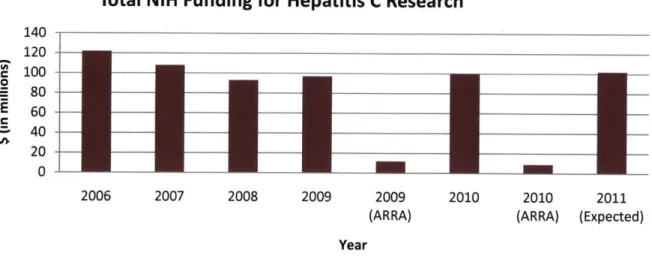
Research for hepatitis C, for both drugs and diagnostic tools, is heavily funded through the NIH (National Institutes of Health) and NCI (National Cancer Institute). The NCI is a division of the NIH. The NIH allotted $100 million for hepatitis C research in 2010. There was also additional funding from the ARRA (American Recovery and Reinvestment Act) in 2009 and 2010.
Funding from the ARRA is about 10% of the total funding for hepatitis C research, but does not extend into 2011. The funding per year is shown below in Figure 2.
Total NIH Funding for Hepatitis C Research
140 120 e 100 0 80 -E 60 -40 -20 0 2006 2007 2008 2009 2009 2010 2010 2011
(ARRA) (ARRA) (Expected) Year
Figure 2 shows the funding for hepatitis C research from 2006 to present and the projected funding for 2011.
The Unmet Need
Proper treatment of hepatitis C (HCV) requires close monitoring of the amount of the virus present in a patient's blood, clinically referred to as the viral load. Currently, to quantify the viral load, a blood sample is taken and the number of viral particles is quantified, usually using PCR (Polymerase Chain Reaction) or TMA (Transcription Mediated Amplification) technology.
The number of viral particles is usually presented as a concentration. The concentration is determined based on the number of viral particles present and the volume of blood sampled. Initial quantification of the patient's viral load can be conducted using commercially available tests because the patient typically has a sufficiently high viral load, one well within the
detectable range of the most commonly used tests. These tests have a quantification range of 30 viral particles to 250,000,000 viral particles per mL.' There are also more sensitive tests
available (as low as 5 viral particles per mL), but they have a small range of quantification and they are only useful when a physician knows that the patient has a low viral load.
Many patients will reach a point where current methods, even the most sensitive tests, are insufficient to determine the viral load. Clinically, this point is referred to as having an
undetectable level of the virus. Physicians have no way of determining if a negative test result is because the patient is cured or because the patient has a level of the virus not detectable with current methods. The latter of those two options is termed a false negative. Unless physicians are able to determine the true point of eradication, and are able to know that a negative result is in fact a true negative, they are only able to treat patients based on clinically established
standards. If they can determine the true point of eradication, physicians could tailor treatments based on each patient's specific response. Continuing treatment "blindly," beyond the point at which the patient achieves a negative test result, subjects many patients to unnecessary
treatment. Physicians have a strong desire to customize treatments and shorten them where possible because of the high cost of treatment, the significant side-effects associated with the treatment and the fact that some patients achieve this undetectable level only 4 weeks, or earlier, into a 48 week treatment.
The Solution
The Cima Lab is developing an in-vivo device to sample large volumes of blood and trap the HCV particles present. This approach overcomes the sampling limitations of current tests that work based on drawing a fixed amount of blood from a patient's arm. The proposed device will be placed in a vein and as viral particles come in contact with it, they will become trapped by antibodies lining the surface of the device. The device will then be removed from the body and 'Concentrations can also be listed in IU/mL. The conversion used for the tests discussed is 2.5 viral particles per IU.
the viral particles will be quantified using one of the commercially available tests. Based on the volume of blood sampled while the device is in the body, the number of viral particles will be converted to a concentration. Physicians can then determine the best course of treatment with this much more sensitive measurement. This in-vivo blood sampling technique allows roughly 40,000 times more blood to be sampled in a 30 minute period than with a traditional blood draw (2 mL).
Antibody/Antigen Interactions
A key component of this device is the interaction between antibodies and viral particles. An antibody is a protein that is naturally created by the body's immune system when it detects a foreign body (i.e. hepatitis viral particles). The HCV viral particles, also referred to as antigens, will bind to the antibodies when they come in contact with each other. While antibodies are produced by the body naturally, they can also be created in a lab. These manufactured antibodies can be attached to the surface of a device and used to trap viral particles. The hepatitis C
antibodies used in this application are specific to the proteins (El and E2) expressed on the surface of the HCV viral particle. These surface proteins are referred to as surface antigens. A schematic of the HCV viral particle is shown in Figure 3.20
HCV Viral Components
Envelope
(E 1 and E2) protein complex
Figure 3 shows a schematic of the hepatitis C viral particle and its surface antigens.
Initial Design Concept
Initially, the focus of the device was on developing a system that could both sample large volumes of blood and quantify viral load
independent of any additional testing M Vtral Partice
equipment. The first concept was to use ,D
magnetic nanoparticles, coated with
*
1antibodies, to capture viral particles in / d S
-the body. The antibody-coated Antad Coatd
Magntic Nanoperticle
nanoparticles would aggregate in the Figure 4 shows the initial design that was considered during presnceof vralpartcle andcaue a this project. Note: image enlarged to show nanoparticles and presence of viral particles and cause a
pore size. change in the magnetic properties of the
surrounding media. This change would then be detectable using either MRI (Magnetic Resonance Imaging) or NMR (Nuclear Magnetic Resonance) technology. The initial concept was that a group of the antibody functionalized nanoparticles would be placed in a polymer membrane (shown in Figure 4). This device would then be placed in a vein, and viral particles would diffuse into the device and cause an aggregation of the nanoparticles. It was hypothesized that this would allow for the sampling of a much larger volume of blood than is currently
possible with a traditional blood draw. The technology, to date, has only been tested in benchtop
in-vitro settings. It had been proven that the nanoparticle technology was capable of trapping
viral particles,2 1 but there were complications associated with implementing the technology in an in-vivo setting. The primary problem was that the group of nanoparticles would need to be
encased in a polymer membrane to keep them from moving throughout the body. This created a large dichotomy between the number of viral particles that came in contact with the surface of the device and the number of viral particles that diffused through the membrane and came in contact with the nanoparticles.
Based on diffusion calculations of the viral particles through the membrane, it was determined that the device would need to be placed in the vessel for a period of up to five hours to match the sampling capabilities of a current blood draw. The device would need to remain in the body for multiple days to have any competitive advantage from a sampling perspective. If the time the device is in the body is longer than one hour, it would require two procedures, one to place the
device and one to remove it. The implications of the cost associated with placement are discussed later, but avoiding a second procedure to remove the device would be advantageous. Also, the longer the device is left in the body, the more important issues such as biocompatibility and biostability become. Increasing pore size to increase the diffusion of viral particles into the device was considered, but determined to be an unsuitable solution. If the pores were made sufficiently large, the nanoparticles would escape from the device.
Another reason for the change in approach was the sensitivity of quantification using MRI or NMR technology. The lowest concentration of viral particles detected with this technique is 500 viral particles/mL. This is roughly 100 times less sensitive that the most sensitive test available on the market today and 17 times less sensitive than the most commonly used test. The device would need to be left in the body for an unsuitable length of time to overcome both the low diffusion rate through the membrane and the lower sensitivity of the quantification associated with this technique.
The approach has shifted from a standalone diagnostic device to a viral particle trapping device that is used in conjunction with currently available diagnostic tests. This new approach
overcomes the problems of the
initial design. The new > cm<_
approach will trap viral particles
Figure 5 shows the second design that was considered for the HCV with antibodies directly on the trapping device.
surface of the device. Exposing
the antibodies directly to viral particles (without a membrane in between) will increase the trapping capabilities of the device. Once the viral particles have been trapped, the device will be removed from the body and a more traditional technique for quantifying viral particles will be employed. This takes advantage of the most sensitive test available and the high sampling rate of an in-vivo device. Additionally, this gives the physician the flexibility to choose which quantification test he/she desires.
Initially, a straight rod design was considered. This straight rod (Figure 5) would be coated with antibodies and passed through a catheter to either the superior vena cava (SVC) or hepatic veins (implantation location will be discussed in detail in a later section). Again the goal was to keep the device in the body for no longer than 30 minutes. It was determined that four rods (3 cm in
length) would be needed to match the blood volume sampling capabilities of a 3 mL blood draw. The low sampling capabilities of this approach were attributed to the strong reliance on having particles diffuse to the surface of the device before binding could occur. It was determined that to justify the cost of placement of the device and the necessary procedure, the sampling volume would need to be higher.
The next design evaluated was the "twisted" coil. This device (a model representing the shape is shown in Figures 6a and 6b) crosses back and forth across the blood vessel, perpendicular to the direction of the blood flow. This allows the device to take advantage of the moving blood to carry the viral particles to the surface of the device. This approach does not rely solely on diffusion as in the straight rod design option, and thus samples far greater volumes of blood. Looking down the vein, one sees the device as represented in Figure 6a. It appears as though a large amount of the vein is blocked. This is not the case as can be seen in Figure 6b. Looking at the device from the side, each time the device crosses the vein it is approximately 0.5 cm from the last place the device crossed the vein. The device does not block more than 4% of the vein at
any one time. This method samples over 85,000 mL of blood in a 30 minute period; this is more than 40,000 times greater than the volume sampled in a current blood draw (2 mL) when placed in the superior vena cava (SVC). When placed in the hepatic veins, the device samples 10,000 times more blood than a traditional blood draw.
Figures 6a and 6b. Figure 6a shows how the device would look if looking down the vein in the direction of flow. Figure 6b shows how the device looks from the side and its spacing. Note: Copper was used for these mockups, but a different material will be used for the actual device (discussed later).
Trapping Capabilities of "Twisted Coil" Design
While the volume of blood sampled is a critical factor when deciding on a design and
implantation location, it is not sufficient to determine if a design is viable based on this criteria. The ability of the viral particles to be captured by the antibodies is equally as important.
Previous research has not explored the binding of moving viral particles and stationary antibodies in blood flowing at the velocities observed in the SVC or hepatic veins. There are, however, examples from microfluidics that examine the binding efficiency of viral particles to antibodies in flowing blood. Researchers explored the ability to capture HPV (Human Papaloma Virus) viral particles with stationary antibodies while exposed to blood containing viral particles. This study explored blood flowing at 0.13 cm/s.2 2 Researchers found that the binding efficiency
at this velocity was determined to be in excess of 30%. This is significantly slower than the rate of blood flow in the SVC (average 22.5 cm/s), but researchers also found that the trapping efficiency plateaued at 30% as blood velocity increased.
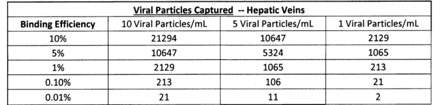
Tables 2 and 3 below show the number of captured viral particles for multiple binding efficiencies and viral particle concentrations during a 30 minute period. The number of viral particles captured must be greater than 30 (the minimum quantifiable about with the Abbott test), to quantify the viral load using the trapping device. This is the ideal situation because the same test can be used at high viral loads without the trapping device and at low viral loads with the trapping device. If the number of particles captured is less that 30 but greater than 5, it can still be quantified, but the LabCorp test would need to be used. This still provides some useful
information, but because it is a different test than the one used at the beginning of treatment there may be some inherent variations. The tables below show the great deal of variation possible in binding efficiency while still having a sufficiently sensitive device for both the SVC and the hepatic veins.
Viral Particles Captured - SVC
Binding Efficiency 10 Viral Particles/mL 5 Viral Particles/mL 1 Viral Particles/mL
10% 85050 42525 8505
5% 42525 21263 4253
1% 8505 4253 851
0.10% 851 425 85
0.01% 85 43 9
Table 2 shows the number of viral particles that could be trapped by the device for three different viral particle concentrations and five different binding efficiencies if the device was placed in the SVC.
Viral Particles Captured -- Hepatic Veins
Binding Efficiency 10 Viral Particles/mL 5 Viral Particles/mL 1 Viral Particles/mL
10% 21294 10647 2129
5% 10647 5324 1065
1% 2129 1065 213
0.10% 213 106 21
0.01% 21 11 2
Table 3 shows the number of viral particles that could be trapped by the device for three different viral particle concentrations and five different binding efficiencies if the device was placed in the hepatic veins.
The last column in both tables shows the number of captured particles if the concentration is 5 times less than what can currently be detected and 30 times less than what can be detected with one of the most commonly used tests. The requirement of capturing 30 viral particles can be achieved at a binding efficiency as low as 0.1% in the SVC. If the device was placed in the hepatic veins, it would still be effective at a binding efficiency of 1% if the concentration in the blood was 1 viral particle per mL. A binding efficiency of 0.1% would be sufficient if the concentration in the blood was 5 viral particles per mL. It is important to remember that the device will only be placed in the hepatic veins if there is a far greater viral concentration than present in the SVC. The number of viral particles captured, as shown in Table 3, does not assume any increased concentration. If the device was placed in the hepatic veins, the number of viral particles captured would actually be much greater than what is shown in Table 3. These calculations assume that the part of the device that crosses the vein is the only part able to trap viral particles.
Based solely on a literature review, an estimation of binding efficiency is difficult. The wide range of possible binding efficiencies makes it risky to predict the sampling capabilities of the device without conducting some in-vitro studies. It is clear from the above tables that success is likely because the device can still be effective at such low binding efficiencies. A technique to conduct these in-vitro tests, as well as in-vivo animal studies, is outlined later.
Implantation Location and Procedure
Implantation Location
Three locations were considered based on the following set of criteria: ease of implantation, size of the vessel and proximity to the liver/viral load. The ease of implantation refers to how easy it is to place and remove the device. The size of the vessel is important because the amount of blood that can be sampled is related to the size of the vessel. The proximity to the liver is relevant because the liver is the site of replication for the hepatitis C virus.
The initial site considered was the hepatic veins. The hepatic veins carry blood from the liver to the inferior vena cava. It has been shown that the liver tissue has a 40 times higher
concentration2 3 of viral RNA than peripheral blood. The comparison of blood close to the liver
to that of peripheral vessels is currently being investigated and the results of this test will dictate the importance of placing the device close to the liver. While this location has the potential for a higher viral load, the implantation procedure is more costly, time consuming and taxing for the patient. There are also space constraints associated with the location. The diameter of the hepatic vein is 1.5 cm. The focus of placement location shifted to the inferior vena cava (IVC) because of these limitations in the hepatic veins.
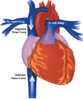
The IVC (shown in Figure 7)24 carries
de-oxygenated blood from the lower part of the body
back to the right atrium. The hepatic veins drain Spi
from the liver and join the IVC. It was
hypothesized that being relatively close to the liver would still provide access to the higher viral load if it existed and, at the same time, avoid the space
constraints associated with the hepatic veins. It was %om determined that this location had a suitable
diameter (2 cm - 3 cm)25 compared to a diameter of
1.5 cm in the hepatic veins. It was also determined
Figure 7 shows the location of the inferior that the blood velocity is 13 cm/s (mean velocity), vena cava and superior vena cava. allowing for sufficient blood sampling in a 30
minute period. The primary problem with placing the device in the IVC is where the hepatic veins meet the IVC. There is not sufficient room in the IVC after the hepatic veins join the IVC before the IVC meets the heart. This limitation made the IVC an unsuitable placement site. The third and final site considered was the SVC (also shown in Figure 7). The superior vena cava carries de-oxygenated blood from the upper half of the body to the right atrium. The superior vena cava has a sufficiently large diameter (3-4 cm) to place the device and a blood velocity (avg. blood velocity = 22.5 cm/s) that will allow for sufficient blood sampling capabilities. The primary drawback to this location is that the device would now be placed further from the liver. As mentioned earlier, the importance of placing the device close to the liver will be determined once clinical measurements of viral load have been taken in blood from the SVC and blood from the hepatic veins.
Implantation Procedure
The procedures to implant the device in the two locations still under consideration are different. It is more complicated to implant the device in the hepatic veins than to place the device in the SVC. The placement of the HCV trapping device in the hepatic veins will be done by an
interventional radiologist. The patient's neck or leg, depending on whether the coronary vein or femoral vein is used, is cleaned and sterilized to prevent infection. The patient lies down during the procedure and a numbing agent is administered at the site of entry into the body. This will prevent the patient from feeling pain during the procedure. A plastic catheter is passed into either the leg or neck, depending on implantation location. An x-ray dye is injected into the catheter and images of the vein are taken. The radiologist then passes the HCV trapping device through the catheter to the deployment site. The catheter will be left in place during sampling (30 minutes). The device will be pulled back through the catheter after the sampling has ended, and the catheter will then be removed from the body.26
If the device is placed in the SVC, the
procedure is much simpler. Placement in the SVC would not require an interventional radiologist. Instead, a peripherally inserted central catheter (PICC) line (shown in Figure
8)27 would be placed by an IV therapist or certified registered nurse. The first method to insert a PICC line is to insert the catheter by feeling the vein in the arm and then confirm the location of the catheter with a chest x-ray. Alternatively, the PICC line could be placed using ultrasound equipment, followed by a chest x-ray to confirm placement. The third option is to use a combination of ultrasound and fluoroscopy. This allows for real-time imaging while placing the PICC line.2 8
PICC
Figure 8 shows the location of the PICC line.
The procedure associated with placing the device in the SVC is significantly easier than the procedure needed to place the device in the hepatic veins. Independent of the viral load present in the two locations, the device would be placed in the SVC. Unless viral load measurements from the hepatic veins are significantly greater, there would be no strategic advantage to placing the device in the hepatic veins. Table 4 outlines the difference between the two locations.
Superior Vena Cava Hepatic Veins
Ease of Placement Easier Harder
Cost of Placement Procedure =$600 -$1400 =$1,700
Personnel Required IV Therapist Interventional Radiologist Proximity to Liver Near Heart Connected to Liver
Diameter of Vessel 3 -4 cm 1.5 cm
Blood Velocity 10 - 35 cm/s 13 cm/s
Comparative Viral Load Roughly Equal to Blood Draw Unknown
Table 4 shows a summary of the benefits and drawbacks of both implantation locations being considered.
Blood Sample Preparation
Current Sample Preparation
The most common types of quantitative HCV tests are Real Time-Polymerase Chain Reaction (RT-PCR) based tests. One of the most popular tests is the COBAS@ AmpliPrep/COBAS@ TaqMan@ HCV test manufactured by Roche. The specific blood specimen preparation technique outlined in this section is for the Roche test. This test measures viral load by quantifying the amount of RNA present in a fixed sample of blood. This system is able to process a maximum of 850 pl of serum or plasma. The HCV viral particles are lysed through
incubation at high temperatures. The incubation occurs with a protease that promotes the lyses of the viral particles to release the RNA. The incubation also occurs in a buffer solution that releases nucleic acids and protects the RNA from RNAases. RNAases is the process in which
the RNA from the viral particles is broken into its smaller component parts. If RNAases occurs, it would distort the true viral load.
Sample Preparation When Using Trapping Device
The preparation of samples collected using the trapping device will not be substantively different than the samples collected using a traditional blood draw. The HCV trapping device would be
removed from the body and placed in blood collected from the patient (2 mL). The blood and trapping device, with bound viral particles, would then be incubated. Incubation would release the RNA contained in the viral particles. Incubation would also occur in a buffer solution to protect the RNA from RNAases as with current sample preparation. The backbone of the device
will be removed from the blood sample and the sample will be processed using the same steps that are used to quantify a viral load in traditional blood draw.
Manufacturing
Materials Selection
Nitinol (NiTi) was chosen as the material for this device because it is highly elastic. The device will be shaped as a "twisted" coil, shown in Figure 6. Given the limited space provided by a catheter (2 mm diameter), the nitinol coil will not be able to reach the site of implantation without being deformed. This necessitates that the material regains its shape once it reaches the deployment site. While biocompatibility is not as important a concern (because the device is
only in the body for 30 minutes), the fact that nitinol is currently used in blood contact applications (stents and blood clot catching devices) reinforces its use in this application.
A polymer coating is applied to the backbone to promote the attachment of the antibodies to the device. The polymer chosen is PDMS (polydimethylsiloxane). PDMS is a highly biocompatible polymer; it is used in contact lenses and in a wide range of other medical applications. Another
reason that PDMS was selected is because of its use in applications involving antibodies. PDMS has been used in microfluidic applications in which antibodies are bound to a PDMS surface which is then in contact with blood.
There are two forms of antibody that could be used to the trap viral particles, humanized and non-humanized antibodies. A humanized antibody is an antibody derived from another species (e.g. primate) and then modified to better match the human version of the antibody. A non-humanized antibody is derived in another species and used without altering it to match the human version. Regardless of which choice is made, the antibody will be specific to the El or E2 antigens present on the envelope of the HCV viral particle. Using a humanized antibody decreases immune response if the antibodies become dislodged from the device while it is in the body. It is, however, possible that a non-humanized version would work because the device is only in the body for 30 minutes.
Manufacturing of Nitinol Backbone
The manufacturing of the nitinol for this application will be a multistep process. Nitinol wire must be made into the desired shape for this device. The process of shaping nitinol is termed shape setting. The wire is set in a fixture or on a mandrel that is in the shape of the desired device, and then a heat treatment process is performed. The heat treatment process can be achieved using an air or vacuum furnace, salt bath, sand bath, heated die or other heating
method. The temperature is raised to a range of 500'C to 550*C. The treatment duration must be long enough for the entire material to reach the desired temperature. The heating of smaller objects, such as the wires used in this device, typically takes less than a minute to reach the desired temperature. The nitinol is then quenched in a water bath. The specific temperature and exact heating time are determined through experimentation prior to manufacturing. The final step in the nitinol manufacturing process is electropolishing. The nitinol, on a microscopic level,
can have crevices on grain boundaries and at the edges. Electropolishing removes the crevices present after machining.2 9
Polymer Coating
The polymer coating process can be conducted by the same company that handles the shape setting of the nitinol. This is one method for coating the device with PDMS. The PDMS is mixed with a curing agent (e.g. Sylgard 184, Dow Coming) and then degassed.3 0 The nitinol wire is dipped in this mixture and then removed and allowed to cure. PDMS has been shown to bind to Ti alloys through a Si - 0 -Ti bond.3' It is placed in a plasma cleaner (e.g. PDC-32F,
Harrick Scientific) after the curing process is completed. The plasma cleaning process serves two purposes, surface sterilization and surface preparation for bonding.32 The preparation of the
PDMS helps to promote binding of the antibodies to the nitinol backbone. The exact process may need to be adjusted based on the final design of the device.
Application ofAntibodies
The application of HCV antibodies to a polymer surface has not yet been explored in a research or commercial application. There are, however, examples of other types of antibodies (cervical cancer) being bound to a polymer surface, specifically PDMS. The surface of the PDMS is treated with a 2% solution of 3-mercaptoproyltrimethosilane in toluene for a one hour period to promote the binding of the antibodies to the polymer. The surface is dried and treated with 2mM GBMS (N-y-maleimidobutyry loxy succinimide ester) for one hour and then rinsed with PBS (phosphate buffered saline). A solution of antibodies is then introduced to the chemically treated PDMS for 30 minutes at room temperature to react with the GBMS. It is important that the application of the antibodies be conducted in a sterile environment because after they bind to the PDMS, the device can no longer be sterilized without damaging the function of the antibodies. The cleanliness of the lab must meet GMP (good manufacturing practice) standards. This includes sterile conditions and regularly maintained equipment to ensure consistent and uniform production. It is expected that the process for the attachment of HCV antibodies will be similar to that of the cervical cancer antibodies.3 3
Packaging and Storage
The specifics of the packaging and storage will be dictated by the antibody choice. The nitinol and the PDMS are able to be stored at room temperature without altering their functions.
However, in general, antibodies must be kept at -200C to avoid damage. The antibodies must also not be thawed and refrozen; this can damage their function.34 Once the device has been
manufactured, it must remain sterilized until it enters the patient's body. It could be preloaded into a PICC line to avoid the physician having to handle the device. This would ensure that the device remains clean and makes the procedure easier for the physician.
Manufacturing Cost
Due to the nature of these components, there are advantages to producing at larger volumes. The production of the nitinol components requires an investment in the mandrel used to shape the nitinol wire into the component. The mandrel is a fixed cost, an investment that must be made regardless of the number of devices produced. The investment in the mandrel will be spread over a larger number of units as the volume produced increases. The cost per device of the polymer coating also decreases as production volume increases. The polymer coating process is conducted in batches of roughly 50 pieces at one time. Additionally, if multiple batches are run consecutively, the changing of tooling is minimized; this is yet another reason for decreased unit costs with increased volume. When producing at the prototype level (less than 10 units), the cost of the nitinol backbone and polymer coating is just under $57 per unit. When producing at production level (greater than 1000 units), the cost per piece decreases to just over $40 per unit. This is a 30% drop in cost per unit for the shaping of the nitinol and polymer coating.
Unit Cost/Device (Quantity Produced)
Manufacturing Step < 10 10-1,000 > 1,000
Nitinol (Material, Finishing and Shape Setting) $47 $37 $31
Polymer Coating (Materials and Process) $10 $10 $9
Antibodies -- Humanized (Materials and $250-$400 $250-$400 $250-$400
Application Process)
Total $307 - $457 $297 -$447 $290-$440
Table 5 shows the unit cost for production of the device at different quantities when using humanized antibodies. Quantities over 1,000 are considered production volume.
Unit Cost/Device (Quantity Produced)
Manufacturing Step < 10 10-1,000 > 1,000
Nitinol (Material, Finishing and Shape Setting) $47 $37 $31
Polymer Coating (Materials and Process) $10 $10 $9
Antibodies -- Non-Humanized (Materials and $50 $50 $50
Application Process)
Total $107 $97 $90
Table 6 shows the unit cost for production of the device at different quantities when using non-humanized antibodies. Quantities over 1,000 are considered production volume.
These costs, shown in Tables 5 and 6, include all equipment, tooling, technicians and associated costs. All prices shown are based on estimates from manufacturers and may change depending
on the final design of the device. The cost for antibodies is the greatest current unknown. It is estimated to be between $50 and $400. This price includes the antibodies and the associated
application process. This cost depends on whether humanized or non-humanized antibodies are used. The cost of antibodies can vary greatly depending on specific factors that will be
determined during the development process. The most suitable and appropriate type of antibody will be determined through future testing. The total cost to manufacture the device at the
prototype level is between $307 and $457 when using humanized antibodies and $107 when using non-humanized antibodies. Once at production volumes, the cost drops to between $290 and $440 when using humanized antibodies and $90 when using non-humanized antibodies. The specific packaging and storage required will be based primarily on the antibody chosen; this cost will be added once the antibody selection process has been completed. There are many
companies that are capable of handling the nitinol shaping and polymer coating portions of the device. Contracting the production of the nitinol shaping and polymer coating processes to a third party avoids the need to make large capital investments (e.g. equipment and facilities). Additionally, the industry is well-established and we would be able to take advantage of the experience and expertise of these existing companies.
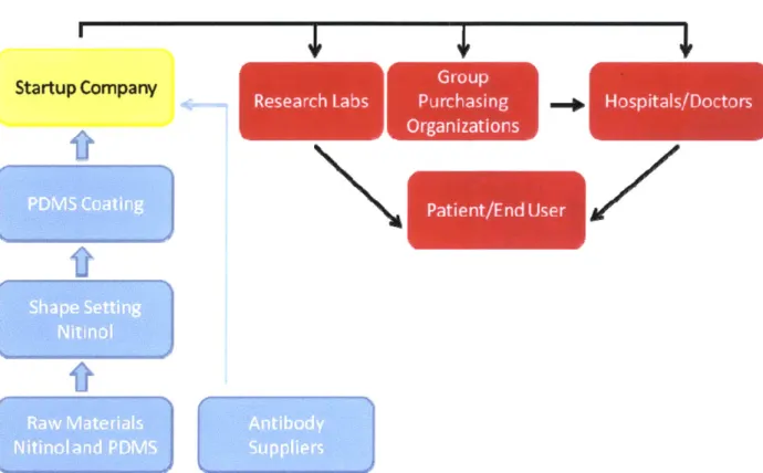
Supply Chain
I
4
4
~1
Startup Company
fr.
mGroup
Research Labs Purchasing
L _AL_ OrganizationsA
Figure 9 shows the existing supply chain , the existing distribution chain and where a startup company would operate.
This device would not be made by the same supply chain as current HCV diagnostic devices. Figure 9 shows how the supply and distribution chains will operate for the manufacturing of this device. Current HCV diagnostic tests are typically PCR or TMA based technologies and rely on chemical reagents. This technology is an antibody-based device. While antibodies are used for detection purposes in microfluidic applications, they are not used for in-vivo applications. The other components, the nitinol backbone and the polymer coating, are also not used for HCV diagnostic devices. This provides a key strategic advantage because the companies that will provide the antibodies, nitinol backbone and polymer are not currently involved in the HCV diagnostics or HCV drug markets. A potential startup company would purchase the nitinol components already shaped and coated and focus on the process of applying the antibodies to the device.
There are two key complementary devices that will need to be employed when using the HCV trapping device, and it is important to understand where they could be purchased. A placement catheter and a vascular access kit are both required. These two devices can be made
... ... ... .. ... ... .. ... ...
" I .... "I'l""I'll""I'll'll""I'llI ... .. "I "I',,',',,, ..... ... ... .. ... ... ... .. .. .... ...
Hospitals/Doctors L Z F_ -- I Patient/Fnd User IL A
independently of the HCV trapping device and do not require a co-manufacturing process. The catheter will be used to place and remove the device, and the vascular access kit will be used to access the vein that the catheter will be placed in. The similarity between the implantation for this device and the placement of an IVC filter allows the same equipment to be used in both procedures. Thus, these two complementary devices can be purchased from manufacturers of
IVC filters. Commercially available catheters for device placement in the IVC are $160 (Vista BRITE TIP@ Guiding Catheter - Cordis) and vascular access kits are $45 (Vascular Access Kit
- Cordis)."
Distribution Chain
Once the device is manufactured, it would be sold into the already existing distribution chain for medical devices. The medical device industry relies heavily on group purchasing organizations (GPOs). These groups pool the purchasing power of hospitals and smaller medical centers to obtain volume discounts from device manufacturers. Today, hospitals purchase about 70% of their equipment through GPOs and 96% to 98% of hospitals use GPOs as part of their purchasing structures. Hospitals are frequently members of more than one GPO; on average, a hospital is a member of 4.2 GPOs. Manufacturers present the technical capabilities and price of the device to the assessment team at each GPO as part of the process to have their device distributed. Terms regarding the agreement between the GPO and a medical device manufacturer are negotiable. Typically, selling to one GPO does not preclude one from selling to another GPO or directly to a hospital.36 The last step of the distribution chain is for the hospitals and researchers to use the
device on a patient or research subject respectively.
Insurance Companies, Medicare and Medicaid
Regardless of whether hospitals and physicians are willing to adopt and use the technology, someone must pay for it. There are three ways that medical expenses are paid for in the United States -- the patient pays out-of-pocket for the treatment or procedure, an insurance company pays for the service or the government pays for it (in the cases of Medicare and Medicaid). The goal of the entity paying for the treatment or procedure is to choose the most cost effective and efficacious treatments available. They will look at both the financial cost of the procedure and device and the benefits of conducting the test or procedure.
A device or procedure must have an associated CPT (Current Procedural Terminology) code, issued by the AMA (American Medical Association), to be covered by an insurance company. When a new medical device is introduced to the market, it must be determined whether the new device or procedure will fall under a current CPT code or if a new one will need to be created. The advantage of using a current CPT code is that there is no new CPT code application that needs to be submitted to the editorial panel at the AMA. The main drawback of using an existing CPT code is that the level of reimbursement is fixed. A new code will be necessary for this device. The procedure required would be above and beyond what is currently covered by CPT codes related to testing for HCV. These CPT codes would not provide a sufficient level of reimbursement for the placement and removal of the device and the cost of the device itself.3 7
It is also important to recognize that recently Medicare and Medicaid have frozen the amount they are willing to reimburse for clinical laboratory testing.3 8 This is particularly relevant
because in the next 10 years physicians are expecting a new wave of HCV infected people over age 60 . People in the United States enter the Medicare program at age 65. A new CPT code would keep the device from being subject to the reimbursement freeze associated with molecular diagnostics.
Potential Customers and Incentives for Adoption
The main incentive to insurance companies is the savings they would experience due to a shorter treatment length. The amount saved through use of the proposed device is discussed in a later section. The primary target customers for this type of device are the physicians. While in some instances patients may request a particular treatment, the majority of times it is the physicians who will determine which tests are most appropriate. There are, however, a growing number of online self-help forums for patients with hepatitis C. Patients discuss matters including which diagnostic tests their physicians have administered and how valuable the test results were for dictating future treatment. These forums have helped to spread the word about new diagnostic tools in the past. Regardless, the decision regarding which tests to administer still falls primarily on the physician, even with these new online forums. The physicians are primarily concerned with effectively treating their patients and receiving reimbursement for their work. Clinical trials to quantify the capabilities of the technology will be required to convince physicians of the effectiveness of this device. The procedural cost associated with this device is $600 -$1,500
(the basis for this number is discussed later). This price is the amount that the physicians would charge the patient, and already includes the physicians' profits. No additional financial incentive would be required. Patients would choose to undergo the procedure because this device could shorten their treatment, saving them the cost of drugs, physician visits, and testing. Additionally, they would avoid the hassle of self-administering medication and the problems associated with the side-effects of treatment.
Market for the HCV Trapping Device
Primary Markets
The primary use of this device is focused on genotype 1 patients who exhibit RVR. The reason for the focus on this segment of the population is the prevalence of genotype 1 (75% US and 60% worldwide) and the lengthy time these patients spend being treated after they achieve RVR. They achieve RVR at week 4, but continue with treatment for another 44 weeks. It has been shown that as many as 78%40 of these patients can achieve SVR (considered cured for HCV) with a shortened treatment. The average savings per genotype 1 patient reaching RVR is
$13,200. It is important to look at the savings per RVR patient rather than per patient of the population who is cured (the 78%) because this test would be conducted indiscriminately on all people who achieve RVR. The savings are associated with the average cost of the drug
($11,500) and the average cost of physicians' visits and tests during the second six months of treatment ($2,100). When setting the price, it is important to include the costs of all associated
equipment, procedures and personnel.
Secondary Markets
The secondary markets for this device will be heavily dictated by the capabilities of the device as determined by the clinical and preclinical trials. There are a number of additional segments of the HCV infected population that could benefit from the device. The first market to focus on is the remainder of the RVR patients who are already having their viral load quantified using the trapping device. These patients, in theory, would have a detectable level of the virus using the
trapping device, but not with traditional blood draw methods. Even though these patients would not be able to stop treatment at 24 weeks, they may be able to end earlier than 48 weeks