Biochemical characterization of zinc-dependent interactions
between p561ck and CD4, and DTT-sensitive interactions
between TGF-beta receptor subunits
byRalph Shih-Ying Lin B.S. Honors
California Institute of Technology (1991)
Submitted in partial fulfillment of the
requirements for the degree of Doctor of Philosophy
at the
Massachusetts Institute of Technology January 1999
0 Ralph Shih-Ying Lin, 1999 All rights reserved
The author hereby grants to MIT permission to reproduce and to distribute copies of this thesis document in whole or in part.
Signature of Author Department of Biology January 1999 Certified by Harvey F. Lodish Thesis Supervisor Accepted by -Terry Orr-Weaver Chairperson, Departmental Committee on Graduate Studies
Biochemical characterization of zinc-dependent interactions between p561ck and CD4, and DTT-sensitive interactions between TGF-beta receptor
subunits by
Ralph Shih-Ying Lin
Submitted to the Department of Biology in January 1999 in partial fulfillment of requirements for the
degree of Doctor of Philosophy Abstract
Binding of the protein tyrosine kinase p5 61ck to T-cell co-receptors CD4 and CD8X is necessary for T-lymphocyte development and activation. Here we demonstrate that Zn2+ is essential for complex formation. In an in vitro binding reaction, Zn2+ mediates p56lck association with a GST fusion protein containing the cytosolic domains of CD4 or CD8U; no other metals tested support binding. Treatment of preformed GST-CD4/p56lck dimers with Zn2+ chelators results in dissociation of GST-CD4 from p561ck suggesting that Zn2+ is contained within these complexes. Furthermore, we show that within live cells,
CD4/p561ck and CD8W/p561ck interactions occur in a zinc dependent fashion. Specifically,
pretreatment of the human T-cell Jurkat cell line with membrane-permeable zinc chelators disrupts CD4/p561ck complexes, and treatment of COS cells co-expressing CD8c and p561ck with such chelators likewise leads to dissociation of CD8o/p561ck complexes. CD4/p561ck and CD8/p561ck represent the first examples of intracellular proteins that require zinc as a bridge for heterodimerization.
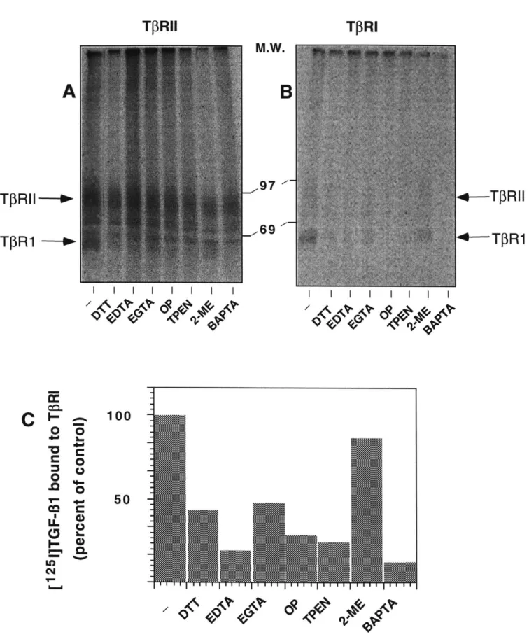
The TGF-s mediate a diversity of biological responses through the binding of a types I (TPRl) and II (TfRII) receptor. Specifically, these cytokines bring about the formation of a signalling complex consisting of a TfRI and TIRII heterocomplex -most likely a heterotetramer. (Gilboa et al., 1998; Henis et al., 1994; Moustakas et al., 1993; Yamashita et al., 1994) However, it has been shown that pre-treatment of cells with DTT abrogates functional recruitment of TPRI to TGF-3 1-bound TfRII rendering the resulting complex unable to signal. (Cheifetz et al., 1990; Wells et al., in press) We further report that similar pre-treatment blocks binding of both TPRI and TfRII to TGF-$2, a ligand distinct from TGF- 1 in that it requires co-expression of both receptors to bind. To identify the properties of DTT required to elicit this phenomenon, we screened a number of chemically related reducing agents for a similar inhibitory effect upon complex formation. Here, we find that 2,3 DMP (2,3 dimercaptopropanol) likewise blocks functional interactions between TPRI and TPRII. Both it and DTT are di-thiol based compounds that
are membrane-permeable. Other thiol-containing compounds lacking either one of these properties exert no effect on complex formation. In addition, DTT does not affect TPRI homodimerization, an upstream event required for TPRIeTPRII hetero-oligomerization. Hence, this would suggest that mechanistically, DTT and similarly, 2,3 DMP, act upon intracellular factor(s) and through chemical properties related to the presence of di-thiols, inhibit TPRIeTPRII association. The cytoplasmic domain of TPRI plays a critical role in these interactions as we find that a cytoplasmically truncated T3RI fails to associate with ligand-bound TPRII. In contrast, the TPRII cytoplasmic domain is dispensable for complex formation, nor is it required to manifest the DTT effect. Therefore, we propose that DTT directly abrogates functional TIRI-TPRII interactions by acting upon structural elements lying within TfRI's intracellular domain that are necessary for it to be recruited by TPRII.
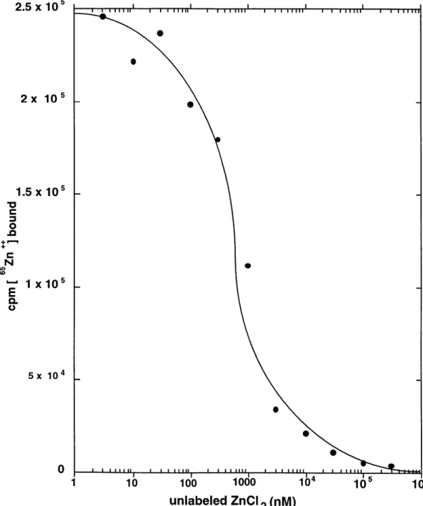
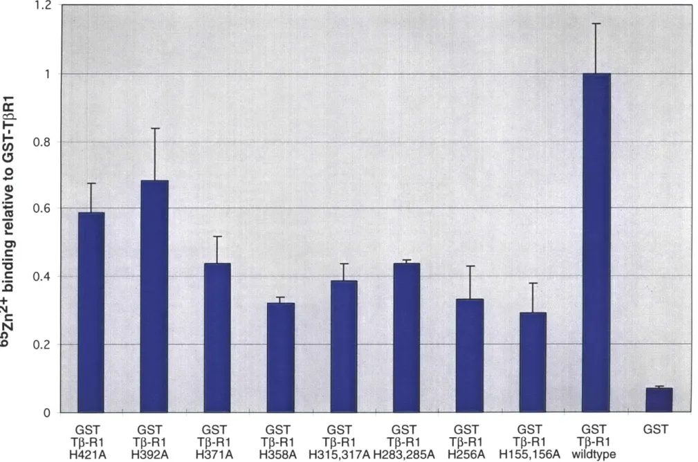
It is thought that DTT, by reducing critical disulfide bonds in the extracellular domain of TfR1, alters the receptor's conformation ultimately preventing TGF-P binding.(Vivien and Wrana, 1995) Other membrane permeable dithiols exerted the same effect. Because in contrast to monothiols, dithiols are metal chelators, we thought it possible that chelation rather than disulfide bond reduction explained their findings. Hence, our model predicts that Zn2+ is essential for the cytoplasmic domain of the type I receptor to assume a particular conformation necessary for interactions with the II receptor. We test this model by examining a number of predictions it makes regarding the structural nature of the type I receptor. First, it predicts that the type I receptor cytosolic domain should bind Zn2+ and its removal by pre-treatment with membrane permeable Zn2+ chelators should block formation of a TGF- 1/types I and II receptor complex. Furthermore, mutations resulting in the loss of zinc binding by the type I receptor should cause loss of recruitment to the ligand bound type II receptor complex and therefore render it unable to signal. Our experimental data however are only consistent with a subset of these predictions. Therefore, our conclusions ultimately do not support an essential role for a divalent metal, namely zinc, in formation of the TGF-P receptor complex.
Acknowledgements
First and foremost, I thank my family for their unconditional support and long-distance encouragement. Without them, this thesis would not have been possible.
I thank Professor Harvey Lodish for very patiently supervising my thesis for the past N years, where N has been arbitrarily large. I have greatly benefited from his unique blend of wisdom,
professionalism, and experience. He has taught me a great deal both about science as well as the many intangibles necessary to succeed in any career.
I thank Professors Paul Matsudaira, David Housman, Bob Weinberg, and Wolfgang Maret for serving on my thesis committee.
I thank all of my fellow graduate students in the Lodish lab, past and present: David Hirsch, Ming Huam-Yuk,. Eugene Kaji, and Herb Lin. Their support, comradeship, advice, and unconventional sense of humor have been great sources of strength for me.
I thank my baymate, Merav Socolovsky, and Philipp Scherer, who continue to serve as role models for me both professionally and personally. Both of them serve as constant reminders that I can always aspire to do better.
I thank all past and present members of the Lodish Lab for making my years here extremely enjoyable and rewarding. I will miss them all.
I thank Eric Clapton, Miles Davis and Sade for keeping my nights in lab and in front of the computer more relaxing and definitely more musical.
Table of Contents Chapter 1: Literature review
Z inc ... . . 7
Biochemistry of zinc...8
Types of zinc binding domains...9
Zinc as a protein-protein bridge ... 10
Zinc physiology...12
CD4 and CD8-mediated T-lymphocyte activation...12
T -cell activation ... 12
Elucidation of CD4 and CD8 function...12
Structural make-up of CD4 and CD8...14
Lck protein tyrosine kinase ... 15
Transforming Growth Factor -
P
(TGF-P) receptors...17TGF- receptor structure and function...17
TGF- signal transduction...19
TGF- responses...22
TGF-ps and human pathogenesis...22
R eferences... 25
F igures ... . .. 38
Chapter 2: Zinc is essential for binding of p561ck to CD4 and CD8X...41
P reface ... 4 2 A bstract ... 43 Introduction ... 44 Experimental Procedures...45 R esu lts ... 4 7 D iscussion ... 49 Acknowledgments ... 52 F igure L egends...53 R eferences ... 56 F igures ... . .. 59
Chapter 3: Chemical dissection of TGF-3 receptor complex formation...65
P reface ... 6 6 A bstract ... 67 Introduction ... 68 Experimental Procedures...70 R esu lts ... 7 3 D iscussion ... 76 Acknowledgments ... 78 F igure L egends...79 F ootnotes ... 8 1 R eferences ... 82 F igures ... . .. 88
Chapter 4: Investigation of a putative role for zinc as an essential component for functional TGF- receptor complex formation...93
P reface ... 94
A bstract ... 95
Introduction ... 96
Experimental Procedures... 98 R esu lts ... 10 2
D iscu ssion ... 10 5
A cknow ledgem ents...107
F igure L egends...108
R eferences ... 112
F ig u res ... 1 17 Chapter 5: Concluding remarks...133
CD4/CD8 interactions with p561ck ... 134
TGF-3 receptor complex formation ... 135
Chapter 1
Literature review:
(1) Role of zinc in protein-protein interactions
(2) CD4/CD8
mediated T-cell activation
This thesis discusses two as of yet largely unstudied problems in protein/protein interactions. First we characterize a novel role for zinc in the mediation of protein-protein interactions. In particular, we examine the role of zinc in bridging the cytoplasmic domain of T-cell co-receptors, CD4 and CD8c, to the lymphocyte specific tyrosine kinase, p561ck. Secondly, we are interested in the process in which transmembrane proteins recognize and ultimately associate with each other to form larger order complexes. Specifically, with respect to the formation of the TGF-P signaling receptor complex, it has been demonstrated that the types I and II TGF-$ receptors homodimerize in the absence of ligand and upon ligand binding, form a types I and II heterotetramer. (Gilboa et al., 1998; Henis et al., 1994; Moustakas et al., 1993; Yamashita et al., 1994) In this thesis, we examine the structural determinants necessary for their oligomerization as well as DTT's mode of action in blocking their formation. This introductory chapter discusses the biochemistry and physiology of zinc and then reviews current models of CD4/CD8 mediated T-cell activation and TGF- receptor signal transduction.
Zinc
Man's use of metals such as copper and iron for medicine and tool-making have long been recognized as critical to the rise of human civilization. Copper for example was discovered in - 9000BC and became widely used by the Greeks as medication for disease as well as nearly all types of tools and jewelry.(Pena et al., 1998) In contrast, the identification and importance of zinc emerged much later. In the production of metal works for example, there is no evidence of zinc being intentionally used prior to the Pax Romana. Indeed, ranking as one of their greatest achievements in metallurgy, the Romans introduced large scale production of brass through the use of calamine (zinc carbonate); this represented zinc's first use as a component for metal production.(Aitchison, 1960)However, elemental zinc remained unknown for at least another thousand years. Because of its low boiling temperature relative to other metals (927TC), it easily vaporized during the smelting process, and hence remained obscure to early metallurgists. It was not until the Chinese developed a method in the thirteenth century to rapidly condense zinc vapour (ZnO) to its liquid state that elemental zinc was first produced. Remarkably, during the Chinese Ming Dynasty (1368-1644), the process became refined enough to produce coins containing over 98% zinc.(Aitchison, 1960; Tylecote, 1992) During the proceeding 400 years, Europeans imported zinc from the Far East for producing brass. In fact, their dependence upon Asian imported zinc did not end until 1738 when the Englishman William
Champion patented a smelting method for producing metallic zinc.(Aitchison, 1960; Tylecote, 1987)
Biochemistry of zinc
Zinc was first characterized as an essential element for plants just over a century ago.(Raulin, 1869) Its role in biochemistry was first recognized in 1940 when Keilin and co-workers purified carbonic anhydrase and showed that the presence of zinc was necessary for catalytic activity.(Keilin and Mann, 1940) In the subsequent 50 years, zinc has emerged as the most ubiquitous metal of the transition group IIB elements in biological systems. Within proteins, it is able to assume a diversity of structural configurations allowing it to play roles in both structural and enzymatic biochemistry. For example, zinc metalloenzymes can be found in all six classes of enzymes established by the International Union of Biochemistry. Secondly, in a structural role, Zn2+ serves as the predominant metal ion stabilizing cross-links within proteins. In fact, it has been estimated that approximately 0.5 - 1% of all human cDNAs encode proteins containing zinc finger motifs, a subset of all zinc binding domains.(Hoovers et al., 1992)
Two intrinsic properties of Zn2+ make it uniquely suitable for the tasks nature assigns to it. First, like many group IIB metals, zinc has an oxidation state of +2 corresponding to the removal of two electrons from their least stable 4s orbitals. Based on hard-soft acid base theory, this positive charge lends it Lewis acid properties. And for Zn2+ in particular, the positive charge yields a borderline Lewis acid capable of ligating a diversity of electron donors including oxygen, nitrogen and sulfur. Secondly, by virtue of its filled d-shell orbital, it is electronically inert and as a result redox stable. In contrast to other divalent metals such as copper and iron, no other oxidation states exist other than +2. Consequently, zinc is uniquely suited to stabilizing cross-links within protein domains without introducing undesired chemical reactivity.
Hence, zinc's versatility arises from two seemingly opposing properties: 1) flexible Lewis acid properties and 2) its high redox stability. In fact, it is the nature of the binding site constructed around the zinc ion that determines which of these two properties becomes manifest. When fulfilling a structural role, zinc is always fully coordinated in a tetrahedral configuration. On the other hand, with an open coordination site ligated to a water molecule, zinc's Lewis acid properties are brought to bear upon enzymatic catalysis.(Vallee and Auld, 1990) For example, in enzymes such as carbonic anhydrase and carboxypeptidase A, it is believed that zinc can lower the pKa of the bound water molecule
hence facilitating formation of a metal-bound hydroxide ion. The resulting hydroxide ion is then capable of nucleophilic attack on a substrate molecule. In other enzymes such as alcohol dehydrogenase, substrate displaces the zinc-bound water. Then zinc, as a Lewis acid, polarizes the substrate molecule thereby priming it for nucleophilic attack. The identity of the other three zinc bound ligands and their spacing appears to determine which mechanistic pathway is used. Indeed, enzymes utilizing zinc to stabilize a metal bound hydroxide ion lack coordinating cysteines and are generally characterized by at least two histidine ligands. In contrast, most zinc alcohol dehydrogenases are characterized by a single histidine and two cysteine ligands. This particular configuration may alter the bound Zn2+'s Lewis acid properties and consequently determine the pathway utilized.
Types of zinc binding domains
Zinc serves as a template around which proteins can form defined tertiary structures. In general, energetic considerations dictate that polypeptides be of at least 50 residues in order to form stable tertiary domains.(Creighton, 1993) However, by using zinc as a cross-link, polypeptides as short as 15 residues are able to fold stably. Nature has put these domains to use in a wide diversity of functions including protein-protein, DNA, RNA and DNA/RNA hybrid binding. In many cases, they participate in the sequence specific binding of nucleic acids as illustrated by the enormous family of zinc finger binding proteins. Numerous transcription factors contain zinc fingers that extend into the major groove of the double helix where residues within the zinc finger interact with DNA bases and backbone phosphates groups.(Pavletich and Pabo, 1991) Each zinc finger can bind 3 contiguous bases of DNA.(Pavletich and Pabo, 1991) Greater sequence specificity is achieved by simply assembling multiple zinc fingers each independently binding their cognate DNA target sequence.(Desjarlais and Berg, 1992; Desjarlais and Berg, 1992)
Aside from their almost exclusive use of cysteines and histidines as coordinating residues, the families of zinc binding domains are structurally diverse. The topologies of the zinc ion ligands is a clear illustration of this structural diversity. Among zinc finger domains, three proteins represent the configurations found in nature thus far. In the primary sequence of the transcription factor TFIIIA, a single zinc ion is coordinated by a pair of cysteines followed by a loop region and a second pair of histidines.(Miller et al.,
1985) Variations on this theme include steroid receptors and LIM domains characterized by
two zinc finger domains placed successively in close proximity to each other with the first through fourth coordinating residues binding the first zinc ion and the fifth through eighth binding the second.(Evans, 1988) In a more complex scheme, RING domains also possess
two zinc binding sites ligated by eight residues. However, the two modules are not discrete, and instead overlap each other.(Barlow et al., 1994; Borden et al., 1995; Freemont, 1993; Saurin et al., 1996) Specifically, the first and third pairs of cysteines coordinate one zinc ion and the intervening second and fourth pairs coordinate a second zinc ion. Third, the transcription activator GAL4, by virtue of having two cysteines each ligating two zinc ions, contains only six residues to coordinate two zinc ions.(Marmorstein et al., 1992)
With respect to function, LIM and RING domains appear to be zinc binding structures specialized for protein/protein interactions. RING domain proteins constitute a family of proteins containing conserved pairs of zinc ligands; however their intervening sequences and spacing are highly variable perhaps reflecting the myriad functions assumed by RING domain proteins. This family of proteins are involved with cell cycle, signal transduction, and viral pathogenesis. Unlike other zinc binding motifs, RING finger proteins appear to be monomeric and hence may not able homo-oligomerize in the absence of other factors. LIM domain proteins are less well characterized biochemically.(Dawid et al., 1998) Many are found in homeodomain proteins and cytoskeletal proteins. Partner proteins include other LIM proteins as well as a variety of other structural motifs.(Jurata and Gill, 1998) While their structures suggest that they may be able to bind DNA, any function in transcription has yet to shown.
Zinc as a protein-protein bridge
Although the paradigm of zinc binding domains forming structures involved in protein-protein interactions has been well established, evidence of zinc directly bridging two proteins has just begun to emerge. In such a role, the interacting interface would possess a zinc binding site containing coordinating residues from each monomer. The HIV Tat protein is a case in point.(Frankel et al., 1988; Frankel et al., 1988) Specifically, Tat in
vitro can form homodimers bridged by zinc. Biophysical measurements suggest the
presence of four zinc ions per dimer. Each monomer contains a cysteine rich domain highly reminiscent in sequence to that of the metal binding sites in metallothionein. In addition, presence of zinc appears to induce structural changes in this cysteine rich region presenting further indication of this region's involvement with metal binding. Overall global folding however appears to be unaffected. In another example, Wells and co-workers show that human growth hormone binds human prolactin receptor with relatively low affinity. However, in the presence of 50gM Zn2+, binding affinity increases by -8000 fold.(Cunningham et al., 1990; Cunningham and Wells, 1991) X-ray crystallographic data
reveals a single zinc ion bridging prolactin receptor residues Asp 217 and His 218 to growth hormone residues His 18 and Glu 174 deep in the binding interface. (Somers et al., 1994) Although the physiological relevance of growth hormone binding the prolactin receptor has yet to be established, this work nevertheless raises the possibility of engineering interfacial zinc binding interfaces to enhance the affinity of protein-protein interactions. In fact, Wells and co-workers, based on sequence alignment and crystallographic data, deduced the corresponding residue in the growth hormone receptor to a zinc coordinating residue in prolactin receptor, His218. Mutation of this specific residue to histidine led to a mutant growth hormone receptor exhibiting similar zinc dependent enhancement (20 fold increase) in ligand binding, lending credence to the feasibilty of such an approach to protein engineering. (Matthews and Wells, 1994)
Zinc physiology
An average adult human contains 2 to 3 grams of zinc, making it one of the most abundant physiological "trace" elements. Furthermore, largely attributed to its lack of chemical reactivity as well as its apparently efficient physiological homeostasis, no known human disorders have been associated with zinc toxicity. Additional evidence pointing to efficient zinc homeostasis has come from studies of the effects of zinc deficiency.(Golub et al., 1995; Golub et al., 1986) Symptoms of moderate zinc deficiency include hypogeusia and a suppressed immune function, and with more severe deficits, anemia and anorexia. However, very low levels of zinc intake are required to manifest these symptoms and as a result, occurrences of such deficiencies are rare. In mice, even the most extreme of dietary regimens induce only transiently decreased levels of zinc in tissues such as the brain. The mechanisms of homeostasis remain unknown. Several putative zinc transporters have been cloned. However, at this time, none have been directly demonstrated to facilitate zinc transport. Total cellular and serum concentrations of zinc are in the 10-lOOiM range.(Falchuk et al., 1995; Hocke et al., 1995; Nomizu et al., 1993; Reyes, 1996;
Swanson and Sharp, 1992; Tiran et al., 1993; Whitehouse et al., 1982) However, most of it is bound to proteins. On the other hand, the free concentration of zinc is reported to be in the picomolar range.(Maret and Vallee, 1996) Under such concentrations, many zinc binding proteins, given their observed affinities for zinc, would incorporate very little of the metal in their structure thus raising the question of how zinc becomes incorporated into proteins. Recently, Vallee and co-workers have proposed that metallothioneins take on the role of zinc storage and its transfer from one protein to another.(Fischer and Davie, 1998; Jacob et al., 1998; Jiang et al., 1998; Maret and Vallee, 1998) Metallothioneins comprise a family of proteins that bind zinc with extremely high affinity ( Kd - 10-13M) and high
stochiometry (up to 7 zinc atoms/molecule). Their model rests on a key observation. Specifically, because of metallothionein's relatively low redox potential (<-366mV), its sulfur ligands can be readily oxidized by mild cellular oxidants such as oxidized glutathione (GSSG) and protein disulfide isomerase.(Maret and Vallee, 1998) Such an oxidation state would destabilize the zinc bound complex, causing its release and presumed transfer to another protein. Therefore, zinc release from metallothionein and its subsequent transfer to another zinc acceptor would be governed by the ratio of reduced to oxidized disulfides. A high reduced/oxidized ratio maintains MT's high affinity for zinc whereas a low one would cause oxidation of MT's sulfur ligands therefore favoring release of bound zinc. Furthermore, their data suggests that this reaction is not strictly reversible suggesting additional factors of regulation.
CD4 and CD8-mediated T-lymphocyte activation
T-cell activationT-cell activation is the process by which a mature T-lymphocyte responds to the presence of an antigen. The changes induced in the T-cell are determined by the developmental history of the cell as well as the nature of the antigen. In particular, these responses include proliferation, programmed cell death, growth factor production, cell migration, cell-cell contact, and an ill-defined process called "memory." (Crabtree and Clipstone, 1994)
Elucidation of CD4 and CD8 function
The development of two technologies in the 1970's, namely monoclonal antibodies and fluorescence activated cell-sorting (FACS), dramatically accelerated efforts aimed at understanding T-lymphocyte biology. Used in concert, they led to the isolation and characterization of T-cell sub-populations based on their expression of a number of cell surface markers. Shortly thereafter, it became evident that these markers could be correlated with a T-lymphocyte's state of differentiation and function.(Reinherz and Schlossman, 1980) Among mature T-cells in particular, two sub-populations of cells exist, each characterized by exclusive expression of a specific membrane glycoprotein: T-helper cells are CD4 positive and T-killer cells are CD8 positive. Because blocking antibodies against CD4 and CD8 could inhibit a variety of functions related to antigen recognition and stimulation, it was clear that they played an essential role in T-cell function.(Dialynas et al., 1983; Haskins et al., 1984; Haskins et al., 1983; Marrack et al., 1983; Marrack et al., 1983; Swain et al., 1984; Wilde et al., 1983) Among such studies, S. Swain provided
evidence suggesting that they interact with specific classes of MHC proteins.(Swain, 1981) She observed that CD8 blocking antibodies inhibited functions specific for the class I major histocompatibility complex (MHC), but had no such effect on class II MHC mediated responses. Similar studies demonstrated CD4 involvement with class II MHC specific functions. Beyond antigen stimulated T-cell activation, these MHC interactions also bear implications in T-cell development as mice homozygously deficient for either glycoprotein exhibit specific deficiencies in the maturation of their respective lineage specific T-cells.(Fung-Leung et al., 1991; Rahemtulla et al., 1991)
Work extending the details of these interactions culminated in the demonstration of physical association betweeen CD4 and class 2 MHC protein by Doyle and co-workers.(Doyle and Strominger, 1987) These results led to the model proposing that CD4 and CD8 act as accessory molecules that increase adhesion between T-cell and APC. It implies that such an accessory molecule and TCR would bind independently to different MHC molecules and as a result increase cell-cell adhesion. CD4 and CD8 then presumably allows the TCR to respond to lower levels of antigen. However, the "accessory molecule" hypothesis fails to explain the strict association of CD4 and CD8 to their respective MHC classes. Indeed, most professional APCs express both classes of MHCs and consequently, either CD4 or CD8 binding to their respective MHCs should fulfil the cell adhesion requirement for T-cell activation. Given that the model limits their roles to cell-cell adhesion, strict segregation of CD4 and CD8 to specific MHC classes would not be necessary.
In 1989, Janeway and co-workers propose an alternative model referred to as the "co-receptor hypothesis."(Janeway, 1991; Janeway, 1992) In contrast to the "accessory molecule hypothesis," they propose that in addition to an adhesion function, these glycoproteins constitute required components of TCR-mediated signal transduction. Janeway and co-workers also predict that upon antigen stimulation, CD4 and CD8 must associate with TCR and bind the same MHC ligand resulting in a complex competent to initiate T-cell signal transduction. Shortly thereafter, physical association between CD4 and TCR was demonstrated providing the first evidence lending credence to the co-receptor model.(Dianzani et al., 1992; Dianzani et al., 1992; Kupfer and Singer, 1988; Kupfer et al., 1987; Kupfer et al., 1987)
Additional evidence of a signalling role emerged from the discovery that the cytosolic domains of both CD4 and CD8x (a subunit of the CD8 complex) associate with the non-receptor tyrosine kinase, Lck (p561ck).(Rudd et al., 1988; Veillette et al., 1988)
Particularly, T-cells expressing CD4 intracellular truncations that do not bind Lck show significantly reduced responses to antigen stimulation.(Miceli and Parnes, 1991; Miceli et al., 1991) Under these circumstances, CD4 is incapable of placing Lck within close proximity of the TCR thus abrogating any co-receptor mediated signal transduction. However, in contrast to mice lacking CD4 or CD8c, transgenic mice expressing truncated forms of CD4 and CD8x do not display complete impairment in T-cell development.(Fung-Leung et al., 1993; Fung-development.(Fung-Leung et al., 1991; Killeen and Littman, 1993; Rahemtulla et al., 1991) This residual response in the organism would suggest the existence of Lck-independent functions for CD4 and CD8, namely cell-cell adhesion. And indeed, ensuing models of co-receptor function incorporate Lck mediated signal transduction as well as MHC binding as factors essential for full T-cell activation.
Structural make-up of CD4 and CD8
While both co-receptors have similar functions, they are structurally different. CD4 is a single transmembrane glycoprotein possessing an extracellular domain folded into four immunoglobulin-like domains, followed by a transmembrane domain and a 38 residue cytoplasmic tail.(Maddon et al., 1985; Wu et al., 1997) Most studies are consistent with CD4 being a monomer on the surface of cells. Under conditions optimized for crystallization however, CD4 can form dimers.(Wu et al., 1997) Recent observations suggest that TCRs can also dimerize raising the possibility of forming a heterotetramer upon binding MHC.(Reich et al., 1997) In such cases, CD4 dimers may be formed.(Vignali et al., 1996) However, whether these structures bear any relevance to signal transduction in T-cells has yet to be determined.
CD4 is also a receptor for HIV. A number of reviews detail its role in infection.(Baltimore, 1995; Chan and Kim, 1998; Choe et al., 1998; Finzi and Silliciano,
1998; James et al., 1996; McCune, 1991; McMichael, 1998)
CD8, on the other hand, exists as a di-sulfide linked dimer.(Norment and Littman, 1988; Snow and Terhorst, 1983) Although homodimers containing two a chains can form, the major dimeric species expressed in mature T-cells consists of an a and
P
chain heterodimer. Both chains are single transmembrane proteins characterized by an extracellular domain containing a single immunoglobulin-like domain.(Kavathas et al., 1984; Littman et al., 1985; Norment and Littman, 1988) CD8c is clearly required for CD8-mediated signal transduction as it is the only subunit that interacts with Lck.(Rudd et al., 1988; Veillette et al., 1988) The role of CD8, on other hand, is not entirely clear. Physicalmeasurements indicate that it exhibits a faster 'on' rate of MHC binding than CD8u.(Garcia et al., 1996) Hence it is thought that CD83 contributes to the stability of the MHC-peptide/TCR complex and in fact, this increased stability correlates with the cX/ heterodimer more efficiently responding to antigen stimulation than the a chain homodimer.(Renard et al., 1996)
Association of Lck with CD4 and CD8x requires two conserved cysteine residues in the cytosolic domain of either co-receptor and two in the amino-terminus of lck.(Turner et al., 1990) In particular, both CD4 and CD8x contain a CXCP motif. These cysteines and their spacing within each domain are highly conserved, yet mutation of adjacent residues has no effect on their association. In CD4, mutation of anyone of these two cysteines results in failure to form a complex with Lck. In contrast, only the mutation of both conserved cysteines in CD8x entirely abolishes its interactions with Lck.
Lck protein tyrosine kinase
Lck was first isolated as a membrane-associated phosphoprotein expressed at high levels in a Moloney Murine Leukemia Virus transformed T-cell lymphoma (LSTRA).(Casnellie et al., 1983; Casnellie et al., 1982; Gacon et al., 1982) It is expressed in all immature and mature T-cell types.(Marth et al., 1985) Other cell types expressing Lck include natural killer cells and activated B-cells.(Einspahr et al., 1990; Taieb et al., 1993) Lck-deficient mice exhibit severe thymic atrophy accompanied by an almost total lack of peripheral T-cells.(Molina et al., 1992) Such profound defects underscore its essential role in thymocyte development. In addition, this phenotype is markedly broader in scope than the phenotype caused by the combined disruption of both CD4 and CD8u. Together with the finding that Lck also associates with the IL-2 receptor
P
chain, it is likely that Lck possesses functions in T-cell development that are co-receptor-independent.(Hatakeyama et al., 1991)Lck is a member of the Src family of protein tyrosine kinases. And as such, it bears a number of stereotypical stuctural features: (1) A myristylated N-terminal domain required for association to the inner aspect of the plasma membrane; (2) an SH3 motif that binds proline-rich peptides; (3) an SH2 motif that binds phosphotyrosine containing peptides; (4) a catalytic tyrosine kinase region; (5) a negative regulatory COOH domain.
However, as mentioned previously, the Lck N-terminal domain is required for association with CD4 and CD8c.(Shaw et al., 1989; Shaw et al., 1990; Turner et al., 1990) Specifically, it possesses two highly conserved cysteines spaced as a CXXC motif,
a feature unique to Lck. Mutation of either cysteine abrogates co-receptor binding. Moreover, although wildtype Src kinase is unable to interact with either co-receptor, a chimeric kinase tagged with only the unique Lck N-terminal domain does. Therefore, this domain is both required and sufficient to mediate CD4 and CD8 association.
Lck activity is regulated by the opposing actions of the p5ocsk kinase and the CD45 phosphatase.(Bergman et al., 1992; Gervais et al., 1993; Koretzky et al., 1990; Nada et al., 1991; Okada et al., 1991; Pingel and Thomas, 1989; Volarevic et al., 1992; Weaver et al., 1991) Their equilibrium ultimately determines the phosphorylation state of tyrosine 505 located in Lck's negative regulatory COOH domain. While it is phosphorylated, the kinase is inactive. In mechanistic terms, it is believed that the SH2 domain binds intramolecularly to the C-terminal phosphotyrosine 505 thereby rendering the kinase domain closed to external substrates. Indeed, deletion of either the SH2 domain or tyrosine 505 results in the loss of repression and as a consequence, constitutive kinase activity.(Amrein et al., 1993; Marth et al., 1988; Reynolds et al., 1992; Veillette et al., 1992)
A model has been proposed to describe the molecular details of Lck involvement in T-cell signal transduction. First, by virtue of associating with CD4 and CD8, antigen stimulation places it within close proximity of the TCR. This in turn leads to CD45-mediated desphosphorylation and hence activation of Lck. In concert with Fyn, another src kinase, Lck is believed to then phosphorylate a number of TCR subunits including the TCR ( chain and the CD3 y, 8, and F chains.(Mustelin et al., 1990; Qian et al., 1993; Samelson
et al., 1986; Straus and Weiss, 1993; Weiss, 1993) This rapid generation of phosphotyrosines is believed to provide a scaffolding to bind a large array of SH2-containing molecules required for the first step of signal amplication including downstream effectors such as Zap70, PLCy, Vav, and Grb-2.(Crabtree and Clipstone, 1994; Ravichandran et al., 1996; Weil and Veillette, 1996) As a consequence of antigen receptor-induced tyrosine phosphorylation, two major signalling pathways are activated leading to transcriptional events necessary for T-cell activation: (1) calcium/calcineurin (Beals et al., 1997; Clipstone and Crabtree, 1992; Timmerman et al., 1996) and (2) ras/raf signal transduction (Cantrell, 1996; Genot et al., 1996; Rayter et al., 1992; Woodrow et al., 1993) (Figure 1). Briefly, calcineurin, a calcium/calmodulin dependent phosphatase, is activated by a rise in intracellular calcium, allowing it to dephosphorylate the transcription factor, NFAT (nuclear factor of activated T-cells). Upon its dephosphorylation, NFAT then translocates to the nucleus, ready to complex with other transcriptional factors. Its transcriptional partners are composed of Fos- and Jun-related protein heterodimers and in contrast to NFAT, their expression is induced via the ras/raf pathway. Indeed, it has
become evident that activation of both the calcium/calcineurin and ras/raf pathways results in the formation of an NFAT/Fos/Jun complex that is competent to activate transcription of an array of genes such interleukins-2 and 4.
Transforming Growth Factor
-
(TGF-)
receptors
TGF-f receptor structure and function
In 1981, Roberts and Sporn isolated a class of growth factors identified as acid-stable polypeptides potentiated by epidermal growth factor to induce transformation upon fibroblasts on soft agar. These initial observations led to their christening as transforming growth factors- (TGF-P1, -P2, -P3) Subsequently however, this initial designation proved to belie its more prevalent effect on epithelial cells: potent growth inhibition.(Holley et al., 1980; Ikeda et al., 1987; Roberts et al., 1985; Wrann et al., 1987) Experimental data collected over the subsequent decade has revealed the complexity of the TGFs-; they represent one of the most multifunctional peptide growth factors yet described, playing roles in growth inhibition, wound healing, immune response, angiogenesis, embryonic development and apoptosis. Moreover, they are the prototypical (founding) members of what has become to be known as the TGF-P superfamily of growth factors including the inhibins, activins, bone morphogenetic proteins, and MUllerian inhibitory substance.
These growth factors bind to two classes of surface receptors termed types I and II. Each ligand binds and activates a particular pair of type I and type II receptors. The types I
and II receptors share a common structural design. Both classes of receptors possess a short cysteine-rich extracellular domain followed by a single transmembrane domain and a serine-threonine kinase-containing cytoplasmic domain. Unique to the type I receptors is a glycine/serine rich region in the juxtamembrane region, termed the GS domain. To date, six type I receptors, termed Alki through 6, have been identified.Although all can bind
TGFs-f
when co-expressed with TPRII, only Alk-5 can signal. (ten Dijke et al., 1994)The TGF-s also exert their effect through the binding of a type III receptor (betaglycan) and its closely related sibling, endoglin. Both are transmembrane proteoglycans with relatively short cytoplasmic domains. In contrast to the types I and II receptors, their cytoplasmic domain lack any similarity to known signalling domains. Hence it has been assumed that these receptors do not play a role in signalling; instead evidence suggests that they bind ligand and subsequently present it to the types I and II receptors, thereby increasing binding affinity of the receptor complex to TGFs-p.(Sankar et al., 1995) Nevertheless, it is clear that they play an essential role in human development, in
particular with respect to angiogenesis. Individuals afflicted with hereditary hemorrhagic telangectasia (HHT) type I possess mutations in the gene encoding endoglin. These individuals suffer from multisystemic vascular dysplasia resulting in gastro-intestinal hemorrhaging and arterio-venous malformations, implicating endoglin involvement in vascular development.(McAllister et al., 1995; McAllister et al., 1994)
Whether there are type III receptors for other TGF-3 ligands has yet to be determined. However, there is evidence consistent with the existence of a type III-like receptor expressed in vascular endothelial cells for activin-A .(McCarthy and Bicknell,
1994)
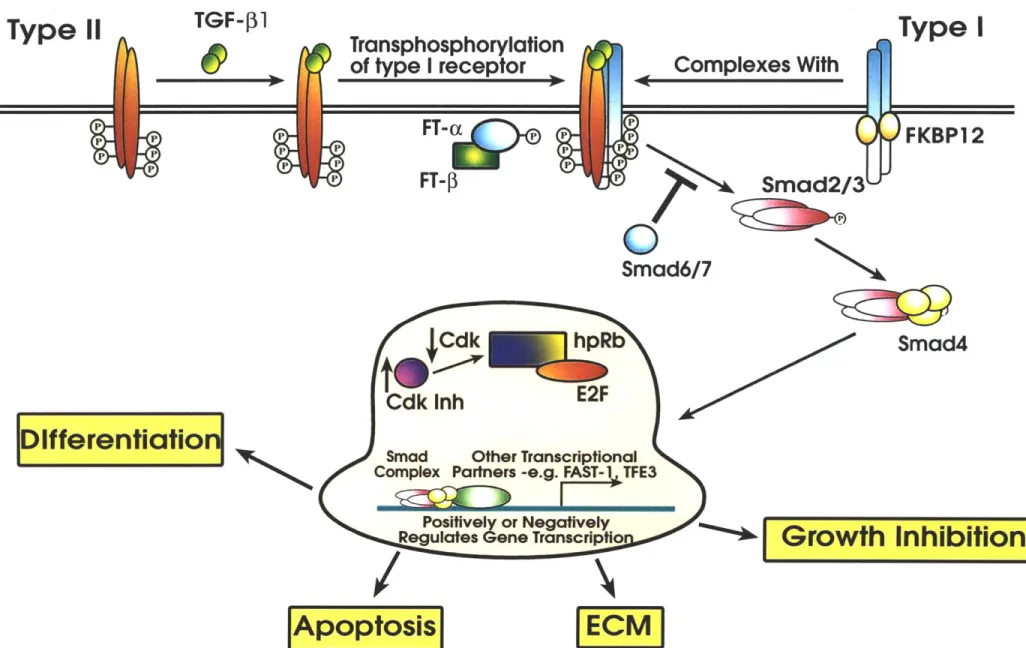
Employing orthogonal approaches, a number of studies have elucidated to a large extent the sequence of events required for activation of the TGF-P receptor complex. Specifically, the types I and II receptors appear to have evolved into two interdependent signalling molecules. Each receptor is required for ligand-induced signalling. In particular, type II is required for ligand binding whereas type I is necessary for initiation of intracellular signalling. In binding and cross-linking studies, it has been established that T RI (Alk-5) requires the presence of the type II receptor to bind the TGF-P ligands.(Wrana et al., 1992) In the case of TGF-1 and TGF-3, TfRII first binds ligand which then forms a ternary complex by recruiting TIRI into the TfRII/ligand complex. On the other hand, TGF-2 appears to require co-expression of both types I and II to bind at high affinities.(Rodriguez et al., 1995)
Studies measuring receptor kinase activity point to how recruitment of TPRI leads to signal initiation.(Wrana et al., 1994) In particular, TPRII maintains high constitutive kinase activity independent of the presence of ligand. In contrast, the T$RI kinase becomes activated only after recruitment to the TPRII/ligand complex. This would suggest that TfRII may be mediating transphosphorylation of TPRI upon formation of the ternary complex. Indeed, when co-expressed with a kinase-defective T$RII, TIRI fails to become phosphorylated. Yet, kinase-deficient and wild-type T3RI phosphorylation patterns are indistinguishable when co-expressed with wild-type TIRII. Therefore, type II kinase activity is necessary for TPRI phosphorylation most likely by direct transphosphorylation of T$RI by RII. Signalling studies further demonsrate that TPRI phosphorylation and therefore its activation constitute events downstream of initial TfRII/ligand binding.(Franzen et al., 1993) Specifically, a clone (RIB) of a TGF-4 responsive mink lung cell line expresses the type II, but not type I receptors; it fails to respond to TGF-$ stimulation as measured by growth inhibition or induction of early response genes such as
plasminogen activator inhibitor-1 (PAI-1). However, transfection of TPRI cDNA into RiBs is sufficient to restore signalling. Furthermore, transfection of a constitutively active T3RI (threonine 204 to aspartate) is sufficient to exert TGF signalling in the absence of TfRII or ligand further exemplifying the effector role of T3RI in the TGF-P receptor complex.
The types I, II and III receptor all form ligand-independent homo-oligomers, most likely dimers and upon TGF-P binding, form a types 1:11 heterotetramer. (Figure 1) (Gilboa et al., 1998; Henis et al., 1994; Moustakas et al., 1993; Yamashita et al., 1994) Two studies demonstrate the functional consequences of this particular receptor stoichiometry. First, Luo and co-workers constructed chimeric receptors consisting of the extracellular domain of the erythropoietin (Epo) receptor fused to the intracellular domain of either the types I or II receptor. They show that Epo-induced hetero-dimerization of a type I chimera with that of a type II can signal growth arrest.(Luo and Lodish, 1996) Moreover, a type I chimera encoding for a constitutively active kinase still requires Epo-induced homodimerization to signal. These results are consistent with the notion that the requirements for TGF- signal transduction include both type I homodimerization as well as its heterodimerization with type II. In another approach, Weis-Garcia et al. observe that a kinase-defectve type I receptor mutant complements an activation-defective mutant when co-expressed in cells. These results further confirm that type I homodimerization is indeed essential for signalling (Weis-Garcia and Massague, 1996).
TGF-P signal transduction
Largely due to classical genetic analysis in Drosophila and Caernorhabtidis
elegans, key components of the signalling pathways emanating from the TGF- receptors
have been identified. Most notably, members of the SMAD family of proteins were initially discovered from Drosophila by a genetic screen in which genes that enhanced the phenotype of a mutation in a BMP2/4 homolog, Decapentaplegic (Dpp), were isolated.(Raftery et al., 1995; Sekelsky et al., 1995) This screen yielded two highly homologous genes: Mothers against dpp (MAD) and Medea. Both encode proteins that act genetically downstream of dpp. Other members of this apparently growing family were later isolated in C. elegans in a similar screen for mutants that exhibited phenotypes similar to that of a type II receptor mutant.(Savage et al., 1996)
Within the past three years, these initial findings have led to rapid progress in the cloning and characterization of eight mammalian SMAD genes.In particular, three
sub-classes of SMADs have emerged: (1) pathway-restricted Smads (Smads 1,2,3,5 and 8), (2) a partner protein that associates with receptor-activated SMADs (Smad 4) and (3) inhibitory SMADs acting as negative regulators of TGF-P signalling (Smads 6,7). Mechanistically, an activated type I receptor phosphorylates a specific SMAD. These receptor-associated SMADs are pathway restricted as each one interacts with only a particular subset of type I receptors. For example, only the TGF- and activin type I receptors can phosphorylate Smads 2 and 3 whereas only the BMP receptors can phosphorylate Smads 1 and presumably 5 and 8.(Baker and Harland, 1996; Chen et al., 1996; Eppert et al., 1996; Holley et al., 1980; Kretzschmar et al., 1997; Liu et al., 1996; Liu et al., 1997; Nakao et al., 1997; Suzuki et al., 1997; Watanabe et al., 1997; Zhang et al., 1996) Following their phosphorylation, these SMADs then associate with Smad4 and translocate to the nucleus.(Lagna et al., 1996; Zhang et al., 1996) By presumably associating with other nuclear factors, the resulting complex then activates transcription of ligand-responsive genes.
Although Smads 2 and 3 are highly homologous and can both associate with the TGF- type I receptor, differences do emerge within the context of the developing organism. Homozygous null Smad 2 mutant mice die early during embryogenesis.(Nomura and Li, 1998; Waldrip et al., 1998) Smad3 mutant mice on the other hand are viable and fertile until four months of age when they begin to develop metastatic colorectal cancer.(Zhu et al., 1998)
Negative regulators, Smads 6 and 7, possess an amino terminal domain highly divergent from other signalling Smads. They inhibit signalling by competing with pathway-restricted SMADs in binding activated type I receptors.(Hayashi et al., 1997; Imamura et al., 1997; Nakao et al., 1997) It has been proposed that by stably binding to these receptors, they then prevent transphosphorylation of the downstream pathway-restricted SMADs. Smads 6 and 7 can bind a number of type I receptors consistent with their inhibition of multiple TGF-3 superfamily signalling pathways. Intriguingly, Smad 6 and 7 mediated inhibiton of TGF- signalling may be autoregulatory as TGF-P stimulation induces their expression. An additional mechanism of inhibition has been proposed for Smad6 as it is capable of binding Smadl and therefore may act as a non-functional Smad 4 decoy.
Several key aspects of Smad signalling require further study. For instance, the SMAD proteins can homo as well as hetero-oligomerize. However, the precise stoichiometry of these complexes remains to be fully determined.In vitro, Smad4 has been
shown to form homotrimers.(Shi et al., 1997) Kawabata and co-workers find that in live cells however, pathway-restricted Smads are monomers and undergo both homo- and hetero-oligomerization upon activation.(Kawabata et al., 1998)
In addition, based on crystallographic data, Shi and co-workers demonstrate that Smad3 binds a specific 4 base pair DNA sequence.(Shi et al., 1998) Because such low DNA binding selectivity would be insufficient for specifically activating transcription of TGF- -responsive genes, it is believed that additional transcription factors act in combination with the SMAD complex to elicit gene expression. A model calling for such a combinatorial approach would allow for the mediation of a wide range of ligand-induced responses despite relatively low DNA sequence binding specificity for the SMAD family of proteins. To date, two such candidate factors appear to fulfill this role. First, the winged helix transcription factor FAST-I has been shown to mediate activin-induced gene expression.(Chen et al., 1996; Chen et al., 1997; Chen et al., 1998) Specifically, FAST-I is a nuclear protein and upon activin stimulation, associates with incoming Smads 2 and 4 to form a complex termed activin-response factor (ARF). The newly formed ARF then activates transcription by binding to DNA sequence elements found within the promoter of the Mix.2 gene, an activin early-response gene. Within the promoter, FAST- 1 binds a six base pair sequence thereby augmenting the DNA binding specificity of the ARF complex. A number of other activin responsive promoters appear to lack FAST-I binding sites in close proximity to those of Smad2. This would suggest that other as of yet unidentified factors may also complex with Smads2 and 4 to induce remaining aspects of the activin induced response. Secondly, with respect to the TGF- pathway, Hua and co-workers show that an E-box binding transcription factor called TFE3 cooperates synergistically with Smads 3 and 4 to activate transcription of the TGF- responsive plasminogen activator inhibitor-I (PAI-1) promoter.(Hua et al., 1998; Westerhausen et al., 1991) By studying the transcriptional activity of PAI-I promoter mutants, they find that TFE3 and Smads3 and 4 bind to adjacent sites within a 36 bp element to induce TGF- dependent trancription. Moreover, TFE-3 binds a specific six base pair DNA sequence within this element. This further demonstrates the involvement of transcription factors that confer greater DNA binding selectivity to SMAD containing complexes, a property presumably necessary for mediating pathway specific responses.
Other molecules have been implicated to mediate TGF-P signalling. The immunophilin, FKBP12, is one of the best characterized examples. Better known as a target for various clinically relevant immunosuppressants, it also binds to a leucine-proline sequence within the type I receptor GS domain preventing transphosphorylation by the type
II receptor. Upon ligand-induced TPRI/T3RII association, it is then thought that FKBP12 dissociates from TfRI.(Chen et al., 1997; Wang et al., 1994; Wang et al., 1996) Mutant type I receptors that are unable to bind FKBP12 exhibit high basal signalling activity consistent with FKBP12's role as a negative regulator of TGF-$ signalling.(Chen et al., 1997) Therefore, its function appears to lie in protecting the cell from spontaneous signalling in the absence of ligand.
Three other proteins have been shown to be capable of directly associating with the TGF- receptors. p21RAS farnesyl protein-transferase a has been shown to bind the type I receptor and both TRIP-1, a WD-domain containing protein, and apolipoprotein J have been shown to bind to the type II receptor.(Chen et al., 1995; Kawabata et al., 1995; Reddy et al., 1996; Ventura et al., 1996; Wang et al., 1996) However, the functional significance of these interactions have yet to be elucidated.
TGF-P responses
The multipotent TGF- signals can be classified into immediate and secondary effects. With regard to immediate effects, the TGFs-3 inhibit growth by arresting the cell cycle at the GI to S transition. Specifically, inhibitory signals are transmitted through two independent pathways: (1) the upregulation of CDK inhibitors, p15 together with either p21 or p27, and (2) the repression of a CDK tyrosine phosphatase, CDC25a.(Figure 2) (Datto et al., 1995; Hannon and Beach, 1994; lavarone and Massague, 1997) Both pathways apparently converge to the negative regulation of cyclin-dependent kinases (CDKs) 4 and CDK6. Activation of CDKs-4 and 6 are necessary for phosphorylation of the retinoblastoma (Rb) protein and subsequent entry to S phase. Therefore, it appears to be at the level of CDK activity, a key junction of the cell cycle machinery, that TGF-P exerts it primary negative growth control.
With certain cell types, TGFs-3 also impinge second order effects including extracellular matrix production, immunosuppresion, and induction of angiogenesis.(Huber et al., 1992; Khanna et al., 1998; Roberts et al., 1986; Tashjian et al., 1985) Under particular circumstances, opposing secondary effects such as the induction of platelet derived growth factor secretion can overcome this block in cell cycle progression hence explaining earlier seemingly contradictory observations documenting the TGF-I induction of transformation upon fibroblasts.
The most striking validation of the current model for TGF- signalling can be illustrated by mutations of genes implicated in the signalling pathway and their resulting disease manifestations. It is believed that cells harboring mutations that render them unresponsive to TGF- are endowed with a significant growth advantage over their responsive counterparts. Indeed, loss of TGF- responsiveness is a common hallmark of various cancers.
On the receptor level, mutations in the type II receptor have been isolated from tumors of the colon, head and neck, T-cell lymphomas and gastric cancers, substantiating their role as tumor suppressors in a variety of cancers.(Garrigue-Antar et al., 1995; Knaus et al., 1996; Lu et al., 1998; Park et al., 1994) Furthermore, type II receptor mutations are found in 90% of all colon cancers exhibiting microsatellite instability.(Markowitz et al.,
1995; Wang et al., 1995) The high mutation frequency observed does not appear to be due
to an inherent susceptibility of the type II receptor gene to mutations. Rather, only gastric/colon cancers exhibit high mutation frequencies whereas other cancers arising from microsatellite instability do not. This would suggest that type II receptor mutations are somehow selected for in gastric and colon cancers. Mutations appear to occur late in tumorigenesis suggesting that the TGF-P pathway plays a role in malignant progression rather than tumor initiation.(Grady et al., 1998) Whether the type I receptor is likewise a
primary target for inactivating tumors in cancers is not as well established. However, Schiemann and co-workers have recently isolated a type I receptor mutation from a TGF-
-resistant T-cell lymphoma [manuscript in preparation]. And indeed, these mutations were demonstrated to result in the loss of type I receptor expression.
The relevance to cancer of the SMAD family of proteins became apparent shortly after their isolation in Drosophila and C. elegans. As one of the first vertebrate SMADs to be identified, SMAD4 was discovered as a gene in which both alleles were deleted in 90% of human pancreatic carcinomas.(Hahn et al., 1996) Subsequent studies in mice reinforce the essential role played by SMAD4 in the TGF-P pathway. Specifically, Takaku and co-workers show that tumors homozygously deficient for both SMAD4 and another tumor suppressor gene, APC, caused larger and more rapidly progressing carcinomas than in mice homozygously deficient for only APC(Hahn et al., 1996) These results therefore are consistent with SMAD4 playing a key role in the rate of tumor progression. Among pathway specific SMADs, SMAD2 has also been shown to be a target of inactivating mutations in colon cancers.(Eppert et al., 1996; Riggins et al., 1997; Uchida et al., 1996)
Intriguingly, despite the fact that SMAD3 knock-out mice develop metastatic colorectal cancer, no mutations have been identified in human cancers thus far.
References
Aitchison, L. (1960). A History of Metals, volume one, Volume 1 (New York, New York: Interscience Publishers, Inc.).
Aitchison, L. (1960). A History of Metals, volume two, Volume 2 (New York, New York: Interscience Publishers, Inc.).
Amrein, K. E., Panholzer, B., Flint, N. A., Bannwarth, W., and Bum, P. (1993). The Src homology 2 domain of the protein-tyrosine kinase p561ck mediates both intermolecular and intramolecular interactions. Proc Natl Acad Sci U S A 90, 10285-9.
Baker, J. C., and Harland, R. M. (1996). A novel mesoderm inducer, Madr2, functions in the activin signal transduction pathway. Genes Dev 10, 1880-9.
Baltimore, D. (1995). The enigma of HIV infection. Cell 82, 175-6.
Barlow, P. N., Luisi, B., Milner, A., Elliott, M., and Everett, R. (1994). Structure of the C3HC4 domain by 1H-nuclear magnetic resonance spectroscopy. A new structural class of zinc-finger. J Mol Biol 237, 201-11.
Beals, C. R., Clipstone, N. A., Ho, S. N., and Crabtree, G. R. (1997). Nuclear localization of NF-ATc by a calcineurin-dependent, cyclosporin-sensitive intramolecular interaction. Genes Dev 11, 824-34.
Bergman, M., Mustelin, T., Oetken, C., Partanen, J., Flint, N. A., Amrein, K. E., Autero, M., Burn, P., and Alitalo, K. (1992). The human p50csk tyrosine kinase phosphorylates p561ck at Tyr-505 and down regulates its catalytic activity. Embo J 11, 2919-24.
Borden, K. L., Boddy, M. N., Lally, J., O'Reilly, N. J., Martin, S., Howe, K., Solomon, E., and Freemont, P. S. (1995). The solution structure of the RING finger domain from the acute promyelocytic leukaemia proto-oncoprotein PML. Embo J 14,
1532-41.
Cantrell, D. (1996). T cell antigen receptor signal transduction pathways. Annu Rev Immunol 14, 259-74.
Casnellie, J. E., Harrison, M. L., Hellstrom, K. E., and Krebs, E. G. (1983). A lymphoma cell line expressing elevated levels of tyrosine protein kinase activity. J Biol Chem 258, 10738-42.
Casnellie, J. E., Harrison, M. L., Hellstrom, K. E., and Krebs, E. G. (1982). A lymphoma protein with an in vitro site of tyrosine phosphorylation homologous to that in pp60src. J Biol Chem 257, 13877-9.
Chan, D. C., and Kim, P. S. (1998). HIV entry and its inhibition. Cell 93, 681-4. Cheifetz, S., Hernandez, H., Laiho, M., ten Dijke, P., Iwata, K. K., and Massague, J. (1990). Distinct transforming growth factor-beta (TGF-beta) receptor subsets as
determinants of cellular responsiveness to three TGF-beta isoforms. J Biol Chem 265, 20533-8.
Chen, R. H., Miettinen, P. J., Maruoka, E. M., Choy, L., and Derynck, R. (1995). A WD-domain protein that is associated with and phosphorylated by the type II TGF-beta receptor. Nature 377, 548-52.
Chen, X., Rubock, M. J., and Whitman, M. (1996). A transcriptional partner for MAD proteins in TGF-beta signalling [published erratum appears in Nature 1996 Dec 19-26; 384(6610):648]. Nature 383, 691-6.
Chen, X., Weisberg, E., Fridmacher, V., Watanabe, M., Naco, G., and Whitman, M. (1997). Smad4 and FAST-I in the assembly of activin-responsive factor. Nature 389, 85-9.
Chen, Y., Lebrun, J. J., and Vale, W. (1996). Regulation of transforming growth factor beta- and activin-induced transcription by mammalian Mad proteins. Proc Natl Acad Sci U
S A 93, 12992-7.
Chen, Y. G., Hata, A., Lo, R. S., Wotton, D., Shi, Y., Pavletich, N., and Massague, J. (1998). Determinants of specificity in TGF-beta signal transduction. Genes Dev 12, 2144-52.
Chen, Y. G., Liu, F., and Massague, J. (1997). Mechanism of TGFbeta receptor inhibition by FKBP 12. Embo J 16, 3866-76.
Choe, H., Martin, K. A., Farzan, M., Sodroski, J., Gerard, N. P., and Gerard, C. (1998). Structural interactions between chemokine receptors, gp120 Env and CD4. Semin Immunol 10, 249-57.
Clipstone, N. A., and Crabtree, G. R. (1992). Identification of calcineurin as a key signalling enzyme in T-lymphocyte activation. Nature 357, 695-7.
Crabtree, G. R., and Clipstone, N. A. (1994). Signal transmission between the plasma membrane and nucleus of T lymphocytes. Annu Rev Biochem 63, 1045-83.
Creighton, T. E. (1993). Proteins, 2 Edition: W.H. Freeman and Company).
Cunningham, B. C., Bass, S., Fuh, G., and Wells, J. A. (1990). Zinc mediation of the binding of human growth hormone to the human prolactin receptor. Science 250, 1709-12. Cunningham, B. C., and Wells, J. A. (1991). Rational design of receptor-specific variants of human growth hormone. Proc Natl Acad Sci U S A 88, 3407-11.
Datto, M. B., Li, Y., Panus, J. F., Howe, D. J., Xiong, Y., and Wang, X. F. (1995). Transforming growth factor beta induces the cyclin-dependent kinase inhibitor p21 through a p53-independent mechanism. Proc Natl Acad Sci U S A 92, 5545-9.
Dawid, I. B., Breen, J. J., and Toyama, R. (1998). LIM domains: multiple roles as adapters and functional modifiers in protein interactions. Trends Genet 14, 156-62. Desjarlais, J. R., and Berg, J. M. (1992). Redesigning the DNA-binding specificity of a zinc finger protein: a data base-guided approach. Proteins 12, 101-4.
Desjarlais, J. R., and Berg, J. M. (1992). Toward rules relating zinc finger protein sequences and DNA binding site preferences. Proc Natl Acad Sci U S A 89, 7345-9.
Dialynas, D. P., Wilde, D. B., Marrack, P., Pierres, A., Wall, K. A., Havran, W., Otten, G., Loken, M. R., Pierres, M., Kappler, J., and et al. (1983). Characterization of the murine antigenic determinant, designated L3T4a, recognized by monoclonal antibody
GK1.5: expression of L3T4a by functional T cell clones appears to correlate primarily with
class II MHC antigen-reactivity. Immunol Rev 74, 29-56.
Dianzani, U., Shaw, A., al-Ramadi, B. K., Kubo, R. T., and Janeway, C. A., Jr. (1992). Physical association of CD4 with the T cell receptor. J Immunol 148, 678-88.
Dianzani, U., Shaw, A., Fernandez-Cabezudo, M., and Janeway, C. A., Jr. (1992). Extensive CD4 cross-linking inhibits T cell activation by anti-receptor antibody but not by antigen. Int Immunol 4, 995-1001.
Doyle, C., and Strominger, J. L. (1987). Interaction between CD4 and class II MHC molecules mediates cell adhesion. Nature 330, 256-9.
Einspahr, K. J., Abraham, R. T., Dick, C. J., and Leibson, P. J. (1990). Protein tyrosine phosphorylation and p561ck modification in IL-2 or phorbol ester-activated human natural killer cells. J Immunol 145, 1490-7.
Eppert, K., Scherer, S. W., Ozcelik, H., Pirone, R., Hoodless, P., Kim, H., Tsui, L. C., Bapat, B., Gallinger, S., Andrulis, I. L., Thomsen, G. H., Wrana, J. L., and Attisano, L.
(1996). MADR2 maps to 18q21 and encodes a TGFbeta-regulated MAD-related protein that is functionally mutated in colorectal carcinoma. Cell 86, 543-52.
Evans, R. M. (1988). The steroid and thyroid hormone receptor superfamily. Science 240, 889-95.
Falchuk, K. H., Montorzi, M., and Vallee, B. L. (1995). Zinc uptake and distribution in Xenopus laevis oocytes and embryos. Biochemistry 34, 16524-31.
Finzi, D., and Silliciano, R. F. (1998). Viral dynamics in HIV-I infection. Cell 93, 665-71.
Fischer, E. H., and Davie, E. W. (1998). Recent excitement regarding metallothionein. Proc Natl Acad Sci U S A 95, 3333-4.
Frankel, A. D., Bredt, D. S., and Pabo, C. 0. (1988). Tat protein from human immunodeficiency virus forms a metal-linked dimer. Science 240, 70-3.
Frankel, A. D., Chen, L., Cotter, R. J., and Pabo, C. 0. (1988). Dimerization of the tat protein from human immunodeficiency virus: a cysteine-rich peptide mimics the normal metal-linked dimer interface. Proc Natl Acad Sci U S A 85, 6297-300.
Franzen, P., ten Dijke, P., Ichijo, H., Yamashita, H., Schulz, P., Heldin, C. H., and Miyazono, K. (1993). Cloning of a TGF beta type I receptor that forms a heteromeric complex with the TGF beta type II receptor. Cell 75, 681-92.
Freemont, P. S. (1993). The RING finger. A novel protein sequence motif related to the zinc finger. Ann N Y Acad Sci 684, 174-92.
Fung-Leung, W. P., Louie, M. C., Limmer, A., Ohashi, P. S., Ngo, K., Chen, L., Kawai, K., Lacy, E., Loh, D. Y., and Mak, T. W. (1993). The lack of CD8 alpha cytoplasmic domain resulted in a dramatic decrease in efficiency in thymic maturation but


![Table 2 Binding of [ 65 Zn] to GST-TpRI-cyt immobilized on glutathione- agarose beads](https://thumb-eu.123doks.com/thumbv2/123doknet/14160804.473237/123.918.67.753.225.437/table-binding-gst-tpri-immobilized-glutathione-agarose-beads.webp)